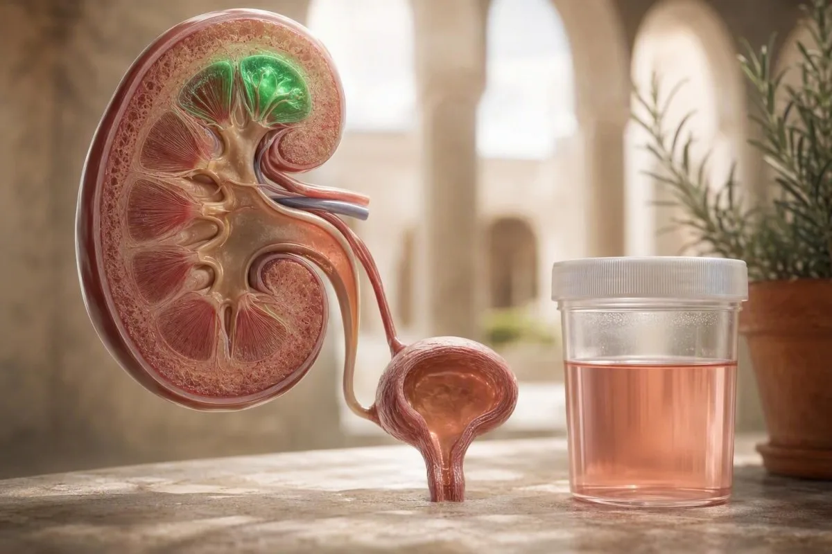
ปัสสาวะสีชมพูหรือสีชาหลังวิ่งเป็นเวลานานมักเป็นการเห็นเลือดในปัสสาวะชั่วคราวที่เกิดจากการออกกำลังกาย แต่ไม่ควรละเลยสีที่มองเห็นได้เสมอไป เวลาที่เกิดขึ้น ผลการตรวจด้วยกล้องจุลทรรศน์ อาการ และการตรวจซ้ำหลังจากพักผ่อนอย่างเพียงพอ จะบอกเรื่องราวที่แท้จริง.


- ปัสสาวะเป็นเลือดที่เกิดจากการออกกำลังกาย มักจะหายไปภายใน 24-72 ชั่วโมงหลังวิ่งอย่างหนัก การคงอยู่นานเกิน 72 ชั่วโมงต้องได้รับการตรวจจากแพทย์.
- เกณฑ์การตรวจด้วยกล้องจุลทรรศน์ ของเม็ดเลือดแดง 3 เซลล์ขึ้นไปต่อสนามกำลังขยายสูงในการเก็บตัวอย่างปัสสาวะที่ถูกต้องตรงตามคำจำกัดความของ AUA สำหรับภาวะเลือดออกในปัสสาวะระดับจุลภาค.
- ความไม่สอดคล้องกันของแถบตรวจ มีความสำคัญ: แผ่นตรวจเลือดเป็นบวกโดยมีเซลล์เม็ดเลือดแดง 0-2 เซลล์ต่อสนามกำลังขยายสูงบ่งชี้ถึงไมโอโกลบินหรือฮีโมโกลบินอิสระ แทนที่จะเป็นการตกเลือดในระบบทางเดินปัสสาวะ.
- ระยะเวลาที่ต้องทำซ้ำ เป็นไปได้ในทางปฏิบัติ: หลีกเลี่ยงการออกกำลังกายหนักเป็นเวลา 48-72 ชั่วโมง ดื่มน้ำตามปกติ จากนั้นให้ตัวอย่างปัสสาวะกลางสาย.
- การประเมินภายในวันเดียวกัน สมเหตุสมผลสำหรับภาวะลิ่มเลือด การไม่สามารถปัสสาวะได้ มีไข้ ปวดเอวรุนแรงข้างเดียว หรือปัสสาวะสีโคล่าพร้อมกับอาการปวดกล้ามเนื้ออย่างรุนแรง.
- รูปแบบเตือนของไต คือเลือดพร้อมกับโปรตีน, เซลล์เม็ดเลือดแดงเป็นก้อน, ความดันโลหิตสูง, หรือ GFR ที่ลดลง—ไม่ใช่เพียงแถบตรวจหลังการแข่งขัน.
- เงื่อนงำของภาวะกล้ามเนื้อลายสลาย คือปัสสาวะสีเข้มผิดปกติร่วมกับอาการปวดกล้ามเนื้ออย่างรุนแรงและระดับ creatine kinase ที่มักสูงกว่า 5 เท่าของค่าสูงสุดที่ยอมรับได้ในห้องปฏิบัติการ.
- บริบทความเสี่ยงมะเร็ง เปลี่ยนแปลงเกณฑ์สำหรับการตรวจสอบ: อายุ, การสัมผัสควันบุหรี่, การฉายรังสีอุ้งเชิงกรานก่อนหน้านี้, และปัสสาวะปนเลือดที่มองเห็นได้ซึ่งไม่หายไป ล้วนมีความสำคัญ.
ทำไมการวิ่งจึงทำให้ปัสสาวะเป็นสีชมพู
ปัสสาวะเป็นเลือดในนักวิ่ง โดยทั่วไปคือการรั่วไหลของเม็ดเลือดแดงในปัสสาวะซึ่งเกิดขึ้นเพียงช่วงสั้นๆ หลังจากการออกกำลังกายอย่างหนัก, ซึ่งมักจะหายไปภายใน 24–72 ชั่วโมงหลังพักผ่อน อย่างไรก็ตาม ปัสสาวะสีชมพู สีแดง หรือสีโค้ลาที่มองเห็นได้สมควรได้รับการทดสอบซ้ำที่เก็บตัวอย่างอย่างเหมาะสม แทนที่จะสันนิษฐานไปเองว่าเกิดจากการวิ่ง.
ในคลินิก รูปแบบที่น่าสบายใจนั้นตรงไปตรงมา: การเปลี่ยนแปลงสีเริ่มขึ้นหลังจากการวิ่งที่ยาวนาน เร็ว หรือลงเนินอย่างไม่ปกติ; ไม่มีไข้, ปวดบั้นเอวบิดเกร็ง, หรือแสบร้อนขณะปัสสาวะ; และผลการตรวจด้วยกล้องจุลทรรศน์กลับสู่ภาวะปกติหลังพักผ่อน. Kantesti คือเครื่องวิเคราะห์การตรวจเลือดด้วย AI ที่สามารถนำเครื่องหมายไตที่เกี่ยวข้อง—creatinine, eGFR, electrolytes, และ creatine kinase—มาพิจารณาร่วมกับประวัติการออกกำลังกาย แทนที่จะปฏิบัติต่อผลลัพธ์หลังการแข่งขันหนึ่งครั้งเป็นการวินิจฉัย.
กลไกที่เสนอ ได้แก่ การสัมผัสผนังกระเพาะปัสสาวะจากแรงกล, การเปลี่ยนแปลงการไหลเวียนเลือดไปที่ไตชั่วคราว, การเข้มข้นของปัสสาวะที่เกิดจากการขาดน้ำ, และบางครั้งการแตกสลายของเม็ดเลือดแดงจากการกระแทกของเท้าซ้ำๆ หลักฐานสำหรับแต่ละกลไกนั้นผสมปนเปกันอย่างตรงไปตรงมา เนื่องจากงานวิจัยส่วนใหญ่มีขนาดเล็กและใช้ระยะทางการแข่งขันที่แตกต่างกัน ในการปฏิบัติงานทางคลินิก 15 ปีของฉัน ประวัติความพยายามครั้งใหม่มีความสำคัญมากกว่าแค่สีของปัสสาวะเพียงอย่างเดียว การวิ่งอัลตร้ามาราธอนครั้งแรกและการวิ่ง 5 กม. ที่คุ้นเคยไม่ได้มีความน่าจะเป็นก่อนการตรวจเท่ากัน.
เลือดที่มองเห็นได้ในปัสสาวะหลังการออกกำลังกาย ไม่ได้มีความหมายเหมือนกับภาวะปัสสาวะเป็นเลือดในนักวิ่งที่ไม่ร้ายแรง แนวทางการพิจารณาภาวะปัสสาวะเป็นเลือดในวงกว้าง มีประโยชน์ เนื่องจากนิ่วในไต, การติดเชื้อ, โรคไตอักเสบ, และมะเร็งทางเดินปัสสาวะ อาจเกิดขึ้นโดยบังเอิญหลังจากวิ่ง.
คำนิยามที่ใช้ได้จริง
ภาวะปัสสาวะเป็นเลือดที่เกิดจากการออกกำลังกาย หมายถึงเม็ดเลือดแดงปรากฏในปัสสาวะหลังการออกแรงอย่างรวดเร็วและหายไปในตัวอย่างที่เก็บหลังพักผ่อน. การทดสอบด้วยแถบสีอย่างเดียวไม่สามารถยืนยันการวินิจฉัยนี้ได้ เนื่องจากรีเอเจนต์สามารถตรวจจับสารสีฮีมจากเซลล์เม็ดเลือดแดงที่สมบูรณ์, ฮีโมโกลบินอิสระ, หรือไมโอโกลบิน.
เป็นเลือดในปัสสาวะ ฮีโมโกลบิน หรือไมโอโกลบิน?
การทดสอบด้วยแถบสีปัสสาวะที่ให้ผลบวกสำหรับเลือด สามารถสะท้อนถึงเม็ดเลือดแดง, ฮีโมโกลบิน, หรือไมโอโกลบิน และการตรวจด้วยกล้องจุลทรรศน์จะแยกพวกมันออกจากกัน. ปัสสาวะสีชมพูที่มีเม็ดเลือดแดงจำนวนมากสนับสนุนภาวะปัสสาวะเป็นเลือด; ปัสสาวะสีน้ำตาลเข้มที่มีเม็ดเลือดแดงน้อยหรือไม่พบเลย ทำให้เกิดความกังวลเกี่ยวกับสารสีจากกล้ามเนื้อบาดเจ็บหรือการแตกสลายของเม็ดเลือดแดง.
การตรวจปัสสาวะด้วยกล้องจุลทรรศน์แสดง เม็ดเลือดแดง 3 เซลล์ขึ้นไปต่อฟิลด์กำลังขยายสูง ยืนยันภาวะจุลโลหิตในปัสสาวะเมื่อเก็บตัวอย่างได้สะอาด ในทางตรงกันข้าม แถบสีให้ผลบวกอย่างชัดเจนโดยมีเม็ดเลือดแดง 0–2 เซลล์ต่อฟิลด์กำลังขยายสูง ควรทำให้แพทย์พิจารณาภาวะไมโอโกลบินในปัสสาวะ โดยเฉพาะอย่างยิ่งหลังการสัมผัสความร้อน, การบาดเจ็บของกล้ามเนื้อแบบบดขยี้, หรืออาการปวดกล้ามเนื้อล่าช้าอย่างรุนแรง สีน้ำตาลหรือสีโค้ลามักเป็นลักษณะของสารสีที่เข้มข้นมากกว่าเลือดที่ออกเล็กน้อยจากกระเพาะปัสสาวะ.
ไมโอโกลบินมีความสำคัญเพราะสามารถเกิดขึ้นร่วมกับภาวะกล้ามเนื้อลายสลายและภาวะไตวายเฉียบพลัน ระดับ creatine kinase ที่สูงกว่า 5 เท่าของค่าสูงสุดที่ยอมรับได้ในห้องปฏิบัติการ มักใช้เป็นเกณฑ์ทางชีวเคมีสำหรับภาวะกล้ามเนื้อลายสลาย แม้ว่าอาการ, โพแทสเซียม, ครีเอตินีน, และปริมาณปัสสาวะจะเป็นตัวกำหนดความเร่งด่วน โปรดดูคำอธิบายที่ใช้ได้จริงของเราเกี่ยวกับ ครีเอทีนไคเนสสูงอย่างอันตราย สำหรับรูปแบบที่ควรรอข้ามคืน.
สีผสมอาหาร บีทรูท แบล็กเบอร์รี่ ไรแฟมพิซิน ฟีนาโซไพรดีน และภาวะขาดน้ำ ก็สามารถทำให้ปัสสาวะมีสีโดยไม่มีเม็ดเลือดแดงได้ การแยกภาวะปัสสาวะสีเข้ม ช่วยหลีกเลี่ยงข้อผิดพลาดทั่วไปในการสันนิษฐานว่าตัวอย่างสีแดงทุกครั้งคือเลือด.
การจำแนกประเภทที่เปลี่ยนแปลงการดูแล
การมีเม็ดเลือดแดงในปัสสาวะบ่งชี้ถึงระบบทางเดินปัสสาวะ ในขณะที่ไมโอโกลบินในปัสสาวะบ่งชี้ถึงกล้ามเนื้อลายและการปกป้องไต. ทั้งสองอย่างสามารถเกิดขึ้นได้หลังจากการออกกำลังกายที่ต้องใช้ความอดทน แต่เฉพาะการตรวจด้วยกล้องจุลทรรศน์และการตรวจเลือดแบบเจาะจงเท่านั้นที่สามารถแยกแยะได้อย่างน่าเชื่อถือ.
วิธีอ่านผลการตรวจปัสสาวะหลังวิ่ง
การตรวจปัสสาวะหลังวิ่งที่มีข้อมูลมากที่สุดรวมถึงการตรวจด้วยแถบทดสอบ การตรวจด้วยกล้องจุลทรรศน์ การวัดโปรตีน และความถ่วงจำเพาะ. เลือดบนแถบทดสอบที่มีเม็ดเลือดแดงชั่วคราวและไม่มีโปรตีนมักมีความเสี่ยงต่ำ เลือดร่วมกับโปรตีนหรือทรงกระบอกต้องได้รับการประเมินไตอย่างรอบคอบมากขึ้น.
ผลการตรวจเลือดด้วยแถบทดสอบจะถูกรายงานเป็น ลบ (negative) ร่องรอย (trace) 1+, 2+, หรือ 3+ แต่เป็นสัญญาณคัดกรองมากกว่าการนับจำนวนเซลล์. AUA กำหนดภาวะไมโครฮีมาตูเรียว่ามีเม็ดเลือดแดงมากกว่า 3 เซลล์ต่อฟิลด์กำลังขยายสูงในการตรวจด้วยกล้องจุลทรรศน์, ไม่ใช่ผลการตรวจด้วยแถบทดสอบเพียงอย่างเดียว (Barocas et al., 2020) การจำแนกประเภทนี้ช่วยหลีกเลี่ยงการส่งนักวิ่งเข้ารับการตรวจหามะเร็งที่ไม่จำเป็นหลังจากการตรวจคัดกรองที่ให้ผลบวกลวง.
ความถ่วงจำเพาะสูงกว่า 1.030 บ่งชี้ว่าปัสสาวะมีความเข้มข้นในห้องปฏิบัติการหลายแห่ง และอาจทำให้สีดูน่าตกใจมากขึ้น แต่ก็ไม่ได้อธิบายถึงเม็ดเลือดแดงที่ยืนยันแล้ว โปรตีนระดับ 1+ หรือสูงกว่าควรกำหนดปริมาณด้วยอัตราส่วนอัลบูมินต่อครีเอตินินในปัสสาวะหรืออัตราส่วนโปรตีนต่อครีเอตินิน โปรตีนจากปัสสาวะจากการออกกำลังกายชั่วคราวมีอยู่จริง แต่ภาวะอัลบูมินในปัสสาวะอย่างต่อเนื่องจะเปลี่ยนการวินิจฉัยแยกโรคไปทางโรคไตส่วนกระจายของเรา คู่มือการตรวจปัสสาวะฉบับสมบูรณ์ อธิบายว่าทำไมตัวบ่งชี้เหล่านี้จึงไม่ควรรวมเข้าเป็นผลลัพธ์เดียว.
Kantesti AI แปลผลครีเอตินินและ eGFR ควบคู่ไปกับผลการตรวจปัสสาวะ เวลาในการออกกำลังกาย และค่าก่อนหน้า ครีเอตินินที่สูงขึ้นเล็กน้อยเพียงครั้งเดียวหลังการแข่งขันที่ร้อนจัดไม่เทียบเท่ากับโรคไตเรื้อรัง ในทางปฏิบัติ ฉันจะสอบถามระยะทาง ภูมิประเทศ อุณหภูมิ การใช้ยาต้านการอักเสบ เวลาของรอบเดือน และว่าได้รับตัวอย่างภายใน 2 ชั่วโมงหลังสิ้นสุดหรือไม่.
เมื่อใดควรตรวจปัสสาวะซ้ำหลังวิ่ง?
การตรวจปัสสาวะซ้ำ 48–72 ชั่วโมงหลังจากการวิ่งหนักครั้งสุดท้ายเป็นค่าเริ่มต้นที่สมเหตุสมผลสำหรับการสงสัยภาวะเลือดออกในปัสสาวะของนักวิ่ง. ใช้ตัวอย่างปัสสาวะตอนเช้าครั้งแรกหรือตัวอย่างกลางสตรีมที่ได้รับน้ำเพียงพอ แต่ไม่ต้องดื่มน้ำปริมาณมากทันทีก่อนหน้านั้น เพราะการเจือจางมากเกินไปอาจบดบังผลการตรวจที่มีประโยชน์.
สำหรับความผิดปกติหลังการแข่งขันตามปกติ ฉันมักจะแนะนำให้งดการวิ่งแบบ Interval, วิ่งขึ้นเนิน, ยกน้ำหนักหนัก หรือการแข่งขันสำหรับ 48 ชั่วโมง; หลังจากวิ่งมาราธอน, อัลตร้ามาราธอน, ภาวะเจ็บป่วยจากความร้อน, หรืออาการปวดเมื่อยผิดปกติ, 72 ชั่วโมง มีเหตุผลมากกว่า การดื่มน้ำตามปกติเพียงพอแล้ว การดื่มน้ำมากเกินไปอาจทำให้ความถ่วงจำเพาะต่ำกว่า 1.005 และบางครั้งทำให้ผลการตรวจที่ก้ำกึ่งยากต่อการตีความ.
หากการตรวจด้วยกล้องจุลทรรศน์ยังคงเป็นบวกหลังจาก 72 ชั่วโมง ให้ทำการตรวจกับแพทย์ปฐมภูมิภายในไม่กี่วัน แทนที่จะตรวจซ้ำหลังการออกกำลังกาย ผลการตรวจที่ต่อเนื่องควรได้รับการวัดความดันโลหิต, ตรวจระดับครีเอตินินในซีรั่ม/eGFR, ตรวจอัตราส่วนอัลบูมินต่อครีเอตินินในปัสสาวะ และพิจารณาการเพาะเชื้อหรือการถ่ายภาพตามความเสี่ยง ความสัมพันธ์ระหว่างการออกแรง, การดื่มน้ำ และค่าประมาณการกรองที่ลดลงชั่วคราวจะกล่าวถึงใน คู่มือ eGFR หลังการออกกำลังกาย.
นักวิ่งมาราธอนสมัครเล่นวัย 52 ปีที่ฉันเคยพบ มีเม็ดเลือดแดง 18 เซลล์ต่อฟิลด์กำลังขยายสูงในเย็นวันหนึ่งหลังการแข่งขันที่อากาศร้อน แต่มีเม็ดเลือดแดง 0–1 เซลล์หลังจากการพักผ่อน 72 ชั่วโมง; ระดับครีเอตินินของเขากลับมาจากการวัด 1.28 mg/dL เป็น 0.96 mg/dL ผลการตรวจเป็นที่น่าพอใจ แต่แผนเดียวกันนี้จะไม่เพียงพอสำหรับผู้ที่สูบบุหรี่วัย 52 ปีที่มีสีปัสสาวะที่มองเห็นได้ซ้ำๆ.
อย่าตรวจที่เส้นชัยหากหลีกเลี่ยงได้
การตรวจทันทีหลังการออกกำลังกายจะเพิ่มโอกาสในการพบเม็ดเลือดแดงชั่วคราว, ปัสสาวะเข้มข้น, และโปรตีน. สิ่งนั้นอาจมีประโยชน์ทางคลินิกในการสงสัยภาวะ Rhabdomyolysis แต่ก็เป็นช่วงเวลาที่ไม่เหมาะสมในการกำหนดค่าพื้นฐานของนักวิ่ง.
รูปแบบเลือดในปัสสาวะหลังวิ่งแบบใดที่มักจะสบายใจได้?
ตอนที่เกิดขึ้นเองหลังจากการวิ่งหนักจะน่าพอใจกว่าเมื่อหายไปอย่างสมบูรณ์, ขาดอาการปวดหรืออาการทั่วไป, และไม่มีโปรตีนรั่วในปัสสาวะหรือการเปลี่ยนแปลงการทำงานของไต. การน่าพอใจไม่ได้หมายความว่าเพิกเฉย; หมายความว่าได้รับการยืนยันด้วยตัวอย่างที่ได้จากการพักผ่อน.
กลุ่มอาการที่มีความเสี่ยงต่ำกว่าคือ นักวิ่งวัยหนุ่มหรือวัยกลางคนที่มีปัจจัยกระตุ้นจากการออกแรงชัดเจน, ความดันโลหิตปกติ, ไม่มีประวัติการสูบบุหรี่, และการตรวจด้วยกล้องจุลทรรศน์ขณะพักผ่อนปกติ ในสถานการณ์นั้น, ผลการตรวจเพียงครั้งเดียวที่เกิดขึ้นชั่วคราว 3–10 RBC/HPF หลังการแข่งขันมักจะหายไปโดยไม่ต้องรักษา ฉันยังคงจะบันทึกไว้, เพราะการเกิดขึ้นซ้ำหลังจากการวิ่งระยะไกลทุกครั้งสมควรได้รับการทบทวนอย่างละเอียดกว่าเหตุการณ์ที่เกิดขึ้นเพียงครั้งเดียว.
อัตราส่วนอัลบูมินต่อครีเอตินินในปัสสาวะที่ปกติโดยทั่วไปคือ ต่ำกว่า 30 mg/g (ต่ำกว่า 3 mg/mmol), แม้ว่าห้องปฏิบัติการและประเทศต่างๆ จะรายงานหน่วยที่แตกต่างกัน เลือดที่ไม่มีอัลบูมิน, แคสต์, หรือเม็ดเลือดแดงผิดรูป บ่งชี้ว่าไม่มีการรั่วซึมจากไต; ไม่ได้พิสูจน์ว่ามาจากกระเพาะปัสสาวะ, แต่เป็นการนำไปสู่คำถามต่อไป คำอธิบายของเราเกี่ยวกับ โปรตีนในปัสสาวะ ครอบคลุมส่วนนี้อย่างชัดเจน.
การบาดเจ็บบริเวณกระเพาะปัสสาวะมีความเป็นไปได้มากขึ้นเมื่อปัสสาวะมีความเข้มข้นสูงและนักวิ่งเข้าเส้นชัยโดยมีกระเพาะปัสสาวะเกือบว่าง ผู้เชี่ยวชาญด้านเวชศาสตร์การกีฬาบางคนแนะนำให้ปัสสาวะก่อนวิ่งระยะไกลและดื่มน้ำเป็นประจำ แต่นี่เป็นคำแนะนำในการป้องกันที่มีหลักฐานการทดลองจำกัด ไม่ใช่สิ่งทดแทนการประเมินเมื่อสีปัสสาวะยังคงอยู่.
ผลลัพธ์ปกติจะมีประทับเวลา
การทดสอบที่ไม่พบเลือด 3 วันหลังการแข่งขันเป็นการยืนยันสำหรับเหตุการณ์นั้น ไม่ใช่การรับประกันตลอดชีวิต. การมีเลือดปนในปัสสาวะที่มองเห็นได้อีกครั้งในอีกหลายเดือนต่อมาควรได้รับการประเมินตามลักษณะเฉพาะของตนเอง โดยเฉพาะอย่างยิ่งหลังอายุ 40 ปี หรือมีการสัมผัสควันบุหรี่.
สัญญาณเตือนที่ทำให้เลือดในปัสสาวะของนักวิ่งมีความเป็นไปได้น้อยลง
การมีเลือดปนในปัสสาวะจำเป็นต้องได้รับการประเมินอย่างเร่งด่วนเมื่อยังคงอยู่หลัง 72 ชั่วโมง กลับมาเป็นซ้ำโดยไม่มีการออกกำลังกายหนัก หรือปรากฏร่วมกับอาการปวด มีไข้ มีลิ่มเลือด มีโปรตีน หรือมีการเปลี่ยนแปลงการทำงานของไต. การรวมกันของผลการตรวจมีความสำคัญมากกว่าตัวอย่างที่ดูน่าตกใจเพียงอย่างเดียว.
ขอคำแนะนำทางการแพทย์ทันเวลาสำหรับปัสสาวะสีแดงที่มองเห็นได้ซึ่งคงอยู่นานเกิน 24 ชั่วโมง, แม้ว่าคุณจะรู้สึกสบายดี โดยเฉพาะอย่างยิ่งสำหรับผู้ที่มีอายุมากกว่า 40 ปี ผู้ที่สูบบุหรี่ในปัจจุบันหรือในอดีต หรือผู้ที่เคยได้รับการฉายรังสีบริเวณอุ้งเชิงกรานมาก่อน แนวทางของ AUA ใช้การจำแนกความเสี่ยงตามอายุ การสูบบุหรี่ ระดับและความคงทนของเลือดในปัสสาวะที่มองด้วยกล้องจุลทรรศน์ และประวัติการมีเลือดปนในปัสสาวะที่มองเห็นได้ก่อนหน้านี้ แทนที่จะปฏิบัติต่อภาวะเลือดปนในปัสสาวะที่มองด้วยกล้องจุลทรรศน์ทุกกรณีเหมือนกัน (Barocas et al., 2020).
เซลล์เม็ดเลือดแดงที่ผิดปกติ (dysmorphic red cells) ท่อปัสสาวะที่มีเซลล์เม็ดเลือดแดง (red-cell casts) โปรตีนอัลบูมินในปัสสาวะปริมาณมาก (albuminuria) ความดันโลหิตสูงชนิดใหม่ หรือ eGFR ที่ลดลง บ่งชี้ว่ามีต้นกำเนิดมาจากตัวกรองไต แทนที่จะเป็นการกระแทกของกระเพาะปัสสาวะ. ท่อปัสสาวะที่มีเซลล์เม็ดเลือดแดง (Red-cell casts) ไม่เคยเป็นผลที่คาดไม่ถึงจากการวิ่งธรรมดา และควรได้รับการประเมินไตอย่างเร่งด่วน ดูภาพรวมของเราเกี่ยวกับ ท่อปัสสาวะแบบเม็ด (granular) และท่อปัสสาวะ (urinary casts) ว่าเหตุใดผลการตรวจตะกอนจึงมีความสำคัญ.
Thomas Klein, MD, แนะนำให้นักวิ่งอย่าประเมินตนเองจากสีปัสสาวะเพียงอย่างเดียว การติดเชื้อทางเดินปัสสาวะอาจทำให้เกิดอาการแสบร้อน ปวดปัสสาวะกะทันหัน มีหนอง และบางครั้งมีเลือดปนในปัสสาวะ ในขณะที่นิ่วจะทำให้เกิดอาการปวดอย่างกะทันหันข้างเดียวและคลื่นไส้ การเพาะเชื้อมีประโยชน์ที่สุดก่อนเริ่มยาปฏิชีวนะเมื่อมีอาการติดเชื้อ.
การผสมผสานระหว่างเลือดและโปรตีน
การมีเลือดปนในปัสสาวะร่วมกับโปรตีนในปัสสาวะที่ยืนยันแล้ว เป็นเรื่องที่น่ากังวลกว่าการมีเลือดปนในปัสสาวะเพียงอย่างเดียว เนื่องจากทั้งสองอย่างอาจมีต้นกำเนิดมาจากตัวกรองไต (glomerular filter). อัตราส่วนอัลบูมินต่อครีเอตินินในปัสสาวะ 30 mg/g หรือสูงกว่า ควรตรวจซ้ำและตีความร่วมกับความดันโลหิตและการทำงานของไต.
เมื่อใดที่ปัสสาวะสีชมพูหลังออกกำลังกายต้องการการดูแลในวันนั้น?
เข้ารับการประเมินเร่งด่วนภายในวันเดียวกัน หากมีลิ่มเลือด ไม่สามารถปัสสาวะได้ ปวดเอวด้านข้างอย่างรุนแรง มีไข้ หน้ามืด ปัสสาวะออกน้อย หรือปัสสาวะสีเข้มร่วมกับอาการปวดกล้ามเนื้อและอ่อนแรงอย่างรุนแรง. ลักษณะเหล่านี้ชี้ให้เห็นถึงภาวะเลือดปนในปัสสาวะที่เกิดจากการออกกำลังกายที่ไม่ซับซ้อน.
ลิ่มเลือดสามารถอุดกั้นการไหลของปัสสาวะ และการไม่สามารถปัสสาวะได้เป็นปัญหาเร่งด่วนโดยไม่คำนึงถึงสมรรถภาพทางกาย ไข้ ไข้ที่ 38.0°C ขึ้นไป ร่วมกับอาการปวดเอว อาจบ่งชี้ถึงการติดเชื้อทางเดินปัสสาวะส่วนบน อาการปวดบีบอย่างรุนแรงพร้อมอาเจียนอาจบ่งชี้ถึงนิ่ว ทั้งสองสถานการณ์ไม่ควรจัดการด้วยการรอทดสอบซ้ำหลังการวิ่ง.
ปัสสาวะสีน้ำตาลเข้มหลังการออกกำลังกายร่วมกับอาการปวดกล้ามเนื้อ กดเจ็บ กล้ามเนื้ออ่อนแรง บวม หรือปัสสาวะออกน้อย เพิ่มความกังวลต่อภาวะกล้ามเนื้อสลาย (rhabdomyolysis) การทดสอบเบื้องต้นมักรวมถึง creatine kinase, creatinine, potassium, calcium, phosphate และการตรวจปัสสาวะ คู่มือ rhabdomyolysis ของ CrossFit มีความเกี่ยวข้องเนื่องจากการออกกำลังกายแบบ Eccentric ที่ไม่คุ้นเคย ไม่ใช่แค่การวิ่งเพียงอย่างเดียว เป็นสาเหตุที่พบบ่อย.
ภาวะขาดน้ำอย่างรุนแรงสามารถทำให้ผลตรวจไตดูแย่ลงได้ แต่ก็สามารถเกิดขึ้นร่วมกับการบาดเจ็บจริงได้ หากคุณมีอาการเวียนศีรษะ สับสน ไม่สามารถดื่มน้ำได้ หรือปัสสาวะน้อยมาก 8–12 ชั่วโมง, อย่าพยายามแก้ไขปัญหาด้วยเครื่องดื่มเกลือแร่ที่บ้าน.
การตั้งครรภ์และยาต้านการแข็งตัวของเลือด
นักวิ่งที่ตั้งครรภ์และผู้ที่รับประทานยาต้านการแข็งตัวของเลือด ควรแจ้งภาวะปัสสาวะเป็นเลือดที่มองเห็นได้ทันที. ยาต้านการแข็งตัวของเลือดสามารถเพิ่มเลือดออกได้ แต่ไม่จำเป็นต้องระบุแหล่งที่มา การตั้งครรภ์ยังเปลี่ยนแปลงเกณฑ์ในการตรวจการติดเชื้อ นิ่ว และโรคไต.
ภาวะที่การวิ่งสามารถเปิดเผยได้มากกว่าที่จะก่อให้เกิด
การวิ่งอาจทำให้ปัญหาทางเดินปัสสาวะที่มีอยู่ปรากฏขึ้นโดยการทำให้ปัสสาวะเข้มข้นขึ้นหรือกระตุ้นให้ทำการทดสอบ แทนที่จะทำให้เกิดภาวะปัสสาวะเป็นเลือดด้วยตนเอง. นิ่ว การติดเชื้อ โรคไตอักเสบจากโกลเมอรูลัส ความผิดปกติของโครงสร้าง และมะเร็ง ยังคงเป็นส่วนหนึ่งของการวินิจฉัยแยกโรค.
นิ่วในไตมักทำให้เกิดอาการปวดสีข้างไปถึงขาหนีบข้างเดียว คลื่นไส้ และพบเซลล์เม็ดเลือดแดงในกล้องจุลทรรศน์ แม้ว่าจะมีนิ่วที่ไม่เจ็บปวดเกิดขึ้นก็ตาม การติดเชื้อในทางเดินปัสสาวะมักทำให้เกิดอาการแสบร้อน ปัสสาวะบ่อย ปวดเบ่ง และพบเซลล์เม็ดเลือดขาว การตรวจไม่พบไนไตรท์ไม่สามารถตัดภาวะนี้ออกได้ คู่มือของเราเกี่ยวกับการตรวจ leukocyte esterase ร่วมกับการตรวจไม่พบไนไตรท์ อธิบายรูปแบบผสมที่พบบ่อยนี้.
โรคไตอักเสบจากโกลเมอรูลัสสามารถเกิดขึ้นได้หลังจากการติดเชื้อในระบบทางเดินหายใจส่วนบน โดยมีปัสสาวะสีโคลา บวม ความดันโลหิตสูง โปรตีนในปัสสาวะ และเซลล์เม็ดเลือดแดงที่ผิดปกติ ในแนวทางการรักษาโรคไตเรื้อรัง KDIGO ปี 2024 โปรตีนในปัสสาวะที่คงอยู่และ eGFR ที่ลดลงถือเป็นตัวบ่งชี้ความเสี่ยงต่อไตที่เสริมกัน ไม่ใช่การวัดที่ใช้แทนกันได้ (KDIGO, 2024).
Kantesti คือเครื่องมือวิเคราะห์ตรวจเลือดที่ขับเคลื่อนด้วย AI ที่ช่วยให้ผู้ใช้จดจำรูปแบบของครีเอตินีน, eGFR, อัลบูมิน, อิเล็กโทรไลต์ และ CBC ได้ แต่ไม่สามารถวินิจฉัยแหล่งทางกายวิภาคของภาวะปัสสาวะเป็นเลือดได้ การถ่ายภาพรังสี การตรวจด้วยกล้องจุลทรรศน์ การเพาะเชื้อ และการตรวจโดยแพทย์ยังคงเป็นส่วนสำคัญในการวินิจฉัย.
มะเร็งพบได้ไม่บ่อยในนักวิ่งอายุน้อย แต่เป็นไปได้ในผู้ใหญ่
ภาวะปัสสาวะเป็นเลือดที่มองเห็นได้ควรได้รับการประเมิน แม้ว่าการออกกำลังกายจะเป็นคำอธิบายที่ชัดเจน โดยเฉพาะในผู้ที่มีประวัติสัมผัสควันบุหรี่หรือมีอาการเรื้อรัง. ความเสี่ยงเพิ่มขึ้นตามอายุและการสัมผัสยาสูบสะสม ซึ่งเป็นเหตุผลว่าทำไมผลปัสสาวะที่เหมือนกันจึงไม่นำไปสู่แผนการตรวจที่เหมือนกัน.
ใครบ้างที่ต้องติดตามผลกับแพทย์ผู้เชี่ยวชาญด้านระบบทางเดินปัสสาวะหรือระบบทางเดินปัสสาวะ?
ภาวะปัสสาวะเป็นเลือดที่คงอยู่หลังจากการเก็บตัวอย่างซ้ำเมื่อพัก ควรประเมินความเสี่ยงสำหรับระบบทางเดินปัสสาวะ ในขณะที่ภาวะปัสสาวะเป็นเลือดร่วมกับโปรตีนในปัสสาวะ, casts, ความดันโลหิตสูง หรือ eGFR ที่ลดลง ชี้ไปทางระบบไต. หลายคนต้องการเพียงการตรวจซ้ำภายใต้การดูแลของแพทย์ปฐมภูมิ การส่งต่อขึ้นอยู่กับรูปแบบ.
การประเมินโดยระบบทางเดินปัสสาวะมีความเหมาะสมเป็นพิเศษสำหรับภาวะปัสสาวะเป็นเลือดที่มองเห็นได้ซ้ำๆ, ภาวะปัสสาวะเป็นเลือดจากกล้องจุลทรรศน์ที่คงอยู่, ประวัติการสูบบุหรี่, การสัมผัสสารอะโรมาติกเอมีนในอาชีพ, การฉายรังสีในอุ้งเชิงกรานก่อนหน้านี้, หรืออาการทางเดินปัสสาวะที่ไม่มีการติดเชื้อ ขึ้นอยู่กับความเสี่ยง แพทย์อาจใช้อัลตราซาวนด์ไตและกระเพาะปัสสาวะ, CT urography, และ cystoscopy การตรวจแบบ trace dipstick หลังการแข่งขันในวัย 24 ปี เป็นสถานการณ์ที่แตกต่างอย่างมากจากปัสสาวะสีแดงซ้ำๆ ในอดีตนักสูบบุหรี่วัย 58 ปี.
การทบทวนโดยระบบไตมีความเป็นไปได้มากขึ้นเมื่ออัตราส่วนอัลบูมินต่อครีเอตินีนในปัสสาวะ ≥30 มก./ก., eGFR ต่ำกว่า ต่ำกว่า 60 mL/min/1.73 m² เป็นเวลา 3 เดือนหรือนานกว่านั้น, red-cell casts, หรือความดันโลหิตที่ควบคุมได้ยาก เกณฑ์เหล่านี้มีความสำคัญเนื่องจากโรคไตเรื้อรังถูกกำหนดโดยระยะเวลาและการวัดผลเพียงครั้งเดียว อ่าน ระยะของโรคไตเรื้อรัง ของเราสำหรับหมวดหมู่ที่ใช้ได้จริง.
บริการตีความผลการตรวจเลือดด้วย AI ของ Kantesti สามารถระบุรูปแบบเลือดและโปรตีนที่ทำซ้ำได้เพื่อปรึกษาหารือ โดยเฉพาะอย่างยิ่งเมื่อผู้ใช้อัปโหลดผลลัพธ์จากห้องปฏิบัติการที่แตกต่างกัน บริการนี้ควรมุ่งสนับสนุน ไม่ใช่แทนที่การตัดสินใจของแพทย์ว่าจะต้องมีการถ่ายภาพทางเดินปัสสาวะ หรือการตรวจโดยผู้เชี่ยวชาญหรือไม่.
อย่าปล่อยให้การใช้ยาต้านการแข็งตัวของเลือดสิ้นสุดการสนทนา
การใช้ยาต้านการแข็งตัวของเลือดเปลี่ยนแปลงความรุนแรงของการตกเลือด แต่ไม่ได้ระบุสาเหตุของปัสสาวะเป็นเลือด. จากประสบการณ์ของผม การอ้างว่าเลือดในปัสสาวะที่กลับมาเป็นซ้ำเกิดจากยา apixaban, warfarin หรือ aspirin โดยไม่มีการตรวจสอบเพิ่มเติม อาจทำให้การวินิจฉัยนิ่ว การติดเชื้อ หรือโรคโครงสร้างล่าช้า.
การตรวจเลือดใดที่ช่วยได้หลังมีเลือดในปัสสาวะที่เกี่ยวข้องกับการออกกำลังกาย?
การตรวจเลือดที่มีประโยชน์ขึ้นอยู่กับอาการ: ครีเอตินีน, eGFR, อิเล็กโทรไลต์ และ creatine kinase ประเมินความเครียดของไตและกล้ามเนื้อ ในขณะที่ CBC ตรวจสอบภาวะโลหิตจาง หรือเบาะแสการติดเชื้อ. แผงการตรวจที่ครอบคลุมตามปกติมีประโยชน์น้อยกว่าการตรวจแบบเจาะจงหลังจากได้ประวัติและการตรวจปัสสาวะที่แม่นยำ.
ครีเอตินีนอาจเพิ่มขึ้นชั่วคราวหลังการออกกำลังกายแบบหนักหน่วงเนื่องจากการเผาผลาญกล้ามเนื้อ, การไหลเวียนเลือดไปไตลดลง และภาวะขาดน้ำ สมการ eGFR มีความน่าเชื่อถือน้อยลงระหว่างการเปลี่ยนแปลงครีเอตินีนอย่างรวดเร็ว ซึ่งเป็นเหตุผลว่าทำไมค่าที่วัดซ้ำหลังจาก 48–72 ชั่วโมง การฟื้นตัวตามปกติมักมีความหมายมากกว่าผลการแข่งขันที่เส้นชัย ทีม แนวทางการตรวจครีเอตินีนซ้ำที่เกี่ยวข้องกับการออกกำลังกาย ให้บริบทสำหรับตัวกระตุ้นความวิตกกังวลที่พบบ่อยนี้.
Creatine kinase มีช่วงหลังการออกกำลังกายที่กว้าง: นักกีฬาระดับสูงอาจแสดงค่าที่สูงขึ้นหลังการแข่งขันโดยไม่มีการบาดเจ็บที่ไต ในขณะที่ค่าที่สูงกว่าขีดจำกัดบนของท้องถิ่นหลายเท่าบวกกับอาการสมควรได้รับการประเมินอย่างเร่งด่วน โพแทสเซียมที่สูงกว่า 6.0 mmol/L อาจเป็นอันตรายและต้องได้รับการประเมินทางคลินิกอย่างเร่งด่วน โดยเฉพาะอย่างยิ่งหากการทำงานของไตบกพร่อง หรือสงสัยว่ามีการบาดเจ็บที่กล้ามเนื้อ.
CBC มีประโยชน์มากที่สุดเมื่อมีอาการอ่อนเพลีย, ซีด, การมีเลือดประจำเดือนมาก, ปัสสาวะเป็นเลือดซ้ำ หรือข้อกังวลเกี่ยวกับอาการป่วยทั่วร่างกาย ฮีโมโกลบินอาจลดลงหลังจากการออกกำลังกายแบบหนักหน่วงจากการขยายตัวของพลาสมา, การสูญเสียธาตุเหล็ก หรือภาวะเม็ดเลือดแดงแตก; ควรตีความค่าเฟอร์ริตินโดยแยกจากภาวะอักเสบเฉียบพลันและการฝึกซ้อมอย่างหนัก นักวิ่งระยะไกลอาจพบ ภาพรวมการตรวจเลือดของนักกีฬา มีประโยชน์สำหรับค่าพื้นฐานที่สมเหตุสมผล.
อย่าวินิจฉัยโรคไตจากแผงการตรวจเพียงครั้งเดียวในช่วงสัปดาห์การแข่งขัน
ค่าครีเอตินีนที่เพิ่มขึ้น 0.3 mg/dL หรือมากกว่า อาจเข้าเกณฑ์ภาวะไตบาดเจ็บเฉียบพลันในกรอบเวลาที่เหมาะสม แต่บริบทและการตรวจซ้ำเป็นสิ่งสำคัญหลังการแข่งขัน. ปริมาณปัสสาวะน้อย, โพแทสเซียมที่สูงขึ้น และค่าที่สูงอย่างต่อเนื่อง มีน้ำหนักมากกว่าผลการตรวจที่ก้ำกึ่งเพียงอย่างเดียว.
วิธีเก็บตัวอย่างปัสสาวะซ้ำที่แพทย์เชื่อถือได้
การเก็บตัวอย่างปัสสาวะในช่วงกลางของการปัสสาวะ (midstream) แบบสะอาด (clean-catch) หลัง 48–72 ชั่วโมงโดยไม่ออกกำลังกายหนัก เป็นวิธีที่ดีที่สุดในการตรวจสอบว่าปัสสาวะเป็นเลือดหายไปหรือไม่. ข้อผิดพลาดในการเก็บตัวอย่าง, การปนเปื้อนจากประจำเดือน และตัวอย่างที่เก่าเกินไป สามารถทำให้ผลลัพธ์คลาดเคลื่อนได้.
ล้างมือ, เริ่มปัสสาวะลงในโถส้วม, จากนั้นเก็บตัวอย่างส่วนกลาง (midstream) โดยไม่สัมผัสด้านในของภาชนะ ส่งตัวอย่างโดยเร็วที่สุด—ควรภายใน 2 ชั่วโมง—หรือปฏิบัติตามคำแนะนำในการแช่เย็นของห้องปฏิบัติการ การทิ้งตัวอย่างไว้นานเกินไปจะเปลี่ยนแปลงลักษณะของเซลล์และทำให้การตรวจด้วยกล้องจุลทรรศน์น่าเชื่อถือน้อยลง.
หลีกเลี่ยงการตรวจระหว่างมีประจำเดือนที่ยังมีการไหลของเลือดอยู่หากเป็นไปได้; หากไม่สามารถรอได้ ให้แจ้งแพทย์และห้องปฏิบัติการ การปนเปื้อนจากช่องคลอดอาจทำให้เห็นเซลล์เม็ดเลือดแดงและโปรตีนที่ปรากฏชัด ในขณะที่เซลล์เยื่อบุผิวแบบแบน (squamous epithelial cells) มักบ่งบอกถึงตัวอย่างที่เก็บไม่ดี แนวทางของเราเกี่ยวกับ เซลล์เยื่อบุผิวในปัสสาวะ อธิบายว่าเหตุใดจึงไม่ใช่เพียงเชิงอรรถในห้องปฏิบัติการที่ไม่สำคัญ.
Thomas Klein, MD, แนะนำให้บันทึกรายละเอียดสี่ประการพร้อมกับสิ่งส่งตรวจ: ครั้งสุดท้ายที่มีการออกกำลังกายอย่างหนัก, ระยะเวลาของกิจกรรม, อุณหภูมิภายนอก และยาที่รับประทานทั้งหมดในช่วง 72 ชั่วโมงก่อนหน้า ผลลัพธ์ที่ไม่มีการระบุเวลาจะตีความได้ยากกว่ามาก โดยเฉพาะอย่างยิ่งเมื่อเก็บปัสสาวะหลังจากออกกำลังกายสองครั้ง.
การทดสอบตอนเช้าตรู่เทียบกับตอนกลางวัน
ตัวอย่างตอนเช้าตรู่มีความเข้มข้นและดีต่อการตรวจหาความผิดปกติเล็กน้อย แต่ตัวอย่างกลางกระแสในตอนกลางวันที่ได้รับน้ำเพียงพอถือว่ายอมรับได้หากการเก็บตัวอย่างสะอาดกว่า. ความสม่ำเสมอมีความสำคัญมากขึ้นสำหรับการเปรียบเทียบเป็นระยะ: ใช้เงื่อนไขที่ใกล้เคียงกันในแต่ละครั้งที่ทำซ้ำ.
การดื่มน้ำ ยาแก้ปวด และผลิตภัณฑ์เสริมอาหารที่เปลี่ยนแปลงภาพรวม
ภาวะขาดน้ำสามารถทำให้ปัสสาวะเข้มข้นและเพิ่มความเครียดของไต ในขณะที่ยา NSAIDs สามารถลดการไหลเวียนโลหิตของไตระหว่างการออกกำลังกายแบบทนทาน. ปัจจัยทั้งสองไม่ได้พิสูจน์สาเหตุของภาวะเลือดออกในปัสสาวะ แต่ทั้งสองปัจจัยส่งผลต่อการตีความผลลัพธ์หลังการวิ่งอย่างรอบคอบของแพทย์.
สำหรับนักวิ่งส่วนใหญ่ การดื่มตามที่รู้สึกกระหายและทดแทนการสูญเสียอย่างค่อยเป็นค่อยไปปลอดภัยกว่าการบังคับดื่มน้ำอย่างเคร่งครัด โซเดียมที่ต่ำมากจากการดื่มน้ำมากเกินไปอาจเป็นอันตรายได้ โซเดียมในเลือดต่ำกว่า 125 mmol/L ที่มีอาการปวดศีรษะ อาเจียน สับสน หรือชัก เป็นภาวะฉุกเฉิน สีของปัสสาวะเป็นสัญญาณคร่าวๆ ของภาวะการได้รับน้ำ ไม่ใช่วัดภาวะการได้รับน้ำที่แม่นยำ.
ไอบูโพรเฟน นาโพรเซน และยา NSAIDs อื่นๆ สามารถลดการไหลเวียนโลหิตของไตที่เกี่ยวข้องกับพรอสตาแกลนดิน โดยเฉพาะอย่างยิ่งในช่วงที่มีความเครียดจากความร้อน อาเจียน หรือกิจกรรมที่ยาวนาน ฉันแนะนำให้ผู้ป่วยหลีกเลี่ยงการใช้ยา NSAIDs ป้องกันตามปกติก่อนการวิ่งมาราธอน เว้นแต่แพทย์ของตนเองจะให้เหตุผลที่ชัดเจน บริบทของยาจะถูกกล่าวถึงใน บทความเกี่ยวกับความปลอดภัยของแนวโน้มในห้องปฏิบัติการของเรา.
วิตามินซีปริมาณสูงสามารถรบกวนปฏิกิริยาบางอย่างของแถบทดสอบปัสสาวะ และผลิตภัณฑ์เสริมอาหารที่วางตลาดเพื่อเพิ่มประสิทธิภาพบางครั้งอาจมีสารกระตุ้นหรือยาขับปัสสาวะที่ไม่ได้ระบุไว้ นำภาชนะบรรจุหรือรูปถ่ายที่ชัดเจนมาในการนัดหมาย มักจะมีประโยชน์มากกว่าการพยายามจำรายการส่วนผสมของผงยี่ห้อ การ์ด คู่มือความปลอดภัยผลิตภัณฑ์เสริมอาหารสำหรับนักกีฬา มีรายการตรวจสอบที่สมเหตุสมผล.
การได้รับน้ำไม่ใช่วิธีรักษาภาวะเลือดออกที่คงอยู่
น้ำอาจแก้ไขความเข้มข้น แต่ไม่สามารถรักษาโรคนิ่ว การติดเชื้อ กระบวนการของไต หรือแหล่งที่มาทางกายวิภาคของทางเดินปัสสาวะได้. หากเซลล์เม็ดเลือดแดงยังคงอยู่เมื่อพักผ่อนและเก็บตัวอย่างอย่างถูกต้อง ควรเพิ่มระดับตามรูปแบบมากกว่าการทดลองเรื่องการได้รับน้ำอีกครั้ง.
คุณสามารถวิ่งต่อไปได้หรือไม่หลังมีเลือดในปัสสาวะจากการออกกำลังกาย?
นักวิ่งส่วนใหญ่สามารถกลับมาวิ่งได้ตามปกติเมื่อสีปัสสาวะกลับสู่ภาวะปกติ อาการหายไป และการตรวจซ้ำเมื่อพักผ่อนให้ผลที่น่าพอใจ. อย่ากลับไปฝึกซ้อมอย่างหนักในขณะที่ปัสสาวะยังคงเป็นสีแดงชัดเจน ผลการตรวจไตผิดปกติ หรืออาการกล้ามเนื้อบ่งชี้ถึงภาวะกล้ามเนื้อสลายตัว.
หลังจากเกิดเหตุการณ์เดี่ยวที่ไม่ซับซ้อน ฉันมักจะแนะนำให้วิ่งเบาๆ เพียงไม่กี่วัน จากนั้นค่อยๆ เพิ่มปริมาณและความเข้มข้นในช่วง 7–14 วัน. นักวิ่งควรหยุดและขอคำแนะนำหากสีกลับมาอีกครั้ง มีอาการปวด หรือปริมาณปัสสาวะลดลง นี่เป็นแนวทางทางคลินิกที่เน้นการปฏิบัติจริง ไม่ใช่ใบสั่งยาที่ผ่านการทดลอง.
ภาวะเลือดออกในปัสสาวะซ้ำๆ หลังจากวิ่งระยะทางเท่าเดิมอาจเข้าเกณฑ์การทดลองเชิงปฏิบัติ: หลีกเลี่ยงการแข่งขันเมื่อขาดน้ำ ปัสสาวะก่อนวิ่งยาว แนะนำการฝึกวิ่งลงเขาอย่างช้าๆ และตรวจสอบการรองรับของรองเท้าหรือพื้นผิวการวิ่ง เก็บบันทึกความเร็ว ความร้อน ระยะเวลา สีปัสสาวะ และผลการตรวจตัวอย่าง คู่มือทางห้องปฏิบัติการสำหรับนักวิ่งมาราธอน สามารถช่วยแยกแยะเครื่องหมายการฟื้นตัวจากเหตุผลในการหยุดพักการฝึกซ้อม.
อย่าฝึกซ้อมต่อไปเพียงเพราะว่ามีรายการแข่งขันที่กำหนดไว้โดยที่ภาวะเม็ดเลือดแดงในปัสสาวะที่มองเห็นได้ไม่สามารถอธิบายสาเหตุได้ นักกีฬามักจะทนต่อความรู้สึกไม่สบายได้ดี ทักษะที่ยากกว่าคือการตระหนักว่าความต่อเนื่องเป็นข้อมูลการวินิจฉัย ไม่ใช่การทดสอบความทรหด.
สร้างค่าพื้นฐานที่แท้จริง
การตรวจปัสสาวะและแผงไตพื้นฐานที่ทำอย่างน้อย 72 ชั่วโมงหลังจากฝึกซ้อมอย่างหนัก จะทำให้ผลการตรวจหลังการแข่งขันในอนาคตตีความได้ง่ายขึ้นมาก. สิ่งนี้มีประโยชน์อย่างยิ่งสำหรับนักวิ่งอัลตร้า นักวิ่งที่มีไตข้างเดียว หรือนักกีฬาที่ใช้ยาที่มีผลต่อการไหลเวียนเลือดไปเลี้ยงไต.
ควรแจ้งแพทย์เกี่ยวกับเลือดในปัสสาวะหลังออกกำลังกายอย่างไร
เส้นทางที่เร็วที่สุดสู่การประเมินภาวะเม็ดเลือดแดงในปัสสาวะที่มีประโยชน์คือลำดับเวลาที่กระชับ: สีปัสสาวะ ปริมาณการออกกำลังกาย อาการ ยา และเวลาในการทดสอบซ้ำ. นำค่าห้องปฏิบัติการจริงมาด้วย แทนที่จะเป็นเพียงข้อความว่าตัวอย่าง “ให้ผลบวกสำหรับเลือด”
ระบุว่าปัสสาวะมีสีชมพู สีแดงสด สีชา หรือมีลิ่มเลือดปรากฏหรือไม่ สีปรากฏก่อน ระหว่าง หรือหลังการออกกำลังกายหรือไม่ และหายไปหลังจากการปัสสาวะครั้งแรกหรือไม่ รวมถึงอาการแสบร้อน ปวดปัสสาวะบ่อย มีไข้ ปวดสีข้าง คลื่นไส้ บวม และปัสสาวะออกน้อย ลำดับเวลาโดยละเอียด อ้างอิงสีปัสสาวะ สามารถช่วยอธิบายการสังเกตการณ์ได้อย่างแม่นยำโดยไม่ตีความมากเกินไป.
นำผลการตรวจปัสสาวะก่อนหน้านี้ ค่า creatinine/eGFR การวัดความดันโลหิต ประวัติการสูบบุหรี่ ประวัติโรคไตในครอบครัว และรายการยา/อาหารเสริมมาด้วย. Kantesti คือแพลตฟอร์มการตีความไบโอมาร์กเกอร์ด้วย AI ที่สามารถจัดระเบียบค่าการตรวจเลือดซ้ำและเน้นการเปลี่ยนแปลงที่มีความหมาย แต่แพทย์จะต้องเชื่อมโยงค่าเหล่านั้นกับการตรวจจุลภาคของปัสสาวะและการตรวจร่างกาย.
ณ วันที่ 3 กันยายน 2569 หลักการทางคลินิกยังคงง่าย: การออกกำลังกายเป็นคำอธิบายที่เป็นไปได้หลังจากผลการตรวจหายไปและไม่มีข้อบ่งชี้ที่ขัดแย้งกัน หากไม่แน่ใจ ให้ถามว่าคุณต้องการการตรวจจุลภาค การเพาะเชื้อ อัตราส่วน albumin-creatinine creatinine ซ้ำ การสร้างภาพ หรือการส่งต่อหรือไม่ ไม่ใช่แค่การทดสอบด้วยแถบกระดาษอีกครั้ง.
คำถามที่ควรสอบถาม
ถามว่าพบเซลล์เม็ดเลือดแดงที่สมบูรณ์หรือไม่ พบโปรตีนหรือสารหล่อหรือไม่ และควรทำการตรวจซ้ำเมื่อใดหลังจากการพักผ่อน. คำตอบทั้งสามนี้มักจะช่วยชี้แจงว่าขั้นตอนต่อไปคือการเฝ้าระวัง การตรวจไต การตรวจการติดเชื้อ หรือการประเมินระบบทางเดินปัสสาวะ.
แผนปฏิบัติการที่สมเหตุสมผลสำหรับเลือดในปัสสาวะของนักวิ่ง
สำหรับอาการไม่เจ็บปวดครั้งเดียวหลังจากการวิ่งอย่างหนัก ให้พักจากการออกกำลังกายที่หนักหน่วงเป็นเวลา 48–72 ชั่วโมง และทำการตรวจปัสสาวะแบบ clean-catch ซ้ำ ขอการดูแลตั้งแต่เนิ่นๆ สำหรับสัญญาณเตือนหรือสีที่มองเห็นได้ต่อเนื่อง. เป้าหมายไม่ใช่การตื่นตระหนกหรือการเพิกเฉย แต่เป็นการพิสูจน์ว่าผลการตรวจหายไป.
ขั้นตอนที่หนึ่ง: หยุดการฝึกซ้อมอย่างหนัก ดื่มน้ำตามปกติ และบันทึกเวลาและอาการ ขั้นตอนที่สอง: ทำการตรวจจุลภาคซ้ำหลัง 48–72 ชั่วโมง. ขั้นตอนที่สาม: หากยังมีเลือดปนอยู่ หรือหากมีโปรตีน สารหล่อ ปวด มีไข้ ลิ่มเลือด ปัสสาวะออกน้อย หรือมีค่าบ่งชี้ไตผิดปกติ ให้รีบไปพบแพทย์เพื่อประเมินโดยเร็วที่สุด ลำดับนี้ป้องกันทั้งการมองข้ามอาการป่วยและการสร้างภาพที่ไม่จำเป็นหลังจากการส่งตัวอย่างที่เส้นชัยเพียงครั้งเดียว.
เมื่อทีมแพทย์ของเราตรวจสอบข้อมูลห้องปฏิบัติการที่อัปโหลด เราจะให้ความสำคัญกับรูปแบบ: ค่า creatinine ที่เพิ่มขึ้นร่วมกับค่า CK ที่สูงและปัสสาวะออกน้อยนั้นเร่งด่วนกว่าค่า creatinine ที่คงที่พร้อมกับการทดสอบด้วยแถบกระดาษปัสสาวะชั่วคราว วิธีการและมาตรการป้องกันของ Kantesti อธิบายไว้ใน ข้อมูลการยืนยันทางคลินิก, และการกำกับดูแลโดยแพทย์ของเรามีรายละเอียดผ่าน คณะกรรมการที่ปรึกษาทางการแพทย์.
ผู้ป่วยส่วนใหญ่พบว่าการทดสอบซ้ำตามเวลาจะเปลี่ยนการเปลี่ยนแปลงสีที่น่าตกใจให้เป็นแผนการที่ชัดเจน แต่ถ้าสัญชาตญาณของคุณบอกว่ามีบางอย่างผิดปกติ โดยเฉพาะอย่างยิ่งหากปัสสาวะยังคงเป็นสีแดงในวันรุ่งขึ้น ให้เชื่อสัญญาณนั้นและไปพบแพทย์เพื่อประเมิน.
กฎหนึ่งประโยค
ปัสสาวะสีชมพูครั้งเดียวหลังจากการวิ่งอย่างหนักอาจเป็นเพียงชั่วคราว ปัสสาวะสีชมพูที่ยังคงอยู่ กลับมาเป็นซ้ำ หรือเกิดขึ้นร่วมกับอาการ ถือเป็นภาวะเม็ดเลือดแดงในปัสสาวะจนกว่าจะได้รับการประเมินอย่างเหมาะสม.
คำถามที่พบบ่อย
ภาวะปัสสาวะปนเลือดในนักวิ่งมักเป็นนานเท่าใด
ภาวะปัสสาวะเป็นเลือดในนักวิ่งมักจะหายไปภายใน 24–72 ชั่วโมงหลังหยุดการออกกำลังกายที่หนักหน่วง การตรวจปัสสาวะซ้ำที่ใช้ได้ผลคือการเก็บปัสสาวะช่วงกลาง (clean-catch, midstream specimen) หลังงดวิ่งหนักหรือยกของหนักเป็นเวลา 48–72 ชั่วโมง หากปัสสาวะมีสีชมพูหรือสีแดงที่มองเห็นได้ต่อเนื่องนานกว่า 24 ชั่วโมง หรือภาวะปัสสาวะเป็นเลือดระดับจุลภาคยังคงอยู่หลังจาก 72 ชั่วโมง ควรได้รับการประเมินจากแพทย์ อาการไข้ ลิ่มเลือด ปวดเอวรุนแรง ปริมาณปัสสาวะลดลง หรือปวดกล้ามเนื้อรุนแรง ถือเป็นข้อบ่งชี้ที่ต้องได้รับการประเมินภายในวันเดียวกัน.
การมีเลือดในปัสสาวะหลังออกกำลังกายเป็นเรื่องปกติหรือไม่
เลือดในปัสสาวะหลังออกกำลังกายอาจเป็นผลชั่วคราวจากการวิ่งอย่างหนัก ภาวะขาดน้ำ การกระทบกระเทือนผนังกระเพาะปัสสาวะ หรือการเปลี่ยนแปลงการไหลเวียนเลือดที่ไตในระยะสั้น แต่ก็ไม่ใช่เรื่องปกติโดยอัตโนมัติ การตรวจด้วยกล้องจุลทรรศน์ที่ยืนยันว่ามีเม็ดเลือดแดง 3 เซลล์ขึ้นไปต่อ High-power field ตรงตามนิยามของ AUA สำหรับภาวะ microhematuria ผลที่หายไปเมื่อพักผ่อนและตรวจซ้ำถือว่าน่าสบายใจ ในขณะที่เลือดที่คงอยู่ โปรตีน สารบด หรืออาการปวด หรืออาการผิดปกติของทางเดินปัสสาวะ จำเป็นต้องได้รับการประเมินเพิ่มเติม สีของปัสสาวะที่มองเห็นได้ควรได้รับการรายงานเสมอ โดยเฉพาะในผู้ใหญ่ที่มีประวัติสูบบุหรี่หรือมีอาการซ้ำ.
อาการปัสสาวะสีชมพูหลังจากการวิ่งเกิดจากการขาดน้ำได้หรือไม่
ภาวะขาดน้ำอาจทำให้ปัสสาวะมีสีเข้มขึ้น และอาจขยายผลการตรวจพบเม็ดเลือดแดงในปัสสาวะหลังการออกกำลังกายโดยการทำให้สารในปัสสาวะเข้มข้นขึ้น แต่ภาวะขาดน้ำเพียงอย่างเดียวไม่สามารถอธิบายการพบเม็ดเลือดแดงที่ยืนยันได้เสมอไป ค่าความถ่วงจำเพาะของปัสสาวะที่สูงกว่า 1.030 มักบ่งชี้ว่าปัสสาวะมีความเข้มข้นในห้องปฏิบัติการส่วนใหญ่ ให้ดื่มน้ำตามปกติเมื่อรู้สึกกระหาย และเก็บตัวอย่างปัสสาวะที่เก็บขณะปัสสาวะครั้งแรกหลังพักผ่อน 48-72 ชั่วโมง แทนที่จะพยายามทำให้ผลตรวจผิดเพี้ยนไปด้วยการดื่มน้ำมากเกินไป อาการกระหายน้ำอย่างรุนแรง เวียนศีรษะ ปัสสาวะออกน้อยมาก หรือสับสน จำเป็นต้องได้รับการตรวจทางการแพทย์อย่างเร่งด่วน.
ความแตกต่างระหว่างภาวะปัสสาวะเป็นเลือด (hematuria) และภาวะปัสสาวะมีไมโอโกลบิน (myoglobinuria) หลังออกกำลังกายคืออะไร
ภาวะปัสสาวะเป็นเลือด (Hematuria) หมายถึง การมีเม็ดเลือดแดงสมบูรณ์ปนอยู่ในปัสสาวะ ในขณะที่ภาวะปัสสาวะมีไมโอโกลบิน (myoglobinuria) สะท้อนถึงเม็ดสีของกล้ามเนื้อที่หลุดออกมาหลังการบาดเจ็บของกล้ามเนื้อ แถบตรวจปัสสาวะ (dipstick) อาจให้ผลบวกต่อเลือดได้ทั้งสองกรณี ดังนั้น การตรวจด้วยกล้องจุลทรรศน์จึงเป็นตัวตัดสิน: ภาวะปัสสาวะมีไมโอโกลบินมักให้ผลบวกของแถบตรวจปัสสาวะด้วยเม็ดเลือดแดง 0–2 เซลล์ต่อสนามกำลังขยายสูง อาการปวดกล้ามเนื้ออย่างรุนแรง อ่อนแรง บวม ปัสสาวะสีเข้มคล้ายน้ำอัดลม หรือค่า creatine kinase สูงกว่า 5 เท่าของขีดจำกัดบนของห้องปฏิบัติการ ทำให้สงสัยภาวะกล้ามเนื้อลายสลาย (rhabdomyolysis) รูปแบบดังกล่าวต้องการการประเมินทางคลินิกเร่งด่วน เนื่องจากระดับโพแทสเซียมและการทำงานของไตอาจเปลี่ยนแปลงได้อย่างรวดเร็ว.
ถ้ามีภาวะปัสสาวะเป็นเลือดที่เกิดจากการออกกำลังกาย ควรหยุดวิ่งหรือไม่?
หยุดการวิ่งที่หนักหน่วงจนกว่าสีปัสสาวะจะกลับสู่ภาวะปกติ และคุณได้ทำการทดสอบซ้ำหลังจากพักผ่อนแล้ว โดยปกติจะใช้เวลา 48–72 ชั่วโมง หากตัวอย่างซ้ำเป็นปกติและคุณไม่มีอาการปวด มีไข้ มีลิ่มเลือด โปรตีนในปัสสาวะ หรือความผิดปกติของการทำงานของไต นักวิ่งส่วนใหญ่สามารถกลับมาวิ่งได้ทีละน้อยในช่วงประมาณ 7–14 วัน อย่ากลับไปฝึกซ้อมอย่างหนักเมื่อปัสสาวะยังคงเป็นสีแดงหรือสีเข้มที่มองเห็นได้ ปริมาณปัสสาวะลดลง หรือมีอาการกล้ามเนื้อรุนแรง หากมีอาการซ้ำหลังจากการออกกำลังกายที่คล้ายคลึงกัน ควรปรึกษาแพทย์แม้ว่าแต่ละครั้งจะหายไปเองก็ตาม.
เมื่อใดที่ภาวะปัสสาวะเป็นเลือดจำเป็นต้องพบแพทย์ผู้เชี่ยวชาญด้านระบบทางเดินปัสสาวะ
ภาวะปัสสาวะเป็นเลือดระดับจุลภาคอย่างต่อเนื่องหลังการตรวจซ้ำขณะพัก ภาวะปัสสาวะเป็นเลือดที่มองเห็นได้เป็นครั้งคราว หรือภาวะปัสสาวะเป็นเลือดในผู้ที่มีประวัติสูบบุหรี่ การฉายรังสีบริเวณอุ้งเชิงกรานมาก่อน หรืออาการทางเดินปัสสาวะ มักจะต้องมีการประเมินความเสี่ยงโดยแพทย์ผู้เชี่ยวชาญด้านระบบทางเดินปัสสาวะ แพทย์ผู้เชี่ยวชาญด้านระบบทางเดินปัสสาวะอาจพิจารณาอัลตราซาวด์ การตรวจเอกซเรย์คอมพิวเตอร์ระบบทางเดินปัสสาวะ และการส่องกล้องกระเพาะปัสสาวะตามอายุและความเสี่ยงโดยรวม แทนที่จะใช้วิธีการตรวจเพียงวิธีเดียวสำหรับทุกคน ภาวะปัสสาวะเป็นเลือดร่วมกับภาวะอัลบูมินูเรีย 30 มก./กรัม หรือมากกว่านั้น มีเซลล์เม็ดเลือดแดงรูปทรงกระบอก ความดันโลหิตสูง หรือ eGFR ต่ำกว่า 60 มล./นาที/1.73 ตร.ม. บ่งชี้ไปทางอายุรแพทย์โรคไตมากขึ้น ความเชี่ยวชาญที่ถูกต้องขึ้นอยู่กับรูปแบบการตรวจปัสสาวะและการตรวจเลือดที่สมบูรณ์.
รับการวิเคราะห์ผลตรวจเลือดด้วย AI วันนี้
เข้าร่วมผู้ใช้งานมากกว่า 2 ล้านคนทั่วโลกที่ไว้วางใจ Kantesti สำหรับการวิเคราะห์ผลตรวจทางห้องแล็บแบบทันทีและแม่นยำ อัปโหลดผลตรวจเลือดของคุณ แล้วรับการอ่านผลตรวจเลือดอย่างครอบคลุมของไบโอมาร์กเกอร์ 15,000+ ภายในไม่กี่วินาที.
📚 งานวิจัยที่อ้างอิง
Klein, T., Mitchell, S., & Weber, H. (2026). AI ช่วยแปลภาษาสำหรับการสนับสนุนการตัดสินใจทางคลินิกเพื่อคัดกรองโรคฮันทาไวรัสระยะเริ่มต้น: การออกแบบ การตรวจสอบความถูกต้องทางวิศวกรรม และการนำไปใช้จริงในรายงานผลเลือดที่แปลแล้วกว่า 50,000 ฉบับ.
Klein, T., Mitchell, S., & Weber, H. (2026). การประเมินเชิงเทคนิคอัตโนมัติแบบลงทะเบียนล่วงหน้าและยึดตามรูบริกของเครื่องมือการตีความผลตรวจเลือด Kantesti บนเคสทดสอบสังเคราะห์ 100,000 รายการ.
📖 อ้างอิงทางการแพทย์ภายนอก
⚕️ ข้อสงวนสิทธิ์ทางการแพทย์
บทความนี้จัดทำเพื่อวัตถุประสงค์ด้านการศึกษาเท่านั้น และไม่ถือเป็นคำแนะนำทางการแพทย์ โปรดปรึกษาผู้ให้บริการด้านสุขภาพที่มีคุณสมบัติเหมาะสมเสมอสำหรับการตัดสินใจด้านการวินิจฉัยและการรักษา.
สัญญาณความน่าเชื่อถือ E-E-A-T
ประสบการณ์
การทบทวนเชิงคลินิกโดยแพทย์ที่นำโดยกระบวนการตีความผลตรวจในห้องแล็บ.
ความเชี่ยวชาญ
โฟกัสด้านเวชศาสตร์ห้องปฏิบัติการเกี่ยวกับพฤติกรรมของไบโอมาร์กเกอร์ในบริบททางคลินิก.
อำนาจ
เขียนโดย ดร. โธมัส ไคลน์ (Dr. Thomas Klein) พร้อมทบทวนโดย ดร. ซาราห์ มิตเชลล์ (Dr. Sarah Mitchell) และ ศ.ดร. ฮันส์ เวเบอร์ (Prof. Dr. Hans Weber).
ความน่าเชื่อถือ
การตีความที่อิงหลักฐาน พร้อมเส้นทางการติดตามที่ชัดเจนเพื่อลดความตื่นตระหนก.