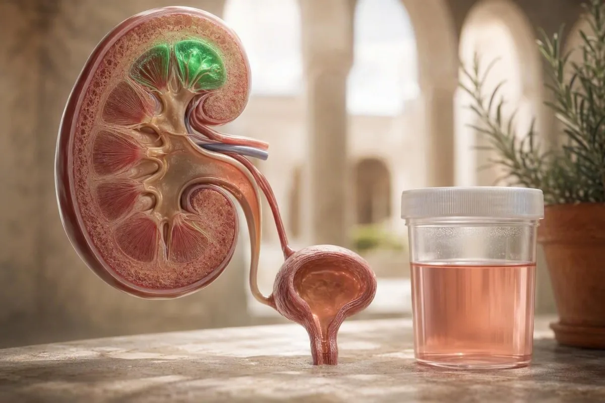
Różowy lub herbaciany kolor próbki moczu po długim biegu jest często tymczasowym krwiomoczem wysiłkowym, ale widoczny kolor nigdy nie powinien być automatycznie ignorowany. Czas, wyniki mikroskopowe, objawy i powtórzone badanie właściwie odpoczętej próbki opowiadają prawdziwą historię.


- Krwinkomocz wysiłkowy zazwyczaj ustępuje w ciągu 24–72 godzin po intensywnym biegu; utrzymywanie się objawów po 72 godzinach wymaga konsultacji lekarskiej.
- Próg mikroskopowy wynoszący 3 lub więcej czerwonych krwinek na pole widzenia pod dużym powiększeniem w prawidłowo pobranej próbce moczu spełnia definicję mikrohematurii wg AUA.
- Niezgodność testu paskowego ma znaczenie: pozytywny wynik testu na krew przy zero do dwóch czerwonych krwinek na pole widzenia pod dużym powiększeniem sugeruje mioglobinę lub wolną hemoglobinę, a nie krwawienie z dróg moczowych.
- Powtórzenie w odpowiednim czasie jest praktyczne: unikać intensywnych ćwiczeń przez 48–72 godzin, nawadniać się normalnie, a następnie dostarczyć próbkę moczu ze środkowego strumienia.
- Ocena tego samego dnia jest rozsądne w przypadku skrzepów, niemożności oddania moczu, gorączki, silnego bólu boku po jednej stronie lub ciemnego moczu z wyraźnym bólem mięśni.
- Wzorzec ostrzegawczy nerek to krew z białkiem, wałeczki krwinkowe, wysokie ciśnienie krwi lub spadający eGFR — nie jest to pojedynczy wynik testu paskowego po zawodach.
- Wskazówka dotycząca rabdomiolizy to nieproporcjonalnie ciemny mocz z ciężkimi objawami mięśniowymi i wynikiem kinazy kreatynowej często przekraczającym 5-krotność górnej granicy laboratorium.
- Kontekst ryzyka nowotworowego zmienia próg diagnostyczny: wiek, narażenie na palenie tytoniu, wcześniejsze napromienianie miednicy i utrzymujące się widoczne krwiomocz mają znaczenie.
Dlaczego bieg może sprawić, że mocz będzie wyglądał na różowy
Krwiomocz biegacza to zazwyczaj krótkotrwałe krwawienie z czerwonych krwinek do moczu po intensywnym wysiłku fizycznym, często ustępujące w ciągu 24–72 godzin odpoczynku. Niemniej jednak, widoczny różowy, czerwony lub przypominający kolor coli mocz zasługuje na prawidłowo pobraną powtórną próbkę, zamiast pochopnego założenia, że bieganie jest przyczyną.
W praktyce klinicznej uspokajający wzorzec jest prosty: zmiana koloru rozpoczyna się po nietypowo długim, szybkim lub zbieżnym biegu; nie ma gorączki, kolkowego bólu boku ani pieczenia podczas oddawania moczu; a mikroskopia normalizuje się po odpoczynku. Kantesti to analizator AI do badań krwi które mogą umieścić powiązane markery nerkowe — kreatyninę, eGFR, elektrolity i kinazę kreatynową — obok historii ćwiczeń, zamiast traktować jeden wynik po wyścigu jako diagnozę.
Proponowane mechanizmy to mechaniczny kontakt ze ścianą pęcherza, tymczasowe przesunięcia przepływu krwi przez nerki, zagęszczenie związane z odwodnieniem i sporadycznie rozpad czerwonych krwinek z powodu powtarzających się uderzeń stopy. Dowody na każdy mechanizm są szczerze mówiąc mieszane, ponieważ większość badań jest mała i dotyczy różnych dystansów wyścigów. W moich 15 latach praktyki klinicznej historia nowego wysiłku jest ważniejsza niż sam kolor; pierwszy ultramaraton i znany mi bieg na 5 km nie niosą takiego samego prawdopodobieństwa przed badaniem.
Widoczna krew w moczu po wysiłku nie jest synonimem łagodnego krwiomoczu biegacza. A szerszy przewodnik po czerwonych flagach krwiomoczu jest przydatny, ponieważ kamienie nerkowe, infekcje, choroby kłębuszkowe i nowotwory dróg moczowych mogą przypadkowo objawić się po biegu.
Praktyczna definicja
Krwiomocz wywołany wysiłkiem oznacza pojawienie się czerwonych krwinek w moczu wkrótce po wysiłku i ich zanik w badanej po odpoczynku próbce. Sam pozytywny wynik testu paskowego nie potwierdza tej diagnozy, ponieważ odczynnik wykrywa barwnik hemu z nieuszkodzonych czerwonych krwinek, wolnej hemoglobiny lub mioglobiny.
Czy to krwiomocz, hemoglobina, czy mioglobina?
Dodatni wynik testu paskowego moczu na obecność krwi może odzwierciedlać obecność czerwonych krwinek, hemoglobiny lub mioglobiny, a mikroskopia je rozróżnia. Różowy mocz z wieloma nieuszkodzonymi czerwonymi krwinkami przemawia za krwiomoczem; ciemnobrązowy mocz z niewielką liczbą czerwonych krwinek lub bez nich budzi obawy o pigmentację spowodowaną urazem mięśni lub hemolizą.
Mikroskopia moczu pokazująca 3 lub więcej czerwonych krwinek w polu widzenia o dużej powiększeniu potwierdza mikrohematurię, gdy próbka jest pobrana czysto. Natomiast silnie dodatni test paskowy z 0–2 czerwonymi krwinkami w polu widzenia o dużej powiększeniu powinien skłonić lekarza do rozważenia mioglobinurii, szczególnie po ekspozycji na ciepło, obciążeniu mięśni typu zgniatającego lub ciężkim opóźnionym bólu mięśni. Brązowy lub cola kolor jest bardziej typowy dla zagęszczonego barwnika niż niewielkiej ilości krwawienia z pęcherza.
Mioglobina ma znaczenie, ponieważ może towarzyszyć rabdomiolizie i ostremu uszkodzeniu nerek. Stężenie kinazy kreatynowej powyżej 5-krotności górnej granicy normy laboratoryjnej jest powszechnie stosowanym progiem biochemicznym dla rabdomiolizy, chociaż objawy, potas, kreatynina i wydalanie moczu określają pilność. Zobacz nasze praktyczne wyjaśnienie niebezpiecznie wysoka kinaza kreatynowa dla wzorców, które nie mogą czekać na noc.
Barwniki spożywcze, buraki, jeżyny, ryfampicyna, fenazopirydyna i odwodnienie mogą również zabarwić mocz bez czerwonych krwinek. diagnostyka ciemnego moczu pomaga uniknąć powszechnego błędu zakładania, że każda czerwona próbka oznacza krew.
Dlaczego rozróżnienie zmienia sposób leczenia
Krwinkomocz skierowuje uwagę na drogi moczowe, podczas gdy mioglobinuria skierowuje uwagę na mięśnie szkieletowe i ochronę nerek. Oba mogą wystąpić po wysiłkach wytrzymałościowych, ale tylko mikroskopia i ukierunkowana analiza krwi mogą je wiarygodnie rozróżnić.
Jak czytać wynik analizy moczu po biegu
Najbardziej pouczające badanie moczu po biegu łączy test paskowy, mikroskopię, pomiar białka i ciężar właściwy. Krew na teście paskowym z przejściowymi czerwonymi krwinkami i bez białka często stanowi niskie ryzyko; krew z białkiem lub wałeczkami wymaga bardziej celowej oceny nerek.
Wynik krwi na teście paskowym jest zgłaszany jako ujemny, śladowy, 1+, 2+ lub 3+, ale jest to sygnał przesiewowy, a nie liczba komórek. AUA definiuje mikrohematurię jako obecność ponad 3 czerwonych krwinek na pole widzenia w wysokim powiększeniu w badaniu mikroskopowym, a nie sam wynik testu paskowego (Barocas et al., 2020). To rozróżnienie pozwala uniknąć niepotrzebnego badania w kierunku raka u biegaczy po fałszywie dodatnim wyniku przesiewowego.
Ciężar właściwy powyżej 1.030 w wielu laboratoriach oznacza zagęszczony mocz i może sprawić, że kolor będzie wyglądał bardziej alarmująco, ale nie wyjaśnia potwierdzonych czerwonych krwinek. Białko w ilości 1+ lub większej powinno być kwantyfikowane za pomocą stosunku albuminy do kreatyniny w moczu lub stosunku białka do kreatyniny; istnieje przejściowa białkomocz wysiłkowy, ale utrzymująca się albuminuria zmienia różnicowanie w kierunku choroby kłębuszkowej. Nasz kompletny przewodnik po analizie moczu wyjaśnia, dlaczego te markery nie powinny być łączone w jeden wynik.
Kantesti AI interpretuje kreatyninę i eGFR wraz z wynikami badania moczu, czasem wysiłku i poprzednimi wartościami; pojedynczy lekko podwyższony poziom kreatyniny po gorącym wyścigu nie jest równoważny z przewlekłą chorobą nerek. W praktyce pytam o dystans, teren, temperaturę, stosowanie leków przeciwzapalnych, czas cyklu menstruacyjnego i czy próbka została pobrana w ciągu 2 godzin od zakończenia.
Kiedy należy powtórzyć badanie moczu po biegu?
Powtórne badanie moczu 48–72 godziny po ostatnim intensywnym biegu jest rozsądnym domyślnym postępowaniem w przypadku podejrzewanej krwiomoczu biegacza. Użyj próbki porannej lub próbki ze środkowego strumienia po dobrym nawodnieniu, ale nie pij litrami wody bezpośrednio przed pobraniem, ponieważ skrajne rozcieńczenie może zaciemnić użyteczne wyniki.
W przypadku rutynowej nieprawidłowości po zawodach zazwyczaj zalecam brak interwałów, podbiegów, ciężkiego podnoszenia ciężarów lub wyścigów przez 48 godzin; po maratonie, ultramaratonie, udarze cieplnym lub nietypowym bólu, 72 godziny jest bardziej uzasadnione. Zwykłe picie do uczucia pragnienia jest wystarczające. Nadmierne nawodnienie może obniżyć ciężar właściwy poniżej 1,005 i czasami utrudnić interpretację granicznych wyników.
Jeśli mikroskopia pozostaje dodatnia po 72 godzinach, należy umówić się na wizytę u lekarza pierwszego kontaktu w ciągu kilku dni, zamiast wielokrotnie powtarzać badania po treningach. Uporczywe wyniki wymagają pomiaru ciśnienia krwi, oceny stężenia kreatyniny w surowicy/eGFR, stosunku albumin do kreatyniny w moczu oraz rozważenia hodowli lub obrazowania w zależności od ryzyka. Związek między wysiłkiem, nawodnieniem a tymczasowo obniżonym szacunkiem filtracji jest omówiony w naszym przewodniku po eGFR po wysiłku.
52-letni amatorski maratończyk, którego widziałem, miał 18 czerwonych krwinek na pole widzenia pod dużym powiększeniem wieczorem po gorącym biegu, ale 0–1 czerwonych krwinek po 72 godzinach odpoczynku; jego kreatynina wróciła z 1,28 mg/dL do 0,96 mg/dL. Wynik był uspokajający, ale ten sam plan nie wystarczyłby dla 52-letniego palacza z powtarzającym się widocznym zabarwieniem moczu.
Unikaj badania na linii mety, jeśli to możliwe.
Badanie natychmiast po wysiłku maksymalizuje szansę na wykrycie tymczasowych czerwonych krwinek, zagęszczonego moczu i białka. Może to być klinicznie przydatne w przypadku podejrzenia rabdomiolizy, ale jest to zły moment na ustalenie poziomu wyjściowego biegacza.
Które wzorce krwiomoczu po biegu są zazwyczaj uspokajające?
Samoograniczający się epizod po intensywnym bieganiu jest bardziej uspokajający, gdy całkowicie ustępuje, nie towarzyszy mu ból ani objawy ogólnoustrojowe, i nie ma białkomoczu ani zmian w funkcji nerek. Uspokajający nie oznacza zignorowany; oznacza potwierdzony próbką pobraną po odpoczynku.
Fenotyp o niższym ryzyku to młodszy lub w średnim wieku biegacz z wyraźnym wyzwalaczem wysiłkowym, prawidłowym ciśnieniem krwi, brakiem historii palenia i prawidłową mikroskopią po odpoczynku. W takich okolicznościach pojedynczy przejściowy wynik 3–10 RBC/HPF po biegu często ustępuje bez leczenia. Nadal bym to dokumentował, ponieważ nawrót po każdym długim biegu zasługuje na dokładniejszą ocenę niż jednorazowe zdarzenie.
Prawidłowy stosunek albumin do kreatyniny w moczu jest zazwyczaj poniżej 30 mg/g (poniżej 3 mg/mmol), chociaż laboratoria i kraje podają jednostki w różny sposób. Krew bez albumin, wałeczków lub dysmorficznych czerwonych krwinek wskazuje na brak przesączania kłębuszkowego; nie dowodzi to pochodzenia z pęcherza moczowego, ale kieruje kolejne pytanie. Nasze wyjaśnienie białka w moczu jasno to obejmuje.
Trauma pęcherza jest bardziej prawdopodobna, gdy mocz jest bardzo skoncentrowany, a biegacz kończy z prawie pustym pęcherzem. Niektórzy klinicyści sportowi sugerują oddawanie moczu przed długim biegiem i regularne picie, ale są to porady profilaktyczne z ograniczonymi dowodami z badań – nie zastępują one oceny, gdy zabarwienie utrzymuje się.
Prawidłowy wynik zawiera sygnaturę czasową.
Negatywny test 3 dni po wyścigu uspokaja w odniesieniu do tego epizodu, ale nie jest gwarancją do końca życia. Nowa widoczna krwiomocz kilka miesięcy później powinna być oceniona na podstawie własnych zasług, szczególnie po 40. roku życia lub po narażeniu na dym tytoniowy.
Ostrzeżenie wskazujące, że krwiomocz biegacza jest mniej prawdopodobny
Krwiomocz wymaga pilnej oceny, gdy utrzymuje się po 72 godzinach, nawraca bez intensywnego wysiłku lub pojawia się z bólem, gorączką, skrzepami, białkiem lub zmianą funkcji nerek. Kombinacja wyników jest bardziej informatywna niż sam dramatycznie wyglądający próbka.
Szukaj terminowej porady medycznej w przypadku widocznego czerwonego moczu, który utrzymuje się przez 24 godziny, nawet jeśli czujesz się dobrze; próg jest niższy dla osób po 40. roku życia, obecnych lub byłych palaczy, lub osób z wcześniejszym napromieniowaniem miednicy. Wytyczne AUA wykorzystują stratyfikację ryzyka opartą na wieku, paleniu, stopniu i utrwalaniu się mikroskopowej krwi oraz wcześniejszym makroskopowym krwiomoczu, zamiast traktować wszystkie mikrohematurie identycznie (Barocas et al., 2020).
Dysmorficzne czerwone krwinki, wałeczki erytrocytarne, znacząca albuminuria, nowe nadciśnienie lub obniżony eGFR sugerują źródło w filtrze nerkowym, a nie wstrząśnięty pęcherz. Wałeczki erytrocytarne nigdy nie są oczekiwaną konsekwencją zwykłego biegania i powinny skłonić do przyspieszonej oceny nerek. Zobacz nasz przegląd wałeczków ziarnistych i moczowych , dlaczego wyniki osadu mają znaczenie.
Thomas Klein, MD, radzi biegaczom, aby nie prowadzili samodzielnej oceny na podstawie samego zabarwienia: infekcja dróg moczowych może powodować pieczenie, pilną potrzebę oddania moczu, ropomocz i czasami krwiomocz, podczas gdy kamienie częściej powodują nagły jednostronny ból i nudności. Posiew jest najbardziej pomocny przed zastosowaniem antybiotyków, gdy występują objawy infekcji.
Kombinacja krew plus białko
Potwierdzony krwiomocz z białkomoczem jest bardziej niepokojący niż izolowany krwiomocz, ponieważ oba mogą pochodzić z filtra kłębuszkowego. Stosunek albuminy do kreatyniny w moczu wynoszący 30 mg/g lub więcej powinien być powtórzony i zinterpretowany wraz z ciśnieniem krwi i funkcją nerek.
Kiedy różowy mocz po wysiłku wymaga opieki tego samego dnia?
Uzyskaj pilną ocenę tego samego dnia w przypadku skrzepów, niemożności oddania moczu, silnego bólu w boku, gorączki, omdleń, zmniejszonej ilości wydalanego moczu lub ciemnego moczu z silnym bólem i osłabieniem mięśni. Te cechy wskazują poza niepowikłanym krwiomoczem spowodowanym wysiłkiem.
Skrzepy mogą blokować przepływ moczu, a niemożność oddania moczu jest problemem pilnym, niezależnie od sprawności fizycznej. Gorączka 38,0°C lub wyższa z bólem w boku może wskazywać na infekcję górnych dróg moczowych; silny kolkowy ból z wymiotami może wskazywać na kamień. Żadna z tych sytuacji nie powinna być zarządzana przez oczekiwanie na powtórne badanie po biegu.
Ciemnobrązowy mocz po wysiłku z wyraźną tkliwością mięśni, osłabieniem, obrzękiem lub zmniejszonym wydalaniem moczu budzi obawy o rabdomiolizę. Początkowe testy zazwyczaj obejmują kinazę kreatynową, kreatyninę, potas, wapń, fosforany i badanie moczu. przewodnik po rabdomiolizie CrossFit jest istotne, ponieważ nieznany wcześniej wysiłek ekscentryczny – a nie samo bieganie – jest częstym czynnikiem wyzwalającym.
Ciężkie odwodnienie może pogorszyć wyniki badań nerek, ale może również współistnieć z rzeczywistym uszkodzeniem. Jeśli odczuwasz zawroty głowy, splątanie, nie możesz utrzymać płynów lub bardzo mało oddajesz moczu przez 8–12 godzin, nie próbuj rozwiązywać problemu domowymi napojami sportowymi.
Ciąża i leki przeciwzakrzepowe
Kobiety w ciąży biegaczki i osoby przyjmujące leki przeciwzakrzepowe powinny niezwłocznie zgłaszać widoczną krwiomoczu. Leki przeciwzakrzepowe mogą nasilać krwawienie, ale nie eliminują potrzeby zidentyfikowania jego źródła; ciąża zmienia również próg badań w kierunku infekcji, kamieni i chorób nerek.
Schorzenia, które bieg może ujawnić, zamiast powodować
Bieganie może ujawnić istniejący problem z drogami moczowymi poprzez zagęszczenie moczu lub skłonienie do wykonania badania, zamiast samoistnie powodować krwiomocz. Kamienie, infekcje, choroby kłębuszków nerkowych, nieprawidłowości strukturalne i nowotwory pozostają częścią diagnostyki różnicowej.
Kamienie nerkowe często powodują jednostronny ból od boku do pachwiny, nudności i obecność czerwonych krwinek w mikroskopii, chociaż występują również kamienie bezbolesne. Infekcje dróg moczowych częściej powodują pieczenie, częstomocz, nagłe parcie i białe krwinki; ujemny wynik na azotyny nie wyklucza ich. Nasz przewodnik po esterazie leukocytów przy ujemnych azotynach wyjaśnia ten powszechny, mieszany wzorzec.
Kłębuszkowe zapalenie nerek może pojawić się po infekcji górnych dróg oddechowych z moczem koloru coli, obrzękami, podwyższonym ciśnieniem krwi, białkomoczem i dysmorficznymi czerwonymi krwinkami. W wytycznych KDIGO 2024 dotyczących CKD, utrwalony albuminuria i obniżony eGFR są traktowane jako uzupełniające markery ryzyka chorób nerek, a nie jako zamienne pomiary (KDIGO, 2024).
Kantesti to narzędzie do analizy badań krwi oparte na AI który pomaga użytkownikom rozpoznać wzorzec w zakresie kreatyniny, eGFR, albumin, elektrolitów i CBC, ale nie może zdiagnozować anatomicznego źródła krwiomoczu. Obrazowanie, mikroskopia, posiew i badanie lekarskie nadal wykonują decydującą pracę.
Rak jest rzadki u młodych biegaczy, ale nie niemożliwy u dorosłych.
Widoczny krwiomocz wymaga oceny, nawet gdy wysiłek fizyczny wydaje się oczywistym wyjaśnieniem, zwłaszcza u osób z narażeniem na palenie tytoniu lub utrzymującymi się objawami. Ryzyko wzrasta wraz z wiekiem i skumulowanym narażeniem na tytoń, dlatego identyczne wyniki badania moczu nie prowadzą do identycznych planów badań.
Kto potrzebuje dalszej opieki urologicznej lub nefrologicznej?
Utrzymujący się krwiomocz po odpoczynku i ponownym pobraniu próbki powinien być oceniony pod kątem ryzyka urologicznego, podczas gdy krwiomocz z białkomoczem, wałeczkami, nadciśnieniem lub upośledzonym eGFR wskazuje na nefrologię. Wiele osób potrzebuje jedynie powtarzanych badań prowadzonych przez lekarza pierwszego kontaktu; skierowanie zależy od wzorca.
Ocena urologiczna jest szczególnie wskazana w przypadku nawracającego widocznego krwiomoczu, utrzymującego się mikroskopowego krwiomoczu, historii palenia tytoniu, narażenia zawodowego na aminy aromatyczne, wcześniejszego napromieniowania miednicy lub objawów ze strony układu moczowego bez infekcji. W zależności od ryzyka, lekarze mogą zastosować ultrasonografię nerek i pęcherza, urografię TK i cystoskopię. Pojedynczy śladowy wynik testu paskowego po wyścigu u 24-latka to zupełnie inna sytuacja niż nawracający czerwony mocz u 58-letniego byłego palacza.
Przegląd nefrologiczny staje się bardziej prawdopodobny przy stosunku albuminy do kreatyniny w moczu ≥30 mg/g, eGFR poniżej 60 ml/min/1,73 m² trwającym 3 miesiące lub dłużej, obecności wałeczków erytrocytarnych lub trudnego do kontrolowania ciśnienia krwi. Te progi są ważne, ponieważ przewlekła choroba nerek jest definiowana zarówno przez czas trwania, jak i pojedynczy pomiar. Przeczytaj nasz przewodnika po stadiach przewlekłej choroby nerek w celu poznania praktycznych kategorii.
Usługa interpretacji badań laboratoryjnych firmy Kantesti oparta na sztucznej inteligencji może sygnalizować powtarzalny wzorzec krwi i białka do omówienia, szczególnie gdy użytkownicy przesyłają wyniki z różnych laboratoriów. Powinna ona wspierać, a nie zastępować, lekarza decydującego o potrzebie wykonania badań obrazowych dróg moczowych lub badania specjalistycznego.
Nie pozwól, aby antykoagulacja zakończyła rozmowę
Stosowanie leków przeciwzakrzepowych zmienia nasilenie krwawienia, ale nie ustala przyczyny krwiomoczu. Moim zdaniem przypisywanie nawracającego krwawienia z dróg moczowych do apiksabanu, warfaryny lub aspiryny bez przeprowadzenia diagnostyki może opóźnić rozpoznanie kamieni, infekcji lub chorób strukturalnych.
Które badania krwi pomagają po krwiomoczu związanym z wysiłkiem?
Przydatne badania krwi zależą od objawów: kreatynina, eGFR, elektrolity i kinaza kreatynowa oceniają obciążenie nerek i mięśni, podczas gdy CBC ocenia niedokrwistość lub wskazówki dotyczące infekcji. Rutynowe szerokie panele są mniej pomocne niż ukierunkowane testy po precyzyjnym wywiadzie i badaniu moczu.
Kreatynina może przejściowo wzrosnąć po intensywnym wysiłku z powodu metabolizmu mięśni, zmniejszonego przepływu krwi przez nerki i odwodnienia. Równania eGFR są mniej wiarygodne podczas szybkiej zmiany kreatyniny, dlatego powtórna wartość po 48–72 godziny zwykłego powrotu do zdrowia jest często bardziej znacząca niż wynik z linii mety. Nasz przewodnik po ponownym sprawdzaniu kreatyniny po wysiłku dostarcza kontekstu dla tego częstego czynnika wywołującego niepokój.
Kinaza kreatynowa ma szeroki zakres po wysiłku: wytrenowani sportowcy mogą wykazywać podwyższenie poziomu po wyścigu bez uszkodzenia nerek, podczas gdy wartości wielokrotnie przekraczające lokalny górny limit, w połączeniu z objawami, wymagają pilnej oceny. Potas powyżej 6,0 mmol/L jest potencjalnie niebezpieczny i wymaga pilnej oceny klinicznej, zwłaszcza jeśli funkcja nerek jest upośledzona lub podejrzewa się uszkodzenie mięśni.
CBC jest najbardziej przydatne, gdy występuje zmęczenie, bladość, obfite krwawienia miesiączkowe, nawracający krwiomocz lub podejrzenie choroby układowej. Hemoglobina może spaść po wysiłku wytrzymałościowym z powodu rozszerzenia objętości osocza, utraty żelaza lub hemolizy; ferrytyna powinna być interpretowana z dala od ostrego stanu zapalnego i intensywnych treningów. Biegacze wytrzymałościowi mogą uznać nasz przegląd badań krwi u sportowców za pomocny w ustaleniu rozsądnej podstawy.
Nie diagnozuj choroby nerek na podstawie jednego panelu z tygodnia wyścigu
Wzrost kreatyniny o 0,3 mg/dl lub więcej może spełniać kryterium ostrego uszkodzenia nerek w odpowiednim przedziale czasowym, ale kontekst i powtórne badania są decydujące po wyścigu. Niska produkcja moczu, wzrost poziomu potasu i utrzymujące się podwyższenie stężenia mają większe znaczenie niż pojedynczy, graniczny wynik.
Jak pobrać powtórzoną próbkę moczu, której lekarze mogą zaufać
Czysta próbka moczu ze środkowego strumienia po 48–72 godzinach bez forsownych ćwiczeń jest najlepszym sposobem na sprawdzenie, czy krwiomocz ustąpił. Błędy w pobieraniu, zanieczyszczenie menstruacyjne i stare próbki mogą dawać mylące wyniki.
Umyj ręce, zacznij oddawać mocz do toalety, a następnie zbierz środkową porcję bez dotykania wnętrza pojemnika. Dostarcz próbkę niezwłocznie – najlepiej w ciągu 2 godzin— lub postępuj zgodnie z instrukcjami laboratorium dotyczącymi przechowywania w lodówce. Długotrwałe przechowywanie zmienia wygląd komórek i może zmniejszyć wiarygodność mikroskopii.
W miarę możliwości unikaj badań podczas aktywnej menstruacji; jeśli badanie nie może czekać, poinformuj lekarza i laboratorium. Zanieczyszczenie przez wydzielinę z pochwy może powodować pojawienie się czerwonych krwinek i białka, podczas gdy płaskie komórki nabłonkowe często sygnalizują źle pobraną próbkę. Nasz przewodnik po komórkach nabłonkowych w moczu wyjaśnia, dlaczego jest to niebanalna uwaga laboratoryjna.
Thomas Klein, MD, zaleca zapisywanie czterech szczegółów wraz z próbką: ostatniego intensywnego wysiłku, czasu trwania wysiłku, temperatury otoczenia i wszystkich leków przyjmowanych w ciągu ostatnich 72 godzin. Wynik bez informacji o czasie jest znacznie trudniejszy do zinterpretowania – zwłaszcza gdy mocz jest pobierany po podwójnym treningu.
Testy z próbki porannej a dziennej
Próbka poranna jest skoncentrowana i dobra do wykrywania drobnych nieprawidłowości, ale prawidłowo nawodniona próbka pobrana w ciągu dnia ze środkowego strumienia jest dopuszczalna, jeśli pobranie jest czystsze. Konsekwencja jest ważniejsza w przypadku porównań seryjnych: należy stosować mniej więcej te same warunki przy każdym powtórzeniu.
Nawodnienie, leki przeciwbólowe i suplementy, które zmieniają obraz
Odwodnienie może zagęścić mocz i zwiększyć obciążenie nerek, podczas gdy NLPZ mogą zmniejszyć przepływ krwi do nerek podczas ćwiczeń wytrzymałościowych. Żaden z tych czynników nie dowodzi przyczyny krwiomoczu, ale oba wpływają na ostrożność, z jaką lekarze interpretują wynik po biegu.
Dla większości biegaczy picie do momentu odczucia pragnienia i stopniowe uzupełnianie strat jest bezpieczniejsze niż rygorystyczne wymuszanie płynów. Bardzo niski poziom sodu wynikający z nadmiernego spożycia wody może być niebezpieczny; poziom sodu w surowicy poniżej 125 mmol/L z bólem głowy, wymiotami, dezorientacją lub drgawkami jest stanem nagłym. Kolor moczu jest zgrubnym wskaźnikiem nawodnienia, a nie precyzyjnym miernikiem.
Ibuprofen, naproksen i inne NLPZ mogą zmniejszać przepływ krwi do nerek zależny od prostaglandyn, zwłaszcza podczas stresu cieplnego, wymiotów lub długotrwałych wysiłków. Radzę pacjentom unikać rutynowego profilaktycznego stosowania NLPZ przed maratonami, chyba że ich lekarz prowadzący podał wyraźny powód. Kontekst leków jest omawiany w naszym artykule o bezpieczeństwie trendów laboratoryjnych.
Duże dawki witaminy C mogą zakłócać niektóre reakcje pasków do badania moczu, a suplementy reklamowane jako poprawiające wydajność czasami zawierają niewymienione stymulanty lub diuretyki. Przynieś opakowania lub wyraźne zdjęcia na wizytę; jest to często bardziej pomocne niż próba zapamiętania listy składników markowego proszku. Nasz poradnik dotyczący bezpieczeństwa suplementów dla sportowców oferuje rozsądną listę kontrolną.
Nawodnienie nie jest leczeniem uporczywej krwiomoczu
Woda może skorygować zagęszczenie, ale nie leczy kamienia, infekcji, procesu kłębuszkowego ani strukturalnego źródła w drogach moczowych. Jeśli czerwone krwinki pozostają w próbce pobranej po odpoczynku i w prawidłowych warunkach, dalsze postępowanie powinno opierać się na schemacie, a nie na kolejnym eksperymencie z nawodnieniem.
Czy można biegać po krwiomoczu wysiłkowym?
Większość biegaczy może stopniowo wrócić do treningów, gdy kolor moczu wróci do normy, objawy ustąpią, a próbka pobrana po odpoczynku będzie uspokajająca. Nie wracaj do ciężkich treningów, gdy mocz jest nadal widocznie czerwony, wyniki badań nerek są nieprawidłowe, lub objawy mięśniowe sugerują rabdomiolizę.
Po niepowikłanym pojedynczym epizodzie zazwyczaj sugeruję lekki bieg przez kilka dni, a następnie stopniowe odbudowywanie objętości i intensywności w ciągu 7–14 dni. Biegacz powinien przerwać trening i szukać pomocy lekarskiej, jeśli kolor moczu powróci, pojawi się ból lub zmniejszy się objętość wydalanego moczu. Jest to pragmatyczne podejście kliniczne, a nie poparta badaniami recepta.
Powtarzający się krwiomocz po tym samym dystansie może uzasadniać praktyczne eksperymenty: unikanie wyścigu w stanie odwodnienia, oddawanie moczu przed długim biegiem, wolniejsze wprowadzanie treningu zbiegowego, a także przegląd dopasowania butów lub terenu. Prowadź dziennik tempa, ciepła, czasu trwania, koloru moczu i wyników badań. przewodnikiem laboratoryjnym dla biegacza maratońskiego może pomóc odróżnić markery regeneracji od powodów do przerwania treningu.
Nie ćwicz przez niewyjaśnioną widoczną krwiomoczu tylko dlatego, że masz zaplanowane wydarzenie. Sportowcy często doskonale tolerują dyskomfort; trudniejszą umiejętnością jest rozpoznanie, że wytrwałość jest informacją diagnostyczną, a nie testem wytrzymałości.
Zbuduj prawdziwy punkt wyjścia
Analiza moczu i panel nerkowy wykonane co najmniej 72 godziny po intensywnym treningu mogą znacznie ułatwić interpretację przyszłych wyników po zawodach. Jest to szczególnie przydatne dla ultrabiegaczy, osób z jedną nerką lub sportowców przyjmujących leki wpływające na perfuzję nerek.
Co powiedzieć swojemu lekarzowi o krwi w moczu po wysiłku
Najszybszą drogą do użytecznej oceny krwiomoczu jest zwięzły harmonogram: kolor moczu, dawka wysiłku, objawy, leki i czas powtórnego badania. Przynieś rzeczywiste wyniki laboratoryjne, a nie tylko informację, że próbka była “dodatnia pod względem obecności krwi”.”
Określ, czy mocz był różowy, jasnoczerwony, barwy herbaty, czy zawierał skrzepy; czy kolor pojawił się przed, w trakcie, czy po wysiłku; oraz czy zniknął po pierwszym oddaniu moczu. Uwzględnij pieczenie, parcie, gorączkę, ból w boku, nudności, obrzęki i zmniejszone oddawanie moczu. Szczegółowy odnośnik koloru moczu może pomóc dokładnie opisać obserwację bez nadmiernej interpretacji.
Przynieś poprzednie analizy moczu, wartości kreatyniny/eGFR, odczyty ciśnienia krwi, historię palenia, historię chorób nerek w rodzinie oraz listę leków/suplementów. Kantesti to platforma do interpretacji biomarkerów AI które mogą organizować powtarzające się wyniki badań krwi i podkreślać znaczące zmiany, ale lekarz musi powiązać te wartości z mikroskopią moczu i badaniem.
Od 3 września 2026 r. zasada kliniczna pozostaje prosta: wysiłek fizyczny jest wiarygodnym wyjaśnieniem dopiero po ustąpieniu wyniku i braku konkurencyjnych wskazówek. Jeśli masz wątpliwości, zapytaj, czy potrzebujesz mikroskopii, hodowli, stosunku albuminy do kreatyniny, powtórnej kreatyniny, obrazowania czy skierowania – a nie tylko kolejnego testu paskowego.
Pytania warte zadania
Zapytaj, czy widziano nienaruszone czerwone krwinki, czy obecne było białko lub wałeczki, i kiedy powinno nastąpić powtórne badanie po odpoczynku. Te trzy odpowiedzi zazwyczaj wyjaśniają, czy następnym krokiem jest uważne monitorowanie, badanie nerek, badanie infekcji czy ocena urologiczna.
Rozsądny plan działania w przypadku krwiomoczu biegacza
W przypadku jednego bezbolesnego epizodu po intensywnym bieganiu należy odpocząć od forsownych ćwiczeń przez 48–72 godziny i powtórzyć analizę moczu z czystej próbki; szukać wcześniejszej pomocy w przypadku sygnałów ostrzegawczych lub utrzymującego się widocznego zabarwienia. Celem nie jest ani panika, ani lekceważenie – celem jest udowodnienie, że wynik ustępuje.
Krok pierwszy: przerwij intensywny trening, pij normalnie i zanotuj czas i objawy. Krok drugi: powtórz mikroskopię po 48–72 godziny. Krok trzeci: jeśli krew się utrzymuje, lub jeśli pojawia się białko, wałeczki, ból, gorączka, skrzepy, zmniejszona ilość oddawanego moczu lub nieprawidłowe markery nerkowe, niezwłocznie umów się na wizytę lekarską. Ta sekwencja zapobiega zarówno pominięciu choroby, jak i niepotrzebnemu obrazowaniu po jednorazowej próbce z linii mety.
Kiedy nasz zespół kliniczny przegląda przesłane dane laboratoryjne, priorytetowo traktujemy wzorce: rosnąca kreatynina w połączeniu z wysoką CK i niską ilością oddawanego moczu jest bardziej pilna niż stabilna kreatynina z przejściowym wynikiem testu moczu na pasku. Metodologia i zabezpieczenia Kantesti są opisane w naszym informacje o walidacji klinicznej, a nasz nadzór lekarski jest szczegółowo opisany w Rada doradcza ds. medycznych.
Większość pacjentów uważa, że powtórzone badanie w ustalonym terminie przekształca alarmującą zmianę koloru w jasny plan. Ale jeśli Twój instynkt podpowiada Ci, że coś jest nie tak – zwłaszcza jeśli mocz jest nadal czerwony jutro – zaufaj temu sygnałowi i zgłoś się na ocenę.
Zasada jednej sentencji
Różowy mocz raz po intensywnym biegu może być przejściowy; różowy mocz, który się utrzymuje, nawraca lub pojawia się z objawami, jest krwiomoczem do czasu właściwej oceny.
Często zadawane pytania
Jak długo trwa krwiomocz wysiłkowy?
Krwinkomocz wysiłkowy zazwyczaj ustępuje w ciągu 24–72 godzin po zaprzestaniu intensywnego wysiłku fizycznego. Praktycznym powtórnym badaniem moczu jest pobranie próbki „środkowego strumienia” z czystego oddania moczu, zebranej po 48–72 godzinach bez ciężkiego biegania lub podnoszenia ciężarów. Utrzymujący się widoczny różowy lub czerwony mocz po 24 godzinach, lub krwiomocz mikroskopowy utrzymujący się po 72 godzinach, powinien zostać oceniony przez lekarza. Gorączka, skrzepy, silny ból w boku, zmniejszone wydalanie moczu lub silny ból mięśni uzasadniają ocenę tego samego dnia.
Czy krew w moczu po wysiłku fizycznym jest normalna?
Krew w moczu po wysiłku może być przemijającym skutkiem intensywnego biegania, odwodnienia, uderzenia o ścianę pęcherza lub krótkotrwałych zmian przepływu krwi w nerkach, ale nie jest to automatycznie normalne. Mikroskopowe potwierdzenie 3 lub więcej czerwonych krwinek na pole widzenia w dużym powiększeniu spełnia definicję mikrogematury AUA. Wynik, który znika przy powtórnym badaniu pobranym po odpoczynku, jest uspokajający, podczas gdy utrzymująca się krew, białko, wałeczki, ból lub objawy ze strony układu moczowego wymagają dalszej oceny. Widoczna zmiana zabarwienia moczu powinna być zawsze zgłaszana, szczególnie u dorosłych z narażeniem na palenie tytoniu lub nawracającymi epizodami.
Czy odwodnienie może powodować różowy mocz po bieganiu?
Odwodnienie może powodować ciemniejszy kolor moczu i może nasilić stwierdzenie krwiomoczu po wysiłku poprzez zagęszczenie zawartości moczu, jednak samo odwodnienie nie wyjaśnia w sposób wiarygodny potwierdzonych czerwonych krwinek. Ciężar właściwy moczu powyżej 1,030 często wskazuje na zagęszczony mocz w wielu laboratoriach. Pij normalnie do momentu odczucia pragnienia i powtórz próbkę czystego strumienia po 48–72 godzinach odpoczynku, zamiast próbować wypłukać wynik nadmierną ilością wody. Silne pragnienie, zawroty głowy, bardzo mała ilość wydalanego moczu lub dezorientacja wymagają natychmiastowej konsultacji lekarskiej.
Jaka jest różnica między krwiomoczem a mioglobinurią po wysiłku fizycznym?
Hematuria oznacza obecność nieuszkodzonych czerwonych krwinek w moczu, podczas gdy mioglobinuria odzwierciedla barwnik mięśniowy uwalniany po urazie mięśni. Pasek testowy może być dodatni pod względem krwi w obu sytuacjach, dlatego mikroskopia jest rozstrzygająca: mioglobinuria często daje dodatni wynik paska testowego przy 0–2 krwinkach czerwonych na pole widzenia w dużym powiększeniu. Silny ból mięśni, osłabienie, obrzęk, ciemny, cola-podobny mocz lub wartość kinazy kreatynowej powyżej 5-krotności górnej granicy laboratoryjnej budzi obawy o rabdomiolizę. Taki obraz kliniczny wymaga pilnej oceny, ponieważ stężenie potasu i czynność nerek mogą szybko ulec zmianie.
Czy powinnam przerwać bieganie, jeśli mam krwiomocz wysiłkowy?
Zaprzestań intensywnych biegów do czasu normalizacji zabarwienia moczu i ukończenia powtórzonego testu sprawnościowego, zwykle po 48–72 godzinach. Jeśli powtórny próbka jest prawidłowa i nie występują bóle, gorączka, skrzepy, białkomocz ani zaburzenia czynności nerek, większość biegaczy może stopniowo wracać do treningów w ciągu około 7–14 dni. Nie wznawiaj intensywnych treningów, gdy mocz pozostaje widocznie czerwony lub ciemny, zmniejsza się jego ilość lub występują silne objawy mięśniowe. Nawracające epizody po podobnych treningach wymagają kontroli lekarskiej, nawet jeśli każdy epizod ustępuje.
Kiedy krwiomocz wymaga konsultacji urologa?
Uporczywa mikroskopowa hematuria po odpoczynku i powtórnym badaniu, nawracająca makroskopowa hematuria lub hematuria u osoby z historią palenia tytoniu, wcześniejszym napromienianiem miednicy lub objawami ze strony układu moczowego często wymaga oceny ryzyka urologicznego. Urolodzy mogą rozważyć ultrasonografię, urografię tomografii komputerowej i cystoskopię w zależności od wieku i ogólnego ryzyka, zamiast stosować jedno badanie dla wszystkich. Hematuria z albuminurią 30 mg/g lub więcej, wałeczkami czerwonokrwinkowymi, nadciśnieniem tętniczym lub eGFR poniżej 60 mL/min/1,73 m² silniej wskazuje na nefrologię. Właściwa specjalność zależy od pełnego obrazu badań moczu i krwi.
Uzyskaj analizę wyników badań krwi zasilaną przez AI już dziś
Dołącz do ponad 2 milionów użytkowników na całym świecie, którzy ufają Kantesti w zakresie natychmiastowej, dokładnej analizy badań laboratoryjnych. Prześlij swoje wyniki badań krwi i otrzymaj kompleksową interpretację biomarkerów 15,000+ w kilka sekund.
📚 Publikacje badawcze z odniesieniami
Klein, T., Mitchell, S., & Weber, H. (2026). Wielojęzyczne wspomaganie klinicznego podejmowania decyzji oparte na sztucznej inteligencji we wczesnym etapie triage'u hantawirusów: projekt, walidacja inżynieryjna i wdrożenie w świecie rzeczywistym dla 50 000 interpretacji wyników badań krwi..Kantesti AI Medical Research.
Klein, T., Mitchell, S., & Weber, H. (2026). Wstępnie zarejestrowany, oparty na rubrykach automatyczny techniczny benchmark interpretacji wyników badań krwi przez silnik Kantesti na 100 000 syntetycznych przypadków testowych.Kantesti AI Medical Research.
📖 Zewnętrzne medyczne źródła odniesienia
⚕️ Zastrzeżenie medyczne
Ten artykuł ma wyłącznie charakter edukacyjny i nie stanowi porady medycznej. Zawsze konsultuj decyzje dotyczące diagnozy i leczenia z wykwalifikowanym pracownikiem ochrony zdrowia.
Sygnały zaufania E-E-A-T
Doświadczenie
Kliniczna weryfikacja procesów interpretacji przez lekarza.
Ekspertyza
Medycyna laboratoryjna skupiona na tym, jak zachowują się biomarkery w kontekście klinicznym.
Autorytatywność
Napisane przez dr. Thomasa Kleina, z recenzją dr Sarah Mitchell i prof. dr. Hansa Webera.
Solidność
Interpretacja oparta na dowodach, z jasnymi ścieżkami dalszego postępowania, aby ograniczyć alarm.