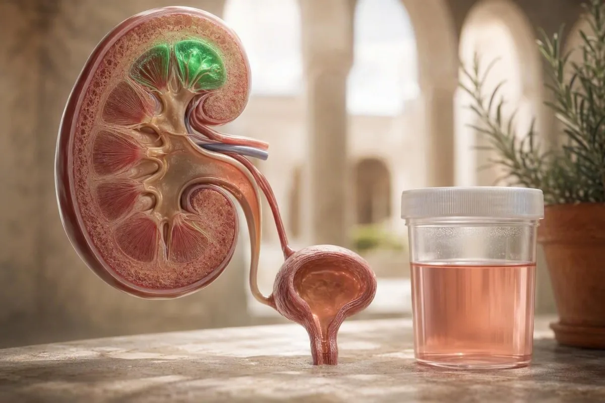
마라톤 완주 후 분홍색 또는 차색 소변은 종종 일시적인 운동 유발 혈뇨이지만, 눈에 보이는 색깔을 자동으로 무시해서는 안 됩니다. 시기, 현미경 검사 결과, 증상, 그리고 충분히 휴식한 후의 재검이 실제 이야기를 들려줍니다.


- 운동 유발성 혈뇨 일반적으로 격렬한 달리기 후 24-72시간 이내에 사라지며, 72시간 이상 지속되면 의학적 검토가 필요합니다.
- 현미경 검사 임계값 적절하게 채취된 소변 샘플에서 고배율 시야당 적혈구 3개 이상은 AUA의 미세 혈뇨 정의를 충족합니다.
- 딥스틱 불일치 중요합니다: 고배율 시야당 적혈구 0~2개에 대해 양성인 혈액 패드는 요로 출혈보다는 미오글로빈 또는 유리 헤모글로빈을 시사합니다.
- 검사 시점 반복 실용적입니다: 48-72시간 동안 격렬한 운동을 피하고, 정상적으로 수분을 섭취한 후, 중간뇨 샘플을 제공합니다.
- 당일 평가 혈전, 소변을 볼 수 없음, 발열, 편측의 심한 옆구리 통증, 또는 심한 근육 통증과 함께 나타나는 콜라 색 소변에 대해서는 합리적입니다.
- 신장 경고 양상 혈액과 단백뇨, 적혈구 원주, 고혈압 또는 eGFR 감소—단순한 경주 후 딥스틱이 아닙니다.
- 횡문근융해증 단서 은 심한 근육 증상과 함께 실험실 상한치의 5배 이상인 크레아틴 키나아제 수치가 나타나는 과도하게 짙은 소변입니다.
- 암 위험 맥락 은 조사 임계값을 변경합니다: 나이, 흡연 노출, 이전 골반 방사선 치료, 지속적인 육안 혈뇨 모두 중요합니다.
달리기가 소변을 분홍색으로 만드는 이유
달리기 선수의 혈뇨는 일반적으로 격렬한 운동 후 짧은 시간 동안 소변으로 적혈구가 새어 나오는 것입니다., 종종 휴식 후 24~72시간 이내에 회복됩니다. 그럼에도 불구하고 분홍색, 붉은색 또는 콜라색 소변은 달리기가 원인이라는 안일한 추측보다는 제대로 채취된 반복 검사를 받을 가치가 있습니다.
임상에서는 안심되는 패턴은 간단합니다: 색상 변화는 비정상적으로 길거나 빠르거나 내리막길 달리기를 한 후에 시작됩니다. 발열, 산통성 옆구리 통증 또는 배뇨 시 작열감이 없습니다. 현미경 검사가 휴식 후 정상화됩니다. Kantesti는 AI 혈액 검사 분석기입니다. 은 진단으로 여기는 대신, 크레아틴, eGFR, 전해질 및 크레아틴 키나아제와 같은 신장 관련 지표를 운동력과 함께 놓고 볼 수 있습니다.
제안된 메커니즘은 기계적 방광 벽 접촉, 일시적인 신장 혈류 변화, 탈수 관련 농축, 그리고 때로는 반복적인 발 충격으로 인한 적혈구 분해입니다. 각각의 메커니즘에 대한 증거는 솔직히 혼합되어 있는데, 대부분의 연구는 규모가 작고 다른 레이스 거리를 사용하기 때문입니다. 제 15년간의 임상 경험에서, 새로운 노력의 병력은 색깔 자체보다 더 중요합니다. 첫 울트라마라톤과 익숙한 5km는 동일한 사전 검사 확률을 갖지 않습니다.
운동 후 소변에서 눈에 띄는 혈액이 반드시 양성 달리기 선수의 혈뇨를 의미하는 것은 아닙니다. 광범위한 혈뇨 위험 신호 안내 는 신장 결석, 감염, 사구체 질환 및 요로암이 우연히 달리기 후에 처음 나타날 수 있으므로 유용합니다.
실용적인 정의
운동 유발성 혈뇨는 운동 직후 소변에서 적혈구가 나타나 휴식 후 반복 검체에서 사라지는 것을 의미합니다. 단지 양성 딥스틱만으로는 이 진단을 확증할 수 없는데, 시약이 온전한 적혈구, 유리 헤모글로빈 또는 미오글로빈에서 헤모색소를 감지하기 때문입니다.
혈뇨인가, 헤모글로빈인가, 아니면 미오글로빈인가?
혈액에 대해 양성 반응을 보이는 소변 딥스틱은 적혈구, 헤모글로빈 또는 미오글로빈을 반영할 수 있으며, 현미경 검사가 이를 구분합니다. 온전한 적혈구가 많은 분홍색 소변은 혈뇨를 뒷받침합니다. 적혈구가 거의 없거나 없는 짙은 갈색 소변은 근육 손상이나 용혈로 인한 색소 침착에 대한 우려를 불러일으킵니다.
소변 현미경 검사에서 고배율 시야당 3개 이상의 적혈구 가 깨끗하게 채취된 검체에서 미세 혈뇨를 확인합니다. 반대로, 고배율 시야당 0-2개의 적혈구가 있는 강한 양성 딥스틱은 임상의가 미오글로빈뇨증을 고려하게 해야 하는데, 특히 열 노출, 압착성 근육 부하 또는 심각한 근육 지연 통증 후에는 더욱 그렇습니다. 갈색 또는 콜라색은 방광 출혈이 적은 양보다 농축된 색소의 특징입니다.
미오글로빈은 횡문근융해증 및 급성 신장 손상을 동반할 수 있으므로 중요합니다. 크레아틴 키나아제 농도가 실험실 정상 상한치의 5배 이상 인 것은 횡문근융해증의 생화학적 임계값으로 흔히 사용되지만, 증상, 칼륨, 크레아틴 및 소변 배출량이 긴급성을 결정합니다. 당사의 실용적인 설명을 참조하십시오. 위험할 정도로 높은 크레아틴 키나아제 야간에 기다릴 필요가 없는 소견에 대하여.
식용 색소, 비트, 블랙베리, 리팜피신, 페나조피리딘, 탈수도 적혈구 없이 소변 색깔을 변화시킬 수 있습니다. A 짙은 소변 감별 모든 붉은색 소변 샘플이 혈액을 나타낸다고 가정하는 일반적인 실수를 피하는 데 도움이 됩니다.
구분이 관리를 바꾸는 이유
적혈구 혈뇨는 비뇨기계에 주의를 집중시키는 반면, 미오글로빈뇨는 골격근 및 신장 보호에 주의를 집중시킵니다. 둘 다 지구력 운동 후에 발생할 수 있지만, 현미경 검사와 표적 혈액 검사만이 이를 신뢰할 수 있게 구별할 수 있습니다.
달리기 후 소변 분석 결과 읽는 법
가장 유익한 달리기 후 소변 검사는 딥스틱, 현미경 검사, 단백질 측정 및 비중을 결합합니다. 일시적인 적혈구와 단백질이 없는 딥스틱상의 혈액은 종종 저위험입니다. 단백질이나 원주와 함께 나타나는 혈액은 보다 신중한 신장 평가가 필요합니다.
딥스틱 혈액 결과는 음성, 미량, 1+, 2+ 또는 3+으로 보고되지만, 세포 수라기보다는 선별 신호입니다. AUA는 현미경 검사에서 고배율 필드당 3개 이상의 적혈구를 미세 혈뇨로 정의합니다., 딥스틱 결과만으로는 그렇지 않습니다(Barocas et al., 2020). 이러한 구분은 잘못된 양성 선별검사 후 달리기 선수들이 불필요한 암 검사를 받지 않도록 합니다.
비중이 1.030 많은 실험실에서 농축된 소변을 나타내며 색깔을 더 경고처럼 보이게 할 수 있지만, 확인된 적혈구를 설명하지는 않습니다. 1+ 이상의 단백질은 소변 알부민-크레아티닌 비율 또는 단백질-크레아티닌 비율로 정량화해야 합니다. 일시적인 운동 단백뇨는 존재하지만, 지속적인 알부민뇨는 미분법을 사구체 질환으로 변경합니다. 우리의 소변검사 완벽 가이드 이 마커들이 하나의 결과로 병합되지 않아야 하는 이유를 설명합니다.
Kantesti AI는 크레아티닌과 eGFR을 소변 검사 결과, 운동 시기 및 이전 값과 함께 해석합니다. 뜨거운 경주 후 약간 상승한 단일 크레아티닌은 만성 신장 질환과 동일하지 않습니다. 실제로는 거리, 지형, 온도, 항염증제 사용, 월경 주기, 그리고 샘플이 완료 후 2시간 이내에 제공되었는지 여부를 묻습니다.
달리기 후 소변 검사를 언제 다시 해야 하나요?
러너의 혈뇨가 의심되는 경우 마지막 고강도 달리기 후 48–72시간 후에 소변 검사를 반복하는 것이 합리적인 기본 절차입니다. 첫 번째 아침 또는 수분 공급이 잘 된 중간 소변 샘플을 사용하되, 극심한 희석이 유용한 소견을 가릴 수 있으므로 직전에 물을 많이 마시게 하지 마십시오.
일상적인 경주 후 이상 소견의 경우, 저는 일반적으로 인터벌, 언덕, 무거운 역기 들기 또는 경주를 권장하지 않습니다. 48시간; 마라톤, 울트라마라톤, 열사병 또는 비정상적인 통증 후에는, 72시간 동안은 격렬한 운동을 피하세요. 더 방어적입니다. 갈증 해소를 위한 일반적인 음료로 충분합니다. 과도한 수분 섭취는 비중을 1.005 미만으로 낮출 수 있으며 때로는 경계선 결과 해석을 어렵게 만들 수 있습니다.
72시간 후에도 현미경 검사가 양성으로 나오면, 운동 후 반복 검사 대신 며칠 내에 일차 진료 검토를 예약하십시오. 지속적인 결과는 혈압 측정, 혈청 크레아티닌/eGFR, 소변 알부민-크레아티닌 비율을 보증하며, 위험에 따라 배양 또는 영상 검사를 고려해야 합니다. 노력, 수분 공급 및 일시적으로 낮은 여과 추정치의 관계는 당사의 운동 후 eGFR 안내서.
52세의 레크리에이션 마라토너 환자는 더운 경주 다음 날 저녁에 고배율 필드당 18개의 적혈구를 보였지만, 72시간 동안 휴식을 취한 후에는 0–1개의 적혈구를 보였으며, 그의 크레아티닌은 1.28 mg/dL에서 0.96 mg/dL로 돌아왔습니다. 결과는 안심할 수 있었지만, 반복되는 가시적인 소변 색깔을 가진 52세의 흡연자에게는 같은 계획이 충분하지 않을 것입니다.
가능하다면 결승선에서 검사하지 마십시오.
운동 직후 검사는 일시적인 적혈구, 농축된 소변 및 단백질을 발견할 가능성을 극대화합니다. 이는 횡문근융해증이 의심될 때 임상적으로 유용할 수 있지만, 러너의 기준선을 설정하기에는 좋지 않은 시기입니다.
달리기 후 혈뇨의 어떤 양상이 보통 안심할 수 있나요?
격렬한 달리기 후 자가 제한적인 에피소드는 완전히 사라지고, 통증이나 전신 증상이 없고, 단백뇨나 신장 기능 변화가 없을 때 더 안심할 수 있습니다. 안심할 수 있다는 것은 무시해도 된다는 뜻이 아니라, 휴식을 취한 샘플로 확인되었다는 뜻입니다.
저위험 표현형은 명확한 운동 유발 요인, 정상 혈압, 흡연 이력 없음, 정상 휴식 현미경 검사를 가진 젊거나 중년의 러너입니다. 그러한 상황에서, 일회성 결과 3–10 RBC/HPF 경주 후 종종 치료 없이 호전됩니다. 나는 그것을 기록할 것입니다. 왜냐하면 매번 장거리 달리기 후 재발은 일회성 사건보다 더 세심한 검토를 받을 가치가 있기 때문입니다.
정상적인 소변 알부민-크레아티닌 비율은 일반적으로 30 mg/g 미만 (3 mg/mmol 미만)이며, 실험실 및 국가마다 단위를 다르게 보고합니다. 알부민, 원주 또는 이형 적혈구 없는 혈액은 사구체 누출에서 멀어집니다. 방광 출처를 증명하지는 않지만 다음 질문을 안내합니다. 당사의 설명 소변 내 단백뇨 는 이 구분을 명확하게 다룹니다.
소변이 매우 농축되어 있고 뛰는 사람이 거의 빈 방광으로 경주를 마칠 때 방광 외상이 더 그럴듯합니다. 일부 스포츠 임상의는 장거리 달리기 전에 소변을 보고 규칙적으로 마시도록 권장하지만, 이는 임상 시험 증거가 제한적인 예방 조언이며, 색깔이 지속될 때 평가를 대체할 수는 없습니다.
정상 결과에는 타임스탬프가 있습니다.
경주 후 3일 된 음성 검사는 해당 에피소드에 대한 안심이며, 평생 보증은 아닙니다. 몇 달 후의 새로운 육안 혈뇨는 별도로 평가해야 하며, 특히 40세 이후 또는 흡연 노출이 있었던 경우 더욱 그렇습니다.
달리기 선수 혈뇨 가능성을 낮추는 경고 신호
혈뇨는 72시간 후에도 지속되거나, 격렬한 운동 없이 재발하거나, 통증, 발열, 혈전, 단백질 또는 신장 기능 변화와 함께 나타날 경우 즉각적인 평가가 필요합니다. 결과의 조합은 그 자체로 극적으로 보이는 샘플보다 더 유익합니다.
다음을 초과하여 지속되는 붉은 소변에 대해 시기적절한 의학적 조언을 구하십시오. 24시간 동안는 격렬한 운동을 피하고,, 기분이 좋더라도; 40세 이상, 현재 또는 이전 흡연자, 또는 골반 방사선 치료 이력이 있는 사람의 경우 역치가 더 낮습니다. AUA 지침은 모든 미세 혈뇨를 동일하게 취급하는 대신 연령, 흡연, 미세 혈뇨의 정도 및 지속성, 이전의 육안 혈뇨를 기반으로 위험 계층화를 사용합니다(Barocas et al., 2020).
이형 적혈구, 적혈구 원주, 상당한 알부민뇨, 새로운 고혈압 또는 감소된 eGFR은 자극받은 방광보다는 신장 여과기 공급원을 시사합니다. 적혈구 원주는 일반적인 달리기의 예상되는 결과가 아닙니다. 신장 평가를 신속하게 받아야 합니다. 의 개요를 참조하십시오. 과립성 및 요로 원주 침전물 소견이 중요한 이유를 설명합니다.
Thomas Klein, MD는 주자들에게 색깔만으로 스스로 진단하지 말라고 조언합니다. 요로 감염은 작열감, 긴박감, 농뇨, 때로는 혈뇨를 유발할 수 있으며, 결석은 더 자주 갑작스러운 편측 통증과 메스꺼움을 유발합니다. 감염 증상이 있는 경우 항생제 투여 전에 배양이 가장 도움이 됩니다.
혈액과 단백질의 조합
단백뇨를 동반한 확인된 혈뇨는 고립된 혈뇨보다 더 우려스럽습니다. 두 가지 모두 사구체 여과기에서 비롯될 수 있기 때문입니다. 소변 알부민-크레아티닌 비율이 30mg/g 이상인 경우 반복하고 혈압 및 신장 기능과 함께 해석해야 합니다.
운동 후 분홍색 소변은 언제 당일 진료가 필요한가요?
혈전, 소변을 볼 수 없음, 심한 옆구리 통증, 발열, 기절, 소변량 감소 또는 심한 근육 통증 및 허약감을 동반한 짙은 소변의 경우 당일 긴급 평가를 받으십시오. 이러한 특징은 합병증 없는 운동 유발성 혈뇨를 넘어섭니다.
혈전은 소변 흐름을 막을 수 있으며, 소변을 통과하지 못하는 것은 운동 능력과 관계없이 긴급한 문제입니다. 옆구리 통증을 동반한 발열은 상부 요로 감염을 나타낼 수 있습니다. 구토를 동반한 심한 산통성 통증은 결석을 나타낼 수 있습니다. 두 상황 모두 달리기 후 반복 검사를 기다려서 관리해서는 안 됩니다. 발열은 와 옆구리 통증은 상부 요로 감염을 나타낼 수 있습니다. 구토를 동반한 심한 산통성 통증은 결석을 나타낼 수 있습니다. 두 상황 모두 달리기 후 반복 검사를 기다려서 관리해서는 안 됩니다.
운동 후 짙은 갈색 소변과 함께 현저한 근육 압통, 허약감, 부기 또는 소변량 감소는 횡문근융해증에 대한 우려를 높입니다. 초기 검사에는 일반적으로 크레아틴 키나아제, 크레아티닌, 칼륨, 칼슘, 인 및 소변 분석이 포함됩니다. CrossFit 횡문근융해증 가이드는 익숙하지 않은 편심 운동—달리기만으로는 아님—이 흔한 유발 요인이기 때문에 관련이 있습니다.
심한 탈수는 신장 결과가 더 나쁘게 보이게 할 수 있지만, 실제 손상과 동시에 존재할 수도 있습니다. 어지럽고, 혼란스럽고, 수분을 섭취할 수 없거나 소변을 거의 보지 못한다면 8–12시간, 집에서 스포츠 음료로 문제를 해결하려고 시도하지 마십시오.
임신 및 항응고제
임신한 여성 주자 및 항응고제를 복용 중인 사람은 눈에 보이는 혈뇨를 즉시 보고해야 합니다. 항응고제는 출혈을 증폭시킬 수 있지만 출처를 식별할 필요성을 제거하지는 못합니다. 임신은 또한 감염, 결석 및 신장 질환을 확인하는 임계값을 변경합니다.
달리기가 유발하는 것이 아니라 드러내는 질환
달리기는 소변을 농축시키거나 검사를 유발함으로써 기존의 요로 문제를 노출시킬 수 있으며, 혈뇨 자체를 유발하는 것은 아닙니다. 결석, 감염, 사구체 질환, 구조적 이상 및 악성 종양은 여전히 감별 진단의 일부입니다.
신장 결석은 종종 편측성 옆구리에서 사타구니 통증, 메스꺼움 및 현미경 검사 상 적혈구를 유발하지만, 통증 없는 결석도 발생합니다. 요로 감염은 더 자주 작열감, 빈뇨, 긴박뇨 및 백혈구를 유발합니다. 니트라이트 음성 결과가 이를 배제하지는 않습니다. 우리의 가이드 류코사이트 에스테라아제와 니트라이트 음성 은 이 흔한 혼합 패턴을 설명합니다.
신장염은 상기도 감염 후 콜라색 소변, 부기, 고혈압, 단백뇨 및 이형 적혈구와 함께 나타날 수 있습니다. KDIGO 2024 CKD 가이드라인에서 지속적인 알부민뇨 및 감소된 eGFR은 상호 교환 가능한 측정치가 아니라 상호 보완적인 신장 위험 표지자로 간주됩니다(KDIGO, 2024).
Kantesti는 AI 기반 혈액검사 분석 도구입니다. 크레아티닌, eGFR, 알부민, 전해질 및 CBC 전반의 패턴을 인식하는 데 도움이 되지만, 혈뇨의 해부학적 원인을 진단할 수는 없습니다. 영상 검사, 현미경 검사, 배양 및 임상의 검진이 여전히 결정적인 역할을 합니다.
암은 젊은 주자에게는 드물지만 성인에게는 불가능하지 않습니다.
눈에 보이는 혈뇨는 운동이 명백한 설명처럼 보일 때도 평가받을 가치가 있으며, 특히 흡연 노출이 있거나 증상이 지속되는 사람들에게 그렇습니다. 위험은 나이와 누적 흡연 노출과 함께 증가하며, 이것이 동일한 소변 결과가 동일한 검사 계획으로 이어지지 않는 이유입니다.
비뇨의학과 또는 신장내과 추적 관찰이 필요한 사람은 누구인가요?
휴식을 취한 후 재검사에서도 혈뇨가 지속되면 비뇨기과 위험 평가를 받아야 하며, 혈뇨와 단백뇨, 원주, 고혈압 또는 eGFR 저하가 동반되면 신장내과를 고려해야 합니다. 많은 사람들에게는 일차 진료 주도의 반복 검사만 필요하며, 의뢰는 패턴에 따라 달라집니다.
비뇨기과 평가는 재발성 가시적 혈뇨, 지속적인 현미경적 혈뇨, 흡연 이력, 직업적 방향족 아민 노출, 이전 골반 방사선 치료 또는 감염 없는 요로 증상이 있는 경우 특히 적절합니다. 위험에 따라 임상의는 신장 및 방광 초음파, CT 요로 조영술 및 방광경 검사를 사용할 수 있습니다. 24세의 젊은이에게서 경미한 추적 검사 딥스틱 한 번은 58세의 전 흡연자에게서 재발하는 붉은 소변과는 매우 다른 상황입니다.
소변 알부민-크레아티닌 비율이 높으면 신장내과 검토 가능성이 높아집니다. 48시간 내 ≥0.3 mg/dL 상승하거나, eGFR이 60 mL/min/1.73 m² 3개월 이상 지속되거나, 적혈구 원주가 있거나, 혈압 조절이 어려운 경우. 이러한 기준점은 만성 신장 질환이 단일 측정뿐만 아니라 기간으로 정의되기 때문에 중요합니다. 실용적인 범주에 대한 당사 가이드를 읽어보십시오. 만성 신장 질환 단계 가이드를 실용적인 범주.
Kantesti의 AI 혈액 검사 해석 서비스는 반복 가능한 혈액 및 단백질 패턴을 논의 대상으로 표시할 수 있으며, 특히 사용자가 다른 실험실의 결과를 업로드할 때 더욱 그렇습니다. 이는 소변 영상 검사 또는 전문의 검사가 필요한지 여부를 결정하는 임상의를 지원해야 하며 대체해서는 안 됩니다.
항응고제 복용 때문에 대화를 중단하지 마십시오.
항응고제 사용은 출혈의 심각성을 변화시키지만 혈뇨의 원인을 확립하지는 않습니다. 제 경험상, 검사 없이 재발성 요혈을 아픽사반, 와파린 또는 아스피린으로 단정하는 것은 결석, 감염 또는 구조적 질환의 인식을 지연시킬 수 있습니다.
운동 관련 혈뇨 후 어떤 혈액 검사가 도움이 되나요?
유용한 혈액 검사는 증상에 따라 달라집니다. 크레아티닌, eGFR, 전해질 및 크레아틴 키나아제는 신장 및 근육 스트레스를 평가하는 반면, CBC는 빈혈 또는 감염 단서를 평가합니다. 일반적인 광범위 패널은 정확한 병력 청취 및 소변 검사 후 표적 검사보다 덜 유용합니다.
크레아티닌은 근육 대사, 신장 관류 감소 및 탈수로 인해 지구력 운동 후 일시적으로 상승할 수 있습니다. eGFR 방정식은 크레아티닌의 급격한 변화 중에는 덜 신뢰할 수 있으며, 이것이 반복값이 48–72시간 일반적인 회복 후의 값이 결승선 결과보다 종종 더 의미있는 이유입니다. 저희의 운동 관련 크레아티닌 재검사 안내서 는 이러한 빈번한 불안 유발 요인에 대한 맥락을 제공합니다.
크레아틴 키나아제는 운동 후 넓은 범위를 가집니다: 훈련된 운동선수는 신장 손상 없이 경주 후 상승을 보일 수 있는 반면, 국소 상한치를 여러 배 초과하는 수치와 증상은 즉각적인 평가가 필요합니다. 칼륨 수치가 6.0 mmol/L를 초과하거나 보다 높으면 위험할 수 있으며, 특히 신장 기능이 저하되었거나 근육 손상이 의심되는 경우 즉각적인 임상 평가가 필요합니다.
CBC는 피로, 창백함, 과다한 월경 출혈, 재발성 혈뇨 또는 전신 질환이 우려될 때 가장 유용합니다. 헤모글로빈은 혈장 용적 팽창, 철분 손실 또는 용혈로 인해 지구력 운동 후 떨어질 수 있습니다; 페리틴은 급성 염증 및 격렬한 훈련과 분리하여 해석해야 합니다. 지구력 주자들은 저희의 운동선수 혈액 검사 개요 가 합리적인 기준선을 제공하는 데 유용하다고 생각할 수 있습니다.
한 번의 경주 주간 패널로 신장 질환을 진단하지 마십시오.
크레아티닌 수치가 0.3mg/dL 이상 상승하면 적절한 시점에서 급성 신장 손상 기준을 충족할 수 있지만, 경주 후에는 맥락과 반복 검사가 결정적입니다. 낮은 소변량, 상승하는 칼륨 수치 및 지속적인 상승은 고립된 경계선 결과보다 더 중요합니다.
의사가 신뢰할 수 있는 재검 소변 샘플 채취 방법
격렬한 운동 없이 48-72시간 후 깨끗한 중류 소변 샘플은 혈뇨가 해소되었는지 확인하는 가장 좋은 방법입니다. 채취 오류, 생리 기간 오염 및 오래된 검체는 오해의 소지가 있는 결과를 초래할 수 있습니다.
손을 씻고 변기에 소변을 보기 시작한 다음, 용기 안쪽을 만지지 않고 중간 소변을 채취합니다. 가능한 한 빨리 검체를 전달하십시오 - 이상적으로는 2시간— 또는 실험실의 냉장 지침을 따르십시오. 오래 서 있으면 세포 모양이 변하고 현미경 검사가 덜 신뢰할 수 있게 될 수 있습니다.
가능하면 생리 중 검사를 피하십시오; 검사를 기다릴 수 없다면 임상의와 실험실에 알리십시오. 질 오염은 적혈구와 단백질이 나타나게 할 수 있으며, 편평 상피 세포는 종종 부적절하게 채취된 샘플을 나타냅니다. 저희의 안내서는 소변의 상피 세포 이 내용이 사소한 실험실 각주가 아닌 이유를 설명합니다.
Thomas Klein, MD는 검체와 함께 마지막 격렬한 운동 세션, 운동 시간, 실외 온도, 그리고 이전 72시간 동안 복용한 모든 약물 등 네 가지 세부 사항을 기록할 것을 권장합니다. 시간 정보가 없는 결과는 해석하기 훨씬 어렵습니다. 특히 이중 운동 후에 소변을 채취한 경우 더욱 그렇습니다.
첫 아침 검체 대 낮 검사
첫 아침 검체는 농축되어 소량의 이상을 감지하는 데 좋지만, 정상적인 수분 섭취 상태에서 채취한 낮 중간 소변 검체도 채취가 더 깨끗하다면 허용됩니다. 반복적인 비교를 위해서는 일관성이 더 중요합니다. 각 반복마다 대략 동일한 조건을 사용하십시오.
수분 공급, 진통제, 보충제가 양상을 바꾸는 방법
탈수는 소변을 농축시키고 신장 스트레스를 증가시킬 수 있으며, NSAID는 지구력 운동 중 신장 혈류를 감소시킬 수 있습니다. 어떤 요인도 혈뇨의 원인을 증명하지는 못하지만, 둘 다 의사가 달리기 후 결과를 얼마나 신중하게 해석하는지에 영향을 미칩니다.
대부분의 달리기 선수에게는 갈증을 느낄 때 마시고 손실을 점진적으로 보충하는 것이 무리하게 수분을 강요하는 것보다 안전합니다. 과도한 물 섭취로 인한 매우 낮은 나트륨 수치는 위험할 수 있습니다. 혈청 나트륨 수치가 125 mmol/L 미만이거나 두통, 구토, 혼란 또는 발작이 동반되는 경우는 응급 상황입니다. 소변 색깔은 대략적인 수분 상태 단서일 뿐, 정확한 수분 측정기는 아닙니다.
이부프로펜, 나프록센 및 기타 NSAID는 특히 열 스트레스, 구토 또는 장시간 운동 중에 프로스타글란딘 매개 신장 혈류를 감소시킬 수 있습니다. 저는 환자들에게 주치의가 명확한 이유를 제시하지 않는 한 마라톤 전에 일상적인 예방적 NSAID 복용을 피하도록 권고합니다. 약물 상황은 저희의 실험실 추세 안전 기사.
고용량 비타민 C는 특정 소변 딥스틱 반응을 방해할 수 있으며, 경기력 향상을 위해 판매되는 보충제에는 때때로 목록에 없는 자극제나 이뇨제가 포함되어 있습니다. 브랜드 분말의 성분 목록을 기억하려 애쓰는 것보다 진료 시 해당 용기나 명확한 사진을 가져오는 것이 종종 더 유용합니다. 저희의 운동 선수를 위한 보충제 안전 가이드 는 합리적인 체크리스트를 제공합니다.
수분 보충은 지속적인 혈액의 치료법이 아닙니다.
물은 농축을 교정할 수는 있지만, 결석, 감염, 사구체 과정 또는 구조적 요로 원인을 치료하지는 않습니다. 휴식을 취하고 제대로 채취한 검체에 적혈구가 계속 남아 있다면, 다른 수분 실험이 아닌 패턴에 따라 추가 조치를 결정해야 합니다.
운동 유발 혈뇨 후 계속 달릴 수 있나요?
대부분의 달리기 선수들은 소변 색깔이 정상으로 돌아오고, 증상이 없고, 휴식을 취한 후 반복 검사 결과가 안심될 때 점진적으로 복귀할 수 있습니다. 소변이 여전히 눈에 띄게 붉거나, 신장 결과가 비정상이거나, 근육 증상이 횡문근융해증을 시사하는 동안에는 격렬한 훈련으로 복귀하지 마십시오.
복잡하지 않은 단일 에피소드 후, 저는 보통 며칠 동안 가벼운 달리기만 하고, 그 다음 볼륨과 강도를 점진적으로 늘려 나갈 것을 제안합니다. 7–14일 이내에 시행합니다.. 색깔이 재발하거나, 통증이 발생하거나, 소변 배출량이 감소하면 달리기 선수는 중단하고 진찰을 받아야 합니다. 이것은 검증된 처방이 아니라 실용적인 임상 접근 방식입니다.
동일한 거리를 달린 후 반복되는 혈뇨는 실용적인 실험을 정당화할 수 있습니다. 탈수 상태에서 경주하지 않고, 장거리 달리기를 하기 전에 소변을 보고, 내리막 훈련을 더 천천히 도입하고, 신발 착용감이나 지형을 검토하십시오. 속도, 열, 지속 시간, 소변 색깔 및 검체 결과를 기록하십시오. 마라톤 주자 검사실 가이드 회복 지표와 훈련을 중단해야 하는 이유를 구별하는 데 도움이 될 수 있습니다.
단순히 예정된 경기가 있다고 해서 설명되지 않는 육안으로 보이는 혈뇨를 통해 훈련하지 마십시오. 운동선수들은 종종 불편함을 잘 견디지만, 더 어려운 기술은 지속성이 진단 정보이지 강인함 테스트가 아님을 인식하는 것입니다.
실제 기준선 설정
격렬한 훈련으로부터 최소 72시간 후에 채취한 기준선 소변 검사 및 신장 패널은 향후 경주 후 결과를 해석하는 데 훨씬 더 쉽게 만듭니다. 이는 특히 초마라토너, 신장이 하나뿐인 사람, 또는 신장 관류에 영향을 미치는 약물을 사용하는 운동선수에게 유용합니다.
운동 후 소변에서 피가 나는 것에 대해 의사에게 어떻게 말해야 하나요?
유용한 혈뇨 평가로 가는 가장 빠른 경로는 간결한 타임라인입니다: 소변 색깔, 운동량, 증상, 약물, 재검사 시점. 샘플이 “혈액 양성”이라는 메시지만이 아니라 실제 실험실 수치를 가져오십시오.”
소변이 분홍색, 밝은 빨간색, 차색이었는지, 또는 혈전이 있었는지; 색깔이 운동 전, 중, 후 또는 첫 배뇨 후 사라졌는지 여부를 명시하십시오. 작열감, 긴박감, 발열, 옆구리 통증, 메스꺼움, 부기 및 소변량 감소를 포함하십시오. 상세한 소변 색깔 참조 은 과도하게 해석하지 않고 관찰을 정확하게 설명하는 데 도움이 될 수 있습니다.
이전 소변 검사, 크레아티닌/eGFR 수치, 혈압 측정치, 흡연 이력, 가족 신장 질환 이력, 약물/보충제 목록을 가져오십시오. Kantesti는 AI 바이오마커 해석 플랫폼입니다. 반복적인 혈액 검사 수치를 정리하고 의미있는 변화를 강조할 수 있지만, 임상의는 해당 수치를 소변 현미경 검사 및 검사와 연결해야 합니다.
2026년 9월 3일 현재, 임상 원칙은 간단하게 유지됩니다: 운동은 결과가 해소되고 경쟁적인 단서가 없을 때만 타당한 설명입니다. 확실하지 않다면, 현미경 검사, 배양, 알부민-크레아티닌 비율, 반복 크레아티닌, 영상 검사 또는 의뢰가 필요한지 물어보십시오. 단순한 딥스틱은 아닙니다.
물어볼 만한 질문
온전한 적혈구가 보였는지, 단백질이나 원주가 존재했는지, 그리고 휴식 후 재검사는 언제 해야 하는지 물어보십시오. 이 세 가지 답변은 일반적으로 다음 단계가 주의 깊은 추적 관찰, 신장 검사, 감염 검사 또는 비뇨기과 평가인지 명확하게 합니다.
달리기 선수 혈뇨에 대한 합리적인 실행 계획
격렬한 달리기 후 통증 없는 한 번의 에피소드에 대해서는 72시간 동안 격렬한 운동을 쉬고 깨끗하게 채취한 소변 검사를 반복하십시오; 적색 경고 신호나 지속적인 육안 색깔에 대해서는 조기에 진료를 받으십시오. 목표는 패닉도 무시도 아닙니다. 그것은 발견이 해소되었음을 증명하는 것입니다.
1단계: 격렬한 훈련을 중단하고, 정상적으로 수분을 섭취하고, 시점과 증상을 기록하십시오. 2단계: 현미경 검사를 반복하십시오. 48–72시간. 3단계: 혈액이 지속되거나, 단백질, 원주, 통증, 발열, 혈전, 소변량 감소 또는 비정상적인 신장 지표가 나타나면 즉시 의료 평가를 받으십시오. 이 순서는 놓친 질병과 일회성 결승선 샘플 후의 불필요한 영상 검사를 모두 방지합니다.
임상팀이 업로드된 실험실 데이터를 검토할 때, 우리는 패턴을 우선시합니다: 상승하는 크레아티닌과 높은 CK 및 낮은 소변량은 일시적인 소변 딥스틱과 함께 안정적인 크레아티닌보다 더 긴급합니다. Kantesti의 방법론과 안전 장치는 저희의 임상적 타당성 정보, 에 설명되어 있으며, 의사 감독은 의료 자문 위원회.
을 통해 상세하게 설명됩니다. 대부분의 환자는 시간별 반복 검사가 경고색 변화를 명확한 계획으로 바꾼다는 것을 알게 됩니다. 그러나 본능적으로 무언가 잘못되었다고 느껴진다면, 특히 소변이 내일도 여전히 붉다면, 그 신호를 믿고 평가를 받으십시오.
한 문장 규칙
격렬한 달리기 후 한 번의 분홍색 소변은 일시적일 수 있습니다; 지속되거나, 재발하거나, 증상과 함께 나타나는 분홍색 소변은 제대로 평가될 때까지 혈뇨입니다.
자주 묻는 질문
달리기 후 혈뇨는 얼마나 오래 지속되나요?
달리기 선수에서 발생하는 혈뇨는 일반적으로 격렬한 운동을 중단한 후 24-72시간 이내에 사라집니다. 실용적인 소변 재검사는 48-72시간 동안 고강도 달리기나 무거운 물건 들기를 하지 않은 후 채취한 깨끗한 중간뇨 표본입니다. 24시간 이상 지속되는 육안으로 보이는 분홍색 또는 붉은 소변, 또는 72시간 후에도 지속되는 현미경적 혈뇨는 의사의 평가를 받아야 합니다. 발열, 응혈, 심한 옆구리 통증, 소변량 감소 또는 심한 근육통은 당일 평가의 근거가 됩니다.
운동 후 소변에 피가 섞여 나오는 것은 정상인가요?
격렬한 달리기, 탈수, 방광벽 충격 또는 일시적인 신장 혈류 변화의 일시적인 효과로 운동 후 소변에서 피가 나올 수 있지만, 자동적으로 정상은 아닙니다. 고배율 시야당 3개 이상의 적혈구를 확인하는 현미경 검사는 AUA의 미세혈뇨 정의를 충족합니다. 휴식을 취한 후 반복 채취한 샘플에서 사라지는 결과는 안심할 수 있지만, 지속적인 혈액, 단백질, 원주, 통증 또는 요로 증상은 추가 평가가 필요합니다. 보이는 소변 색깔은 항상 보고해야 하며, 특히 흡연 노출이 있거나 재발성 에피소드가 있는 성인의 경우 더욱 그렇습니다.
탈수 증상이 있으면 달리기 후 소변 색깔이 분홍색으로 변할 수 있나요?
탈수는 소변을 더 어둡게 보이게 하고 소변 내용물을 농축시켜 운동 후 혈뇨 소견을 증폭시킬 수 있지만, 탈수만으로는 확인된 적혈구를 확실하게 설명할 수는 없습니다. 많은 검사실에서 소변 비중이 1.030 이상이면 소변이 농축되었음을 나타냅니다. 과도한 물로 결과를 없애려고 시도하기보다는 갈증에 따라 정상적으로 마시고 48-72시간 휴식 후 깨끗하게 받은 검체를 다시 채취하십시오. 심한 갈증, 현기증, 소변 배출량 매우 적음 또는 혼란은 즉각적인 의학적 검토가 필요합니다.
운동 후 혈뇨와 미오글로빈뇨의 차이점은 무엇인가요?
혈뇨(hematuria)는 소변에 온전한 적혈구가 존재하는 것을 의미하는 반면, 근육 손상 후 방출되는 근육 색소는 미오글로빈뇨(myoglobinuria)로 나타납니다. 두 상황 모두 딥스틱 검사에서 혈액 양성으로 나올 수 있으므로, 현미경 검사가 결정적입니다. 미오글로빈뇨는 종종 고배율 시야당 0-2개의 적혈구 수치에도 딥스틱 양성 반응을 보입니다. 심한 근육통, 근력 약화, 부종, 콜라색 소변, 또는 실험실 상한값의 5배를 초과하는 크레아틴 키나아제(creatine kinase) 수치는 횡문근융해증(rhabdomyolysis)을 의심하게 합니다. 해당 패턴은 칼륨 및 신장 기능이 빠르게 변할 수 있으므로 긴급한 임상 평가가 필요합니다.
운동 유발성 혈뇨가 있을 때 달리기를 중단해야 하나요?
소변 색깔이 정상으로 돌아오고 보통 48–72시간 후에 휴식을 취한 후 반복 검사를 완료할 때까지 격렬한 달리기를 중단하십시오. 반복 검체에서 이상이 없고 통증, 발열, 혈전, 단백뇨 또는 신장 기능 이상이 없다면, 대부분의 달리기 선수들은 약 7–14일에 걸쳐 점진적으로 복귀할 수 있습니다. 소변이 눈에 띄게 붉거나 어둡게 남아 있거나, 소변량이 감소했거나, 심한 근육 증상이 있는 경우에는 강렬한 훈련을 재개하지 마십시오. 유사한 운동 후에 재발하는 에피소드는 각 에피소드가 해결되더라도 의학적 검토가 필요합니다.
혈뇨는 비뇨의학과 의사 진료가 언제 필요합니까?
반복되는 육안적 혈뇨, 흡연력이 있거나 골반부 방사선 치료 이력 또는 비뇨기 증상이 있는 환자의 현미경적 혈뇨는 비뇨기과적 위험 평가를 필요로 하는 경우가 많습니다. 비뇨기과 의사는 모든 환자에게 동일한 검사를 적용하기보다는 연령과 전반적인 위험도에 따라 초음파, CT 요로 조영술, 방광경 검사를 고려할 수 있습니다. 알부민뇨 30 mg/g 이상, 적혈구 원주, 고혈압 또는 eGFR 60 mL/min/1.73 m² 미만의 혈뇨는 신장내과적 평가를 더 강력하게 시사합니다. 정확한 진료과는 완전한 소변 및 혈액 검사 패턴에 따라 결정됩니다.
오늘 AI 기반 혈액검사 분석 받기
즉시 정확한 검사 분석을 위해 Kantesti를 신뢰하는 전 세계 200만 명 이상의 사용자와 함께하세요. 혈액검사 결과를 업로드하고 몇 초 만에 15,000+ 바이오마커에 대한 포괄적인 해석을 받아보세요.
📚 참고된 연구 출판물
Klein, T., Mitchell, S., & Weber, H. (2026). 다국어 AI 기반 임상 의사 결정 지원 시스템을 이용한 한타바이러스 조기 분류: 50,000건의 해석된 혈액 검사 보고서에 대한 설계, 엔지니어링 검증 및 실제 배포. Kantesti AI 의학 연구.
Klein, T., Mitchell, S., & Weber, H. (2026). Kantesti 혈액검사 해석 엔진의 100,000개의 합성 테스트 케이스에 대한 사전등록, 루브릭 기반 자동 기술 벤치마크. Kantesti AI 의학 연구.
📖 외부 의학 참고문헌
⚕️ 의료 면책 조항
이 글은 교육 목적만을 위한 것이며 의학적 조언을 구성하지 않습니다. 진단 및 치료 결정에는 항상 자격을 갖춘 의료 전문가와 상담하세요.
E-E-A-T 신뢰 신호
경험
의사가 주도하는 검사 해석 워크플로 임상 검토.
전문적 지식
임상 맥락에서 바이오마커가 어떻게 거동하는지에 대한 검사실 의학 중심.
권위
Dr. Thomas Klein이 작성했으며 Dr. Sarah Mitchell과 Prof. Dr. Hans Weber가 검토했습니다.
신뢰성
경고를 줄이기 위한 명확한 후속 경로가 포함된 근거 기반 해석.