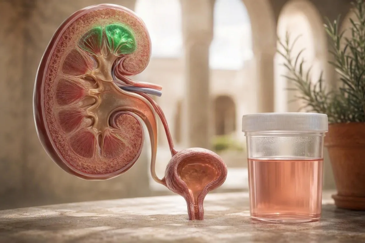
შარდის ვარდისფერი ან ჩაისფერი ნიმუში ხანგრძლივი სირბილის შემდეგ ხშირად არის დროებითი, ვარჯიშით გამოწვეული ჰემატურია, მაგრამ ხილვადი ფერი არასოდეს არ უნდა იყოს უგულებელყოფილი. დრო, მიკროსკოპული მონაცემები, სიმპტომები და სათანადოდ დასვენებული განმეორებითი ნიმუში ამბობს ნამდვილ ამბავს.


- ვარჯიშით გამოწვეული ჰემატურია ჩვეულებრივ ქრება მძიმე სირბილიდან 24-72 საათის შემდეგ; 72 საათზე მეტი ხნის შემდეგ საჭიროა სამედიცინო შეფასება.
- მიკროსკოპიის ზღვარი 3 ან მეტი ერითროციტი მაღალი სიმძლავრის ველზე, სათანადოდ აღებულ შარდის ნიმუშში, შეესაბამება AUA-ს მიკროჰემატურიის განმარტებას.
- დიპსტიკის შეუსაბამობა მნიშვნელოვანია: დადებითი სისხლის ბალიში ნულოვანი ან ორი ერითროციტით მაღალი სიმძლავრის ველზე მიუთითებს მიოგლობინზე ან თავისუფალ ჰემოგლობინზე, შარდში სისხლდენასთან შედარებით.
- განმეორების დრო პრაქტიკულია: მოერიდეთ მძიმე ვარჯიშს 48-72 საათის განმავლობაში, ნორმალურად დალიეთ სითხე, შემდეგ აიღეთ შარდის შუა ნაკადის ნიმუში.
- იმავე დღის შეფასება გონივრულია თრომბების, შარდვის შეუძლებლობის, ცხელების, ცალმხრივი ძლიერი ტკივილის გვერდში ან კოლას ფერის შარდის და კუნთების ძლიერი ტკივილის დროს.
- თირკმლის გამაფრთხილებელი ნიმუში არის სისხლი პლუს ცილა, ერითროციტული ცილინდრები, მაღალი არტერიული წნევა ან დაცემული eGFR - არა იზოლირებული პოსტრეის დიპსტიკი.
- ნიშანი რაბდომიოლიზის შესახებ არის არაპროპორციულად მუქი შარდი მძიმე კუნთოვანი სიმპტომებით და კრეატინ კინაზას მაჩვენებლით, რომელიც ხშირად აღემატება ლაბორატორიული ზედა ზღვრის 5-ჯერ.
- კიბოს რისკის კონტექსტი ცვლის კვლევის ზღვარს: ასაკი, მოწევის ისტორია, მენჯის ღრუს წინა დასხივება და ჰემატურიის მუდმივი ხილვადობა ყველაფერი მნიშვნელოვანია.
რატომ შეიძლება შარდს ვარდისფერი შეფერვა ჰქონდეს სირბილის შემდეგ
მორბენალის ჰემატურია, როგორც წესი, არის სისხლის წითელი უჯრედების შარდში მოკლევადიანი გაჟონვა ინტენსიური ვარჯიშის შემდეგ., რომელიც ხშირად ქრება დასვენებიდან 24–72 საათის განმავლობაში. მიუხედავად ამისა, ვარდისფერი, წითელი ან კოკა-კოლას ფერის შარდი იმსახურებს სათანადოდ შეგროვებულ განმეორებით ტესტს, ვიდრე შემთხვევითი ვარაუდი, რომ ამას სირბილი იწვევს.
კლინიკაში დამამშვიდებელი სურათი მარტივია: ფერის ცვლილება იწყება არადამ parâტარად ხანგრძლივი, სწრაფი ან დაღმართზე სირბილის შემდეგ; არ არის ცხელება, კოლიკური ტკივილი წელის არეში ან შარდვისას წვა; და მიკროსკოპია ნორმალიზდება დასვენების შემდეგ. Kantesti არის AI სისხლის ანალიზის ანალიზატორი , რომელიც საშუალებას აძლევს დაკავშირებულ თირკმლის მარკერებს—კრეატინინს, eGFR, ელექტროლიტებს და კრეატინ კინაზას—განთავსდეს ვარჯიშის ისტორიის გვერდით, ნაცვლად იმისა, რომ რბოლის შემდგომი შედეგი დიაგნოზად ჩაითვალოს.
შემოთავაზებული მექანიზმებია ბუშტის კედლის მექანიკური კონტაქტი, თირკმლის სისხლის ნაკადის დროებითი ცვლილებები, დეჰიდრატაციასთან დაკავშირებული კონცენტრაცია და ზოგჯერ ერითროციტების დაშლა ფეხის განმეორებითი დარტყმისგან. თითოეული მექანიზმის მტკიცებულებები გულწრფელად შერეულია, რადგან უმეტესობა კვლევები მცირეა და იყენებს სხვადასხვა დისტანციებს რბოლებში. ჩემს 15 წლიან კლინიკურ პრაქტიკაში, ახალი დატვირთვის ისტორია უფრო მნიშვნელოვანია, ვიდრე მხოლოდ ფერი; პირველი ულტრამარათონი და ნაცნობი 5 კმ არ ატარებენ ერთსა და იმავე პრე-ტესტ ალბათობას.
შარდში სისხლის ხილვადობა ვარჯიშის შემდეგ არ არის სინონიმი კეთილთვისებიანი მორბენალის ჰემატურიისთვის. ჰემატურიის წითელი დროშების ფართო სახელმძღვანელო სასარგებლოა, რადგან თირკმლის ქვები, ინფექცია, გლომერულური დაავადება და შარდ-სასქესო გზების სიმსივნეები შეიძლება შემთხვევით გამოვლინდნენ სირბილის შემდეგ.
პრაქტიკული განმარტება
ვარჯიშით გამოწვეული ჰემატურია ნიშნავს, რომ სისხლის წითელი უჯრედები შარდში ჩნდება ძალისხმევის შემდეგ და ქრება დასვენებულ მდგომარეობაში აღებულ განმეორებით ნიმუშში. მხოლოდ დადებითი ტესტ-ზოლი არ ამტკიცებს ამ დიაგნოზს, რადგან რეაგენტი აღმოაჩენს ჰემის პიგმენტს მთლიანი სისხლის წითელი უჯრედებიდან, თავისუფალი ჰემოგლობინიდან ან მიოგლობინიდან.
ჰემატურია, ჰემოგლობინი თუ მიოგლობინი?
შარდის ტესტ-ზოლი, რომელიც დადებითად რეაგირებს სისხლზე, შეიძლება ასახავდეს სისხლის წითელ უჯრედებს, ჰემოგლობინს ან მიოგლობინს, ხოლო მიკროსკოპია განასხვავებს მათ. ვარდისფერი შარდი მრავალი მთლიანი სისხლის წითელი უჯრედებით ადასტურებს ჰემატურიას; მუქი ყავისფერი შარდი რამდენიმე ან სისხლის წითელი უჯრედების გარეშე იწვევს შეშფოთებას კუნთების დაზიანების ან ჰემოლიზის პიგმენტზე.
შარდის მიკროსკოპია, რომელიც აჩვენებს 3 ან მეტ სისხლის წითელ უჯრედს მაღალი გადიდების ველზე ადასტურებს მიკროჰემატურიას, როდესაც ნიმუში სუფთად არის შეგროვებული. განსხვავებით, ძლიერ დადებითი ტესტ-ზოლი 0–2 სისხლის წითელი უჯრედით მაღალი გადიდების ველზე, ექიმს უნდა აფიქრებინოს მიოგლობინურია, განსაკუთრებით სიცხის ზემოქმედების, კუნთების დატვირთვის მსგავსი დაზიანების ან კუნთების ძლიერი შემაჯამებელი ტკივილის შემდეგ. ყავისფერი ან კოკა-კოლას ფერი უფრო ტიპიურია შემაჯამებელი პიგმენტისთვის, ვიდრე ბუშტის სისხლდენის მცირე რაოდენობისთვის.
მიოგლობინს მნიშვნელობა აქვს, რადგან ის შეიძლება თან ახლდეს რაბდომიოლიზს და თირკმლის მწვავე დაზიანებას. კრეატინ კინაზას კონცენტრაცია, რომელიც აღემატება ლაბორატორიული ნორმის ზედა ზღვრის 5-ჯერ ხშირად გამოიყენება, როგორც ბიოქიმიური ზღვარი რაბდომიოლიზისთვის, თუმცა სიმპტომები, კალიუმი, კრეატინინი და შარდის გამოყოფა განსაზღვრავენ გადაუდებლობას. იხილეთ ჩვენი პრაქტიკული ახსნა საშიშროებად მაღალი კრეატინ კინაზა იმ ნიმუშებისთვის, რომლებსაც არ უნდა ველოდოთ მთელი ღამით.
საკვების საღებავებს, ჭარხალს, მაყვლის კენკრას, რიფამპიცინს, ფენაზოპირედინს და დეჰიდრატაციას ასევე შეუძლია შარდის შეფერადება წითელი უჯრედების გარეშე. ა მუქი შარდის დიფერენციალი ხელს უწყობს გავრცელებული შეცდომის თავიდან აცილებას, ყოველგვარი წითელი ნიმუშის სისხლად მიჩნევისას.
რატომ იცვლება დიაგნოსტიკა
ერითროციტული ჰემატურია მიუთითებს საშარდე გზებზე, მაშინ როდესაც მიოგლობინურია მიუთითებს ჩონჩხის კუნთებსა და თირკმელების დაცვაზე. ორივე შეიძლება მოხდეს გამძლეობის ღონისძიებების შემდეგ, მაგრამ მხოლოდ მიკროსკოპია და მიზანმიმართული სისხლის ანალიზი შეიძლება საიმედოდ განასხვავოს ისინი.
როგორ წავიკითხოთ შარდის ანალიზის შედეგები სირბილის შემდეგ
ყველაზე ინფორმაციული პოსტ-გაშვების შარდის ტესტი აერთიანებს დიპსტიკს, მიკროსკოპიას, ცილის გაზომვას და სპეციფიკურ სიმკვრივეს. დიპსტიკზე სისხლი ტრანზიტორული ერითროციტებით და ცილის გარეშე ხშირად დაბალი რისკია; სისხლი პლუს ცილა ან კასტები საჭიროებს უფრო დეტალურ თირკმელების შეფასებას.
დიპსტიკის სისხლის შედეგი ფასდება როგორც უარყოფითი, კვალი, 1+, 2+, ან 3+, მაგრამ ეს არის სკრინინგის სიგნალი და არა უჯრედების რაოდენობა. AUA მიკროჰემატურიას განსაზღვრავს, როგორც 3-ზე მეტ ერითროციტს მაღალი გადიდების ველზე მიკროსკოპული გამოკვლევისას., არა მხოლოდ დიპსტიკის შედეგი (Barocas et al., 2020). ეს განსხვავება არ აიძულებს მორბენალებს ზედმეტი კიბოს კვლევისკენ ცრუ-დადებითი სკრინინგის შემდეგ.
სპეციფიკური სიმკვრივე აღემატება 1.030 მიუთითებს კონცენტრირებულ შარდზე ბევრ ლაბორატორიაში და შეიძლება შარდი უფრო საგანგაშოდ გამოიყურებოდეს, მაგრამ ეს არ ხსნის დადასტურებულ ერითროციტებს. 1+ ან უფრო მაღალი ცილა უნდა იყოს კვანტიფიცირებული შარდის ალბუმინ-კრეატინინის თანაფარდობით ან ცილა-კრეატინინის თანაფარდობით; არსებობს ტრანზიტორული ვარჯიშის ცილოვანია, თუმცა მუდმივი ალბუმინურია ცვლის დიფერენციალს გლომერულური დაავადებისკენ. ჩვენი შარდის ანალიზის სრული სახელმძღვანელო ხსნის, რატომ არ უნდა გაერთიანდეს ეს მარკერები ერთ შედეგში.
Kantesti AI ინტერპრეტაციას უკეთებს კრეატინინს და eGFR-ს შარდის მონაცემებთან, ვარჯიშის დროთან და წინა მნიშვნელობებთან ერთად; ცხელ რბოლის შემდეგ ერთჯერადი ოდნავ მომატებული კრეატინინი არ არის ექვივალენტური ქრონიკული თირკმლის დაავადებასთან. პრაქტიკაში, მე ვეკითხები მანძილს, რელიეფს, ტემპერატურას, ანთების საწინააღმდეგო პრეპარატების გამოყენებას, მენსტრუალური ციკლის დროულობას და იმას, იქნა თუ არა ნიმუში აღებული დასრულებიდან 2 საათის განმავლობაში.
როდის უნდა გავიმეოროთ შარდის ტესტი სირბილის შემდეგ?
შარდის ხელახალი ტესტირება 48–72 საათის შემდეგ ბოლო მძიმე გარბენის შემდეგ არის გონივრული სტანდარტი ეჭვმიტანილი მორბენლის ჰემატურიისთვის. გამოიყენეთ პირველი დილის ან კარგად დატენიანებული შუა ნაკადის ნიმუში, მაგრამ ნუ დალევთ ლიტრ წყალს უშუალოდ მანამდე, რადგან უკიდურესმა განზავებამ შეიძლება დაფაროს სასარგებლო აღმოჩენები.
ჩვეულებრივი რბოლის შემდგომი პათოლოგიისთვის, მე ჩვეულებრივ გირჩევთ არანაირი ინტერვალები, აღმართები, მძიმე აწევა ან რბოლა 48 საათში; მარათონის, ულტრამარათონის, სითბური დაავადების ან უჩვეულო ტკივილის შემდეგ, 72 საათის განმავლობაში უფრო დასაბუთებულია. ჩვეულებრივი დალევა წყურვილის მიხედვით საკმარისია. ზედმეტი დატენიანება ამცირებს სპეციფიკურ სიმძიმეს 1.005-ზე დაბლა და ზოგჯერ ართულებს საზღვრულ შედეგის ინტერპრეტაციას.
თუ მიკროსკოპია დადებითი რჩება 72 საათის შემდეგ, დანიშნეთ პირველადი სამედიცინო დახმარების მიმოხილვა დღეების განმავლობაში, ვიდრე განმეორებითი ტესტირება ვარჯიშების შემდეგ. მდგრადი შედეგები მოითხოვს არტერიული წნევის გაზომვას, შრატის კრეატინინის/eGFR, შარდის ალბუმინ-კრეატინინის თანაფარდობას და კულტურის ან ვიზუალიზაციის გათვალისწინებას რისკის მიხედვით. ექსერციის, ჰიდრატაციის და დროებით დაბალი ფილტრაციის შეფასების ურთიერთმიმართება მოცემულია ჩვენს პოსტ-ვარჯიშის eGFR სახელმძღვანელოში.
52 წლის მოყვარულმა მარათონელმა, რომელიც ვნახე, ჰქონდა 18 წითელი უჯრედი მაღალი სიმძლავრის ველზე ცხელი რბოლის საღამოს, მაგრამ 0–1 წითელი უჯრედი 72 დასვენებული საათის შემდეგ; მისი კრეატინინი დაუბრუნდა 1,28 მგ/დლ-დან 0,96 მგ/დლ-მდე. შედეგი დამაიმედებელი იყო, მაგრამ იგივე გეგმა არ იქნებოდა საკმარისი 52 წლის მწეველისთვის განმეორებითი ხილული შარდის ფერით.
ნუ გააკეთებთ ტესტირებას ფინიშის ხაზზე, თუ ამას მოერიდებით
ტესტირება ვარჯიშისთანავე ზრდის შანსს დროებითი წითელი უჯრედების, კონცენტრირებული შარდის და ცილის აღმოჩენის. ეს შეიძლება იყოს კლინიკურად სასარგებლო ეჭვმიტანილი რაბდომიოლიზის შემთხვევაში, მაგრამ ეს ცუდი დროა მორბენლის ბაზის დასადგენად.
რომელი პოსტრანინგული ჰემატურიის ნიმუშებია ჩვეულებრივ დამაიმედებელი?
ინტენსიური სირბილის შემდეგ თვითშეზღუდული ეპიზოდი უფრო დამაიმედებელია, როდესაც ის მთლიანად ქრება, არ აქვს ტკივილი ან სისტემური სიმპტომები და არ აქვს პროტეინურია ან თირკმლის ფუნქციის ცვლილება. დამაიმედებელი არ ნიშნავს უგულებელყოფას; ეს ნიშნავს დადასტურებას დასვენებული ნიმუშით.
დაბალი რისკის ფენოტიპი არის ახალგაზრდა ან საშუალო ასაკის მორბენალი მკაფიო ექსერციული ტრიგერით, ნორმალური არტერიული წნევით, მოწევის ისტორიის გარეშე და ნორმალური დასვენებული მიკროსკოპით. ამ გარემოებაში, ერთჯერადი გარდამავალი შედეგი 3–10 RBC/HPF რბოლის შემდეგ ხშირად მთავრდება მკურნალობის გარეშე. მე მაინც დავაფიქსირებდი მას, რადგან ყოველ გრძელ გარბენის შემდეგ გამეორება იმსახურებს უფრო ფრთხილად განხილვას, ვიდრე ერთჯერადი მოვლენა.
შარდის ალბუმინ-კრეატინინის ნორმალური თანაფარდობა ზოგადად არის 30 მგ/გ-ზე ნაკლები (3 მგ/mmol-ზე ნაკლები), თუმცა ლაბორატორიები და ქვეყნები განსხვავებულად იუწყებიან ერთეულებს. სისხლი ალბუმინის, კასტების ან დისმორფული წითელი უჯრედების გარეშე არ მიუთითებს გლომერულარულ გაჟონვაზე; ეს არ ამტკიცებს შარდის ბუშტის წყაროს, მაგრამ ეს ხელმძღვანელობს შემდეგ კითხვას. ჩვენი ახსნა ცილა შარდში ფარავს ამ განყოფილებას ნათლად.
შარდის ბუშტის ტრავმა უფრო სავარაუდოა, როდესაც შარდი ძალიან კონცენტრირებულია და მორბენალი თითქმის ცარიელი შარდის ბუშტით ასრულებს. ზოგიერთი სპორტული კლინიცისტი გვირჩევს შარდვას ხანგრძლივი სირბილის წინ და რეგულარულ დალევას, მაგრამ ეს არის პრევენციის რჩევა შეზღუდული საცდელი მტკიცებულებით — არა შეფასების შემცვლელი, როდესაც ფერი შენარჩუნებულია.
ნორმალურ შედეგს აქვს დროის ბეჭედი
ნეგატიური ტესტი რბოლის 3 დღის შემდეგ ამ ეპიზოდისთვის ამშვიდებს, არა სიცოცხლის გარანტიას. ახალი ხილული ჰემატურია რამდენიმე თვის შემდეგ უნდა შეფასდეს მისი დამსახურების მიხედვით, განსაკუთრებით 40 წლის ასაკის შემდეგ ან მოწევის ზემოქმედებით.
გამაფრთხილებელი ნიშნები, რომლებიც ამცირებს მორბენლის ჰემატურიის ალბათობას
ჰემატურია საჭიროებს სასწრაფო შეფასებას, როდესაც ის გრძელდება 72 საათის შემდეგ, მეორდება მძიმე ვარჯიშის გარეშე, ან ჩნდება ტკივილით, ცხელებით, შედედებით, პროტეინით ან თირკმლის ფუნქციის ცვლილებით. დასკვნების კომბინაცია უფრო ინფორმატიულია, ვიდრე თავისთავად დრამატულად ნაჩვენები ნიმუში.
დროულად მიმართეთ სამედიცინო რჩევისთვის ხილული წითელი შარდისთვის, რომელიც გრძელდება 24 საათის განმავლობაში, მაშინაც კი, თუ თავს კარგად გრძნობთ; ზღურბლი უფრო დაბალია 40 წელზე უფროსი ასაკის ადამიანებისთვის, მწეველთათვის ან ყოფილი მწეველთათვის, ან მათთვის, ვისაც აქვს წინა მენჯის დასხივება. AUA სახელმძღვანელო იყენებს რისკის სტრატიფიკაციას ასაკის, მოწევის, მიკროსკოპული სისხლის ხარისხისა და მდგრადობის, და წინა გროს ჰემატურიის მიხედვით, ნაცვლად იმისა, რომ ყველა მიკროჰემატურია ერთნაირად მოექცეს (Barocas et al., 2020).
დისმორფული ერითროციტები, ერითროციტების ცილინდრები, მნიშვნელოვანი ალბუმინურია, ახალი ჰიპერტენზია ან შემცირებული eGFR მიუთითებს თირკმლის ფილტრის წყაროზე, ვიდრე შერყეული შარდის ბუშტი. ერითროციტების ცილინდრები არასოდეს არის ჩვეულებრივი სირბილის მოსალოდნელი შედეგი და უნდა გამოიწვიოს თირკმელების სასწრაფო შეფასება. იხილეთ ჩვენი მიმოხილვა მარცვლოვანი და შარდის ცილინდრები იმის შესახებ, თუ რატომ არის მნიშვნელოვანი ნალექის დასკვნები.
თომას კლაინი, MD, ურჩევს მორბენლებს, არ მოახდინონ თვითდიაგნოსტიკა მხოლოდ ფერის მიხედვით: შარდსასქესო სისტემის ინფექციამ შეიძლება გამოიწვიოს წვა, შარდვის იმპერატიულობა, პიურია და ზოგჯერ ჰემატურია, ხოლო ქვებს უფრო ხშირად იწვევს მოულოდნელი ცალმხრივი ტკივილი და გულისრევა. კულტურა ყველაზე სასარგებლოა ანტიბიოტიკებამდე, როდესაც ინფექციის სიმპტომებია.
სისხლის პლუს ცილის კომბინაცია
დადასტურებული ჰემატურია პროტეინურიასთან ერთად უფრო შემაშფოთებელია, ვიდრე იზოლირებული ჰემატურია, რადგან ორივე შეიძლება წარმოიშვას გლომერულური ფილტრიდან. შარდის ალბუმინ-კრეატინინის თანაფარდობა 30 მგ/გ ან მეტი უნდა განმეორდეს და შეფასდეს სისხლის წნევასთან და თირკმლის ფუნქციასთან ერთად.
როდის საჭიროებს ვარჯიშის შემდეგ ვარდისფერი შარდი იმავე დღეს მკურნალობას?
მიიღეთ იმავე დღეს სასწრაფო შეფასება შედედების, შარდვის შეუძლებლობის, ძლიერი გვერდითი ტკივილის, ცხელების, გულის წასვლის, შარდის გამოყოფის შემცირების, ან მუქი შარდის ძლიერი კუნთების ტკივილითა და სისუსტით. ეს მახასიათებლები მიუთითებს არა-გართულებულ ვარჯიშით გამოწვეულ ჰემატურიაზე.
შედედებამ შეიძლება შეაფერხოს შარდის ნაკადი, ხოლო შარდის გამოყოფის შეუძლებლობა არის გადაუდებელი პრობლემა სპორტული ფიტნესის მიუხედავად. ცხელება 38.0°C ან უფრო მაღალი გვერდითი ტკივილით შეიძლება მიუთითებდეს ზედა შარდის ინფექციაზე; ძლიერი კოლიკური ტკივილი ღებინებით შეიძლება მიუთითებდეს ქვას. არცერთი სიტუაცია არ უნდა იქნას მართული პოსტრანის განმეორებითი ტესტის მოლოდინში.
მუქი ყავისფერი შარდი დაძაბვის შემდეგ პლუს კუნთების გამოხატული მგრძნობელობა, სისუსტე, შეშუპება ან შემცირებული გამოყოფა ზრდის რაბდომიოლიზის შეშფოთებას. საწყისი ტესტირება ჩვეულებრივ მოიცავს კრეატინ კინაზას, კრეატინინს, კალიუმს, კალციუმს, ფოსფატს და შარდის ანალიზს. CrossFit რაბდომიოლიზის გზამკვლევი
relevant რადგან უჩვეულო ექსცენტრული ვარჯიში - არა მარტო სირბილი - ხშირი გამომწვევია.
.მწვავე დეჰიდრატაციამ შეიძლება თირკმელების შედეგები გააუარესოს, მაგრამ მას ასევე რეალურ ტრავმასთან ერთად შეუძლია თანაარსებობა. თუ გაბრუებული ხართ, გაქვთ ცნობიერების არევა, არ შეგიძლიათ სითხის მიღება ან ძალიან ცოტა შარდავთ
8–12 საათს,, არ სცადოთ სპორტული სასმელებით სახლში პრობლემის მოგვარება.
.ორსულობა და ანტიკოაგულანტები
ორსულმა მორბენალებმა და ანტიკოაგულანტების მიმღებმა ადამიანებმა დაუყოვნებლივ უნდა იტყობინონ მაკროსკოპული ჰემატურია.
ანტიკოაგულანტებს შეუძლიათ სისხლდენის გაძლიერება, მაგრამ არ გამორიცხავენ სისხლდენის წყაროს იდენტიფიცირების აუცილებლობას; ორსულობა ასევე ცვლის ინფექციის, ქვების და თირკმელების დაავადების შემოწმების ზღვარს.
.მდგომარეობები, რომლებიც სირბილმა შეიძლება გამოავლინოს და არა გამოიწვიოს
სირბილმა შეიძლება გამოამჟღავნოს არსებული საშარდე პრობლემა შარდის კონცენტრაციის ან ტესტის გამოძახების გზით, და არა თავად ჰემატურიის გამოწვევის.
ქვები, ინფექცია, გლომერულური დაავადება, სტრუქტურული ანომალიები და სიმსივნე რჩება დიფერენციული დიაგნოზის ნაწილად.
.
გაშვებამ შეიძლება გამოააშკარავოს საშარდე პირობები, რომლებიც არ არის გამოწვეული ვარჯიშით.
.თირკმლის ქვები ხშირად იწვევს ერთმხრივ ტკივილს წელიდან საზარდულამდე, გულისრევას და სისხლის წითელი უჯრედების მიკროსკოპიას, თუმცა უმტკივნეულო ქვებიც გვხვდება. შარდის ინფექცია უფრო ხშირად იწვევს წვას, ხშირ შარდვას, აუცილებლობას და თეთრი უჯრედების არსებობას; ნიტრიტის უარყოფითი შედეგი არ გამორიცხავს მას. ჩვენი სახელმძღვანელო
ლეიკოციტების ესთერაზა ნიტრიტის გარეშე
ხსნის ამ გავრცელებულ შერეულ ნიმუშს.
.გლომერულონეფრიტი შეიძლება გამოვლინდეს ზედა სასუნთქი გზების ინფექციის შემდეგ ქოლას მსგავსი შარდით, შეშუპებით, მომატებული სისხლის წნევით, პროტეინურიით და დისმორფული ერითროციტებით. KDIGO 2024 CKD სახელმძღვანელოში, მდგრადი ალბუმინურია და დაქვეითებული eGFR განიხილება როგორც თირკმლის რისკის დამატებითი მარკერები, და არა ურთიერთშემცვლელი საზომები (KDIGO, 2024).
.Kantesti არის AI-ზე დაფუძნებული სისხლის ანალიზის ინტერპრეტაციის ინსტრუმენტი
რომელიც ეხმარება მომხმარებლებს ამოიცნონ კრეატინინის, eGFR, ალბუმინის, ელექტროლიტების და CBC-ის ნიმუში, მაგრამ მას არ შეუძლია ჰემატურიის ანატომიური წყაროს დიაგნოსტიკა. გამოსახულების კვლევა, მიკროსკოპია, კულტურა და კლინიცისტის გამოკვლევა კვლავ ასრულებს გადამწყვეტ სამუშაოს.
.კიბო იშვიათია ახალგაზრდა მორბენალებში, მაგრამ შეუძლებელი არ არის მოზრდილებში
მაკროსკოპული ჰემატურია საჭიროებს შეფასებას მაშინაც კი, როცა ვარჯიში აშკარა ახსნას ჰგავს, განსაკუთრებით მწეველებში ან სიმპტომების მქონე პირებში.
რისკი იზრდება ასაკთან და თამბაქოს დაგროვებასთან ერთად, ამიტომ იგივე შარდის შედეგები არ იწვევს ერთნაირ ტესტირების გეგმებს.
.ვის სჭირდება უროლოგიური ან ნეფროლოგიური მეთვალყურეობა?
დასვენების შემდეგ განმეორებითი სინჯის შემთხვევაში მდგრადი ჰემატურია უნდა შეფასდეს უროლოგიური რისკისთვის, ხოლო ჰემატურია პლუს პროტეინურია, ცილინდრები, ჰიპერტენზია ან დაქვეითებული eGFR მიუთითებს ნეფროლოგიურ პრობლემაზე.
ბევრ ადამიანს ესაჭიროება მხოლოდ პირველადი ჯანდაცვის მიერ ჩატარებული განმეორებითი ტესტირება; მიმართვა დამოკიდებულია ნიმუშზე.
.
შარდის ნალექი და თირკმლის მარკერები ხელს უწყობს უროლოგიური ან ნეფროლოგიური მიმოხილვის მიმართულებას.
.უროლოგიური შეფასება განსაკუთრებით მიზანშეწონილია განმეორებითი მაკროსკოპული ჰემატურიის, მდგრადი მიკროსკოპული ჰემატურიის, მოწევის ისტორიის, არომატული ამინებით პროფესიული ზემოქმედების, მენჯის ღრუს წინა დასხივების ან შარდვის სიმპტომების დროს ინფექციის გარეშე. რისკიდან გამომდინარე, კლინიცისტებმა შეიძლება გამოიყენონ თირკმელებისა და ბუშტის ულტრაბგერითი კვლევა, CT-უროგრაფია და ცისტოსკოპია. 24 წლის ასაკის პირისთვის რბოლისშემდგომი კვალიანი დიპსტრიკი ძალიან განსხვავებული სცენარია, ვიდრე 58 წლის ყოფილი მწეველისთვის განმეორებითი წითელი შარდი.
.ნეფროლოგიური მიმოხილვა უფრო სავარაუდოა შარდის ალბუმინ-კრეატინინის თანაფარდობის შემთხვევაში
≥30 მგ/გ,eGFR-ის დაქვეითება
60 მლ/წთ/1.73 მ²3 თვის ან მეტი ხანგრძლივობით, ერითროციტების ცილინდრები ან ძნელად კონტროლირებადი სისხლის წნევა. ეს ზღვრები მნიშვნელოვანია, რადგან ქრონიკული თირკმელების დაავადება განისაზღვრება ხანგრძლივობით, ისევე როგორც ერთი საზომით. წაიკითხეთ ჩვენი
ქრონიკული თირკმლის დაავადების სტადიების სახელმძღვანელოპრაქტიკული კატეგორიებისთვის.
.Kantesti-ის AI ლაბორატორიული ტესტის ინტერპრეტაციის სერვისს შეუძლია განმეორებადი სისხლისა და ცილის ნიმუშის გამოვლენა განსახილველად, განსაკუთრებით მაშინ, როდესაც მომხმარებლები სხვადასხვა ლაბორატორიიდან ატვირთავს შედეგებს. მან ხელი უნდა შეუწყოს, მაგრამ არ ჩაანაცვლოს კლინიცისტის გადაწყვეტილება, საჭიროა თუ არა შარდ-სასქესო სისტემის ვიზუალიზაცია ან სპეციალისტის გამოკვლევა.
ნუ მისცემთ ანტიკოაგულაციას საუბრის დასასრულს
ანტიკოაგულაციური საშუალებების გამოყენება ცვლის სისხლდენის სიმძიმეს, მაგრამ არ იძლევა ჰემატურიის მიზეზს. ჩემი გამოცდილებით, განმეორებადი შარდის სისხლის მიზეზის ატრიბუტირება აპქსაბანის, ვარფარინის ან ასპირინის მიმართ, გამოკვლევის გარეშე, შეიძლება შეაფერხოს კენჭების, ინფექციის ან სტრუქტურული დაავადების ამოცნობა.
რომელი სისხლის ანალიზები გვეხმარება ვარჯიშით გამოწვეული ჰემატურიის შემდეგ?
სასარგებლო სისხლის ანალიზები დამოკიდებულია სიმპტომებზე: კრეატინინი, eGFR, ელექტროლიტები და კრეატინ კინაზა აფასებენ თირკმელებისა და კუნთების სტრესს, ხოლო CBC აფასებს ანემიის ან ინფექციის ნიშნებს. რუტინული ფართო პანელები ნაკლებად სასარგებლოა, ვიდრე მიზანმიმართული ტესტირება ზუსტი ისტორიისა და შარდის ანალიზის შემდეგ.
კრეატინინი შეიძლება დროებით გაიზარდოს გამძლეობის ვარჯიშის შემდეგ კუნთების მეტაბოლიზმის, თირკმელების პერფუზიის შემცირებისა და დეჰიდრატაციის გამო. eGFR განტოლებები ნაკლებად სანდოა კრეატინინის სწრაფი ცვლილების დროს, ამიტომ განმეორებითი მნიშვნელობა შემდეგ 48–72 საათით ჩვეულებრივი აღდგენის შემდეგ ხშირად უფრო მნიშვნელოვანია, ვიდრე ფინიშის ხაზის შედეგი. ჩვენი ვარჯიშთან დაკავშირებული კრეატინინის ხელახალი შემოწმების სახელმძღვანელო აძლევს კონტექსტს ამ ხშირი შფოთვის გამომწვევისთვის.
კრეატინ კინაზას აქვს ფართო პოსტ-ვარჯიშის დიაპაზონი: გაწვრთნილ სპორტსმენებს შეიძლება აღენიშნებოდეთ მატება რბოლის შემდეგ თირკმელების დაზიანების გარეშე, ხოლო ადგილობრივი ზედა ზღვრის მრავალჯერადი მნიშვნელობები სიმპტომებთან ერთად საჭიროებს სასწრაფო შეფასებას. კალიუმი ზემოთ 6.0 მმოლ/ლ-ს არის პოტენციურად საშიში და საჭიროებს სასწრაფო კლინიკურ შეფასებას, განსაკუთრებით თუ თირკმელების ფუნქცია დაქვეითებულია ან საეჭვოა კუნთების დაზიანება.
CBC ყველაზე სასარგებლოა, როდესაც არის დაღლილობა, ფერმკრთალობა, მძიმე მენსტრუალური სისხლდენა, განმეორებითი ჰემატურია ან სისტემური დაავადების ეჭვი. ჰემოგლობინი შეიძლება დაეცეს გამძლეობის მოვლენების შემდეგ პლაზმის მოცულობის გაფართოების, რკინის დაკარგვის ან ჰემოლიზის გამო; ფერიტინი უნდა იქნას განმარტებული მწვავე ანთებისა და ინტენსიური ვარჯიშისგან განცალკევებით. გამძლეობის მორბენლებს შეიძლება გამოადგეთ ჩვენი სპორტსმენის სისხლის ანალიზის მიმოხილვა გონივრული ბაზისისთვის.
ნუ დაუსვამთ თირკმელების დაავადების დიაგნოზს ერთი სარბოლო კვირის პანელით
კრეატინინის 0,3 მგ/დლ ან მეტი მატება შეიძლება შეესაბამებოდეს მწვავე თირკმელების დაზიანების კრიტერიუმს შესაბამის დროის ჩარჩოში, მაგრამ კონტექსტი და განმეორებითი ტესტირება გადამწყვეტია რბოლის შემდეგ. შარდის დაბალი გამოყოფა, კალიუმის მატება და მდგრადი მომატება უფრო მნიშვნელოვანია, ვიდრე იზოლირებული სასაზღვრო შედეგი.
როგორ ავიღოთ განმეორებითი შარდის ნიმუში, რომელსაც ექიმები ენდობიან
სუფთა შუა ნაკადის შარდის ნიმუში 48-72 საათის შემდეგ, ძლიერი ვარჯიშის გარეშე, საუკეთესო საშუალებაა იმის შესამოწმებლად, აღმოიფხვრა თუ არა ჰემატურია. შეგროვების შეცდომებმა, მენსტრუალური დაბინძურებამ და ძველმა ნიმუშმა შეიძლება შექმნას მცდარი შედეგები.
დაიბანეთ ხელები, დაიწყეთ შარდვა ტუალეტში, შემდეგ შეაგროვეთ შუა ნაკადის ნაწილი კონტეინერის შიგთავსის შეხების გარეშე. მიიტანეთ ნიმუში დაუყოვნებლივ - იდეალურად ფარგლებში 2 საათამდე—ან დაიცავით ლაბორატორიის მაცივარში შენახვის ინსტრუქციები. ხანგრძლივი დგომა ცვლის უჯრედების გარეგნობას და შეიძლება გახდეს მიკროსკოპია ნაკლებად სანდო.
მოერიდეთ ტესტირებას აქტიური მენსტრუაციის დროს, თუ ეს შესაძლებელია; თუ ტესტირება არ შეიძლება გადაიდოს, აცნობეთ კლინიცისტს და ლაბორატორიას. ვაგინალური დაბინძურება შეიძლება გამოიწვიოს აშკარა სისხლის წითელი უჯრედები და ცილა, ხოლო ბრტყელი ეპითელური უჯრედები ხშირად მიუთითებს ცუდად შეგროვებულ ნიმუშზე. ჩვენი სახელმძღვანელო შარდის ეპითელური უჯრედების შესახებ ხსნის, თუ რატომ არ არის ეს უმნიშვნელო ლაბორატორიული სქოლიო.
Thomas Klein, MD, გვირჩევს ნიმუშთან ერთად ოთხი დეტალის ჩაწერას: ბოლო ინტენსიური ვარჯიში, აქტივობის ხანგრძლივობა, გარემოს ტემპერატურა და ყველა მედიკამენტი, მიღებული წინა 72 საათის განმავლობაში. დროის მითითების გარეშე მიღებული შედეგის ინტერპრეტაცია გაცილებით რთულია — განსაკუთრებით მაშინ, როდესაც შარდი ორმაგი ვარჯიშის შემდეგ არის შეგროვებული.
პირველი დილის ტესტირება დღისით ჩატარებულ ტესტირებასთან შედარებით
პირველი დილის ნიმუში არის კონცენტრირებული და კარგია მცირე დარღვევების გამოსავლენად, თუმცა ნორმალურად ჰიდრატირებული დღის შუადღის შუა შარდის ნიმუში მისაღებია, თუ შეგროვება უფრო სუფთაა. თანმიმდევრულობა უფრო მნიშვნელოვანია სერიული შედარებისთვის: ყოველი გამეორებისთვის გამოიყენეთ დაახლოებით იგივე პირობები.
ჰიდრატაცია, ტკივილგამაყუჩებლები და დანამატები, რომლებიც ცვლიან სურათს
დეჰიდრატაცია აკონცენტრირებს შარდს და ზრდის თირკმელების დატვირთვას, ხოლო NSAID-ები ამცირებს თირკმელების სისხლის ნაკადს გამძლეობის ვარჯიშის დროს. არცერთი ფაქტორი არ ამტკიცებს ჰემატურიის მიზეზს, მაგრამ ორივე გავლენას ახდენს იმაზე, თუ რამდენად ფრთხილად აფასებენ კლინიცისტები სირბილის შემდგომ შედეგს.
უმეტესი მორბენალებისთვის, წყურვილის მიხედვით დალევა და სითხის თანდათანობით შევსება უფრო უსაფრთხოა, ვიდრე მკაცრად სითხის მიღების იძულება. ძალიან დაბალი ნატრიუმი ჭარბი წყლის მიღებიდან შეიძლება საშიში იყოს; შრატის ნატრიუმი ქვემოთ 125 მმოლ/ლ-ზე თავის ტკივილთან, ღებინებასთან, დაბნეულობასთან ან კრუნჩხვებთან ერთად არის საგანგებო მდგომარეობა. შარდის ფერი არის ჰიდრატაციის უხეში მინიშნება, არა ზუსტი ჰიდრატაციის მრიცხველი.
იბუპროფენი, ნაპროქსენი და სხვა NSAID-ები შეიძლება ამცირებდნენ პროსტაგლანდინით შუამავალ თირკმელების სისხლის ნაკადს, განსაკუთრებით სითბური სტრესის, ღებინების ან ხანგრძლივი აქტივობების დროს. მე ვურჩევ პაციენტებს, თავიდან აიცილონ პროფილაქტიკური NSAID-ების რუტინული გამოყენება მარათონების წინ, თუ მათი ექიმი მკაფიო მიზეზს არ მისცემს. მედიკამენტების კონტექსტი განხილულია ჩვენს ლაბორატორიული ტენდენციების უსაფრთხოების სტატიაში.
მაღალი დოზით ვიტამინი C-მ შეიძლება ხელი შეუშალოს შარდის ტესტ-ზოლის გარკვეულ რეაქციებს, ხოლო შესრულებისთვის განკუთვნილი დანამატები ზოგჯერ შეიცავს დაუფარავ სტიმულატორებს ან შარდმდენებს. მოიტანეთ კონტეინერები ან მკაფიო ფოტოები შეხვედრაზე; ეს ხშირად უფრო სასარგებლოა, ვიდრე ბრენდირებული ფხვნილის ინგრედიენტების სიის დამახსოვრების მცდელობა. ჩვენი სპორტსმენებისთვის განკუთვნილი დანამატების უსაფრთხოების სახელმძღვანელო გთავაზობთ გონივრულ ჩამონათვალს.
ჰიდრატაცია არ არის მდგრადი სისხლის მკურნალობა
წყალმა შეიძლება გამოასწოროს კონცენტრაცია, მაგრამ არ მკურნალობს ქვას, ინფექციას, გლომერულ პროცესს ან შარდსასქესო სისტემის სტრუქტურულ წყაროს. თუ დასვენებულ, კარგად შეგროვებულ ნიმუშზე რჩება სისხლის წითელი უჯრედები, ესკალაცია უნდა ეფუძნებოდეს ნიმუშს და არა კიდევ ერთ ჰიდრატაციის ექსპერიმენტს.
შეგიძლიათ თუ არა გააგრძელოთ სირბილი ვარჯიშით გამოწვეული ჰემატურიის შემდეგ?
უმეტეს მორბენალს შეუძლია თანდათანობით დაბრუნება, მას შემდეგ რაც შარდის ფერი ნორმალიზდება, სიმპტომები გაქრება და დასვენებულის განმეორებითი ნიმუში დამაიმედებელია. არ დაუბრუნდეთ მძიმე ვარჯიშს, სანამ შარდი რჩება ხილულად წითელი, თირკმელების შედეგები არანორმალურია, ან კუნთების სიმპტომები მიუთითებს რაბდომიოლიზზე.
არასამედიცინო ეპიზოდის შემდეგ, მე ჩვეულებრივ ვურჩევ რამდენიმე დღის განმავლობაში მხოლოდ მსუბუქ სირბილს, შემდეგ კი მოცულობისა და ინტენსივობის თანდათანობით აღდგენას 7–14 დღის განმავლობაში. მორბენალმა უნდა შეწყვიტოს და მიმართოს ექიმს, თუ ფერი განმეორდება, განვითარდება ტკივილი, ან შარდის გამოყოფა შემცირდება. ეს არის პრაგმატული კლინიკური მიდგომა, არა კვლევით დამტკიცებული რეცეპტი.
განმეორებითი ჰემატურია იგივე დისტანციის შემდეგ შეიძლება გაამართლოს პრაქტიკული ექსპერიმენტები: მოერიდეთ დეჰიდრატირებულ მდგომარეობაში რბოლას, მოშარდეთ ხანგრძლივ სირბილამდე, უფრო ნელა დანერგეთ დაღმართზე სირბილი და შეამოწმეთ ფეხსაცმლის მორგებულობა ან რელიეფი. აწარმოეთ ჩანაწერი ტემპის, სიცხის, ხანგრძლივობის, შარდის ფერის და ნიმუშის შედეგების შესახებ. მარათონის მორბენალის ლაბორატორიული სახელმძღვანელო შეუძლია დაეხმაროს აღდგენის მარკერების გარჩევას იმ მიზეზებისგან, რომლებიც საჭიროებენ ვარჯიშის შეჩერებას.
ნუ გაივლით მკურნალობას მხოლოდ იმის გამო, რომ გაქვთ დაგეგმილი ღონისძიება და შეინიშნება აუხსნელი ჰემატურია. სპორტსმენები ხშირად კარგად იტანენ დისკომფორტს; უფრო რთული უნარია იმის გაცნობიერება, რომ ეს არის დიაგნოსტიკური ინფორმაცია და არა სიმტკიცის გამოცდა.
შექმენით ნამდვილი ბაზისური მაჩვენებლები
შარდის ანალიზის და თირკმელების პანელის ბაზისური მაჩვენებლები, აღებული მძიმე ვარჯიშის დასრულებიდან სულ მცირე 72 საათის შემდეგ, საშუალებას იძლევა მომავალში რბოლის შემდგომი შედეგების ინტერპრეტაცია ბევრად გაადვილდეს. ეს განსაკუთრებით სასარგებლოა ულტრამარათონელებისთვის, ადამიანებისთვის ერთი თირკმლით, ან სპორტსმენებისთვის, რომლებიც იყენებენ მედიკამენტებს, რომლებიც გავლენას ახდენენ თირკმლის პერფუზიაზე.
რა უნდა ვუთხრათ ექიმს ვარჯიშის შემდეგ შარდში სისხლის შესახებ
ჰემატურიის შეფასების ყველაზე სწრაფი გზა არის ლაკონური დროის მონაცემები: შარდის ფერი, ვარჯიშის დოზა, სიმპტომები, მედიკამენტები და განმეორებითი ტესტირების დრო. წარმოადგინეთ ლაბორატორიული მონაცემები, ვიდრე მხოლოდ შეტყობინება, რომ ნიმუში იყო “სისხლიანი”.”
მიუთითეთ, იყო თუ არა შარდი ვარდისფერი, ნათელი წითელი, ჩაისფერი, ან შეიცავდა თუ არა შედედებულ სისხლს; გამოჩნდა თუ არა ფერი ვარჯიშის დაწყებამდე, მის დროს, თუ მის შემდეგ; და გაქრა თუ არა ის პირველი შარდვის შემდეგ. შეიტანეთ წვა, შარდვის მოთხოვნილება, ცხელება, ტკივილი წელის არეში, გულისრევა, შეშუპება და შარდის გამოყოფის შემცირება. დეტალური შარდის ფერის მითითება დაგეხმარებათ დაკვირვების ზუსტად აღწერაში მის გადამეტებულ ინტერპრეტაციაზე გადასვლის გარეშე.
წარმოადგინეთ წინა შარდის ანალიზები, კრეატინინის/eGFR მნიშვნელობები, არტერიული წნევის მაჩვენებლები, მოწევის ისტორია, ოჯახის თირკმელების ისტორია და მედიკამენტების/დამატებების სია. Kantesti არის AI ბიომარკერების ინტერპრეტაციის პლატფორმა რომელიც შეიძლება ორგანიზებული იყოს განმეორებითი სისხლის ტესტის მნიშვნელობების და მნიშვნელოვანი ცვლილებების ხაზგასასმელად, მაგრამ კლინიცისტმა უნდა დააკავშიროს ეს მნიშვნელობები შარდის მიკროსკოპიასთან და გამოკვლევასთან.
2026 წლის 3 სექტემბრის მონაცემებით, კლინიკური პრინციპი მარტივი რჩება: ვარჯიში არის სავარაუდო ახსნა მხოლოდ შედეგის გაქრობის და სხვა მიზეზების არარსებობის შემდეგ. თუ ეჭვი გეპარებათ, იკითხეთ, გჭირდებათ თუ არა მიკროსკოპია, კულტურა, ალბუმინ-კრეატინინის თანაფარდობა, განმეორებითი კრეატინინი, ვიზუალიზაცია, თუ რეფერალი — არა მხოლოდ კიდევ ერთი დიპსტიკი.
დასასმელი კითხვები
იკითხეთ, შეინიშნებოდა თუ არა მთლიანი ერითროციტები, იყო თუ არა ცილა ან კასტები, და როდის უნდა მოხდეს დასვენების შემდეგ განმეორებითი გამოკვლევა. ეს სამი პასუხი ჩვეულებრივ განმარტავს, არის თუ არა შემდეგი ნაბიჯი დაკვირვებითი მონიტორინგი, თირკმელების ტესტირება, ინფექციის ტესტირება, თუ უროლოგიური შეფასება.
მორბენლის ჰემატურიის რაციონალური სამოქმედო გეგმა
ერთი უმტკივნეულო ეპიზოდის შემდეგ მძიმე სირბილის შემდეგ, დაისვენეთ ინტენსიური ვარჯიშისგან 48–72 საათის განმავლობაში და გაიმეორეთ სუფთა შარდის ნიმუშის ანალიზი; ეძიეთ უფრო ადრეული დახმარება წითელი დროშების ან მდგრადი ხილული ფერის შემთხვევაში. მიზანი არ არის პანიკა ან უგულებელყოფა — ეს არის იმის დამტკიცება, რომ ეს მაჩვენებელი ქრება.
ნაბიჯი პირველი: შეწყვიტეთ მძიმე ვარჯიში, დალიეთ ნორმალური რაოდენობით წყალი და აღნიშნეთ დრო და სიმპტომები. ნაბიჯი მეორე: გაიმეორეთ მიკროსკოპია შემდეგ 48–72 საათით. ნაბიჯი მესამე: თუ სისხლი არ ქრება, ან თუ ჩნდება ცილა, კასტები, ტკივილი, ცხელება, შედედებული სისხლი, შარდის დაბალი გამოყოფა, ან თირკმელების პათოლოგიური მარკერები, დაუყოვნებლივ მოაწყვეთ სამედიცინო შეფასება. ეს თანმიმდევრობა ხელს უშლის როგორც გამოტოვებულ დაავადებას, ასევე უაზრო ვიზუალიზაციას ერთჯერადი ფინიშის ხაზის შემდეგ.
როდესაც ჩვენი კლინიკური გუნდი მიმოიხილავს ატვირთულ ლაბორატორიულ მონაცემებს, ჩვენ ვამუშავებთ ნიმუშებს: მზარდი კრეატინინი პლუს მაღალი CK და შარდის დაბალი გამოყოფა უფრო გადაუდებელია, ვიდრე სტაბილური კრეატინინი გარდამავალი შარდის დიპსტიკით. Kantesti-ის მეთოდოლოგია და უსაფრთხოების ზომები აღწერილია ჩვენს კლინიკური ვალიდაციის ინფორმაცია, და ჩვენი ექიმის ზედამხედველობა დეტალურად არის აღწერილი სამედიცინო საკონსულტაციო საბჭო.
უმეტეს პაციენტებს მიაჩნიათ, რომ დროული განმეორებითი ტესტი აქცევს საგანგაშო ფერის ცვლილებას ნათელ გეგმად. მაგრამ თუ თქვენი ინსტინქტი ამბობს, რომ რაღაც არ არის რიგზე — განსაკუთრებით თუ შარდი ხვალაც წითელია — ენდეთ ამ სიგნალს და მიმართეთ ექიმს.
ერთსიტყვიანი წესი
ვარდისფერი შარდი ერთხელ მძიმე სირბილის შემდეგ შეიძლება იყოს გარდამავალი; ვარდისფერი შარდი, რომელიც გრძელდება, მეორდება, ან ჩნდება სიმპტომებთან ერთად, არის ჰემატურია სათანადო შეფასებამდე.
ხშირად დასმული კითხვები
რამდენი ხანი გრძელდება მორბენალთა ჰემატურია?
სპორტსმენის ჰემატურია, როგორც წესი, ქრება ძლიერი ფიზიკური დატვირთვის შეწყვეტიდან 24–72 საათის შემდეგ. შარდის განმეორებითი პრაქტიკული ანალიზი არის სუფთა შარდის ნიმუშის აღება შუა ნაკადით, 48–72 საათის შემდეგ ყოველგვარი ინტენსიური სირბილისა და სიმძიმეების აწევის გარეშე. 24 საათზე მეტხანს მომატებული ვარდისფერი ან წითელი შარდი, ან მიკროსკოპული ჰემატურია, რომელიც 72 საათის შემდეგაც არ გაქრა, უნდა შეფასდეს ექიმის მიერ. ცხელება, შედედებული სისხლი, ძლიერი ტკივილი წელის არეში, შარდის გამოყოფის შემცირება ან ძლიერი კუნთოვანი ტკივილი მოითხოვს იმავე დღეს შეფასებას.
სისხლდენა შარდში ვარჯიშის შემდეგ ნორმალურია?
შარდში სისხლი ვარჯიშის შემდეგ შეიძლება იყოს აერობული ვარჯიშის, დეჰიდრატაციის, შარდის ბუშტის კედლის ტრავმის ან თირკმელების სისხლის მიმოქცევის დროებითი ცვლილებების შედეგი, თუმცა ეს ავტომატურად ნორმალური არ არის. მიკროსკოპიული კვლევა, რომელიც ადასტურებს 3 ან მეტ ერითროციტს მაღალი გადიდების ველზე, შეესაბამება AUA-ს მიკროჰემატურიის განმარტებას. განმეორებით აღებულ დასვენებულ ნიმუშში გაქრობილი შედეგი დამამშვიდებელია, ხოლო მდგრადი სისხლი, ცილა, ცილინდრები, ტკივილი ან შარდვის სიმპტომები საჭიროებს შემდგომ შეფასებას. შარდის ხილვადი ფერი ყოველთვის უნდა ეცნობოს, განსაკუთრებით მწეველ მოზრდილებში ან განმეორებითი ეპიზოდებისას.
დეჰიდრატაციამ შეიძლება თუ არა გამოიწვიოს ვარდისფერი შარდი სირბილის შემდეგ?
გაუწყლოებამ შეიძლება შარდი მუქი გახადოს და ვარჯიშის შემდგომი ჰემატურიის შედეგის გაძლიერება გამოიწვიოს შარდის კომპონენტების კონცენტრაციის გზით, თუმცა გაუწყლოება თავისთავად არ ხსნის დადასტურებულ ერითროციტებს. შარდის სპეციფიკური სიმძიმე 1.030-ზე მეტი ხშირად მიუთითებს კონცენტრირებულ შარდზე ბევრ ლაბორატორიაში. დალიეთ ნორმალურად წყურვილის დაკმაყოფილებამდე და გაიმეორეთ სუფთად აღებული ნიმუში 48–72 საათიანი დასვენების შემდეგ, ვიდრე ეცდებით შედეგის ჩამორეცხვას ჭარბი წყლის საშუალებით. ძლიერი წყურვილი, თავბრუსხვევა, შარდის ძალიან დაბალი გამოყოფა ან დაბნეულობა საჭიროებს დაუყოვნებელ სამედიცინო შეფასებას.
რა არის ჰემატურიასა და მიოგლობინურიას შორის განსხვავება ვარჯიშის შემდეგ?
ჰემატურია ნიშნავს შარდში სისხლის წითელი უჯრედების არსებობას, ხოლო მიოგლობინურია კუნთის დაზიანების შემდეგ გამოყოფილი კუნთის პიგმენტის ანარეკლს წარმოადგენს. ორივე შემთხვევაში შარდის ტესტ-ზოლს შეუძლია სისხლის დადებითი შედეგის გამოვლენა, ამიტომ გადამწყვეტი მნიშვნელობა აქვს მიკროსკოპიას: მიოგლობინურია ხშირად იძლევა ტესტ-ზოლის დადებით შედეგს 0–2 ერითროციტის არსებობით მაღალი გადიდების ველზე. კუნთების ძლიერი ტკივილი, სისუსტე, შეშუპება, მუქი, კოლას ფერის შარდი ან კრეატინკინაზას (CK) მნიშვნელობა, რომელიც 5-ჯერ აღემატება ლაბორატორიის ზედა ზღვრულ მაჩვენებელს, ზრდის რაბდომიოლიზის ეჭვს. ამ სურათს საჭიროებს სასწრაფო კლინიკური შეფასება, რადგან კალიუმის და თირკმელების ფუნქციის სწრაფი ცვლილებებია მოსალოდნელი.
უნდა შევწყვიტო სირბილი, თუ მაქვს ვარჯიშით გამოწვეული ჰემატურია?
შეწყვიტეთ დატვირთული სირბილი, სანამ შარდის ფერი ნორმას არ დაუბრუნდება და არ ჩაატარებთ დასვენებულ განმეორებით ტესტს, რომელიც ჩვეულებრივ ტარდება 48-72 საათის შემდეგ. თუ განმეორებითი სინჯი ნორმალურია და არ გაქვთ ტკივილი, ცხელება, შედედება, პროტეინურია ან თირკმლის ფუნქციის დარღვევა, მორბენალთა უმეტესობას შეუძლია თანდათანობით დაუბრუნდეს დატვირთვას დაახლოებით 7-14 დღის განმავლობაში. არ განაახლოთ ინტენსიური ვარჯიში, როდესაც შარდი კვლავ ჩანს წითლად ან მუქად, შარდის გამოყოფა შემცირებულია, ან არსებობს კუნთოვანი სიმპტომების მძიმე მიმდინარეობა. მსგავსი ვარჯიშების შემდეგ განმეორებითი ეპიზოდები საჭიროებს სამედიცინო შეფასებას, მაშინაც კი, თუ თითოეული ეპიზოდი ალაგდება.
როდის საჭიროებს ჰემატურია უროლოგს?
მდგრადი მიკროსკოპული ჰემატურია დასვენების შემდეგ განმეორებითი სინჯის აღებისას, განმეორებითი ხილული ჰემატურია ან ჰემატურია მწეველ პირებში, მენჯის არეში წინა დასხივებით ან შარდსასქესო სისტემის სიმპტომებით ხშირად მოითხოვს უროლოგიურ რისკის შეფასებას. უროლოგებმა შეიძლება განიხილონ ულტრაბგერითი კვლევა, CT-ურიოგრაფია და ცისტოსკოპია ასაკისა და საერთო რისკის მიხედვით, ვიდრე ერთი ტესტის გამოყენებით ყველასთვის. ჰემატურია ალბუმინურიით 30 მგ/გ ან მეტი, ერითროციტული ცილინდრები, ჰიპერტენზია ან GFR 60 მლ/წთ/1.73 მ²-ზე ნაკლები უფრო ძლიერ მიუთითებს ნეფროლოგიაზე. სწორი სპეციალობა დამოკიდებულია შარდისა და სისხლის სრული ანალიზის სურათზე.
მიიღეთ AI-ით გაძლიერებული სისხლის ანალიზის განმარტება დღეს
შეუერთდით 2 მილიონზე მეტ მომხმარებელს მთელ მსოფლიოში, რომლებიც ენდობიან Kantesti-ს ლაბორატორიული ანალიზების მყისიერ და ზუსტ განმარტებაში. ატვირთეთ თქვენი სისხლის ანალიზის შედეგები და მიიღეთ 15,000+ ბიომარკერების ყოვლისმომცველი ინტერპრეტაცია წამებში.
📚 მითითებული კვლევითი პუბლიკაციები
კლაინი, ტ., მიტჩელი, ს., და ვებერი, ჰ. (2026). მრავალენოვანი AI-ით მხარდაჭერილი კლინიკური გადაწყვეტილების მხარდაჭერა ჰანტავირუსის ადრეული ტრიაჟისთვის: დიზაინი, ინჟინერული ვალიდაცია და რეალურ პირობებში განთავსება 50,000 ინტერპრეტირებული სისხლის ტესტის ანგარიშზე. Kantesti AI სამედიცინო კვლევა.
კლაინი, ტ., მიტჩელი, ს., და ვებერი, ჰ. (2026). Kantesti სისხლის ანალიზის ინტერპრეტაციის ძრავის ტექნიკური ავტომატიზებული ბენჩმარკი, რუბრიკაზე დაფუძნებული, წინასწარ რეგისტრირებული — 100,000 სინთეზური ტესტ-შემთხვევის საფუძველზე. Kantesti AI სამედიცინო კვლევა.
📖 გარე სამედიცინო წყაროები
⚕️ სამედიცინო პასუხისმგებლობის შეზღუდვა
ეს სტატია მხოლოდ საგანმანათლებლო მიზნებისთვისაა და არ წარმოადგენს სამედიცინო რჩევას. დიაგნოზისა და მკურნალობის გადაწყვეტილებებისთვის ყოველთვის მიმართეთ კვალიფიციურ ჯანდაცვის სპეციალისტს.
E-E-A-T სანდოობის სიგნალები
გამოცდილება
ექიმის ხელმძღვანელობით კლინიკური მიმოხილვა ლაბორატორიული ინტერპრეტაციის სამუშაო პროცესებზე.
ექსპერტიზა
ლაბორატორიული მედიცინის აქცენტი იმაზე, როგორ იქცევიან ბიომარკერები კლინიკურ კონტექსტში.
ავტორიტეტულობა
დაწერილია დოქტორ თომას კლაინის მიერ, მიმოხილვა: დოქტორ სარა მიტჩელისა და პროფესორ დოქტორ ჰანს ვებერის მიერ.
სანდოობა
მტკიცებულებებზე დაფუძნებული ინტერპრეტაცია, მკაფიო შემდგომი ნაბიჯებით, რათა შემცირდეს განგაშის დონე.