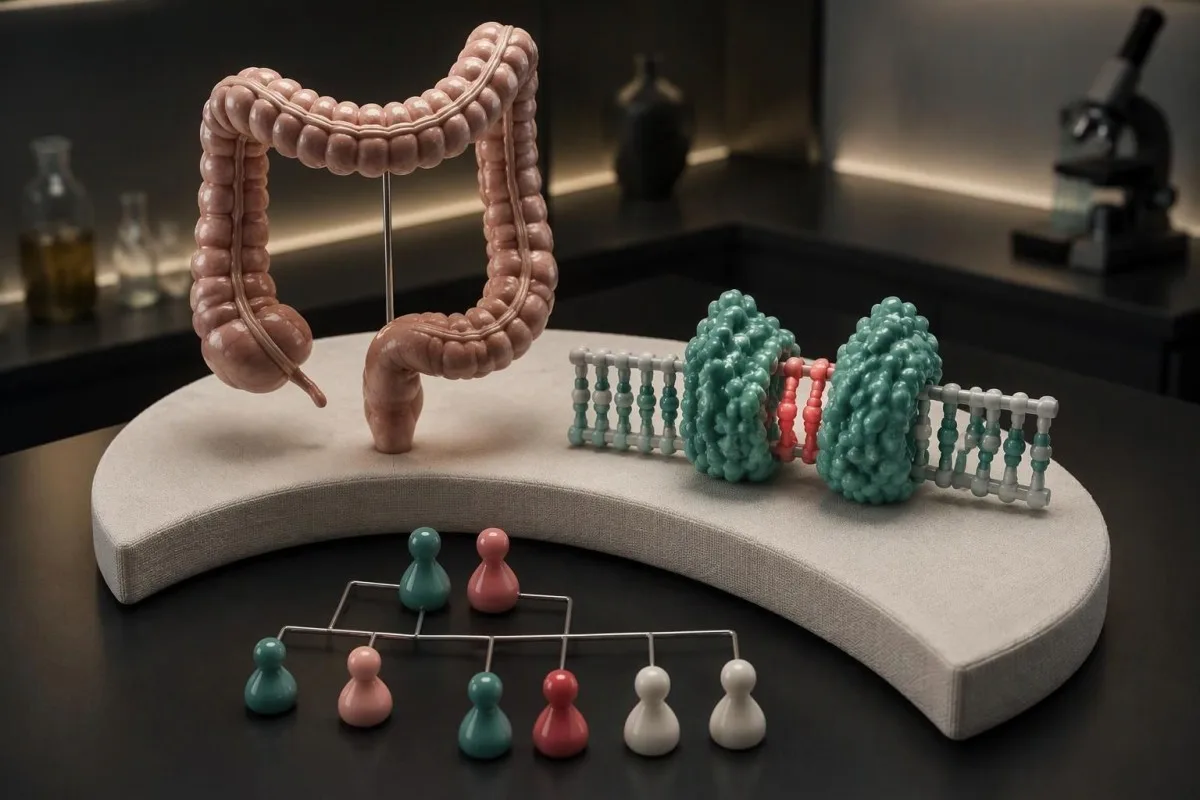
A positive inherited-risk result means a cancer predisposition—not a cancer diagnosis. A negative result depends on what was tested, while an uncertain variant should not determine treatment; tumor MSI and mismatch-repair screening answer different questions.

Galvenie secinājumi
- Positive germline result means a pathogenic or likely pathogenic variant associated with Lynch syndrome was identified; it does not mean cancer is present.
- Core gene panel usually covers MLH1, MSH2, MSH6, PMS2 and relevant EPCAM deletions—5 genes, but not 5 interchangeable risk profiles.
- True negative means the laboratory excluded a known familial pathogenic variant; a negative panel without an identified family variant may remain uninformative.
- Uncertain variant is a variant of uncertain significance, or VUS; this classification is not a positive Lynch syndrome diagnosis.
- Tumor screening uses MSI testing or immunohistochemistry for 4 mismatch-repair proteins; an abnormal tumor result does not prove an inherited variant.
- Colonoscopy planning is gene-specific: the 2020 UK hereditary colorectal cancer guideline recommends starting at 25 for MLH1/MSH2 and 35 for MSH6/PMS2, generally every 2 years.
- Iedzimtība ģimenē gives each child of a person with a heterozygous Lynch-associated pathogenic variant a 50% chance of inheriting that variant.
- Aspirin prevention requires an individual safety discussion: CAPP2 tested 600 mg daily, but that trial dose is not a self-treatment recommendation.
What do Lynch syndrome test results actually mean?
Lynch syndrome genetic testing identifies an inherited predisposition, not whether you currently have cancer. A pathogenic or likely pathogenic germline variant supports a Lynch syndrome diagnosis; a negative result has 2 different interpretations, and a variant of uncertain significance does not establish inherited cancer risk.
Vārds pozitīvu needs its specimen attached. A positive germline result concerns DNA from a constitutional sample, usually blood or saliva; a positive tumor screen concerns an existing cancer's characteristics. These are 2 separate findings, and only the first ordinarily establishes the inherited predisposition relevant to predictive testing in relatives.
A report may contain 5 classification terms: pathogenic, likely pathogenic, uncertain significance, likely benign and benign. Laboratories sometimes summarize these as positive, inconclusive or negative, which can hide the distinction between an uncertain finding and no actionable finding. Read the variant classification line—not just a portal's colored status flag.
For example, imagine 2 siblings without cancer: one has the family's pathogenic MSH2 variant and the other tests negative specifically for it. The first needs a prevention plan; the second generally does not need Lynch-specific surveillance for that familial variant. Neither result, by itself, tells you whether either sibling currently has a polyp.
Kantesti ir AI asins analīžu analizators that helps explain routine laboratory findings, but a normal complete blood count cannot exclude any of the 5 core Lynch-associated genes. Our organizācija un klīniskais tvērums are distinct from a diagnostic genetics laboratory: hereditary-risk decisions require the original report and qualified clinical interpretation.
Germline testing, MSI and mismatch-repair screening differ
Germline testing asks whether a Lynch-associated variant is present in constitutional DNA, while MSI un mismatch-repair immunohistochemistry examine cancer tissue. Immunohistochemistry assesses 4 proteins; MSI measures instability in repetitive DNA sequences. Neither tumor screening method alone proves Lynch syndrome.

Mismatch-repair immunohistochemistry, usually abbreviated IHC, examines MLH1, PMS2, MSH2 and MSH6 protein expression. Loss of 1 or more proteins can point toward a repair defect, but the pathologist must confirm that non-cancerous cells provide valid internal staining controls. Failed controls can make an apparent loss uninterpretable rather than genuinely abnormal.
An MSI-high result means the tested cancer shows substantial microsatellite instability. Some assays use 5 markers, while sequencing-based methods assess many more sites; their cutoffs are assay-specific. An MSI percentage is not your percentage chance of inheriting Lynch syndrome, a distinction also relevant to kvalitatīvi un kvantitatīvi rezultāti.
Blood-based germline testing does not measure circulating cancer DNA unless a different test was specifically ordered. Constitutional DNA is usually obtained from white cellular elements, and saliva contains cells suitable for many genetics assays. After an allogeneic stem-cell transplant, these 2 specimen types can be problematic because donor-derived DNA may affect interpretation.
Cancer tissue can contain both inherited variants and changes acquired only by that cancer. Paired tumor–normal sequencing compares 2 specimens to help separate those possibilities, although interpretation still depends on assay coverage and quality. Ask whether your report describes a constitutional finding, a tissue-only finding, or an unresolved possibility requiring a separate germline test.
What should a Lynch syndrome gene panel include?
A Lynch syndrome gene panel usually includes MLH1, MSH2, MSH6, PMS2 and relevant EPCAM deletions. Testing should assess both small sequence changes and larger deletions or duplications where appropriate; a list of 5 gene names alone does not establish complete analytical coverage.
MLH1, MSH2, MSH6 and PMS2 encode the 4 mismatch-repair proteins commonly assessed by IHC. EPCAM is different: certain deletions near its end can silence the neighboring MSH2 gene. An EPCAM finding therefore needs its exact deletion boundaries reviewed; not every change in EPCAM causes Lynch syndrome.
Two technical blind spots deserve explicit questions: copy-number changes and difficult-to-sequence regions. PMS2 has a closely related pseudogene that complicates some analyses, so specialist methods may be necessary. The broader lesson—that copy count and sequence findings are not interchangeable—is explained in our copy-number screening guide, although SMA and Lynch syndrome have different inheritance.
A broader hereditary cancer panel may examine 20, 50 or more genes because overlapping family histories can suggest another syndrome. More genes can produce more uncertain findings and incidental risks unrelated to the original concern. Before testing, ask which additional findings would change surveillance and whether you can discuss their implications before receiving results.
A negative sequencing result may not exclude every mechanism associated with inherited risk. Rare constitutional MLH1 methylation changes, mosaicism, or structural changes outside the assay's scope can require additional investigation. If an older report lists only 1 gene or lacks deletion analysis, a genetics service can decide whether updated testing would answer a genuinely unresolved question.
Who should consider testing and genetic counseling?
Testing is most informative when a relative has a known Lynch-associated variant, a cancer has suspicious mismatch-repair findings, or personal and family histories suggest inherited risk. A pedigree covering 3 generations helps determine who should be tested first and how a negative result would be interpreted.

Young-onset colorectal or endometrial cancer, multiple Lynch-associated cancers in 1 person, and cancers across successive generations can justify assessment. However, a small family, adoption, early deaths or few relatives with a uterus can hide the pattern. Absence of a striking family history is therefore weaker evidence than a confirmed negative test for a known familial variant.
When possible, test an affected relative before an unaffected relative. If a hypothetical parent had colorectal cancer at 43 and stored tissue is available, their tumor findings and germline result may be more informative than a broad panel in a healthy child. Our guide to ģimenes slimību vēža testēšanu explains why routine laboratory screening is not a substitute.
Clinical prediction tools can estimate the probability of carrying a mismatch-repair variant. A PREMM5 score of at least 2.5% is a commonly used threshold for considering genetic evaluation, but referral criteria vary and tumor findings can justify assessment independently. A risk score selects people for evaluation; it does not convert a 2.5% estimate into a diagnosis.
Before ordering a test, discuss at least 3 practical issues: possible uncertain or incidental findings, implications for relatives, and local insurance or employment rules. Kantesti's informāciju par privātumu un datiem can help you review our handling of uploaded records, but genetics laboratories and healthcare services have their own consent, retention and disclosure policies.
Positive results and MSH6 mutation meaning
A pathogenic or likely pathogenic MSH6 germline variant supports a diagnosis of Lynch syndrome; it does not diagnose cancer. For MSH6 mutation meaning, distinguish 3 details: the exact variant, its laboratory classification and whether it was found in constitutional DNA or only in cancer tissue.
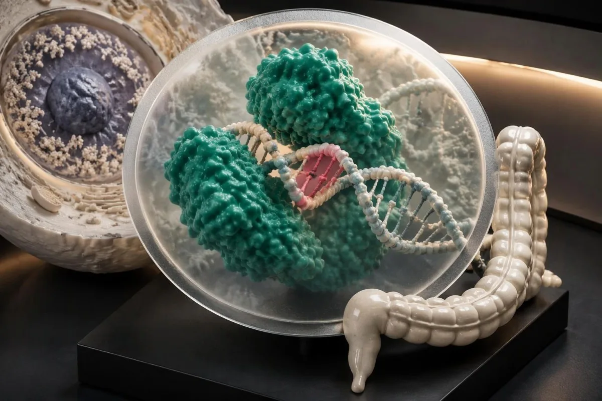
Lynch syndrome usually involves a pathogenic variant in 1 of the 2 copies of a mismatch-repair gene. Cancer development commonly requires additional changes in affected cells, including loss of the remaining functional repair pathway. This explains why inherited predisposition can be present from birth while cancer may develop much later—or never develop.
Risk differs across genes, organs, ages and family backgrounds. MSH6-associated colorectal cancer often develops later than MLH1- or MSH2-associated colorectal cancer, which helps explain different surveillance starting ages. For someone with a uterus, endometrial risk deserves a separate discussion; 1 combined label such as 'moderate lifetime cancer risk' can obscure very different organ-specific decisions.
Vārds likely does not make a likely pathogenic variant equivalent to a VUS. The ACMG/AMP framework describes likely pathogenic as greater than 90% certainty that a variant is disease-causing, not a greater than 90% chance that its carrier will develop cancer (Richards et al., 2015). That distinction prevents a classification probability from becoming a misleading personal risk estimate.
Kantesti ir AI asins analīžu rezultātu platforma that can help contextualize hemoglobin and ferritin, but those 2 measurements cannot calculate penetrance for an MSH6 variant. Our validācijas un klīniskās robežas should not be interpreted as validation for germline variant classification. Ask your genetics team for a gene-specific surveillance letter rather than a generic positive-result summary.
When does a negative result genuinely reduce risk?
A true negative excludes the specific pathogenic variant already established in your family; an uninformative negative finds no explanatory variant when the family's genetic cause remains unknown. These 2 negative results can lead to very different surveillance recommendations despite identical wording in a patient portal.
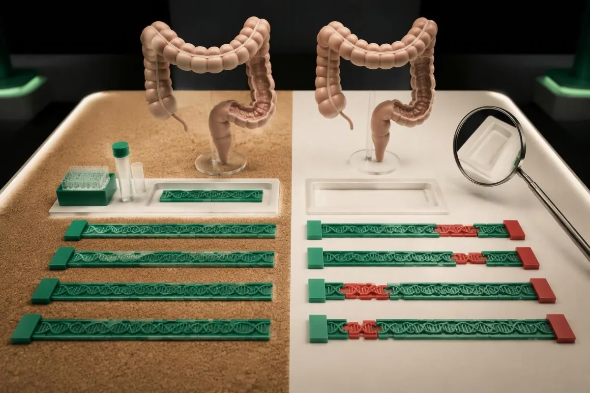
For a hypothetical adult whose mother has a confirmed pathogenic MLH1 variant, a validated negative test for that exact variant usually removes the mother's Lynch-associated inherited risk. It does not remove unrelated risks from the other parent, personal polyps or lifestyle. General-population screening—or another family-history schedule—may still apply; 'negative' never means zero lifetime cancer risk.
An unaffected person with 2 close relatives who had colorectal cancer may receive a negative broad panel without anyone identifying the family's cause. That result cannot establish whether the family has an undetected genetic mechanism, another inherited condition, or shared non-genetic risk. Surveillance should continue to reflect the documented pedigree rather than automatically reverting to the population schedule.
Three report details can change interpretation: the familial variant identifier, the test's detection methods and its stated limitations. Confirm that the laboratory actually tested the same variant found in the relative, particularly when reports use different transcript versions. Our medicīnisko pārskatu avota pārbaudes explain why the original laboratory wording matters more than an automated paraphrase.
Kantesti cannot make a negative inherited-risk report more conclusive by combining it with normal routine laboratory values. Even 10 normal blood-test results do not repair missing gene coverage or exclude a family-specific variant. A genetics review is more useful than repeating the same panel annually unless technology, the pedigree or the original interpretation has materially changed.
What should you do with an uncertain genetic variant?
A nenoteiktas nozīmes variants, or VUS, means evidence is insufficient to classify a genetic change as disease-causing or benign. A VUS is 1 of 5 standard classification categories and should not, by itself, trigger Lynch-specific surgery, intensified surveillance or predictive testing that labels relatives as affected.

ACMG/AMP guidance states that a VUS should not be used in clinical decision-making (Richards et al., 2015). A person with a VUS may still need earlier colonoscopy because of 2 relatives with young-onset colorectal cancer, but the reason is the family history—not proof that the uncertain variant causes Lynch syndrome. Keep those rationales separate in the surveillance letter.
Laboratories evaluate several evidence types, including population frequency, functional experiments, segregation within families and consistency with established disease mechanisms. A computer prediction alone is not enough to establish pathogenicity. Even agreement between 2 prediction tools may reflect similar underlying training data rather than 2 independent pieces of biological evidence.
Reclassification can move a VUS toward benign or pathogenic, but there is no universal 12-month deadline for resolution. Ask who will contact you if the laboratory changes its classification and whether your contact details are linked to a recontact process. Our medicīniskā satura standarti also distinguish unresolved evidence from clinically actionable conclusions.
Testing selected relatives can sometimes help a laboratory study an uncertain variant, but that is different from routine predictive cascade testing. If 1 relative carries the VUS and another does not, those results alone should not decide who needs surgery or who can stop screening. Request written clarification when a family study is being performed for evidence gathering rather than diagnosis.
Why can tumor screening be abnormal without Lynch syndrome?
Abnormal tumor mismatch repair can result from sporadic MLH1 methylation, acquired changes in both copies of a repair gene, or inherited Lynch syndrome. These are at least 3 distinct explanations; tumor follow-up tests help distinguish them, especially when germline testing finds no pathogenic variant.
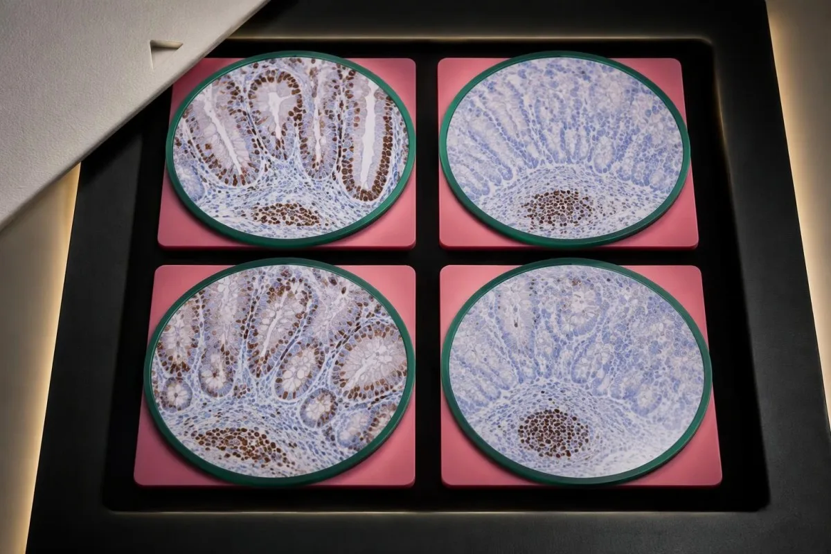
Loss of MLH1 and PMS2 often occurs together because these 2 proteins function as a pair. In colorectal cancer, MLH1 promoter methylation testing and sometimes BRAF V600E testing can support a sporadic explanation. BRAF is not the corresponding exclusion tool for endometrial cancer, and unusually young onset or a strong pedigree may still warrant specialist review.
Loss of MSH2 with MSH6, or isolated loss of MSH6 or PMS2, can guide germline assessment toward particular genes. However, these 4-protein patterns are clues, not a guaranteed gene diagnosis: staining artifacts, treatment effects and unusual biological mechanisms can complicate interpretation. A retained stain also does not prove that a protein functions normally.
Double-somatic findings describe 2 acquired events affecting mismatch repair within the cancer, without an explanatory germline variant identified. Paired tumor–normal analysis can support that conclusion and avoid incorrectly labeling an entire family with Lynch syndrome. An unresolved mismatch-repair-deficient cancer is not automatically double-somatic; management may still depend on family history and the completeness of testing.
Tumor markers such as CEA, reported in ng/mL, cannot establish whether mismatch-repair loss is inherited. Our explanation of elevated CEA limitations separates monitoring markers from hereditary diagnosis. Ask for 2 summaries when relevant: one explaining cancer treatment implications, including possible immunotherapy relevance, and another explaining what relatives should do.
How does a positive result change colonoscopy surveillance?
Confirmed Lynch syndrome generally requires regular colonoscopy, with starting age and interval tailored to the gene and family history. The 2020 BSG/ACPGBI/UKCGG guideline recommends starting at 25 years for MLH1/MSH2 un 35 years for MSH6/PMS2, with colonoscopy generally every Ja ir spēcīga ģimenes anamnēze vai vielmaiņas risks, selektīvu testēšanu var sākt no.
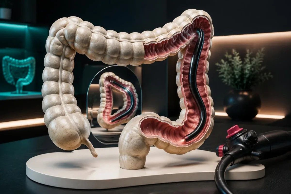
UK recommendations illustrate why a positive MSH6 result does not necessarily generate the same schedule as a positive MLH1 result (Monahan et al., 2020). US guidance commonly starts MLH1/MSH2 surveillance at 20–25 years and MSH6/PMS2 surveillance at 30–35 years, with intervals ranging from 1–3 years depending on gene and guideline. EPCAM-related surveillance is commonly aligned with MSH2.
A family member's unusually early cancer, previous colorectal cancer, adenomas or an inadequate examination can justify a different schedule. A normal colonoscopy means no concerning finding was identified at that examination; it does not remove the inherited predisposition. If your recommended interval is 2 years, ask whether the next date is measured from the completed high-quality examination or a planned repeat.
FIT and FOBT do not replace Lynch surveillance colonoscopy. Stool tests detect blood rather than reliably finding and removing precancerous polyps, so a negative stool test cannot reset a 1–2-year colonoscopy plan. Our pozitīvās FIT izmeklējumu rezultātu izmeklēšanas vadlīnijas explains why an abnormal stool result still needs diagnostic assessment.
Preparation quality, complete examination and polyp removal matter alongside the calendar interval. Keep the endoscopy report, including whether the colon was fully examined and whether any polyp requires an earlier repeat. The limitations described in our FOBT result interpretation also explain why new symptoms need review between scheduled procedures rather than waiting for the next surveillance date.
What changes for endometrial and ovarian cancer prevention?
Lynch syndrome can increase endometrial and, for some genes, ovarian cancer risk, but screening and preventive surgery are separate decisions. Some US guidance considers endometrial tissue sampling every 1–2 years from age 30–35; a survival benefit from routine screening has not been established.

A Pap test or HPV test does not screen the endometrium or ovaries. These 2 tests primarily support cervical cancer prevention, so a reassuring cervical result cannot exclude a Lynch-associated endometrial problem. Our HPV un Pap testa rezultātu interpretācijā helps distinguish which organ a screening result actually addresses.
New bleeding after menopause, persistent bleeding between periods, or a meaningful change from your usual menstrual pattern deserves prompt assessment rather than waiting 12 months for surveillance. Premenopausal symptoms have many benign explanations, but an inherited-risk history changes the threshold for investigation. Our Ieteikumi par stipru menstruālo asiņošanu also identifies circumstances needing urgent care.
Risk-reducing hysterectomy can prevent endometrial cancer, but removing the ovaries is a separate choice with different benefits and harms. Timing depends on the gene, completed childbearing, previous cancer and preferences; removal before natural menopause can affect bone, cardiovascular and sexual health. For PMS2, ovarian risk is not clearly increased, so treating all 4 mismatch-repair genes identically can lead to unnecessary intervention.
Screening practice differs internationally because detection studies have not established the same benefit as prevention by surgery. Transvaginal ultrasound and CA-125, reported in U/mL, have not been proven to reliably prevent ovarian cancer deaths in Lynch syndrome. Ask for a written plan covering symptoms, any proposed tissue sampling, fertility goals and the specific rationale for considering 1 operation rather than another.
Which other cancers need a gene-specific discussion?
Additional Lynch surveillance depends on the gene, family history and local guidance; it is not a universal annual scan of every organ. Upper urinary tract, stomach, small bowel and certain skin cancers deserve consideration, while the benefit of several proposed screening tests remains uncertain across the 4 mismatch-repair genes.
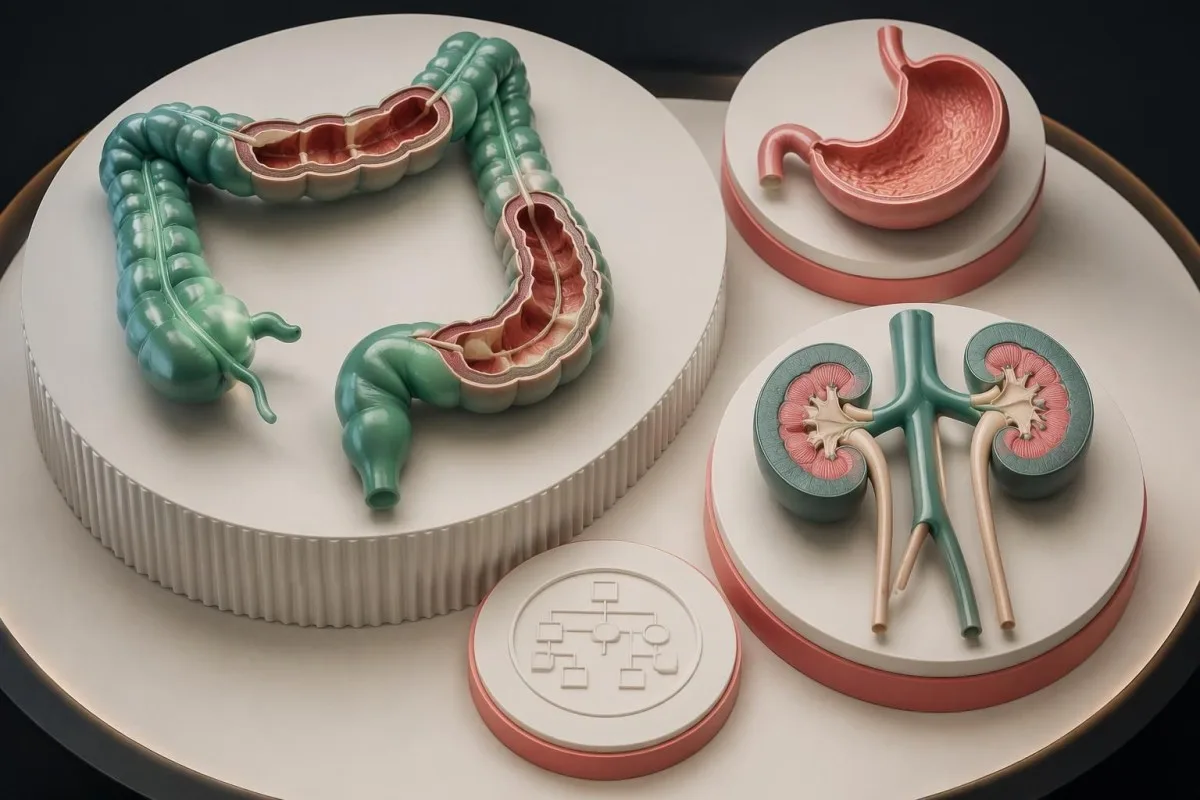
Upper urinary tract cancer involves the renal pelvis or ureter and is particularly relevant to some MSH2 families. Visible blood in urine needs assessment even after 1 apparently benign episode, although infections and stones are common alternatives. Our urinary cancer warning signs address symptom urgency; bladder and upper-tract cancers are not anatomically identical.
Routine urinalysis or urine cytology has not been shown to provide a clear mortality benefit for every person with Lynch syndrome. Some services consider annual testing when family history or gene-specific risk warrants it, while others emphasize symptoms. A negative urine dipstick cannot guarantee the absence of a small upper-tract cancer, and a positive result still has more than 1 possible cause.
Upper gastrointestinal surveillance is another area of disagreement. Some US protocols consider endoscopy from age 30–40 at 2–4-year intervals for selected people, whereas the 2020 UK guideline does not recommend routine gastric or small-bowel surveillance outside a clinical trial (Monahan et al., 2020). Helicobacter pylori testing and eradication are a distinct preventive discussion, not proof that endoscopy is unnecessary.
Sebaceous skin growths can be part of the Muir–Torre phenotype, a Lynch-associated presentation that may prompt dermatological assessment. Pancreatic screening is generally restricted to selected higher-risk people, often involving a relevant family history and a specialist program. Rather than requesting 10 unrelated annual tests, ask which additional organ has a documented risk increase in your gene and family.
Can aspirin or routine blood tests help prevent cancer?
Aspirin can reduce colorectal cancer risk in Lynch syndrome, but dose and suitability require a clinician's assessment. CAPP2 randomized 861 participants uz 600 mg aspirin daily or placebo and found a long-term colorectal cancer benefit; routine blood tests cannot replace genetic testing or colonoscopy.

CAPP2 reported colorectal cancer in 40 of 427 aspirin-assigned participants versus 58 of 434 placebo-assigned participants during long-term follow-up, with a hazard ratio of 0.65 (Burn et al., 2020). Benefit was delayed rather than immediate. The trial's 600 mg dose is evidence about that regimen, not permission to start high-dose aspirin without reviewing bleeding risk and other medicines.
Aspirin planning should consider at least 3 safety factors: previous gastrointestinal bleeding or ulcer disease, anticoagulant use and allergy or intolerance. Lower-dose approaches are used in practice, but the optimal dose for an individual is not determined by their gene name alone. Ask the prescriber to explain the evidence behind the chosen dose, intended duration and any stomach-protection strategy.
Iron deficiency may develop before anemia and can justify investigation even when a recent complete blood count was reassuring. A ferritin below about 15 ng/mL strongly supports depleted iron stores in an otherwise uncomplicated adult, but inflammation can make ferritin less reliable. Our zema feritīna simptomu ceļvedis explains why replacing iron does not remove the need to identify its cause.
Kantesti can help explain routine hemoglobin and iron-panel trends, but neither a normal hemoglobin in g/dL nor 1 normal ferritin result rules out colorectal cancer. Our dzelzs izmeklējumu interpretācija is useful for understanding iron physiology, not diagnosing Lynch syndrome. Nutrition and exercise support general health; no supplement repairs an inherited pathogenic mismatch-repair variant.
What should relatives do after a positive result?
Each child of a person with a heterozygous Lynch-associated pathogenic variant has a 50% chance of inheriting that variant, regardless of sex. Adult relatives should be offered targeted cascade testing when appropriate, using the exact familial variant rather than assuming that every relative needs a broad panel.
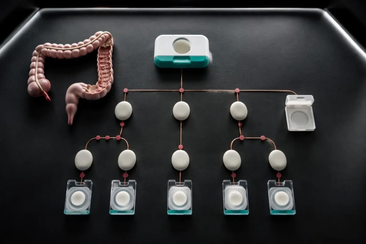
The 50% probability applies independently to each pregnancy; it does not mean exactly half the children in a particular family will inherit the variant. A sibling's probability depends on the family structure and which parent carries it. If a shared parent has the variant, each sibling generally has a 50% chance; if the variant arose newly in one sibling, the calculation changes.
A family letter should identify the gene, exact variant, laboratory classification and route to genetic counseling. Sharing the relevant genetics report can prevent a relative from being tested for the wrong target. Our drošu ģimenes rezultātu kopīgošanu describes consent-based workflows, but sending a report is not the same as another person consenting to genetic testing.
Predictive testing is usually deferred until adulthood because routine Lynch surveillance generally begins later; exceptionally early family cancers can change that discussion. Two pathogenic variants affecting both copies of the same mismatch-repair gene can cause constitutional mismatch repair deficiency, a different condition with childhood implications. If both reproductive partners carry pathogenic variants in the same gene, each pregnancy can have a 25% chance of that biallelic condition.
Kantesti's family-history features can support organization of health information, but they cannot infer that an untested relative carries the variant. Our vadlīnijas par daudzgadu piekrišanu explains why 1 person's result should not automatically populate another person's record as a diagnosis. Relatives may need time to decide about testing; offer accurate information without presenting inherited predisposition as an inevitable cancer outcome.
Your next steps and the scope of supporting research
The next step is to match the result to 3 records: the original genetics report, relevant tumor testing and a verified family history. Then obtain a written surveillance plan that states which recommendations follow from a pathogenic variant and which follow from personal or family history.
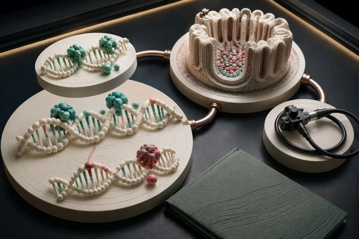
Bring 5 questions to the follow-up appointment: Is this germline or tumor-only? What is the exact classification? Was the familial variant excluded? Does further tumor analysis remain necessary? What should relatives do? Request specific dates for surveillance rather than 'regular checks,' and identify who owns the plan if genetics, gastroenterology and oncology services are all involved.
Kantesti ir ar AI balstīts asins analīzes rīks that can support understanding of routine laboratory reports, not a service that confirms any of the 5 Lynch-associated genes. Our atbildīgas MI izmantošanas robežas explain why original-source verification and clinician review matter. Do not allow an automated summary to override a laboratory's VUS classification or change a specialist's surveillance interval.
The 2 Zenodo publications listed below are background resources on routine iron and coagulation testing; neither is a Lynch syndrome genetic-testing study. The koagulācijas testu rezultātu interpretācijas ceļvedis may clarify laboratory terminology during medication discussions, but a normal aPTT in seconds does not establish that aspirin is safe or exclude gastrointestinal bleeding risk.
New symptoms require diagnostic assessment even if your next surveillance examination is 1 year away. Persistent rectal bleeding, unexplained iron deficiency, visible blood in urine or postmenopausal bleeding should not be dismissed because an inherited-risk result was negative. Heavy bleeding with fainting, severe abdominal pain or black tarry stool with weakness warrants urgent medical care rather than waiting for genetic clarification.
Bieži uzdotie jautājumi
Vai pozitīvs ģenētiskais tests uz Linča sindromu nozīmē, ka man ir vēzis?
0 Pozitīvs dzimumšūnu līnijas Linča sindroma tests liecina par iedzimtu noslieci uz vēzi, nevis par jau esošu vēzi. Parasti rezultāts atklāj patogēnu vai, visticamāk, patogēnu variantu, kas ietekmē vienu ar Linča sindromu saistīta gēna kopiju. Vēža risks atšķiras atkarībā no gēna, vecuma, orgāna un ģimenes anamnēzes, tāpēc pozitīvs rezultāts nenozīmē, ka vēzis ir neizbēgams. Atbilstošais nākamais solis ir ģenētiskā konsultēšana un konkrētajam gēnam pielāgots uzraudzības plāns, nevis pieņēmums, ka nepieciešama vēža ārstēšana.
0 Vai negatīvs ģenētiskais tests var izslēgt Linča sindromu?
A negative test is most conclusive when it excludes the exact pathogenic variant already confirmed in the family. There are 2 main negative-result situations: a true negative for that familial variant and an uninformative negative when the family's cause remains unknown. A negative panel may not exclude mechanisms outside its analytical coverage or another explanation for a strong cancer history. Screening should therefore follow the genetics team's interpretation of the report, personal history and pedigree.
Ko nozīmē MSH6 mutācija?
Patogēns vai, visticamāk, patogēns MSH6 variants konstitucionālajā DNS apstiprina Linča sindroma diagnozi, savukārt MSH6 VUS to neapstiprina. MSH6 ir viens no 4 neatbilstību labošanas proteīnu gēniem, ko parasti izvērtē Linča sindroma noteikšanai. Kolorektālais vēzis ar MSH6 saistīta Linča sindroma gadījumā bieži attīstās vēlāk nekā ar MLH1 vai MSH2 saistītas slimības gadījumā, taču arī endometrija vēža riskam jāpievērš atsevišķa uzmanība. Atrade, kas konstatēta tikai vēža audos, pirms to var raksturot kā iedzimtu, ir papildus jāizvērtē.
Vai MSI augsta pakāpe ir tas pats, kas Linča sindroms?
MSI-augsts raksturo atkārtotu DNS sekvenču nestabilitāti pārbaudītajā audzējā un nav sinonīms iedzimtam Linča sindromam. Audzēja skrīningā var izmantot arī imūnhistoķīmiju 4 proteīniem: MLH1, PMS2, MSH2 un MSH6. Sporforiska metilācija vai iegūtas izmaiņas var izraisīt neatbilstošu DNS reparāciju bez iedzimta patogēna varianta. Dzimumšūnu līnijas izvērtēšana un, ja indicēts, papildu audzēja testēšana nosaka, ko šis atradums nozīmē radiniekiem.
Vai radinieki ir jāpārbauda attiecībā uz Linča sindroma VUS?
A Lynch-associated VUS should not ordinarily be used for predictive testing that determines whether relatives are affected or can stop surveillance. A VUS is 1 of 5 standard variant classification categories and does not establish pathogenicity. A genetics laboratory may sometimes request selected family testing to gather evidence, but that is a different purpose from diagnostic cascade testing. Relatives' surveillance should reflect their personal and family histories while the variant remains uncertain.
Cik bieži cilvēkiem ar Linča sindromu jāveic kolonoskopija?
Colonoscopy intervals in Lynch syndrome depend on the gene, examination findings, family history and local guidance. The 2020 UK hereditary colorectal cancer guideline recommends colonoscopy generally every 2 years, starting at 25 for MLH1/MSH2 and 35 for MSH6/PMS2. US schedules commonly use intervals between 1 and 3 years, with earlier starting ages for MLH1/MSH2 than for MSH6/PMS2. A negative FIT or FOBT does not replace the recommended colonoscopy schedule.
Should I start taking aspirin if my Lynch syndrome test is positive?
Do not start aspirin solely because a Lynch syndrome result is positive without discussing benefits and harms with a clinician. CAPP2 tested 600 mg daily in 861 participants and demonstrated a delayed long-term colorectal cancer benefit, but that dose is not a universal prescribing recommendation. Previous gastrointestinal bleeding, ulcer disease, anticoagulants and intolerance can change the decision. The chosen dose and duration should be documented alongside surveillance, because aspirin does not replace colonoscopy.
Uzziniet, kā lasīt asins analīžu rezultātus
Augšupielādējiet laboratorijas pārskatu, un Kantesti mākslīgais intelekts dažu sekunžu laikā vienkāršā valodā izskaidros katru biomarķieri. To izmanto vairāk nekā 2 miljoni cilvēku visā pasaulē.
Atsauces
- Klein, T., Mitchell, S., & Weber, H. (2026). Dzelzs izmeklējumu ceļvedis: TIBC, dzelzs piesātinājums un saistīšanas spēja. Kantesti mākslīgā intelekta medicīniskie pētījumi. DOI
- Klein, T., Mitchell, S., & Weber, H. (2026). aPTT normālais diapazons: D-dimēra un C proteīna rokasgrāmata par asins recēšanu. Kantesti mākslīgā intelekta medicīniskie pētījumi. DOI
- Monahan KJ et al. (2020). Guidelines for the management of hereditary colorectal cancer from the British Society of Gastroenterology (BSG)/Association of Coloproctology of Great Britain and Ireland (ACPGBI)/United Kingdom Cancer Genetics Group (UKCGG). Gut. DOI
- Richards S et al. (2015). Standarti un vadlīnijas sekvenču variantu interpretācijai: Amerikas Medicīniskās ģenētikas un genomikas koledžas un Molekulārās patoloģijas asociācijas kopīgs konsensa ieteikums. Medicīniskā ģenētika. DOI
- Burn J et al. (2020). Cancer prevention with aspirin in hereditary colorectal cancer (Lynch syndrome), 10-year follow-up and registry-based 20-year data in the CAPP2 study: a double-blind, randomised, placebo-controlled trial. The Lancet. DOI
- 2M+Analizētie testi
- 127+Valstis
- 100+Valodas
Par medicīnas komandu


E-E-A-T uzticēšanās signāli
Pieredze
Ārstu vadīta klīniskā laboratorijas interpretācijas darbplūsmu pārskatīšana.
Ekspertīze
Laboratorijas medicīnas fokuss uz to, kā biomarķieri uzvedas klīniskā kontekstā.
Autoritāte
Medicīniski recenzējis Dr. Thomas Klein, piedaloties Dr. Sarah Mitchell un Prof. Dr. Hans Weber.
Uzticamība
Uz pierādījumiem balstīta interpretācija ar skaidriem turpmākās rīcības ceļiem, lai mazinātu trauksmi.




Medicīniskā atruna
Šis raksts ir paredzēts tikai izglītojošiem nolūkiem un nav medicīnisks padoms. Vienmēr konsultējieties ar kvalificētu veselības aprūpes speciālistu, lai pieņemtu lēmumus par diagnostiku un ārstēšanu.
Publicēts · Sazinieties ar redakcijas komandu
Publicējis Kantesti Ltd, reģistrēts Anglijā un Velsā, uzņēmuma reģistrācijas Nr. 17090423, Londona, Apvienotā Karaliste.
