
Niski wynik aktywności enzymu GAA nasuwa podejrzenie choroby Pompego, ale samo badanie przesiewowe nie może jej potwierdzić. Powtórzenie badania w odpowiednim materiale i analiza genetyczna GAA pomagają odróżnić rzeczywisty niedobór, nosicielstwo i pseudoniedobór.

Najważniejsze wnioski
- Niska aktywność enzymu GAA jest sygnałem wskazującym na potrzebę dalszej diagnostyki, a nie rozpoznaniem; 1 nieprawidłowy wynik z suchej kropli krwi wymaga potwierdzenia pod kierunkiem specjalisty.
- Zakresy referencyjne zależą od materiału i metody oznaczenia; wartości w nmol/h/mL nie można bezpośrednio porównywać z wartościami w nmol/h/mg białka.
- Resztkowa aktywność enzymu często wynosi poniżej 1% w klasycznej niemowlęcej postaci choroby Pompego, ale wartość ta nie jest uniwersalnym progiem badania przesiewowego.
- Pseudoniedobór może obniżać laboratoryjnie oznaczaną aktywność GAA bez wywoływania choroby Pompego; powtórzenie tego samego oznaczenia może dać ponownie niski wynik.
- Badanie genu GAA poszukuje wariantów chorobotwórczych dotyczących obu kopii genu; 2 warianty należy zinterpretować pod kątem klasyfikacji i fazy.
- Warianty o niepewnym znaczeniu nie potwierdzają choroby Pompego; 1 lub 2 warianty o niepewnym znaczeniu wymagają dodatkowych dowodów.
- Ryzyko rodzinne wynosi 25% dla każdego dziecka dotkniętego chorobą w każdej ciąży, gdy oboje rodzice są nosicielami wariantu GAA wywołującego chorobę.
- Ocena pilna jest potrzebna u niemowlęcia z niską aktywnością GAA oraz trudnościami w karmieniu, znacznym osłabieniem lub podejrzeniem zajęcia serca; nie należy czekać na rutynowe ponowne badanie.
Co tak naprawdę oznacza niski wynik aktywności enzymu GAA?
A Badanie w kierunku choroby Pompego wykazujące niską aktywność GAA oznacza, że laboratorium zmierzyło obniżoną aktywność kwaśnej alfa-glukozydazy w warunkach zastosowanego testu. Nie potwierdza to choroby Pompego: 1 niski wynik badania przesiewowego może wynikać z rzeczywistego niedoboru, pseudoniedoboru, nakładania się wyników u nosicieli lub problemu z próbką.

Aktywność GAA mierzy funkcję enzymu, a nie po prostu ilość obecnego białka enzymatycznego. Pierwsze 3 kwestie, które należy sprawdzić, to rodzaj próbki, jednostki miary i wartość graniczna przyjęta przez laboratorium; wyniku określonego jako niski, bez tych informacji, nie można wiarygodnie zinterpretować w odniesieniu do liczby znalezionej w internecie.
Powód wykonania badania zmienia znaczenie dodatniego wyniku przesiewowego. Noworodek zidentyfikowany w ramach badań przesiewowych populacji i 45-latek z postępującym osłabieniem mięśni bioder mają różne prawdopodobieństwo wyjściowe choroby Pompego, nawet jeśli podane wartości aktywności enzymu są takie same; dlatego objawy i wyniki badania przedmiotowego należy rozpatrywać razem z wynikiem laboratoryjnym.
Dodatni wynik badania przesiewowego nie jest tym samym co potwierdzenie choroby. Jeśli 2 laboratoria stosują różne substraty lub sposoby przygotowania próbek, ich wyniki liczbowe mogą się różnić, mimo że żadne z nich nie popełniło błędu; należy zapytać, czy podczas kolejnej wizyty zostanie wykonany kolejny pomiar przesiewowy, czy rzeczywiście potwierdzający test aktywności enzymu.
Kantesti jest Analizator do badań krwi AI które może pomóc uporządkować wynik badania GAA wraz z rutynowymi wynikami laboratoryjnymi, ale nie może potwierdzić rozpoznania choroby Pompego na podstawie 1 wartości oznaczonej jako nieprawidłowa. Nasza informacje o firmie wyjaśnia organizację usługi; podejrzenie dziedzicznego niedoboru enzymu nadal wymaga konsultacji ze specjalistą chorób metabolicznych lub nerwowo-mięśniowych.
Dlaczego niedobór GAA powoduje problemy z mięśniami i sercem?
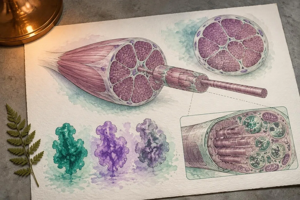
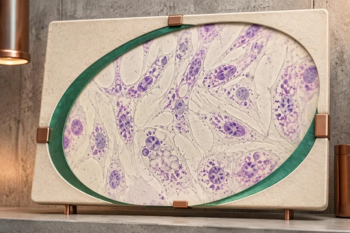
Choroba Pompego wynika z niedoboru lizosomalnej kwaśnej alfa-glukozydazy, enzymu rozkładającego glikogen w komórkowych przedziałach recyklingowych. GAA działa na wiązania glukozy alfa-1,4 i alfa-1,6; niewystarczająca aktywność powoduje gromadzenie się glikogenu, szczególnie w mięśniach szkieletowych i sercu.

Klasyczna niemowlęca postać choroby Pompego często wiąże się z aktywnością GAA poniżej 1% normy w diagnostycznych testach tkanek. W postaci o późnym początku może utrzymywać się większa aktywność, czasami około 2–40% w określonych testach; te orientacyjne zakresy pokrywają się z ograniczeniami testów i nie mogą zastępować zatwierdzonej przez laboratorium interpretacji diagnostycznej.
Wytyczne Kishnaniego i wsp. z 2006 r., opublikowane w Genetics in Medicine, opisują związek między niedoborem GAA, spichrzaniem glikogenu i oceną wieloukładową. Rozróżnienie kliniczne nie sprowadza się jedynie do tego, czy aktywność jest niższa niż 1%: wiek wystąpienia objawów, wyniki dotyczące serca i czynność mięśni oddechowych również pomagają określić postać i pilność leczenia choroby.
Rutynowy wynik stężenia glukozy nie mierzy rozkładu glikogenu w lizosomach. Na przykład u hipotetycznej osoby dorosłej stężenie glukozy na czczo może wynosić 92 mg/dL, mimo dziedzicznego zaburzenia spichrzania glikogenu w mięśniach; prawidłowe stężenie glukozy nie dowodzi, że glikogen w lizosomach mięśni jest prawidłowo przetwarzany.
Inne choroby lizosomalne dotyczą odmiennych enzymów i genów, dlatego ich wartości granicznych nie można stosować w badaniach w kierunku choroby Pompego. Nasze wyjaśnienie potwierdzania choroby Gauchera badaniem aktywności enzymu przedstawia szerszy schemat postępowania od badania aktywności enzymu do badań genetycznych, wskazując zarazem, dlaczego testu enzymatycznego w kierunku jednej choroby nie można zastąpić testem w kierunku innej choroby.
Czy istnieje zakres normy dla aktywności enzymu GAA?
There is no single universal normal range for GAA enzyme activity. Dried blood spots, leukocytes and cultured fibroblasts use different preparations and reporting units; a number expressed in nmol/h/mL cannot be directly compared with a result expressed in nmol/h/mg protein.

A screening cutoff separates samples needing follow-up from samples passing that screen. It does not necessarily separate affected people from unaffected people with complete accuracy; laboratories may use different fluorometric or tandem mass spectrometry methods, and 2 screening programs may therefore select different recall thresholds.
For a clearly hypothetical example, suppose a laboratory uses a DBS screening cutoff of 1.5 nmol/h/mL and reports 0.7 nmol/h/mL. The result is below that invented laboratory threshold, but dividing 0.7 by 1.5 gives 47% of the cutoff—not 47% of normal enzyme activity and not a severity score.
The lower limit of detection is different from the clinical cutoff. A report reading less than 0.1 in its stated units may mean the method cannot quantify lower activity reliably; it does not prove absolute absence of enzyme, and repeating measurements close to that analytical floor may produce proportionally large variation.
Screening performance also depends on prevalence. In a hypothetical group with 10 true-positive and 100 false-positive results, only about 9% of positive screens represent disease; those invented counts are not Pompe screening statistics, but explain why wyniki jakościowe w porównaniu z ilościowymi require different interpretation.
| Poziom | Zakres | Co to oznacza |
|---|---|---|
| Passes hypothetical screen | ≥1.5 nmol/h/mL | Illustrative DBS cutoff only; not a universal normal range and not an absolute exclusion in a symptomatic person. |
| Below hypothetical cutoff | <1.5 nmol/h/mL | Illustrative follow-up threshold only; true deficiency, pseudodeficiency and specimen effects still need separation. |
Dlaczego należy powtórzyć badanie GAA w innym materiale?
Repeat GAA testing checks whether low activity is reproducible and whether specimen quality or assay interference explains it. Confirmation usually moves beyond 1 screening card to a fresh, appropriately handled specimen, often with GAA measurement in leukocytes or another specialist-selected cell preparation.

Heat, humidity and inadequate drying can reduce enzyme activity in a dried blood spot. A low result after 2 days of poorly controlled transport may deserve a fresh collection, but transport history cannot be used to dismiss a result automatically; the laboratory should assess card quality and its stability requirements.
Acarbose is used in some GAA assay methods to suppress interfering maltase-glucoamylase activity. That interference can make measured activity appear higher and potentially obscure deficiency; checking how a laboratory isolates GAA activity can therefore matter more than obtaining a second number with the same unresolved methodological limitation.
Two low DBS results still do not automatically confirm Pompe disease. Pseudodeficiency can reproduce across repeated measurements, including some confirmatory methods; when enzyme and genetic findings disagree, a specialist laboratory may recommend cultured fibroblast testing or additional molecular investigation rather than simply ordering a third screening card.
Give the laboratory the dates of collection, recent transfusions or transplantation, and any enzyme replacement treatment already received. Our guide to zakłóceń związanych z jakością próbki explains the general distinction between biological findings and specimen problems; GAA testing has its own handling rules, so do not import another test's redraw interval.
Czym różni się pseud niedobór GAA od choroby Pompego?
GAA pseudodeficiency means an inherited variant lowers measured enzyme activity without causing the glycogen-storage disorder Pompe disease. Two commonly discussed variants are c.1726G>A, p.Gly576Ser, and c.2065G>A, p.Glu689Lys; their interpretation depends on the complete genotype and the assay used.
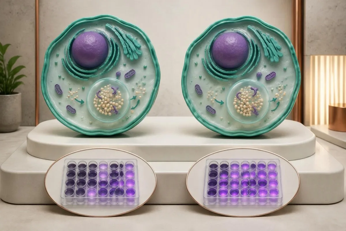
Pseudodeficiency is a biochemical testing issue, not an early stage of Pompe disease. A person with only pseudodeficiency variants does not need Pompe treatment because of 1 low activity result; the specialist must first verify that disease-causing variants or another explanation have not been missed.
The p.Gly576Ser and p.Glu689Lys variants are often found together on 1 gene copy and are particularly relevant in some East Asian ancestry populations. Ancestry changes how often laboratories encounter this pattern, but cannot establish or exclude pseudodeficiency in an individual; people from other backgrounds can carry these variants too.
Pseudodeficiency can remain low on repeat enzyme testing. The practical distinction is whether the complete biochemical and genetic evidence supports disease-causing GAA deficiency, not whether a second result falls below an arbitrary percentage; specialists may compare different assay substrates or cell preparations when the pattern remains ambiguous.
Pseudodeficiency variants can also coexist with a disease-causing variant, making a short report misleading if it lists everything under mutations. Our Fabry enzyme and genetics guide shows another situation where enzyme results need genetic context, although Fabry's X-linked inheritance differs from Pompe's 2-copy recessive mechanism.
Czego należy szukać w badaniu genu GAA?
GAA gene testing investigates whether disease-causing variants affect both copies of the GAA gene. Pompe disease is autosomal recessive, and the GAA gene is located on chromosome 17; interpretation requires variant classification, adequate test coverage and, when needed, confirmation that variants affect opposite gene copies.
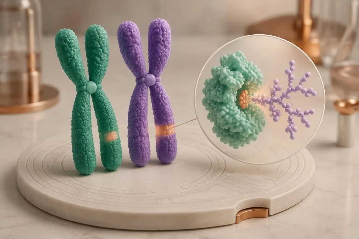
Two pathogenic or likely pathogenic GAA variants in trans support molecular confirmation of Pompe disease. In trans means the variants affect opposite gene copies; 2 variants in cis affect the same copy and do not, by themselves, demonstrate a recessive disease genotype.
Standard sequencing detects many small substitutions and insertions or deletions, but does not answer every possible genetic question. If only 1 disease-causing variant is found despite convincing enzyme deficiency, ask whether deletion/duplication analysis was included and whether poorly covered or deep intronic regions require further investigation.
A common later-onset-associated GAA variant is c.-32-13T>G, which affects splicing. Finding this variant on 1 copy does not diagnose Pompe disease, and finding it with another disease-causing variant does not precisely predict the age symptoms will begin; residual splicing and other biological factors influence presentation.
Parental testing can help establish phase when a laboratory detects 2 different variants but cannot determine their arrangement. Our guide to inherited disease genetic reports explains why inheritance and family testing matter, while the actual gene coverage and diagnostic criteria must remain specific to GAA.
Jak interpretować wyniki patogenne, niepewne i ujemne?
A pathogenic result, a variant of uncertain significance and a negative result have different diagnostic meanings. Genetic laboratories commonly use 5 categories: pathogenic, likely pathogenic, uncertain significance, likely benign and benign; a VUS is not equivalent to a disease-causing GAA variant.
Richards et al.'s 2015 ACMG/AMP consensus established the widely used 5-category sequence-variant framework and states that a VUS should not guide clinical decision-making as though pathogenic. For Pompe evaluation, low enzyme activity can contribute evidence, but its value depends on whether pseudodeficiency and assay limitations have been addressed.
One pathogenic variant plus 1 VUS is an unresolved combination, not automatic confirmation or exclusion. Depending on the enzyme and clinical findings, specialists may investigate family segregation, RNA effects or variant-specific functional evidence; a diagnostic enzyme result may support disease even while the second molecular finding remains incompletely characterized.
A negative sequencing report means the test found no reportable variants within its scope. It does not prove that every GAA-related mechanism was examined; 1 negative limited test may warrant expanded testing when properly performed enzyme studies and the clinical pattern remain strongly suggestive.
Kantesti jest platforma do interpretacji wyników badań krwi AI that can help separate a report's 5 classification terms from its numerical enzyme findings, without adjudicating an uncertain genotype. Our standardy techniczne i kliniczne describe interpretation boundaries; our uncertain gene-result explanation provides a related example of why variants and diagnoses are not interchangeable.
Czy jeden wariant GAA oznacza nosicielstwo czy chorobę?
One confirmed disease-causing GAA variant usually indicates carrier status if a second disease-causing variant and diagnostic enzyme deficiency are absent. Carriers do not typically develop Pompe disease; when both parents are carriers, each pregnancy has a 25% affected, 50% carrier and 25% non-carrier probability.
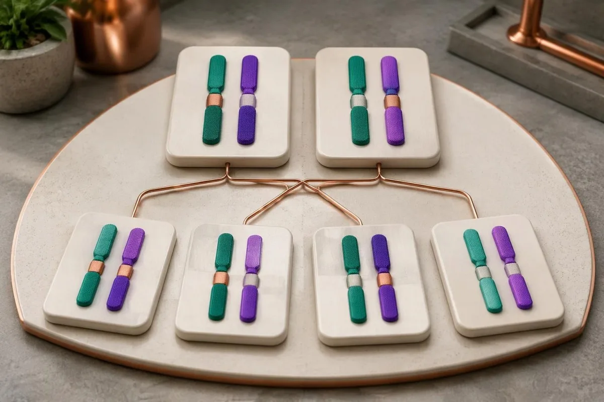
Carrier enzyme values can overlap with values in unaffected people. GAA activity is therefore not a reliable standalone carrier test, and a carrier's activity should not be assumed to equal exactly 50% of a laboratory's normal mean; targeted genetic testing is generally more informative when a familial variant is known.
The 25% affected-child probability applies independently to each pregnancy when both parents carry disease-causing variants capable of producing Pompe disease together. Having 1 unaffected child does not reduce the next pregnancy's Mendelian probability, and pseudodeficiency variants should not be substituted for pathogenic variants in that calculation.
Family testing is most useful after the affected person's variants are clarified. If a symptomatic adult has only 1 identified pathogenic variant, relatives should not be reassured that the entire family question is settled until the specialist considers whether the second variant was missed by the original assay.
For a hypothetical couple, one partner carrying a pathogenic GAA variant and the other carrying only a pseudodeficiency variant does not create the same 25% Pompe risk as 2 pathogenic-variant carriers. Our carrier screening limitations guide explains why a report's coverage and residual risk deserve discussion before reproductive decisions.
Które inne wyniki badań laboratoryjnych przemawiają za diagnostyką choroby Pompego?
Creatine kinase, AST, ALT and urinary glucose tetrasaccharide can support Pompe evaluation, but none confirms the diagnosis alone. Urinary glucose tetrasaccharide may be reported as Glc4 or within a Hex4 assay; these 2 report terms require method-specific interpretation rather than assumption that every laboratory measures exactly the same thing.
Normal CK does not exclude later-onset Pompe disease. In a hypothetical adult with progressive difficulty rising from a chair, CK of 140 U/L may fall within that laboratory's interval despite clinically meaningful weakness; the combination of symptoms and low confirmed GAA activity matters more than a reassuring CK flag.
AST and ALT can rise because of muscle involvement, not only liver disease. A hypothetical AST of 86 U/L with elevated CK and muscle weakness deserves a muscle-aware assessment; our aldolase and muscle marker guide explains why additional muscle enzymes may help characterize the pattern without identifying its cause.
Urinary Glc4 is a supportive glycogen-related biomarker, not a Pompe-specific diagnostic test. Reference limits vary with age and method, and elevation can occur in other circumstances; 1 elevated result should be interpreted alongside enzyme findings, genetics and the laboratory's creatinine-normalized units.
A falling CK or Glc4 after treatment does not automatically prove that strength or breathing has improved by the same percentage. Our discussion of AST interpretation beyond liver disease illustrates why 2 related markers can move together while still answering different questions about tissue injury and function.
Dlaczego przy nieprawidłowym wyniku badania w kierunku choroby Pompego należy ocenić oddychanie?
Pompe disease can weaken the diaphragm and other respiratory muscles, sometimes before obvious walking difficulty. Assessment may include seated and supine forced vital capacity, respiratory muscle pressures and sleep-related testing; a daytime oxygen saturation of 97% does not rule out overnight hypoventilation.
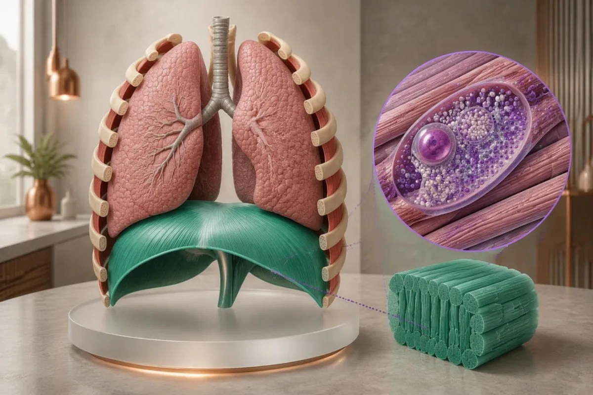
A position-related fall in vital capacity can suggest diaphragm weakness. For example, a hypothetical seated FVC of 3.0 L and supine FVC of 2.4 L represents a 20% decline; the specialist interprets that difference with technique, predicted values and symptoms, rather than treating it as a Pompe-specific cutoff.
Morning headaches, unrefreshing sleep and difficulty breathing when lying flat can justify evaluation for sleep-related hypoventilation. These symptoms do not identify Pompe disease on their own; our przewodnik po testowaniu duszności explains why laboratory results cannot replace respiratory examination and physiological testing.
Arterial carbon dioxide is normally about 35–45 mmHg in adults, with context-dependent variation. A value above 45 mmHg indicates hypercapnia, but its urgency depends on pH, symptoms and the clinical setting; our blood gas interpretation guide explains why oxygen and carbon dioxide answer different questions.
Severe breathlessness, confusion, blue coloration or rapidly worsening weakness warrants emergency care, not an online interpretation session. A confirmed molecular result affecting 2 GAA copies does not measure current respiratory safety; clinicians need direct functional assessment to decide whether ventilation support or urgent monitoring is necessary.
Co dzieje się po dodatnim wyniku badania przesiewowego noworodka w kierunku choroby Pompego?
A positive newborn Pompe screen requires prompt specialist assessment, confirmatory enzyme testing and genetic evaluation. It does not mean every infant has Pompe disease; the immediate task is to distinguish pseudodeficiency or carrier findings from infantile disease and a later-onset-associated genotype affecting both GAA copies.

Burton et al.'s 2017 Pediatrics newborn-screening algorithms describe an initial evaluation integrating GAA confirmation, molecular testing and clinical assessment. If an infant has feeding difficulty, marked hypotonia or suspected cardiomyopathy, do not wait 2 weeks for a routine repeat appointment; the screening team should arrange urgent metabolic and cardiac evaluation.
Classic infantile Pompe disease commonly involves hypertrophic cardiomyopathy. A newborn's echocardiogram and electrocardiogram help assess risk that cannot be estimated from 1 enzyme number; absence of obvious heart symptoms at home does not replace the specialist examination after an abnormal screen.
CRIM status concerns whether cross-reactive GAA protein is present and helps specialists plan immune-risk management around enzyme replacement therapy. Less than 1% enzyme activity does not by itself establish CRIM-negative status, because an inactive protein may still be present; this is a protein-expression question, not just an activity threshold.
Later-onset-associated findings in a newborn require a monitoring plan, not an invented age-of-onset prediction. Even 2 classified disease-causing variants may not reveal when symptoms will emerge; our wytyczne dotyczące bezpieczeństwa pediatrycznych zastosowań AI explains why pediatric results require age-specific context and clinician-led decisions.
Jak specjaliści wyjaśniają sprzeczne wyniki badań u dorosłych?
Conflicting adult Pompe results are resolved by checking specimen quality, confirmatory enzyme methods, variant classification and clinical fit. One low screen plus 1 genetic finding rarely tells the whole story; specialists investigate whether the apparent conflict comes from pseudodeficiency, an incomplete molecular test or another cause of weakness.

Consider a hypothetical 52-year-old with mild fatigue, low DBS GAA activity and only the 2 commonly discussed pseudodeficiency variants. If specialist testing supports pseudodeficiency and no disease-causing genotype, the low screen does not explain the fatigue; other causes should be assessed rather than attaching a Pompe label to an incidental biochemical finding.
A second hypothetical adult has progressive hip weakness, a 20% positional FVC decline, confirmed cellular GAA deficiency and 2 pathogenic variants in trans. Together, those findings support Pompe disease; a normal routine glucose or modest CK result would not outweigh the converging biochemical, genetic and functional evidence.
Convincing enzyme deficiency with only 1 identified pathogenic variant deserves further investigation. The appropriate next step may be deletion/duplication analysis, RNA studies or another specialist assay—not automatically calling the person a carrier; the investigation should also check whether non-Pompe neuromuscular conditions better explain the examination.
Variant-related disease monitoring is not the same as predicting a person's future from their DNA. Our inherited neuromuscular risk guide offers a related family-testing perspective, but 2 conditions can differ substantially in inheritance, penetrance and treatment triggers.
Co należy zabrać na wizytę u specjalisty choroby Pompego?
Bring the complete enzyme report, complete genetic report and a dated symptom history to a Pompe specialist appointment. Three report details are especially useful: specimen type, assay units and variant classification; screenshots showing only a red flag or the word mutation can omit information that changes the interpretation.

Ask whether confirmatory testing has already been completed and whether the laboratory measured GAA specifically in an appropriate cell preparation. If 2 variants were found, ask whether they are in trans; if only 1 was found, ask what variant types and regions the test could detect.
Do not try to raise GAA activity with supplements or diet before confirmation. No vitamin or food corrects inherited pathogenic variants affecting both GAA copies; nutrition may support general care after diagnosis, but cannot substitute for enzyme confirmation or specialist treatment planning.
Kantesti jest Narzędzie do analizy wyników badań krwi oparte na AI that can help organize report terminology and associated laboratory trends, not decide whether 2 uncertain variants establish Pompe disease. Our przewodnik weryfikacji źródła raportu explains why the original document remains the reference when an automated explanation and a specialist interpretation differ.
Genetic documents can contain information relevant to relatives as well as the tested person. Before sharing 1 family member's report, check consent and remove unnecessary identifiers; Kantesti's informacje o prywatności i danych describes our handling approach, while sharing results with relatives should follow a genetic counselor's guidance.
Publikacje naukowe i ograniczenia interpretacji wyników badań w kierunku choroby Pompego
Pompe test interpretation rests on disease-specific enzyme, genetic and clinical evidence—not on general laboratory publications or an AI explanation. Kantesti includes 3 directly relevant external sources below and 2 separate educational publications; the iron and coagulation publications are not evidence for diagnosing Pompe disease.
The 2006 Kishnani guideline provides disease-specific diagnostic and management context, while the 2017 Burton newborn-screening paper addresses confirmation after population screening. Their recommendations should be applied with the specialist laboratory's validated methods; neither supplies 1 universally transferable DBS enzyme cutoff for every instrument and population.
Richards et al.'s 2015 ACMG/AMP publication explains the 5-category variant framework used to distinguish pathogenic findings from uncertainty. Classification can change when new evidence becomes available, so ask the genetics service whether an unresolved result has a reanalysis pathway rather than assuming every VUS will eventually become pathogenic.
Nasz materiał edukacyjny publikacja o badaniach żelaza addresses a different laboratory domain. The separate publikacja o badaniach krzepnięcia also covers unrelated markers; their 2 DOI records are provided for publication transparency, not as supporting citations for GAA deficiency or pseudodeficiency.
Materiały Kantesti standardy treści medycznych distinguish patient education from diagnosis and treatment instructions. The practical endpoint is a documented interpretation of 3 evidence streams—enzyme testing, genetic analysis and clinical assessment—with urgent care taking priority whenever infantile disease or respiratory compromise is suspected.
Często zadawane pytania
0 Niska aktywność enzymu GAA zawsze oznacza chorobę Pompego?
Niska aktywność enzymu GAA nie zawsze oznacza chorobę Pompego. Jeden nieprawidłowy wynik badania przesiewowego może wskazywać na rzeczywisty niedobór, pseudoniedobór, degradację próbki lub nakładanie się na aktywność związaną z nosicielstwem. Potwierdzenie wymaga interpretacji przez specjalistę odpowiedniego oznaczenia aktywności enzymu, wyników badań genetycznych GAA oraz kontekstu klinicznego. Powtórzenie wyłącznie tego samego badania przesiewowego może ponownie wykazać pseudoniedobór, zamiast rozwiać wątpliwości.
Jaki jest prawidłowy poziom enzymu GAA?
There is no universal normal GAA enzyme level because specimen preparation, assay substrate and reporting units differ between laboratories. A value in nmol/h/mL cannot be directly compared with a value in nmol/h/mg protein. Classic infantile Pompe disease often has less than 1% of normal activity in diagnostic tissue assays, but this is not a universal newborn-screening cutoff. Use the reference information printed on the original report and the specialist laboratory's interpretation.
Czy dwa niskie wyniki badań GAA nadal mogą wskazywać na pseudoniedobór?
Dwa niskie wyniki badań aktywności GAA nadal mogą wskazywać na pseudoniedobór, zwłaszcza gdy zastosowano tę samą lub podobnie podatną na zakłócenia metodę oznaczenia. Warianty pseudoniedoboru obniżają mierzoną aktywność, nie powodując same w sobie choroby Pompego, czyli glikogenozy. Specjaliści odróżniają ten wzorzec, analizując pełny genotyp GAA, charakterystykę metody oznaczenia oraz, w razie potrzeby, badając inną próbkę komórek lub wykonując badanie czynnościowe. Dwa powtarzające się niskie wyniki nie są zatem równoznaczne z dwoma niezależnymi potwierdzeniami choroby.
Czy dwa warianty genu GAA potwierdzają chorobę Pompego?
Dwa warianty genu GAA nie potwierdzają automatycznie choroby Pompego. Dwa warianty patogenne lub prawdopodobnie patogenne w układzie trans, wpływające na przeciwne kopie genu, przemawiają za potwierdzeniem molekularnym, podczas gdy dwa warianty w układzie cis wpływają tylko na jedną kopię. Wariantów o niepewnym znaczeniu i wariantów pseudoniedoboru nie można uznawać za potwierdzone czynniki chorobotwórcze. Dowody enzymatyczne i kliniczne pozostają szczególnie przydatne, gdy klasyfikacja lub faza dziedziczenia nie zostały ustalone.
Czy prawidłowy poziom CK może wykluczyć późno ujawniającą się chorobę Pompego?
Normal CK cannot rule out late-onset Pompe disease. A hypothetical CK of 140 U/L may fall within a laboratory's reference interval despite progressive proximal weakness or diaphragm involvement. CK measures muscle injury-related enzyme release, not lysosomal GAA function. Persistent compatible symptoms deserve specialist assessment even when routine muscle markers are reassuring.
Jak pilne jest uzyskanie dodatniego wyniku badania przesiewowego noworodka w kierunku choroby Pompego?
Dodatni wynik badania przesiewowego noworodka w kierunku choroby Pompego wymaga niezwłocznego kontaktu z zespołem ds. badań przesiewowych lub zespołem metabolicznym, a niemowlę z trudnościami w karmieniu, znaczną hipotonią lub podejrzeniem zajęcia serca wymaga pilnej oceny. Jeden dodatni wynik badania przesiewowego nie potwierdza rozpoznania, ale przy podejrzeniu klasycznej postaci niemowlęcej nie należy czekać na rutynową wizytę za kilka tygodni. Specjaliści organizują potwierdzające badanie aktywności enzymu, ocenę genetyczną i ocenę kardiologiczną, często równolegle. Poważne trudności w oddychaniu lub obniżona reaktywność wymagają pilnej pomocy medycznej.
Jakie jest ryzyko dla dzieci, jeśli oboje rodzice są nosicielami choroby Pompego?
Jeśli oboje rodzice są nosicielami wariantów genu GAA wywołujących chorobę, w każdej ciąży prawdopodobieństwo urodzenia chorego dziecka wynosi 25%, prawdopodobieństwo urodzenia dziecka będącego nosicielem wynosi 50%, a prawdopodobieństwo urodzenia dziecka, które nie odziedziczy żadnego z wariantów rodzinnych, wynosi 25%. Prawdopodobieństwa te dotyczą niezależnie każdej ciąży. Wariantów pseudoniedoboru nie można w tych obliczeniach traktować zamiennie z wariantami patogennymi. Poradnictwo genetyczne pomaga potwierdzić wyniki badań rodziców i wyjaśnić dostępne opcje badań.
Dowiedz się, jak czytać wyniki badań krwi
Prześlij wynik badania laboratoryjnego, a sztuczna inteligencja Kantesti w kilka sekund objaśni prostym językiem każdy biomarker. Z narzędzia korzysta ponad 2 miliony osób na całym świecie.
Odniesienia
- Klein, T., Mitchell, S. i Weber, H. (2026). Przewodnik po badaniach gospodarki żelazowej: TIBC, wysycenie żelazem i zdolność wiązania. Badania medyczne z wykorzystaniem sztucznej inteligencji Kantesti. DOI
- Klein, T., Mitchell, S. i Weber, H. (2026). Prawidłowy zakres aPTT: przewodnik po D-dimerze i białku C w krzepnięciu krwi. Badania medyczne z wykorzystaniem sztucznej inteligencji Kantesti. DOI
- Kishnani PS et al. (2006). Pompe disease diagnosis and management guideline. Genetics in Medicine. DOI PubMed
- Burton BK et al. (2017). The Initial Evaluation of Patients After Positive Newborn Screening: Recommended Algorithms Leading to a Confirmed Diagnosis of Pompe Disease. Pediatrics. DOI
- Richards S i wsp. (2015). Standardy i wytyczne dotyczące interpretacji wariantów sekwencji: wspólne zalecenie konsensusu American College of Medical Genetics and Genomics oraz Association for Molecular Pathology. Genetics in Medicine. DOI PubMed
- 2 mln+Analizowane badania
- 127+Kraje
- 100+Języki
O zespole medycznym


Sygnały zaufania E-E-A-T
Doświadczenie
Kliniczna weryfikacja procesów interpretacji przez lekarza.
Ekspertyza
Medycyna laboratoryjna skupiona na tym, jak zachowują się biomarkery w kontekście klinicznym.
Autorytatywność
Recenzja medyczna: dr Thomas Klein, przy współpracy dr Sarah Mitchell i prof. dr. Hansa Webera.
Solidność
Interpretacja oparta na dowodach, z jasnymi ścieżkami dalszego postępowania, aby ograniczyć alarm.





Zastrzeżenie medyczne
Ten artykuł ma wyłącznie charakter edukacyjny i nie stanowi porady medycznej. Zawsze konsultuj decyzje dotyczące diagnozy i leczenia z wykwalifikowanym pracownikiem ochrony zdrowia.
Opublikowano · Skontaktuj się z zespołem redakcyjnym
Opublikowano przez Kantesti Ltd, zarejestrowaną w Anglii i Walii, numer firmy. 17090423, Londyn, Wielka Brytania.
