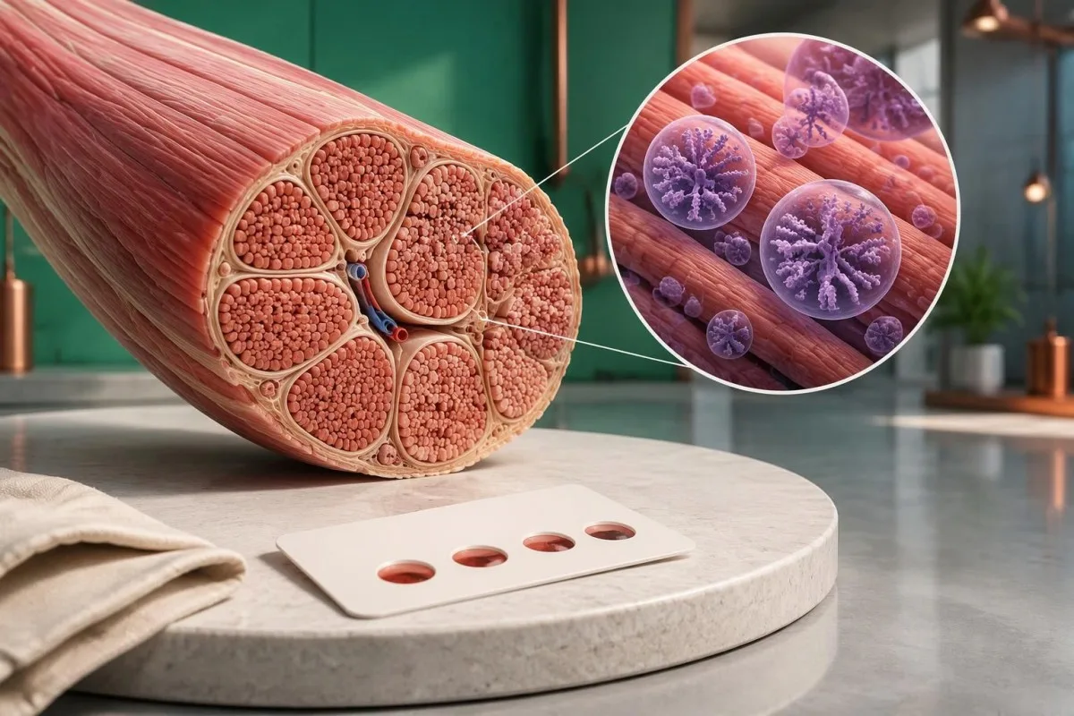
Lág niðurstaða fyrir GAA-ensím vekur grun um Pompe-sjúkdóm, en skimun ein og sér getur ekki staðfest hann. Endurtekin prófun á viðeigandi sýni og erfðagreining á GAA hjálpa til við að greina á milli raunverulegs ensímskorts, arfberastöðu og sýndarskorts.

Helstu niðurstöður
- Lítil virkni GAA-ensíms er vísbending um að fylgja þurfi málinu eftir, ekki greining; 1 óeðlilegt þurrblóðsýni þarf staðfestingu samkvæmt fyrirmælum sérfræðings.
- Viðmiðunargildi fer eftir sýninu og mæliaðferðinni; nmól/klst./ml er ekki hægt að bera beint saman við nmól/klst./mg próteins.
- Afgangsensímvirkni er oft undir 1% við klassískan ungbarnagerðar Pompe-sjúkdóm, en sú tala er ekki algilt viðmiðunarmark fyrir skimun.
- Sýndarskortur getur lækkað GAA-virkni á rannsóknarstofu án þess að valda Pompe-sjúkdómi; endurtekin mæling með sömu aðferð getur skilað aftur lágri niðurstöðu.
- Prófun á GAA-geninu leitar að sjúkdómsvaldandi afbrigðum sem hafa áhrif á bæði eintök gensins; túlka þarf 2 afbrigði með tilliti til flokkunar og arfgerðar.
- Óviss afbrigði útiloka ekki Pompe-sjúkdóm; 1 eða 2 afbrigði með óvissa þýðingu krefjast frekari gagna.
- Áhætta fyrir fjölskyldu er 25% fyrir að barn verði haldið sjúkdómnum í hverri meðgöngu þegar báðir foreldrar bera sjúkdómsvaldandi GAA-afbrigði.
- Bráð mat er nauðsynlegt fyrir ungbarn með lágt GAA-gildi ásamt erfiðleikum við næringu, greinilegum vöðvaslappleika eða grun um hjartaþátttöku; ekki bíða eftir hefðbundnu endurprófi.
Hvað þýðir lág niðurstaða fyrir GAA-ensím í raun?
A Próf fyrir Pompe-sjúkdómi sem sýnir litla GAA-virkni þýðir að rannsóknarstofan mældi skerta virkni sýru-alfa-glúkósídasa við skilyrði sín fyrir mælinguna. Það sannar ekki Pompe-sjúkdóm: 1 lágt skimunarniðurstaða getur endurspeglað raunverulegan skort, gerviskort, skörun við arfbera eða vandamál með sýni.

Mæling á GAA-virkni metur ensímvirkni, ekki einfaldlega magn ensímpróteins. Fyrstu 3 atriðin sem þarf að athuga eru tegund sýnis, mælieiningar og viðmiðunarmörk rannsóknarstofunnar; niðurstöðu sem lýst er sem lágri án þessara upplýsinga er ekki hægt að túlka á áreiðanlegan hátt út frá tölu sem finnst á netinu.
Ástæðan fyrir prófuninni breytir merkingu jákvæðrar skimunar. Nýfætt barn sem greinist við almenna skimun og 45 ára einstaklingur með vaxandi máttleysi í mjöðmum hafa mismunandi upphaflegar líkur á Pompe-sjúkdómi, jafnvel þótt ensímgildi þeirra séu þau sömu; því þarf að skoða einkenni og niðurstöður læknisskoðunar samhliða rannsóknarniðurstöðunni.
Jákvæð skimun er ekki það sama og staðfestur sjúkdómur. Ef 2 rannsóknarstofur nota mismunandi hvarfefni eða undirbúning sýna geta tölulegar niðurstöður þeirra verið ósamhljóða án þess að hvorug rannsóknarstofan hafi gert mistök; spyrðu hvort næsti tími sé fyrir aðra skimunarmælingu eða raunverulega staðfestandi ensímpróf.
Kantesti er AI blóðprufugreiningartæki sem getur hjálpað til við að skipuleggja GAA-niðurstöðu samhliða hefðbundnum rannsóknarniðurstöðum, en getur ekki staðfest greiningu á Pompe-sjúkdómi út frá 1 niðurstöðu sem er merkt óeðlileg. Okkar fyrirtækjaupplýsingar okkar útskýrir skipulagið að baki þjónustunni; grunur um arfgengan ensímskort krefst áfram mats sérfræðings í efnaskiptasjúkdómum eða tauga- og vöðvasjúkdómum.
Af hverju veldur GAA-skortur vöðva- og hjartavandamálum?
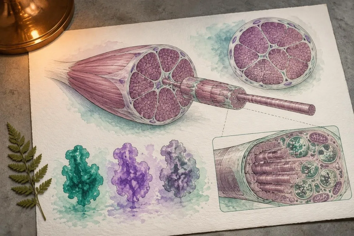
Pompe-sjúkdómur stafar af skorti á súrum alfa-glúkósídasa í leysikornum, ensími sem brýtur niður glýkógen inni í niðurbrotsrýmum frumna. GAA verkar á bæði alfa-1,4- og alfa-1,6-glúkósíðtengi; ófullnægjandi virkni veldur uppsöfnun glýkógens, einkum í beinagrindarvöðvum og hjartavöðva.
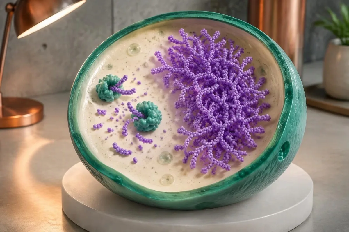
Í klassískum ungbarnastigs Pompe-sjúkdómi er GAA-virkni í greiningarsýnum oft undir 11% af eðlilegu gildi. Sjúkdómur sem kemur fram síðar á ævinni getur haft meiri varðveitta virkni, stundum um 2–1% í tilteknum mælingum; þessi lýsandi viðmið skarast við takmarkanir mæliaðferða og mega ekki koma í stað staðfestrar greiningartúlkunar rannsóknarstofu.
Leiðbeiningar Kishnani o.fl. frá 2006 í Genetics in Medicine lýsa tengslum GAA-skorts, glýkógengeymslu og mats á mörgum líffærakerfum. Klínísk aðgreining felst ekki aðeins í því hvort virkni sé undir 11%: aldur við einkennaframkomu, hjartaniðurstöður og starfsemi öndunarvöðva hjálpa einnig til við að ákvarða form sjúkdómsins og hversu brýnt er að bregðast við.
Hefðbundin mæling á glúkósa mælir ekki niðurbrot glýkógens í leysikornum. Til dæmis getur ímyndaður fullorðinn einstaklingur haft fastandi blóðsykur 92 mg/dL en jafnframt arfgengan glýkógengeymslusjúkdóm í vöðvum; eðlilegur blóðsykur sýnir ekki fram á að glýkógen inni í leysikornum vöðva sé unnið á eðlilegan hátt.
Aðrir leysikornasjúkdómar fela í sér önnur ensím og gen og því er ekki hægt að yfirfæra viðmiðunarmörk þeirra á Pompe-prófanir. Útskýring okkar á staðfestingu Gaucher-ensíms sýnir almenna verkferlið frá ensímprófi til erfðaprófs, en jafnframt hvers vegna ensímpróf fyrir einn sjúkdóm getur ekki komið í stað ensímprófs fyrir annan.
Er til eðlilegt viðmiðunargildi fyrir virkni GAA-ensíms?
There is no single universal normal range for GAA enzyme activity. Dried blood spots, leukocytes and cultured fibroblasts use different preparations and reporting units; a number expressed in nmol/h/mL cannot be directly compared with a result expressed in nmol/h/mg protein.

A screening cutoff separates samples needing follow-up from samples passing that screen. It does not necessarily separate affected people from unaffected people with complete accuracy; laboratories may use different fluorometric or tandem mass spectrometry methods, and 2 screening programs may therefore select different recall thresholds.
For a clearly hypothetical example, suppose a laboratory uses a DBS screening cutoff of 1.5 nmol/h/mL and reports 0.7 nmol/h/mL. The result is below that invented laboratory threshold, but dividing 0.7 by 1.5 gives 47% of the cutoff—not 47% of normal enzyme activity and not a severity score.
The lower limit of detection is different from the clinical cutoff. A report reading less than 0.1 in its stated units may mean the method cannot quantify lower activity reliably; it does not prove absolute absence of enzyme, and repeating measurements close to that analytical floor may produce proportionally large variation.
Screening performance also depends on prevalence. In a hypothetical group with 10 true-positive and 100 false-positive results, only about 9% of positive screens represent disease; those invented counts are not Pompe screening statistics, but explain why eigindlegar samanborið við megindlegar niðurstöður require different interpretation.
| Styrkur | Bil | Merking |
|---|---|---|
| Passes hypothetical screen | ≥1.5 nmol/h/mL | Illustrative DBS cutoff only; not a universal normal range and not an absolute exclusion in a symptomatic person. |
| Below hypothetical cutoff | <1.5 nmol/h/mL | Illustrative follow-up threshold only; true deficiency, pseudodeficiency and specimen effects still need separation. |
Af hverju á að endurtaka GAA-prófun á öðru sýni?
Repeat GAA testing checks whether low activity is reproducible and whether specimen quality or assay interference explains it. Confirmation usually moves beyond 1 screening card to a fresh, appropriately handled specimen, often with GAA measurement in leukocytes or another specialist-selected cell preparation.

Heat, humidity and inadequate drying can reduce enzyme activity in a dried blood spot. A low result after 2 days of poorly controlled transport may deserve a fresh collection, but transport history cannot be used to dismiss a result automatically; the laboratory should assess card quality and its stability requirements.
Acarbose is used in some GAA assay methods to suppress interfering maltase-glucoamylase activity. That interference can make measured activity appear higher and potentially obscure deficiency; checking how a laboratory isolates GAA activity can therefore matter more than obtaining a second number with the same unresolved methodological limitation.
Two low DBS results still do not automatically confirm Pompe disease. Pseudodeficiency can reproduce across repeated measurements, including some confirmatory methods; when enzyme and genetic findings disagree, a specialist laboratory may recommend cultured fibroblast testing or additional molecular investigation rather than simply ordering a third screening card.
Give the laboratory the dates of collection, recent transfusions or transplantation, and any enzyme replacement treatment already received. Our guide to truflanir vegna sýnagæða explains the general distinction between biological findings and specimen problems; GAA testing has its own handling rules, so do not import another test's redraw interval.
Hvernig er sýndarskortur á GAA frábrugðinn Pompe-sjúkdómi?
GAA pseudodeficiency means an inherited variant lowers measured enzyme activity without causing the glycogen-storage disorder Pompe disease. Two commonly discussed variants are c.1726G>A, p.Gly576Ser, and c.2065G>A, p.Glu689Lys; their interpretation depends on the complete genotype and the assay used.
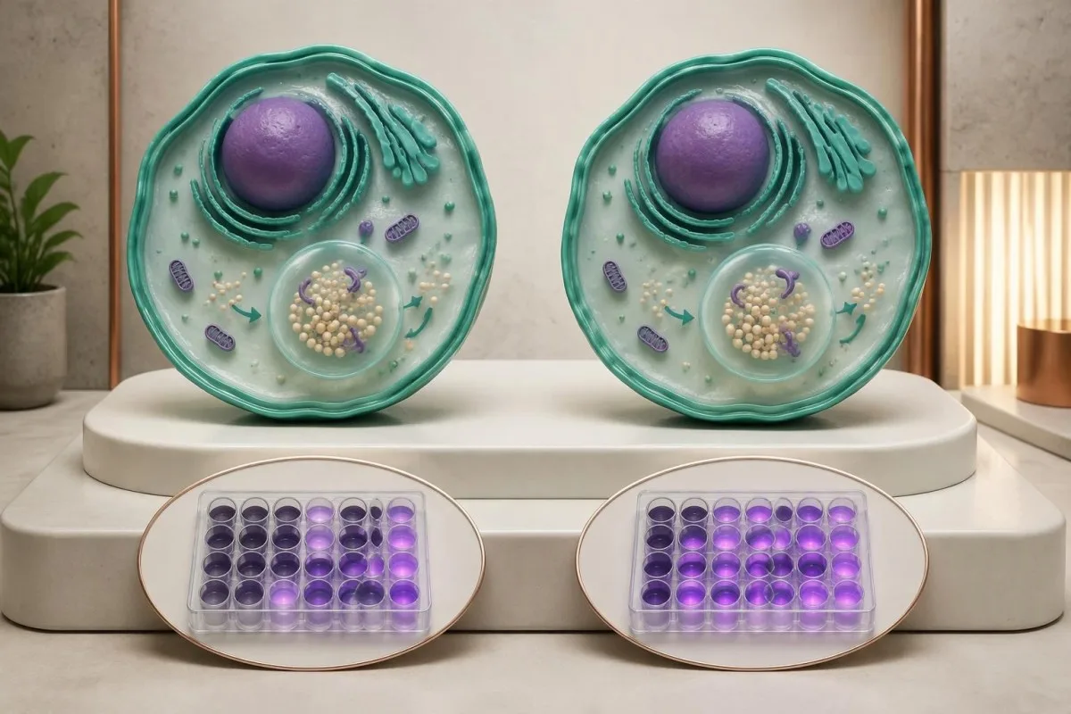
Pseudodeficiency is a biochemical testing issue, not an early stage of Pompe disease. A person with only pseudodeficiency variants does not need Pompe treatment because of 1 low activity result; the specialist must first verify that disease-causing variants or another explanation have not been missed.
The p.Gly576Ser and p.Glu689Lys variants are often found together on 1 gene copy and are particularly relevant in some East Asian ancestry populations. Ancestry changes how often laboratories encounter this pattern, but cannot establish or exclude pseudodeficiency in an individual; people from other backgrounds can carry these variants too.
Pseudodeficiency can remain low on repeat enzyme testing. The practical distinction is whether the complete biochemical and genetic evidence supports disease-causing GAA deficiency, not whether a second result falls below an arbitrary percentage; specialists may compare different assay substrates or cell preparations when the pattern remains ambiguous.
Pseudodeficiency variants can also coexist with a disease-causing variant, making a short report misleading if it lists everything under mutations. Our Fabry enzyme and genetics guide shows another situation where enzyme results need genetic context, although Fabry's X-linked inheritance differs from Pompe's 2-copy recessive mechanism.
Að hverju ætti að leita í prófun á GAA-geninu?
GAA gene testing investigates whether disease-causing variants affect both copies of the GAA gene. Pompe disease is autosomal recessive, and the GAA gene is located on chromosome 17; interpretation requires variant classification, adequate test coverage and, when needed, confirmation that variants affect opposite gene copies.
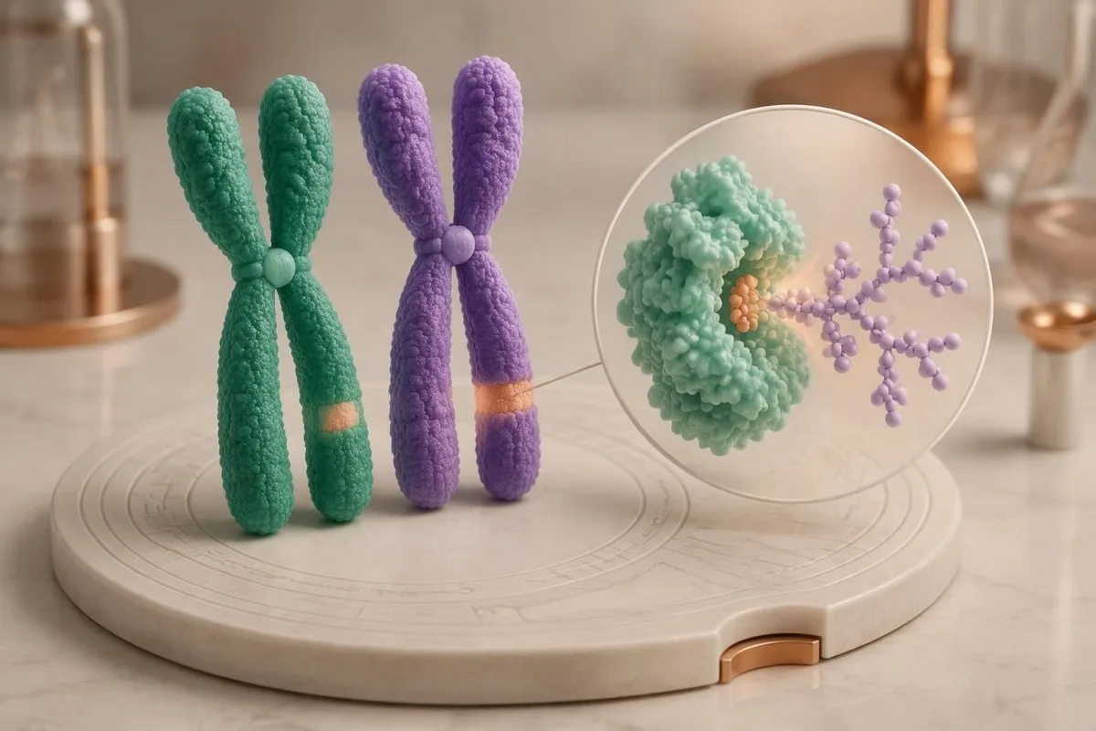
Two pathogenic or likely pathogenic GAA variants in trans support molecular confirmation of Pompe disease. In trans means the variants affect opposite gene copies; 2 variants in cis affect the same copy and do not, by themselves, demonstrate a recessive disease genotype.
Standard sequencing detects many small substitutions and insertions or deletions, but does not answer every possible genetic question. If only 1 disease-causing variant is found despite convincing enzyme deficiency, ask whether deletion/duplication analysis was included and whether poorly covered or deep intronic regions require further investigation.
A common later-onset-associated GAA variant is c.-32-13T>G, which affects splicing. Finding this variant on 1 copy does not diagnose Pompe disease, and finding it with another disease-causing variant does not precisely predict the age symptoms will begin; residual splicing and other biological factors influence presentation.
Parental testing can help establish phase when a laboratory detects 2 different variants but cannot determine their arrangement. Our guide to inherited disease genetic reports explains why inheritance and family testing matter, while the actual gene coverage and diagnostic criteria must remain specific to GAA.
Hvernig ber að túlka sjúkdómsvaldandi, óvissar og neikvæðar niðurstöður?
A pathogenic result, a variant of uncertain significance and a negative result have different diagnostic meanings. Genetic laboratories commonly use 5 categories: pathogenic, likely pathogenic, uncertain significance, likely benign and benign; a VUS is not equivalent to a disease-causing GAA variant.
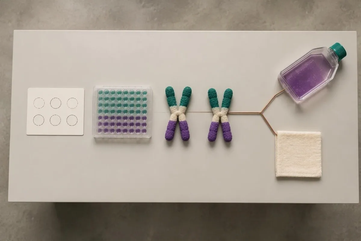
Richards et al.'s 2015 ACMG/AMP consensus established the widely used 5-category sequence-variant framework and states that a VUS should not guide clinical decision-making as though pathogenic. For Pompe evaluation, low enzyme activity can contribute evidence, but its value depends on whether pseudodeficiency and assay limitations have been addressed.
One pathogenic variant plus 1 VUS is an unresolved combination, not automatic confirmation or exclusion. Depending on the enzyme and clinical findings, specialists may investigate family segregation, RNA effects or variant-specific functional evidence; a diagnostic enzyme result may support disease even while the second molecular finding remains incompletely characterized.
A negative sequencing report means the test found no reportable variants within its scope. It does not prove that every GAA-related mechanism was examined; 1 negative limited test may warrant expanded testing when properly performed enzyme studies and the clinical pattern remain strongly suggestive.
Kantesti er AI blóðrannsóknartúlkunarvettvangur that can help separate a report's 5 classification terms from its numerical enzyme findings, without adjudicating an uncertain genotype. Our tæknilegir og klínískir staðlar describe interpretation boundaries; our uncertain gene-result explanation provides a related example of why variants and diagnoses are not interchangeable.
Þýðir eitt GAA-afbrigði arfberastöðu eða sjúkdóm?
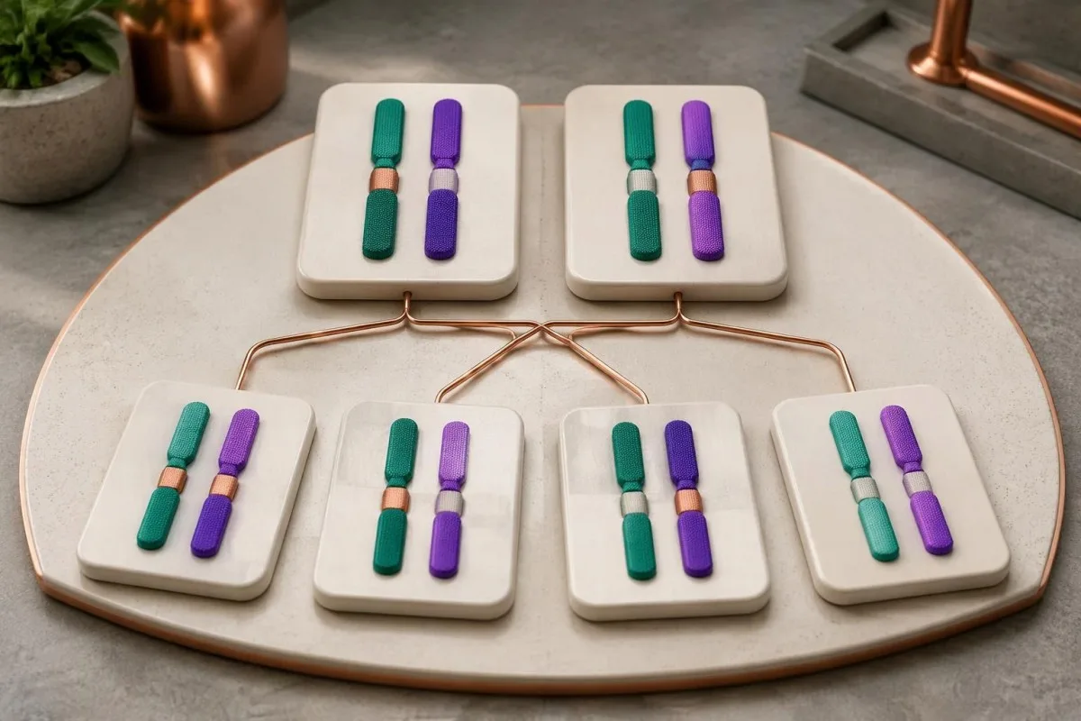
One confirmed disease-causing GAA variant usually indicates carrier status if a second disease-causing variant and diagnostic enzyme deficiency are absent. Carriers do not typically develop Pompe disease; when both parents are carriers, each pregnancy has a 25% affected, 50% carrier and 25% non-carrier probability.
Carrier enzyme values can overlap with values in unaffected people. GAA activity is therefore not a reliable standalone carrier test, and a carrier's activity should not be assumed to equal exactly 50% of a laboratory's normal mean; targeted genetic testing is generally more informative when a familial variant is known.
The 25% affected-child probability applies independently to each pregnancy when both parents carry disease-causing variants capable of producing Pompe disease together. Having 1 unaffected child does not reduce the next pregnancy's Mendelian probability, and pseudodeficiency variants should not be substituted for pathogenic variants in that calculation.
Family testing is most useful after the affected person's variants are clarified. If a symptomatic adult has only 1 identified pathogenic variant, relatives should not be reassured that the entire family question is settled until the specialist considers whether the second variant was missed by the original assay.
For a hypothetical couple, one partner carrying a pathogenic GAA variant and the other carrying only a pseudodeficiency variant does not create the same 25% Pompe risk as 2 pathogenic-variant carriers. Our carrier screening limitations guide explains why a report's coverage and residual risk deserve discussion before reproductive decisions.
Hvaða aðrar niðurstöður úr rannsóknarstofuprófum styðja mat á Pompe-sjúkdómi?
Creatine kinase, AST, ALT and urinary glucose tetrasaccharide can support Pompe evaluation, but none confirms the diagnosis alone. Urinary glucose tetrasaccharide may be reported as Glc4 or within a Hex4 assay; these 2 report terms require method-specific interpretation rather than assumption that every laboratory measures exactly the same thing.
Normal CK does not exclude later-onset Pompe disease. In a hypothetical adult with progressive difficulty rising from a chair, CK of 140 U/L may fall within that laboratory's interval despite clinically meaningful weakness; the combination of symptoms and low confirmed GAA activity matters more than a reassuring CK flag.
AST and ALT can rise because of muscle involvement, not only liver disease. A hypothetical AST of 86 U/L with elevated CK and muscle weakness deserves a muscle-aware assessment; our aldolase and muscle marker guide explains why additional muscle enzymes may help characterize the pattern without identifying its cause.
Urinary Glc4 is a supportive glycogen-related biomarker, not a Pompe-specific diagnostic test. Reference limits vary with age and method, and elevation can occur in other circumstances; 1 elevated result should be interpreted alongside enzyme findings, genetics and the laboratory's creatinine-normalized units.
A falling CK or Glc4 after treatment does not automatically prove that strength or breathing has improved by the same percentage. Our discussion of AST interpretation beyond liver disease illustrates why 2 related markers can move together while still answering different questions about tissue injury and function.
Af hverju á að meta öndun þegar Pompe-próf er óeðlilegt?
Pompe disease can weaken the diaphragm and other respiratory muscles, sometimes before obvious walking difficulty. Assessment may include seated and supine forced vital capacity, respiratory muscle pressures and sleep-related testing; a daytime oxygen saturation of 97% does not rule out overnight hypoventilation.
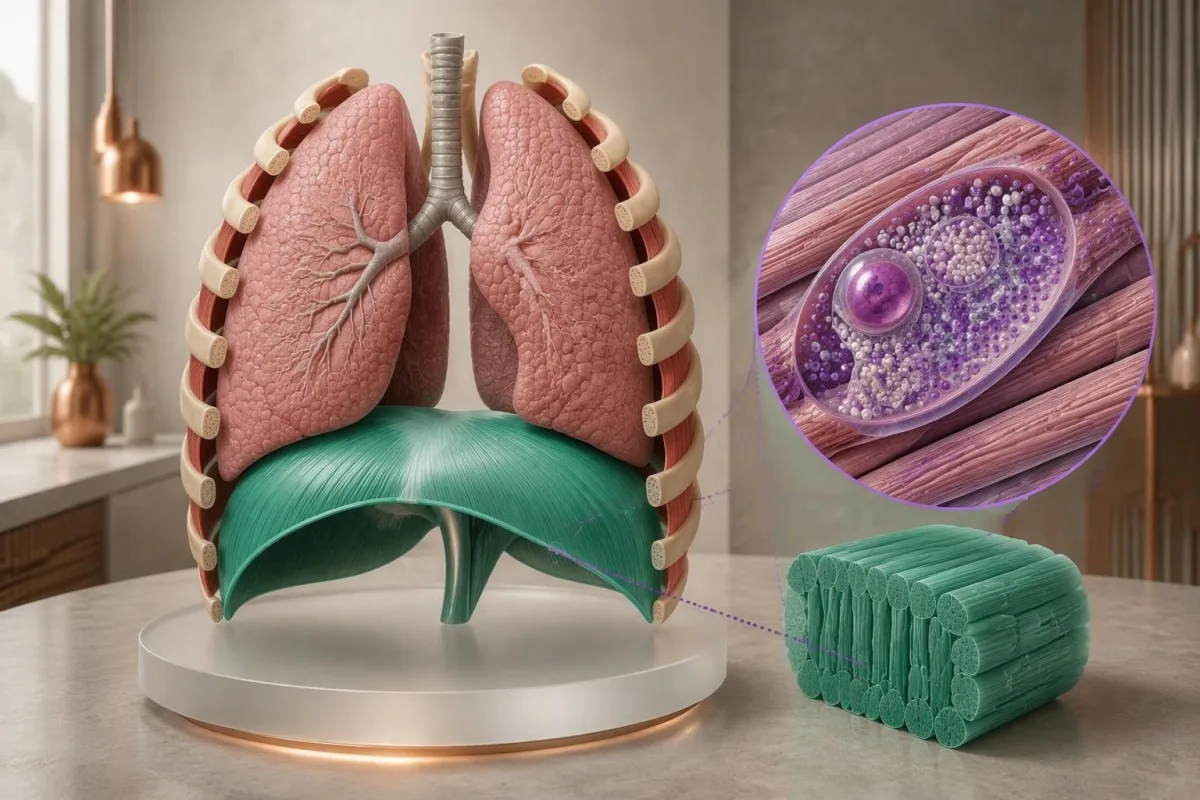
A position-related fall in vital capacity can suggest diaphragm weakness. For example, a hypothetical seated FVC of 3.0 L and supine FVC of 2.4 L represents a 20% decline; the specialist interprets that difference with technique, predicted values and symptoms, rather than treating it as a Pompe-specific cutoff.
Morning headaches, unrefreshing sleep and difficulty breathing when lying flat can justify evaluation for sleep-related hypoventilation. These symptoms do not identify Pompe disease on their own; our prófunarleiðbeiningar um andnauð explains why laboratory results cannot replace respiratory examination and physiological testing.
Arterial carbon dioxide is normally about 35–45 mmHg in adults, with context-dependent variation. A value above 45 mmHg indicates hypercapnia, but its urgency depends on pH, symptoms and the clinical setting; our blood gas interpretation guide explains why oxygen and carbon dioxide answer different questions.
Severe breathlessness, confusion, blue coloration or rapidly worsening weakness warrants emergency care, not an online interpretation session. A confirmed molecular result affecting 2 GAA copies does not measure current respiratory safety; clinicians need direct functional assessment to decide whether ventilation support or urgent monitoring is necessary.
Hvað gerist eftir jákvæða Pompe-skimun hjá nýbura?
A positive newborn Pompe screen requires prompt specialist assessment, confirmatory enzyme testing and genetic evaluation. It does not mean every infant has Pompe disease; the immediate task is to distinguish pseudodeficiency or carrier findings from infantile disease and a later-onset-associated genotype affecting both GAA copies.

Burton et al.'s 2017 Pediatrics newborn-screening algorithms describe an initial evaluation integrating GAA confirmation, molecular testing and clinical assessment. If an infant has feeding difficulty, marked hypotonia or suspected cardiomyopathy, do not wait 2 weeks for a routine repeat appointment; the screening team should arrange urgent metabolic and cardiac evaluation.
Classic infantile Pompe disease commonly involves hypertrophic cardiomyopathy. A newborn's echocardiogram and electrocardiogram help assess risk that cannot be estimated from 1 enzyme number; absence of obvious heart symptoms at home does not replace the specialist examination after an abnormal screen.
CRIM status concerns whether cross-reactive GAA protein is present and helps specialists plan immune-risk management around enzyme replacement therapy. Less than 1% enzyme activity does not by itself establish CRIM-negative status, because an inactive protein may still be present; this is a protein-expression question, not just an activity threshold.
Later-onset-associated findings in a newborn require a monitoring plan, not an invented age-of-onset prediction. Even 2 classified disease-causing variants may not reveal when symptoms will emerge; our öryggisleiðbeiningar fyrir gervigreind í barnalækningum explains why pediatric results require age-specific context and clinician-led decisions.
Hvernig leysa sérfræðingar úr ósamræmi í niðurstöðum prófana hjá fullorðnum?
Conflicting adult Pompe results are resolved by checking specimen quality, confirmatory enzyme methods, variant classification and clinical fit. One low screen plus 1 genetic finding rarely tells the whole story; specialists investigate whether the apparent conflict comes from pseudodeficiency, an incomplete molecular test or another cause of weakness.

Consider a hypothetical 52-year-old with mild fatigue, low DBS GAA activity and only the 2 commonly discussed pseudodeficiency variants. If specialist testing supports pseudodeficiency and no disease-causing genotype, the low screen does not explain the fatigue; other causes should be assessed rather than attaching a Pompe label to an incidental biochemical finding.
A second hypothetical adult has progressive hip weakness, a 20% positional FVC decline, confirmed cellular GAA deficiency and 2 pathogenic variants in trans. Together, those findings support Pompe disease; a normal routine glucose or modest CK result would not outweigh the converging biochemical, genetic and functional evidence.
Convincing enzyme deficiency with only 1 identified pathogenic variant deserves further investigation. The appropriate next step may be deletion/duplication analysis, RNA studies or another specialist assay—not automatically calling the person a carrier; the investigation should also check whether non-Pompe neuromuscular conditions better explain the examination.
Variant-related disease monitoring is not the same as predicting a person's future from their DNA. Our inherited neuromuscular risk guide offers a related family-testing perspective, but 2 conditions can differ substantially in inheritance, penetrance and treatment triggers.
Hvað ættirðu að taka með í viðtal hjá sérfræðingi í Pompe-sjúkdómi?
Bring the complete enzyme report, complete genetic report and a dated symptom history to a Pompe specialist appointment. Three report details are especially useful: specimen type, assay units and variant classification; screenshots showing only a red flag or the word mutation can omit information that changes the interpretation.

Ask whether confirmatory testing has already been completed and whether the laboratory measured GAA specifically in an appropriate cell preparation. If 2 variants were found, ask whether they are in trans; if only 1 was found, ask what variant types and regions the test could detect.
Do not try to raise GAA activity with supplements or diet before confirmation. No vitamin or food corrects inherited pathogenic variants affecting both GAA copies; nutrition may support general care after diagnosis, but cannot substitute for enzyme confirmation or specialist treatment planning.
Kantesti er AI-knúið blóðprófunargreiningartól that can help organize report terminology and associated laboratory trends, not decide whether 2 uncertain variants establish Pompe disease. Our leiðarvísir um að kanna uppruna niðurstaðna explains why the original document remains the reference when an automated explanation and a specialist interpretation differ.
Genetic documents can contain information relevant to relatives as well as the tested person. Before sharing 1 family member's report, check consent and remove unnecessary identifiers; Kantesti's upplýsingar um friðhelgi einkalífs og gögn describes our handling approach, while sharing results with relatives should follow a genetic counselor's guidance.
Rannsóknarrit og takmarkanir við túlkun Pompe-prófa
Pompe test interpretation rests on disease-specific enzyme, genetic and clinical evidence—not on general laboratory publications or an AI explanation. Kantesti includes 3 directly relevant external sources below and 2 separate educational publications; the iron and coagulation publications are not evidence for diagnosing Pompe disease.
The 2006 Kishnani guideline provides disease-specific diagnostic and management context, while the 2017 Burton newborn-screening paper addresses confirmation after population screening. Their recommendations should be applied with the specialist laboratory's validated methods; neither supplies 1 universally transferable DBS enzyme cutoff for every instrument and population.
Richards et al.'s 2015 ACMG/AMP publication explains the 5-category variant framework used to distinguish pathogenic findings from uncertainty. Classification can change when new evidence becomes available, so ask the genetics service whether an unresolved result has a reanalysis pathway rather than assuming every VUS will eventually become pathogenic.
Fræðsluefni okkar járnrannsóknarútgáfa addresses a different laboratory domain. The separate grein um storkupróf also covers unrelated markers; their 2 DOI records are provided for publication transparency, not as supporting citations for GAA deficiency or pseudodeficiency.
Efni Kantesti staðlar fyrir læknisfræðilegt efni distinguish patient education from diagnosis and treatment instructions. The practical endpoint is a documented interpretation of 3 evidence streams—enzyme testing, genetic analysis and clinical assessment—with urgent care taking priority whenever infantile disease or respiratory compromise is suspected.
Algengar spurningar
Þýðir lítil virkni GAA-ensíms alltaf Pompe-sjúkdóm?
Lítil virkni GAA-ensíms þýðir ekki alltaf Pompe-sjúkdóm. Ein óeðlileg niðurstaða úr skimun getur endurspeglað raunverulegan skort, gerviskort, niðurbrot sýnis eða skörun við virkni sem tengist arfberum. Staðfesting krefst túlkunar sérfræðings á viðeigandi ensímprófi, erfðafræðilegum niðurstöðum fyrir GAA og klínísku samhengi. Endurtekning á sama skimunarprófi ein og sér getur endurskapað gerviskort í stað þess að leysa úr málinu.
Hvert er eðlilegt magn GAA-ensíms?
There is no universal normal GAA enzyme level because specimen preparation, assay substrate and reporting units differ between laboratories. A value in nmol/h/mL cannot be directly compared with a value in nmol/h/mg protein. Classic infantile Pompe disease often has less than 1% of normal activity in diagnostic tissue assays, but this is not a universal newborn-screening cutoff. Use the reference information printed on the original report and the specialist laboratory's interpretation.
Geta tvö lág GAA-próf samt verið fölsk ensímskerðing?
Tvö lág GAA-próf geta enn endurspeglað gerviskort, einkum þegar notaðar eru sömu eða svipaðar prófunaraðferðir sem verða fyrir áhrifum. Gerviskortafbrigði lækka mælda virkni án þess að valda sjálf Pompe-glýkógengeymslusjúkdómi. Sérfræðingar greina þetta mynstur með því að skoða allt GAA-erfðamengið, eiginleika prófunarinnar og, þegar þörf krefur, aðra frumugerð eða virknirannsókn. Tvær endurteknar lágar niðurstöður jafngilda því ekki tveimur óháðum staðfestingum á sjúkdómnum.
Staðfesta tvö GAA-genafbrigði Pompes-sjúkdóm?
0 Tvö GAA-afbrigði staðfesta ekki Pompe-sjúkdóm sjálfkrafa. Tvö sjúkdómsvaldandi eða líklega sjúkdómsvaldandi afbrigði í trans, sem hafa áhrif á gagnstæð genaeintök, styðja sameindafræðilega staðfestingu, en tvö afbrigði í cis hafa aðeins áhrif á eitt eintak. Afbrigði með óvissa þýðingu og afbrigði sem valda gerviskorti á ensími geta ekki talist staðfestar niðurstöður sem valda sjúkdómi. Ením- og klínískar vísbendingar eru áfram sérstaklega gagnlegar þegar flokkun eða fasi er óútkljáður.
Getur eðlilegt CK-gildi útilokað Pompe-sjúkdóm með síðkomnu upphafi?
Normal CK cannot rule out late-onset Pompe disease. A hypothetical CK of 140 U/L may fall within a laboratory's reference interval despite progressive proximal weakness or diaphragm involvement. CK measures muscle injury-related enzyme release, not lysosomal GAA function. Persistent compatible symptoms deserve specialist assessment even when routine muscle markers are reassuring.
Hversu áríðandi er jákvæð skimun nýbura fyrir Pompe-sjúkdómi?
Jákvætt nýburaskimunarpróf fyrir Pompe-sjúkdómi krefst þess að haft sé tafarlaust samband við skimunar- eða efnaskiptateymið, og barn með erfiðleika við að nærast, verulega vöðvaslekju eða grun um hjartavanda þarfnast bráðrar skoðunar. Eitt jákvætt skimunarkort staðfestir ekki greininguna, en ekki ætti að bíða í nokkrar vikur eftir hefðbundnum tíma ef grunur er um klassískan ungbarna-Pompe-sjúkdóm. Sérfræðingar sjá um staðfestandi ensímpróf, erfðafræðilega rannsókn og mat á hjarta, oft samhliða. Alvarlegir öndunarerfiðleikar eða skert viðbragð krefst bráðrar læknisaðstoðar.
Hver er áhættan fyrir börn ef báðir foreldrar eru berar Pompe-sjúkdóms?
Ef báðir foreldrar bera sjúkdómsvaldandi GAA-afbrigði eru 25% líkur á að barn verði fyrir áhrifum, 50% líkur á að barn verði arfberi og 25% líkur á að barn erfi hvorugt ættlæga afbrigðið í hverri meðgöngu. Þessar líkur gilda óháð hver annarri fyrir hverja meðgöngu. Gerviskortssýnisafbrigði koma ekki í stað sjúkdómsvaldandi afbrigða í þessum útreikningi. Erfðaráðgjöf hjálpar til við að staðfesta niðurstöður hjá foreldrunum og útskýra valkosti varðandi erfðarannsóknir.
Fáðu útskýringar á niðurstöðum blóðrannsókna
Hladdu upp rannsóknarskýrslunni þinni og AI Kantesti útskýrir hvert lífmerki á mannamáli á nokkrum sekúndum. Yfir 2 milljónir manna um allan heim nota þjónustuna.
Heimildir
- Klein, T., Mitchell, S., & Weber, H. (2026). Leiðarvísir um járnrannsóknir: TIBC, járnmettun og járnbindigeta. Kantesti AI Medical Research. DOI
- Klein, T., Mitchell, S., & Weber, H. (2026). aPTT-viðmiðunarmörk: leiðarvísir um D-dímer og prótein C í blóðstorknun. Kantesti AI Medical Research. DOI
- Kishnani PS et al. (2006). Pompe disease diagnosis and management guideline. Erfðafræði í læknisfræði. DOI PubMed
- Burton BK et al. (2017). The Initial Evaluation of Patients After Positive Newborn Screening: Recommended Algorithms Leading to a Confirmed Diagnosis of Pompe Disease. Pediatrics. DOI
- Richards S o.fl. (2015). Staðlar og leiðbeiningar um túlkun raðafbrigða: sameiginleg samhljóða tilmæli frá American College of Medical Genetics and Genomics og Association for Molecular Pathology. Erfðafræði í læknisfræði. DOI PubMed
- 2M+Greindar rannsóknir
- 127+Lönd
- 100+Tungumál
Um læknateymið


E-E-A-T traustmerki
Reynsla
Læknastýrð klínísk yfirferð á vinnuferlum við túlkun rannsóknarniðurstaðna.
Sérþekking
Áhersla á rannsóknarstofulækningar: hvernig lífmarkarar hegða sér í klínísku samhengi.
Yfirvald
Dr. Thomas Klein fór yfir efnið læknisfræðilega, með framlagi frá Dr. Sarah Mitchell og Prof. Dr. Hans Weber.
Traustleiki
Rökstudd túlkun byggð á gögnum með skýrum eftirfylgnileiðum til að draga úr ávörun.





Fyrirvari um læknisfræðilegt efni
Þessi grein er eingöngu til fræðslu og felur ekki í sér læknisráðgjöf. Leitaðu alltaf til hæfs heilbrigðisstarfsmanns vegna ákvarðana um greiningu og meðferð.
Gefið út · Hafðu samband við ritstjórnarteymið
Gefið út af Kantesti Ltd, skráðu í Englandi og Wales, fyrirtækisnúmer. 17090423, London, Bretlandi.
