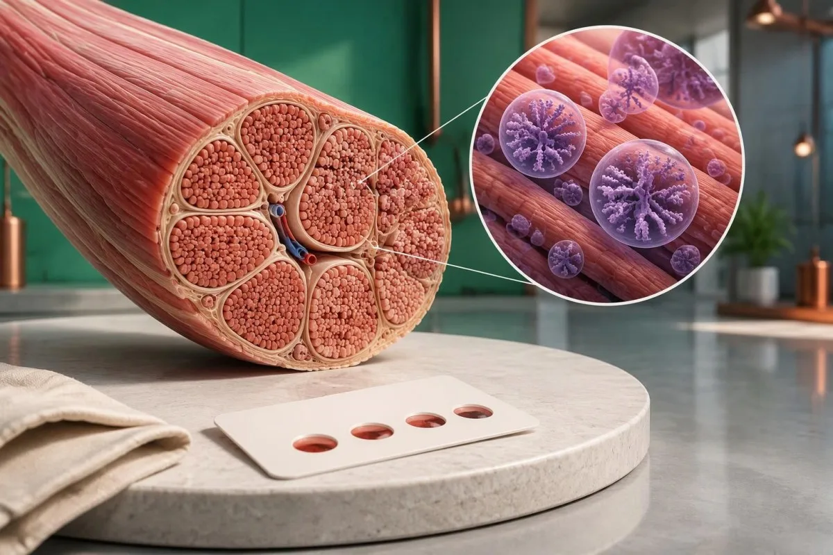
ผลเอนไซม์ GAA ต่ำทำให้สงสัยโรค Pompe แต่การคัดกรองเพียงอย่างเดียวไม่สามารถยืนยันโรคได้ การตรวจซ้ำจากสิ่งส่งตรวจที่เหมาะสมและการวิเคราะห์พันธุกรรม GAA ช่วยแยกภาวะพร่องเอนไซม์จริง ภาวะพาหะ และภาวะพร่องเทียม.

ประเด็นสำคัญ
- การทำงานของเอนไซม์ GAA ต่ำ เป็นสัญญาณให้ติดตามต่อ ไม่ใช่การวินิจฉัย; จุดเลือดแห้งที่ผิดปกติ 1 จุดต้องได้รับการยืนยันตามคำแนะนำของผู้เชี่ยวชาญ.
- ช่วงค่าอ้างอิง ขึ้นอยู่กับสิ่งส่งตรวจและวิธีตรวจ; ไม่สามารถเปรียบเทียบ nmol/h/mL กับ nmol/h/mg โปรตีนโดยตรงได้.
- การทำงานของเอนไซม์ที่ยังคงเหลือ มักต่ำกว่า 1% ในโรค Pompe ชนิดคลาสสิกที่เริ่มในวัยทารก แต่ตัวเลขนี้ไม่ใช่เกณฑ์คัดกรองสากล.
- ภาวะพร่องเทียม อาจทำให้กิจกรรม GAA ในห้องปฏิบัติการต่ำโดยไม่ก่อให้เกิดโรค Pompe; การตรวจซ้ำด้วยวิธีตรวจเดิมอาจให้ผลต่ำซ้ำ.
- การตรวจยีน GAA มองหาตัวแปรก่อโรคที่ส่งผลต่อสำเนายีนทั้งสองชุด; ต้องแปลผลตัวแปร 2 ตำแหน่งเพื่อจำแนกและระยะของยีน.
- แปรผันที่ไม่แน่ชัด ไม่ยืนยันว่าเป็นโรค Pompe; การพบตัวแปรที่มีนัยสำคัญไม่แน่ชัด 1 หรือ 2 ตัวแปรต้องอาศัยหลักฐานเพิ่มเติม.
- ความเสี่ยงในครอบครัว คือ 25% ที่บุตรแต่ละคนจะได้รับผลกระทบในแต่ละครรภ์ เมื่อบิดาและมารดาต่างเป็นพาหะของตัวแปร GAA ที่ก่อให้เกิดโรค.
- ต้องประเมินอย่างเร่งด่วน จำเป็นสำหรับทารกที่มี GAA ต่ำร่วมกับปัญหาการกินนม กล้ามเนื้ออ่อนแรงอย่างชัดเจน หรือสงสัยว่าหัวใจได้รับผลกระทบ อย่ารอการตรวจซ้ำตามกำหนดปกติ.
ผลเอนไซม์ GAA ต่ำหมายความว่าอย่างไรกันแน่?
A การตรวจโรค Pompe ที่แสดงกิจกรรม GAA ต่ำ หมายความว่าห้องปฏิบัติการตรวจพบการทำงานของ acid alpha-glucosidase ลดลงภายใต้เงื่อนไขการทดสอบของตน ผลนี้ไม่ได้พิสูจน์ว่าเป็นโรค Pompe: ผลคัดกรองต่ำ 1 ครั้งอาจสะท้อนภาวะพร่องเอนไซม์จริง ภาวะพร่องเอนไซม์เทียม ค่าที่ทับซ้อนกับผู้เป็นพาหะ หรือปัญหาของตัวอย่างตรวจ.

การวัดกิจกรรม GAA เป็นการวัดการทำงานของเอนไซม์ ไม่ใช่เพียงปริมาณโปรตีนเอนไซม์ที่มีอยู่. รายละเอียด 3 ข้อแรกที่ควรตรวจสอบคือชนิดตัวอย่าง หน่วยวัด และเกณฑ์ตัดสินของห้องปฏิบัติการ ผลที่ระบุว่าต่ำโดยไม่มีรายละเอียดเหล่านี้ไม่สามารถแปลผลให้เทียบกับตัวเลขที่พบทางออนไลน์ได้อย่างน่าเชื่อถือ.
เหตุผลในการตรวจส่งผลต่อความหมายของผลคัดกรองที่เป็นบวก ทารกแรกเกิดที่ตรวจพบจากการคัดกรองประชากรกับผู้มีอายุ 45 ปีที่มีอาการกล้ามเนื้อสะโพกอ่อนแรงลงเรื่อย ๆ มีความน่าจะเป็นเบื้องต้นที่จะเป็นโรค Pompe ต่างกัน แม้ค่ากิจกรรมของเอนไซม์ที่รายงานจะเท่ากันก็ตาม ดังนั้นควรพิจารณาอาการและผลตรวจร่างกายประกอบกับผลตรวจทางห้องปฏิบัติการ.
ผลคัดกรองเป็นบวกไม่เหมือนกับผลยืนยันว่าเป็นโรค. หากห้องปฏิบัติการ 2 แห่งใช้สารตั้งต้นหรือการเตรียมตัวอย่างต่างกัน ผลตัวเลขอาจไม่ตรงกันโดยไม่ได้หมายความว่าห้องปฏิบัติการแห่งใดทำผิดพลาด ให้สอบถามว่าการนัดครั้งถัดไปเป็นการตรวจวัดคัดกรองอีกครั้งหรือเป็นการตรวจเอนไซม์เพื่อยืนยันผลโดยเฉพาะ.
คันเตสตีเป็น เครื่องวิเคราะห์ผลเลือด AI ซึ่งช่วยจัดระเบียบรายงาน GAA ควบคู่กับผลตรวจทางห้องปฏิบัติการตามปกติได้ แต่ไม่สามารถยืนยันการวินิจฉัยโรค Pompe จากค่าที่ถูกทำเครื่องหมายไว้เพียง 1 ค่าได้ บริการของเรา ข้อมูลบริษัท อธิบายโครงสร้างการดำเนินงานเบื้องหลังบริการ ส่วนภาวะพร่องเอนไซม์ที่สงสัยว่าเป็นโรคถ่ายทอดทางพันธุกรรมยังคงต้องได้รับการประเมินจากแพทย์ผู้เชี่ยวชาญด้านเมแทบอลิซึมหรือระบบประสาทและกล้ามเนื้อ.
เหตุใดภาวะพร่อง GAA จึงทำให้เกิดปัญหาที่กล้ามเนื้อและหัวใจ?
โรค Pompe เกิดจากภาวะพร่องเอนไซม์ lysosomal acid alpha-glucosidase, ซึ่งเป็นเอนไซม์ที่ย่อยสลายไกลโคเจนภายในช่องรีไซเคิลของเซลล์ GAA ออกฤทธิ์กับพันธะกลูโคสทั้งแบบ alpha-1,4 และ alpha-1,6; กิจกรรมเอนไซม์ที่ไม่เพียงพอทำให้ไกลโคเจนสะสม โดยเฉพาะในกล้ามเนื้อลายและกล้ามเนื้อหัวใจ.

โรค Pompe ชนิดรุนแรงที่เริ่มในวัยทารกมักมีกิจกรรม GAA ต่ำกว่า 1% ของค่าปกติในการทดสอบเนื้อเยื่อเพื่อการวินิจฉัย. โรคที่เริ่มแสดงอาการภายหลังอาจยังคงมีกิจกรรมเอนไซม์สูงกว่า โดยในบางการทดสอบอาจอยู่ที่ประมาณ 2–40%; ช่วงค่าเชิงพรรณนาเหล่านี้ทับซ้อนกับข้อจำกัดของการทดสอบ และต้องไม่ใช้แทนการแปลผลเพื่อการวินิจฉัยที่ผ่านการตรวจสอบความถูกต้องของห้องปฏิบัติการ.
แนวทางของ Kishnani และคณะใน Genetics in Medicine ปี 2006 อธิบายความเชื่อมโยงระหว่างภาวะพร่อง GAA การสะสมไกลโคเจน และการประเมินหลายระบบ ความแตกต่างทางคลินิกไม่ได้พิจารณาเพียงว่ากิจกรรมเอนไซม์ต่ำกว่า 1% หรือไม่: อายุเมื่อเริ่มแสดงอาการ ผลตรวจหัวใจ และการทำงานของกล้ามเนื้อช่วยหายใจก็ช่วยระบุรูปแบบและความเร่งด่วนของโรคได้เช่นกัน.
ผลตรวจกลูโคสตามปกติไม่ได้วัดการกำจัดไกลโคเจนในไลโซโซม. ตัวอย่างเช่น ผู้ใหญ่สมมติรายหนึ่งอาจมีระดับกลูโคสขณะอดอาหาร 92 mg/dL แต่มีโรคสะสมไกลโคเจนในกล้ามเนื้อที่ถ่ายทอดทางพันธุกรรม ระดับกลูโคสปกติไม่ได้แสดงว่าไกลโคเจนภายในไลโซโซมของกล้ามเนื้อถูกนำไปผ่านกระบวนการตามปกติ.
โรคไลโซโซมชนิดอื่นเกี่ยวข้องกับเอนไซม์และยีนที่ต่างกัน จึงนำค่าเกณฑ์ของโรคเหล่านั้นมาใช้กับการตรวจ Pompe ไม่ได้ คำอธิบายของเราเกี่ยวกับ การยืนยันเอนไซม์ในโรค Gaucher แสดงขั้นตอนการตรวจจากเอนไซม์ไปสู่พันธุศาสตร์ในภาพรวม พร้อมชี้ให้เห็นว่าเหตุใดการทดสอบเอนไซม์ของโรคหนึ่งจึงใช้แทนการทดสอบของอีกโรคหนึ่งไม่ได้.
มีช่วงปกติของการทำงานของเอนไซม์ GAA หรือไม่?
There is no single universal normal range for GAA enzyme activity. Dried blood spots, leukocytes and cultured fibroblasts use different preparations and reporting units; a number expressed in nmol/h/mL cannot be directly compared with a result expressed in nmol/h/mg protein.

A screening cutoff separates samples needing follow-up from samples passing that screen. It does not necessarily separate affected people from unaffected people with complete accuracy; laboratories may use different fluorometric or tandem mass spectrometry methods, and 2 screening programs may therefore select different recall thresholds.
For a clearly hypothetical example, suppose a laboratory uses a DBS screening cutoff of 1.5 nmol/h/mL and reports 0.7 nmol/h/mL. The result is below that invented laboratory threshold, but dividing 0.7 by 1.5 gives 47% of the cutoff—not 47% of normal enzyme activity and not a severity score.
The lower limit of detection is different from the clinical cutoff. A report reading less than 0.1 in its stated units may mean the method cannot quantify lower activity reliably; it does not prove absolute absence of enzyme, and repeating measurements close to that analytical floor may produce proportionally large variation.
Screening performance also depends on prevalence. In a hypothetical group with 10 true-positive and 100 false-positive results, only about 9% of positive screens represent disease; those invented counts are not Pompe screening statistics, but explain why ผลเชิงคุณภาพเทียบกับผลเชิงปริมาณ require different interpretation.
| ระดับ | ช่วง | ความหมาย |
|---|---|---|
| Passes hypothetical screen | ≥1.5 nmol/h/mL | Illustrative DBS cutoff only; not a universal normal range and not an absolute exclusion in a symptomatic person. |
| Below hypothetical cutoff | <1.5 nmol/h/mL | Illustrative follow-up threshold only; true deficiency, pseudodeficiency and specimen effects still need separation. |
เหตุใดจึงต้องตรวจ GAA ซ้ำจากสิ่งส่งตรวจชนิดอื่น?
Repeat GAA testing checks whether low activity is reproducible and whether specimen quality or assay interference explains it. Confirmation usually moves beyond 1 screening card to a fresh, appropriately handled specimen, often with GAA measurement in leukocytes or another specialist-selected cell preparation.

Heat, humidity and inadequate drying can reduce enzyme activity in a dried blood spot. A low result after 2 days of poorly controlled transport may deserve a fresh collection, but transport history cannot be used to dismiss a result automatically; the laboratory should assess card quality and its stability requirements.
Acarbose is used in some GAA assay methods to suppress interfering maltase-glucoamylase activity. That interference can make measured activity appear higher and potentially obscure deficiency; checking how a laboratory isolates GAA activity can therefore matter more than obtaining a second number with the same unresolved methodological limitation.
Two low DBS results still do not automatically confirm Pompe disease. Pseudodeficiency can reproduce across repeated measurements, including some confirmatory methods; when enzyme and genetic findings disagree, a specialist laboratory may recommend cultured fibroblast testing or additional molecular investigation rather than simply ordering a third screening card.
Give the laboratory the dates of collection, recent transfusions or transplantation, and any enzyme replacement treatment already received. Our guide to การรบกวนจากคุณภาพตัวอย่าง explains the general distinction between biological findings and specimen problems; GAA testing has its own handling rules, so do not import another test's redraw interval.
ภาวะพร่อง GAA เทียมแตกต่างจากโรค Pompe อย่างไร?
GAA pseudodeficiency means an inherited variant lowers measured enzyme activity without causing the glycogen-storage disorder Pompe disease. Two commonly discussed variants are c.1726G>A, p.Gly576Ser, and c.2065G>A, p.Glu689Lys; their interpretation depends on the complete genotype and the assay used.
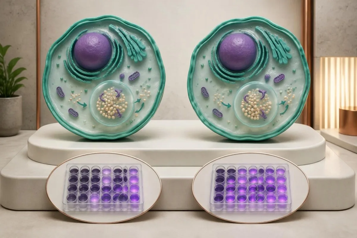
Pseudodeficiency is a biochemical testing issue, not an early stage of Pompe disease. A person with only pseudodeficiency variants does not need Pompe treatment because of 1 low activity result; the specialist must first verify that disease-causing variants or another explanation have not been missed.
The p.Gly576Ser and p.Glu689Lys variants are often found together on 1 gene copy and are particularly relevant in some East Asian ancestry populations. Ancestry changes how often laboratories encounter this pattern, but cannot establish or exclude pseudodeficiency in an individual; people from other backgrounds can carry these variants too.
Pseudodeficiency can remain low on repeat enzyme testing. The practical distinction is whether the complete biochemical and genetic evidence supports disease-causing GAA deficiency, not whether a second result falls below an arbitrary percentage; specialists may compare different assay substrates or cell preparations when the pattern remains ambiguous.
Pseudodeficiency variants can also coexist with a disease-causing variant, making a short report misleading if it lists everything under mutations. Our Fabry enzyme and genetics guide shows another situation where enzyme results need genetic context, although Fabry's X-linked inheritance differs from Pompe's 2-copy recessive mechanism.
การตรวจยีน GAA ควรมองหาอะไร?
GAA gene testing investigates whether disease-causing variants affect both copies of the GAA gene. Pompe disease is autosomal recessive, and the GAA gene is located on chromosome 17; interpretation requires variant classification, adequate test coverage and, when needed, confirmation that variants affect opposite gene copies.
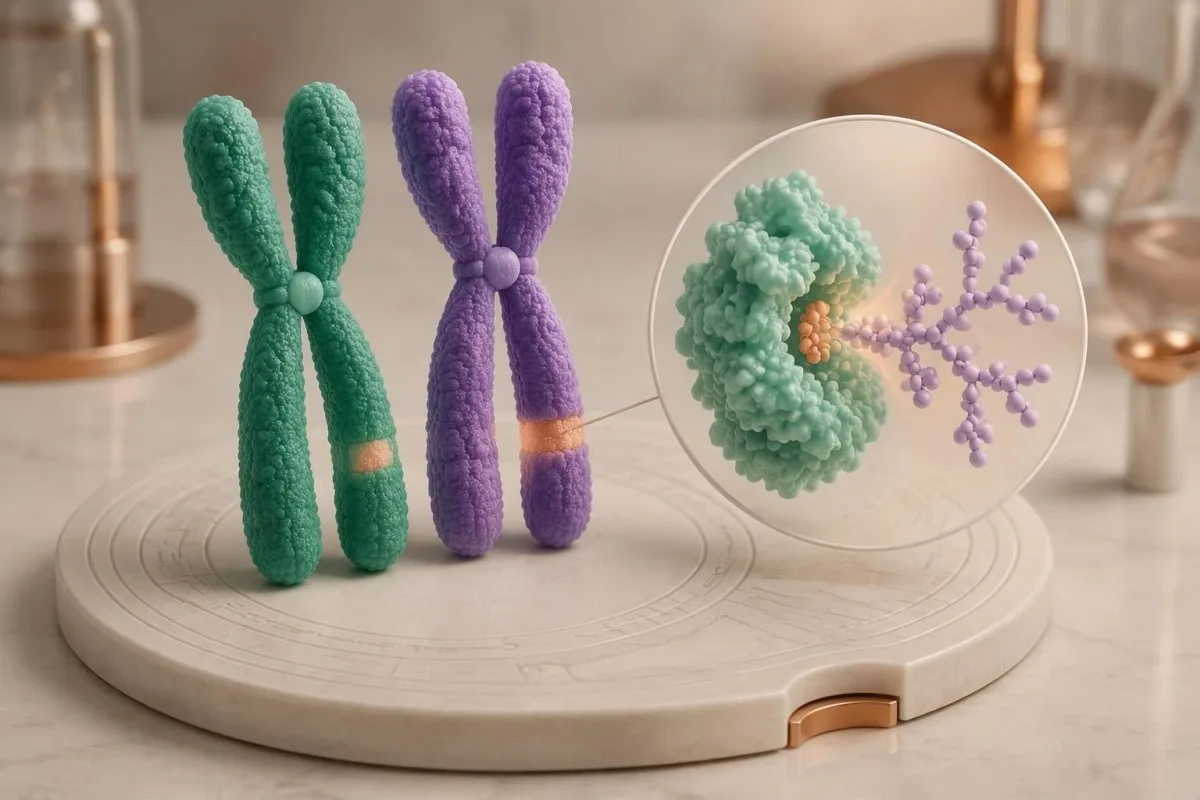
Two pathogenic or likely pathogenic GAA variants in trans support molecular confirmation of Pompe disease. In trans means the variants affect opposite gene copies; 2 variants in cis affect the same copy and do not, by themselves, demonstrate a recessive disease genotype.
Standard sequencing detects many small substitutions and insertions or deletions, but does not answer every possible genetic question. If only 1 disease-causing variant is found despite convincing enzyme deficiency, ask whether deletion/duplication analysis was included and whether poorly covered or deep intronic regions require further investigation.
A common later-onset-associated GAA variant is c.-32-13T>G, which affects splicing. Finding this variant on 1 copy does not diagnose Pompe disease, and finding it with another disease-causing variant does not precisely predict the age symptoms will begin; residual splicing and other biological factors influence presentation.
Parental testing can help establish phase when a laboratory detects 2 different variants but cannot determine their arrangement. Our guide to inherited disease genetic reports explains why inheritance and family testing matter, while the actual gene coverage and diagnostic criteria must remain specific to GAA.
ควรแปลผลลัพธ์ที่ก่อโรค ไม่แน่ชัด และผลลบอย่างไร?
A pathogenic result, a variant of uncertain significance and a negative result have different diagnostic meanings. Genetic laboratories commonly use 5 categories: pathogenic, likely pathogenic, uncertain significance, likely benign and benign; a VUS is not equivalent to a disease-causing GAA variant.
Richards et al.'s 2015 ACMG/AMP consensus established the widely used 5-category sequence-variant framework and states that a VUS should not guide clinical decision-making as though pathogenic. For Pompe evaluation, low enzyme activity can contribute evidence, but its value depends on whether pseudodeficiency and assay limitations have been addressed.
One pathogenic variant plus 1 VUS is an unresolved combination, not automatic confirmation or exclusion. Depending on the enzyme and clinical findings, specialists may investigate family segregation, RNA effects or variant-specific functional evidence; a diagnostic enzyme result may support disease even while the second molecular finding remains incompletely characterized.
A negative sequencing report means the test found no reportable variants within its scope. It does not prove that every GAA-related mechanism was examined; 1 negative limited test may warrant expanded testing when properly performed enzyme studies and the clinical pattern remain strongly suggestive.
คันเตสตีเป็น แพลตฟอร์มการแปลผลตรวจเลือดของ AI that can help separate a report's 5 classification terms from its numerical enzyme findings, without adjudicating an uncertain genotype. Our มาตรฐานทางเทคนิคและทางคลินิกของเรา describe interpretation boundaries; our uncertain gene-result explanation provides a related example of why variants and diagnoses are not interchangeable.
การมีตัวแปร GAA หนึ่งตำแหน่งหมายถึงเป็นพาหะหรือเป็นโรค?
One confirmed disease-causing GAA variant usually indicates carrier status if a second disease-causing variant and diagnostic enzyme deficiency are absent. Carriers do not typically develop Pompe disease; when both parents are carriers, each pregnancy has a 25% affected, 50% carrier and 25% non-carrier probability.
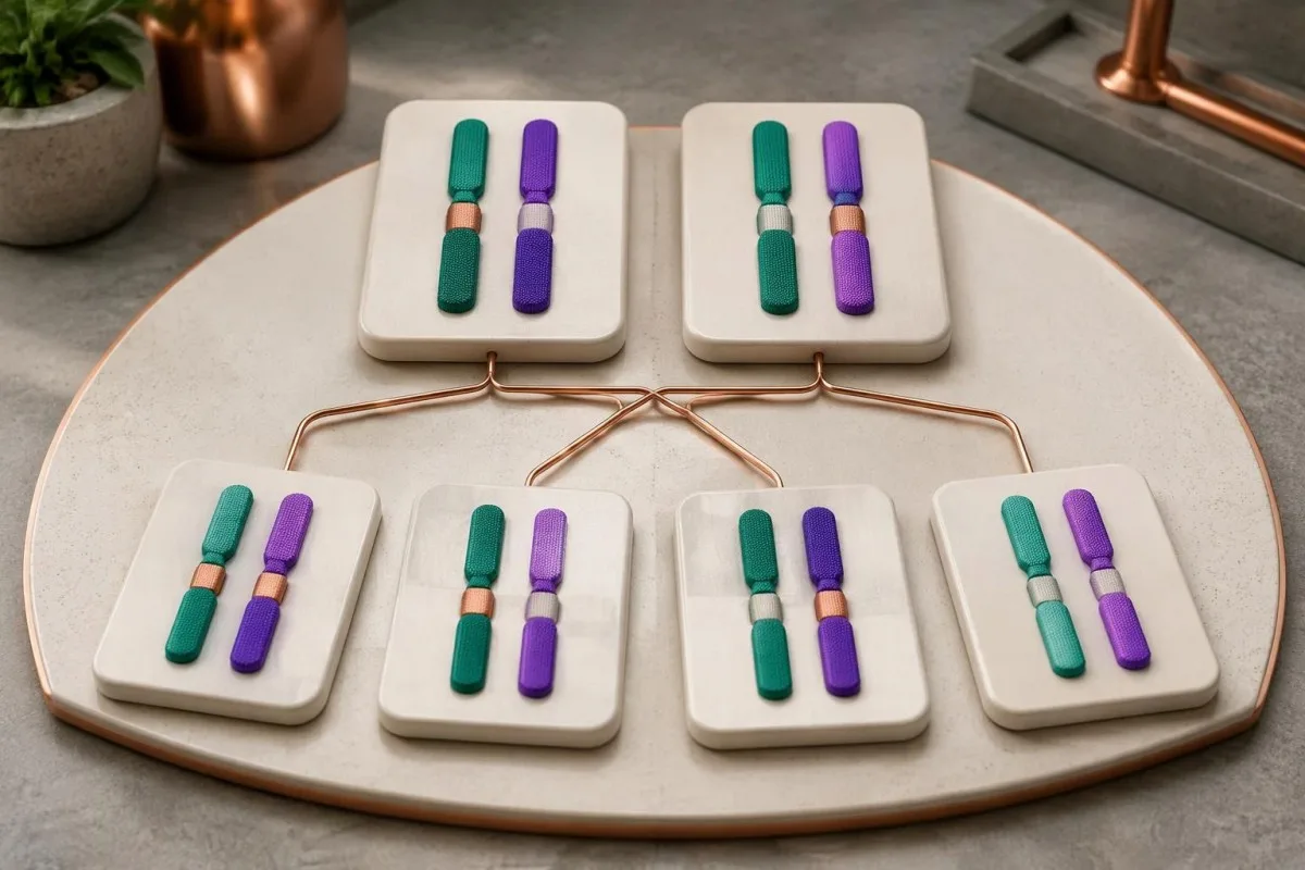
Carrier enzyme values can overlap with values in unaffected people. GAA activity is therefore not a reliable standalone carrier test, and a carrier's activity should not be assumed to equal exactly 50% of a laboratory's normal mean; targeted genetic testing is generally more informative when a familial variant is known.
The 25% affected-child probability applies independently to each pregnancy when both parents carry disease-causing variants capable of producing Pompe disease together. Having 1 unaffected child does not reduce the next pregnancy's Mendelian probability, and pseudodeficiency variants should not be substituted for pathogenic variants in that calculation.
Family testing is most useful after the affected person's variants are clarified. If a symptomatic adult has only 1 identified pathogenic variant, relatives should not be reassured that the entire family question is settled until the specialist considers whether the second variant was missed by the original assay.
For a hypothetical couple, one partner carrying a pathogenic GAA variant and the other carrying only a pseudodeficiency variant does not create the same 25% Pompe risk as 2 pathogenic-variant carriers. Our carrier screening limitations guide explains why a report's coverage and residual risk deserve discussion before reproductive decisions.
ผลตรวจทางห้องปฏิบัติการอื่นใดสนับสนุนการประเมินโรค Pompe?
Creatine kinase, AST, ALT and urinary glucose tetrasaccharide can support Pompe evaluation, but none confirms the diagnosis alone. Urinary glucose tetrasaccharide may be reported as Glc4 or within a Hex4 assay; these 2 report terms require method-specific interpretation rather than assumption that every laboratory measures exactly the same thing.
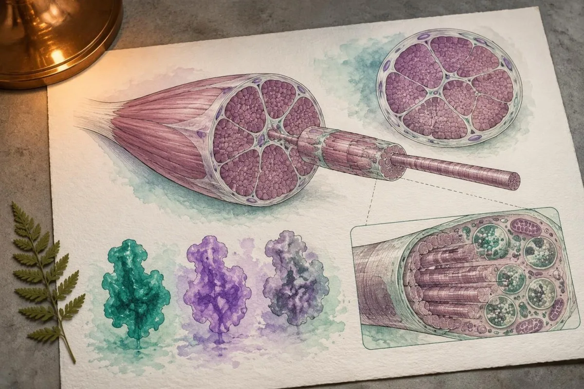
Normal CK does not exclude later-onset Pompe disease. In a hypothetical adult with progressive difficulty rising from a chair, CK of 140 U/L may fall within that laboratory's interval despite clinically meaningful weakness; the combination of symptoms and low confirmed GAA activity matters more than a reassuring CK flag.
AST and ALT can rise because of muscle involvement, not only liver disease. A hypothetical AST of 86 U/L with elevated CK and muscle weakness deserves a muscle-aware assessment; our aldolase and muscle marker guide explains why additional muscle enzymes may help characterize the pattern without identifying its cause.
Urinary Glc4 is a supportive glycogen-related biomarker, not a Pompe-specific diagnostic test. Reference limits vary with age and method, and elevation can occur in other circumstances; 1 elevated result should be interpreted alongside enzyme findings, genetics and the laboratory's creatinine-normalized units.
A falling CK or Glc4 after treatment does not automatically prove that strength or breathing has improved by the same percentage. Our discussion of AST interpretation beyond liver disease illustrates why 2 related markers can move together while still answering different questions about tissue injury and function.
เหตุใดจึงต้องประเมินการหายใจเมื่อผลตรวจ Pompe ผิดปกติ?
Pompe disease can weaken the diaphragm and other respiratory muscles, sometimes before obvious walking difficulty. Assessment may include seated and supine forced vital capacity, respiratory muscle pressures and sleep-related testing; a daytime oxygen saturation of 97% does not rule out overnight hypoventilation.
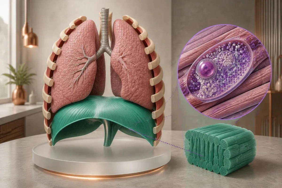
A position-related fall in vital capacity can suggest diaphragm weakness. For example, a hypothetical seated FVC of 3.0 L and supine FVC of 2.4 L represents a 20% decline; the specialist interprets that difference with technique, predicted values and symptoms, rather than treating it as a Pompe-specific cutoff.
Morning headaches, unrefreshing sleep and difficulty breathing when lying flat can justify evaluation for sleep-related hypoventilation. These symptoms do not identify Pompe disease on their own; our คู่มือการทดสอบอาการหายใจถี่ explains why laboratory results cannot replace respiratory examination and physiological testing.
Arterial carbon dioxide is normally about 35–45 mmHg in adults, with context-dependent variation. A value above 45 mmHg indicates hypercapnia, but its urgency depends on pH, symptoms and the clinical setting; our blood gas interpretation guide explains why oxygen and carbon dioxide answer different questions.
Severe breathlessness, confusion, blue coloration or rapidly worsening weakness warrants emergency care, not an online interpretation session. A confirmed molecular result affecting 2 GAA copies does not measure current respiratory safety; clinicians need direct functional assessment to decide whether ventilation support or urgent monitoring is necessary.
เกิดอะไรขึ้นหลังผลคัดกรอง Pompe ในทารกแรกเกิดเป็นบวก?
A positive newborn Pompe screen requires prompt specialist assessment, confirmatory enzyme testing and genetic evaluation. It does not mean every infant has Pompe disease; the immediate task is to distinguish pseudodeficiency or carrier findings from infantile disease and a later-onset-associated genotype affecting both GAA copies.

Burton et al.'s 2017 Pediatrics newborn-screening algorithms describe an initial evaluation integrating GAA confirmation, molecular testing and clinical assessment. If an infant has feeding difficulty, marked hypotonia or suspected cardiomyopathy, do not wait 2 weeks for a routine repeat appointment; the screening team should arrange urgent metabolic and cardiac evaluation.
Classic infantile Pompe disease commonly involves hypertrophic cardiomyopathy. A newborn's echocardiogram and electrocardiogram help assess risk that cannot be estimated from 1 enzyme number; absence of obvious heart symptoms at home does not replace the specialist examination after an abnormal screen.
CRIM status concerns whether cross-reactive GAA protein is present and helps specialists plan immune-risk management around enzyme replacement therapy. Less than 1% enzyme activity does not by itself establish CRIM-negative status, because an inactive protein may still be present; this is a protein-expression question, not just an activity threshold.
Later-onset-associated findings in a newborn require a monitoring plan, not an invented age-of-onset prediction. Even 2 classified disease-causing variants may not reveal when symptoms will emerge; our คำแนะนำด้านความปลอดภัยของ AI สำหรับเด็ก explains why pediatric results require age-specific context and clinician-led decisions.
ผู้เชี่ยวชาญหาข้อสรุปอย่างไรเมื่อผลตรวจในผู้ใหญ่ขัดแย้งกัน?
Conflicting adult Pompe results are resolved by checking specimen quality, confirmatory enzyme methods, variant classification and clinical fit. One low screen plus 1 genetic finding rarely tells the whole story; specialists investigate whether the apparent conflict comes from pseudodeficiency, an incomplete molecular test or another cause of weakness.

Consider a hypothetical 52-year-old with mild fatigue, low DBS GAA activity and only the 2 commonly discussed pseudodeficiency variants. If specialist testing supports pseudodeficiency and no disease-causing genotype, the low screen does not explain the fatigue; other causes should be assessed rather than attaching a Pompe label to an incidental biochemical finding.
A second hypothetical adult has progressive hip weakness, a 20% positional FVC decline, confirmed cellular GAA deficiency and 2 pathogenic variants in trans. Together, those findings support Pompe disease; a normal routine glucose or modest CK result would not outweigh the converging biochemical, genetic and functional evidence.
Convincing enzyme deficiency with only 1 identified pathogenic variant deserves further investigation. The appropriate next step may be deletion/duplication analysis, RNA studies or another specialist assay—not automatically calling the person a carrier; the investigation should also check whether non-Pompe neuromuscular conditions better explain the examination.
Variant-related disease monitoring is not the same as predicting a person's future from their DNA. Our inherited neuromuscular risk guide offers a related family-testing perspective, but 2 conditions can differ substantially in inheritance, penetrance and treatment triggers.
คุณควรนำอะไรไปพบผู้เชี่ยวชาญด้านโรค Pompe?
Bring the complete enzyme report, complete genetic report and a dated symptom history to a Pompe specialist appointment. Three report details are especially useful: specimen type, assay units and variant classification; screenshots showing only a red flag or the word mutation can omit information that changes the interpretation.

Ask whether confirmatory testing has already been completed and whether the laboratory measured GAA specifically in an appropriate cell preparation. If 2 variants were found, ask whether they are in trans; if only 1 was found, ask what variant types and regions the test could detect.
Do not try to raise GAA activity with supplements or diet before confirmation. No vitamin or food corrects inherited pathogenic variants affecting both GAA copies; nutrition may support general care after diagnosis, but cannot substitute for enzyme confirmation or specialist treatment planning.
คันเตสตีเป็น เครื่องมือวิเคราะห์ผลตรวจเลือดที่ขับเคลื่อนด้วย AI that can help organize report terminology and associated laboratory trends, not decide whether 2 uncertain variants establish Pompe disease. Our คู่มือตรวจสอบแหล่งที่มาของรายงาน explains why the original document remains the reference when an automated explanation and a specialist interpretation differ.
Genetic documents can contain information relevant to relatives as well as the tested person. Before sharing 1 family member's report, check consent and remove unnecessary identifiers; Kantesti's ข้อมูลความเป็นส่วนตัวและข้อมูล describes our handling approach, while sharing results with relatives should follow a genetic counselor's guidance.
งานวิจัยที่ตีพิมพ์และข้อจำกัดของการแปลผลตรวจ Pompe
Pompe test interpretation rests on disease-specific enzyme, genetic and clinical evidence—not on general laboratory publications or an AI explanation. Kantesti includes 3 directly relevant external sources below and 2 separate educational publications; the iron and coagulation publications are not evidence for diagnosing Pompe disease.
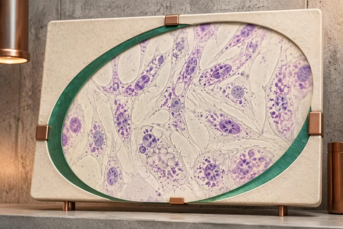
The 2006 Kishnani guideline provides disease-specific diagnostic and management context, while the 2017 Burton newborn-screening paper addresses confirmation after population screening. Their recommendations should be applied with the specialist laboratory's validated methods; neither supplies 1 universally transferable DBS enzyme cutoff for every instrument and population.
Richards et al.'s 2015 ACMG/AMP publication explains the 5-category variant framework used to distinguish pathogenic findings from uncertainty. Classification can change when new evidence becomes available, so ask the genetics service whether an unresolved result has a reanalysis pathway rather than assuming every VUS will eventually become pathogenic.
เนื้อหาให้ความรู้ของเรา สิ่งพิมพ์ด้านการตรวจธาตุเหล็ก addresses a different laboratory domain. The separate เอกสารตีพิมพ์เกี่ยวกับการตรวจการแข็งตัวของเลือด also covers unrelated markers; their 2 DOI records are provided for publication transparency, not as supporting citations for GAA deficiency or pseudodeficiency.
ของ Kantesti มาตรฐานเนื้อหาทางการแพทย์ distinguish patient education from diagnosis and treatment instructions. The practical endpoint is a documented interpretation of 3 evidence streams—enzyme testing, genetic analysis and clinical assessment—with urgent care taking priority whenever infantile disease or respiratory compromise is suspected.
คำถามที่พบบ่อย
การทำงานของเอนไซม์ GAA ต่ำหมายความว่าเป็นโรคปอมเปเสมอไปหรือไม่?
กิจกรรมของเอนไซม์ GAA ต่ำไม่ได้หมายความว่าเป็นโรคพอมพ์เสมอไป ผลคัดกรองที่ผิดปกติเพียงครั้งเดียวอาจสะท้อนถึงภาวะพร่องเอนไซม์จริง ภาวะพร่องเอนไซม์เทียม การเสื่อมสภาพของสิ่งส่งตรวจ หรือกิจกรรมของเอนไซม์ที่ทับซ้อนกับกิจกรรมที่เกี่ยวข้องกับการเป็นพาหะ การยืนยันผลต้องอาศัยผู้เชี่ยวชาญแปลผลการตรวจเอนไซม์ที่เหมาะสม ผลตรวจทางพันธุกรรมของ GAA และบริบททางคลินิก การตรวจคัดกรองด้วยวิธีเดิมซ้ำเพียงอย่างเดียวอาจทำให้พบภาวะพร่องเอนไซม์เทียมซ้ำ แทนที่จะช่วยคลี่คลายข้อสงสัย.
ระดับเอนไซม์ GAA ปกติคือเท่าใด?
There is no universal normal GAA enzyme level because specimen preparation, assay substrate and reporting units differ between laboratories. A value in nmol/h/mL cannot be directly compared with a value in nmol/h/mg protein. Classic infantile Pompe disease often has less than 1% of normal activity in diagnostic tissue assays, but this is not a universal newborn-screening cutoff. Use the reference information printed on the original report and the specialist laboratory's interpretation.
ผลตรวจ GAA ต่ำสองครั้งยังอาจเป็นภาวะพร่องเทียมได้หรือไม่?
ผลการตรวจ GAA ต่ำสองครั้งยังอาจสะท้อนภาวะ pseudodeficiency โดยเฉพาะเมื่อใช้วิธีตรวจแบบเดียวกันหรือที่ได้รับผลกระทบคล้ายกัน Variant ของ pseudodeficiency ทำให้ค่ากิจกรรมที่วัดได้ลดลง โดยตัว variant เองไม่ได้ก่อให้เกิดโรคสะสมไกลโคเจนชนิด Pompe ผู้เชี่ยวชาญแยกรูปแบบนี้โดยพิจารณาจีโนไทป์ GAA ทั้งหมด คุณลักษณะของวิธีตรวจ และการตรวจด้วยเซลล์ชนิดอื่นหรือการตรวจการทำงานเพิ่มเติมเมื่อจำเป็น ดังนั้นค่าต่ำสองครั้งที่ตรวจซ้ำจึงไม่เทียบเท่ากับการยืนยันโรคอย่างเป็นอิสระสองครั้ง.
การพบความแปรผันของยีน GAA สองตำแหน่งยืนยันว่าเป็นโรคพอมพีหรือไม่?
การพบแปรผันของยีน GAA สองตำแหน่งไม่ได้ยืนยันโรค Pompe โดยอัตโนมัติ การพบแปรผันที่ก่อโรคหรือมีแนวโน้มก่อโรคสองตำแหน่งบนอัลลีลคนละข้าง ซึ่งส่งผลต่อสำเนายีนคนละชุด สนับสนุนการยืนยันระดับโมเลกุล ขณะที่แปรผันสองตำแหน่งบนอัลลีลเดียวกันส่งผลต่อสำเนาเพียงชุดเดียวเท่านั้น แปรผันที่มีนัยสำคัญไม่แน่ชัดและแปรผันที่ทำให้เกิดภาวะเทียมพร่องเอนไซม์ไม่อาจนับเป็นข้อค้นพบที่ยืนยันแล้วว่าก่อโรคได้ หลักฐานด้านเอนไซม์และอาการทางคลินิกยังคงมีประโยชน์อย่างยิ่งเมื่อยังไม่สามารถสรุปการจัดประเภทหรือเฟสของแปรผันได้.
ค่า CK ปกติสามารถตัดโรค Pompe ที่เริ่มมีอาการในวัยผู้ใหญ่ได้หรือไม่?
Normal CK cannot rule out late-onset Pompe disease. A hypothetical CK of 140 U/L may fall within a laboratory's reference interval despite progressive proximal weakness or diaphragm involvement. CK measures muscle injury-related enzyme release, not lysosomal GAA function. Persistent compatible symptoms deserve specialist assessment even when routine muscle markers are reassuring.
ผลตรวจโรคปอมเปในทารกแรกเกิดเป็นบวกเร่งด่วนแค่ไหน?
ผลตรวจคัดกรองโรคพอมพีในทารกแรกเกิดเป็นบวกจำเป็นต้องติดต่อทีมคัดกรองหรือทีมเมแทบอลิกโดยเร็ว และทารกที่มีปัญหาในการกิน กล้ามเนื้ออ่อนแรงอย่างชัดเจน หรือสงสัยว่ามีภาวะหัวใจเกี่ยวข้อง จำเป็นต้องได้รับการประเมินอย่างเร่งด่วน ผลตรวจคัดกรองที่เป็นบวกเพียงครั้งเดียวยังไม่ยืนยันการวินิจฉัย แต่หากสงสัยว่าเป็นโรคชนิดคลาสสิกที่เริ่มในวัยทารก ไม่ควรรอการนัดตรวจตามปกติในอีกหลายสัปดาห์ต่อมา ผู้เชี่ยวชาญจะจัดการตรวจยืนยันการทำงานของเอนไซม์ การประเมินทางพันธุกรรม และการประเมินหัวใจ ซึ่งมักดำเนินการควบคู่กันไป หากหายใจลำบากอย่างรุนแรงหรือการตอบสนองลดลง จำเป็นต้องเข้ารับการดูแลฉุกเฉิน.
หากพ่อและแม่เป็นพาหะของโรคปอมเปทั้งคู่ เด็กจะมีความเสี่ยงอย่างไร?
หากบิดาและมารดาต่างเป็นพาหะของยีนแปรผัน GAA ที่ก่อโรค การตั้งครรภ์แต่ละครั้งมีโอกาส 25% ที่บุตรจะได้รับผลกระทบ โอกาส 50% ที่บุตรจะเป็นพาหะ และโอกาส 25% ที่บุตรจะไม่ได้รับยีนแปรผันในครอบครัวทั้งสองชนิด โอกาสเหล่านี้ใช้แยกกันอย่างเป็นอิสระกับการตั้งครรภ์แต่ละครั้ง ยีนแปรผันที่ทำให้เกิดภาวะบกพร่องเทียมไม่สามารถใช้แทนยีนแปรผันก่อโรคในการคำนวณนี้ได้ การให้คำปรึกษาทางพันธุศาสตร์ช่วยยืนยันผลตรวจของบิดามารดาและอธิบายทางเลือกในการตรวจ、】【.
อ่านผลตรวจเลือดอย่างไร
อัปโหลดรายงานผลตรวจของคุณ แล้ว AI ของ Kantesti จะอธิบายตัวบ่งชี้ทางชีวภาพแต่ละรายการด้วยภาษาที่เข้าใจง่ายภายในไม่กี่วินาที มีผู้ใช้ทั่วโลกมากกว่า 2 ล้านคน.
เอกสารอ้างอิง
- ไคลน์, ที., มิตเชลล์, เอส., & เวเบอร์, เอช. (2026). คู่มือการตรวจธาตุเหล็ก: TIBC ความอิ่มตัวของธาตุเหล็ก และความสามารถในการจับธาตุเหล็ก. Kantesti AI การวิจัยทางการแพทย์. โดอิ
- ไคลน์, ที., มิตเชลล์, เอส., & เวเบอร์, เอช. (2026). ช่วงค่าปกติของ aPTT: คู่มือ D-Dimer และ Protein C เกี่ยวกับการแข็งตัวของเลือด. Kantesti AI การวิจัยทางการแพทย์. โดอิ
- Kishnani PS et al. (2006). Pompe disease diagnosis and management guideline. วารสาร Genetics in Medicine. โดอิ พับเมด
- Burton BK et al. (2017). The Initial Evaluation of Patients After Positive Newborn Screening: Recommended Algorithms Leading to a Confirmed Diagnosis of Pompe Disease. Pediatrics. โดอิ
- Richards S และคณะ (2015). มาตรฐานและแนวทางสำหรับการแปลผลตัวแปรของลำดับ: ข้อเสนอแนะฉันทามติร่วมของ American College of Medical Genetics and Genomics และ Association for Molecular Pathology. วารสาร Genetics in Medicine. โดอิ พับเมด
- 2 ล้าน+การตรวจที่วิเคราะห์
- 127+ประเทศ
- 100+ภาษา
เกี่ยวกับทีมแพทย์


สัญญาณความน่าเชื่อถือ E-E-A-T
ประสบการณ์
การทบทวนเชิงคลินิกโดยแพทย์ที่นำโดยกระบวนการตีความผลตรวจในห้องแล็บ.
ความเชี่ยวชาญ
โฟกัสด้านเวชศาสตร์ห้องปฏิบัติการเกี่ยวกับพฤติกรรมของไบโอมาร์กเกอร์ในบริบททางคลินิก.
อำนาจ
ทบทวนทางการแพทย์โดย นพ. Thomas Klein พร้อมผลงานจาก นพ. Sarah Mitchell และศาสตราจารย์ ดร. Hans Weber.
ความน่าเชื่อถือ
การตีความที่อิงหลักฐาน พร้อมเส้นทางการติดตามที่ชัดเจนเพื่อลดความตื่นตระหนก.





ข้อจำกัดความรับผิดชอบทางการแพทย์
บทความนี้จัดทำเพื่อวัตถุประสงค์ด้านการศึกษาเท่านั้น และไม่ถือเป็นคำแนะนำทางการแพทย์ โปรดปรึกษาผู้ให้บริการด้านสุขภาพที่มีคุณสมบัติเหมาะสมเสมอสำหรับการตัดสินใจด้านการวินิจฉัยและการรักษา.
เผยแพร่แล้ว · ติดต่อทีมบรรณาธิการ
เผยแพร่โดย Kantesti Ltd จดทะเบียนในอังกฤษและเวลส์ เลขทะเบียนบริษัท. 17090423, ลอนดอน สหราชอาณาจักร.
