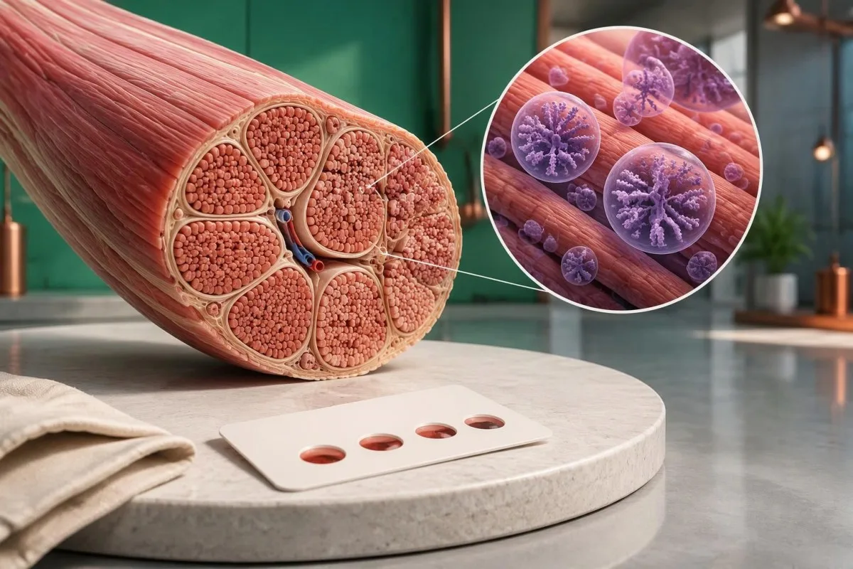
GAA酵素の測定値が低いとポンペ病が疑われますが、スクリーニングだけでは確定できません。適切な検体で再検査を行い、GAA遺伝子解析を実施することで、真の欠損、保因者状態、偽欠損を区別できます。.

要点
- GAA酵素活性の低値 は診断ではなく、追加評価の必要性を示す所見です。異常な乾燥血液スポット検体は、専門医の指示による確認検査が必要です。.
- 参考範囲 は検体と測定法によって異なります。nmol/h/mLとnmol/h/mg proteinを直接比較することはできません。.
- 残存酵素活性 は古典的乳児型ポンペ病では1%未満であることが多いですが、この数値は普遍的なスクリーニングカットオフではありません。.
- 偽欠損 はポンペ病を引き起こすことなく検査上のGAA活性を低下させることがあります。同じ測定法で再検査しても、低値が再現される場合があります。.
- GAA遺伝子検査 は、遺伝子の両方のコピーに影響を及ぼす疾患原因バリアントを調べます。2つのバリアントについて、分類と位相を解釈する必要があります。.
- 意義不明のバリアント ポンペ病を確定するものではありません。不確実な意義の変異が1つまたは2つある場合は、追加のエビデンスが必要です。.
- 家族のリスク は、両親ともに疾患原因となるGAAバリアントを保有している場合、妊娠ごとに罹患児が生まれる確率として25%です。.
- 緊急の評価 GAA低値に加え、哺乳困難、顕著な筋力低下、または心臓病変の疑いがある乳児には、ルーチンの再検査を待たずに必要です。.
GAA酵素の測定値が低いとは、実際にはどういう意味ですか?
A ポンペ病検査 GAA活性低値を示す結果は、検査室がその測定法の条件下で酸性α-グルコシダーゼ活性の低下を測定したことを意味します。ポンペ病を証明するものではありません。スクリーニングで低値が1回認められても、真の欠損、偽欠損、保因者に関連する値の重なり、または検体の問題が反映されている可能性があります。.

GAA活性は酵素の機能を測定するものであり、酵素タンパク質の量だけを測定するものではありません。. まず確認すべき3つの詳細は、検体の種類、測定単位、検査室の判定限界です。これらの詳細を伴わずに低値と記載された結果は、オンラインで見つけた数値と確実に比較して解釈することはできません。.
検査の理由によって、陽性スクリーニングの意味は変わります。集団スクリーニングで見つかった新生児と、進行性の股関節周囲の筋力低下がある45歳の人では、報告された酵素値が同じでも、ポンペ病の事前確率は異なります。そのため、症状と診察所見も検査結果と併せて考慮する必要があります。.
スクリーニング陽性は、疾患陽性と同じではありません。. 2つの検査室で異なる基質や検体調製法を使用している場合、どちらの検査室にも誤りがなくても数値が一致しないことがあります。次の予約が別のスクリーニング測定なのか、確定診断のための酵素検査なのかを確認してください。.
カンテスティは AI血液検査分析装置 GAAの報告を通常の検査所見と併せて整理する助けにはなりますが、フラグの付いた値が1つあるだけではポンペ病の診断を確定できません。当社の 会社情報は、より広範な医療AIのミッション、認証、および製品の範囲を説明しています。実用的な約束はシンプルです。迅速な解釈であっても、慎重な解釈であるべきです。 は、サービスの仕組みを説明するものです。遺伝性酵素欠損が疑われる場合は、引き続き代謝疾患または神経筋疾患の専門医による評価が必要です。.
GAA欠損はなぜ筋肉や心臓の問題を引き起こすのですか?
ポンペ病はリソソーム酸性α-グルコシダーゼの欠損によって生じます, 。この酵素は、細胞内のリサイクル区画でグリコーゲンを分解します。GAAはα-1,4およびα-1,6グルコース結合の両方に作用し、活性が不十分だと、特に骨格筋と心筋にグリコーゲンが蓄積します。.

古典的乳児型ポンペ病では、診断用組織検査におけるGAA活性が正常値の1%未満であることが多いです。. 遅発型では、より高い活性が保たれる場合があり、特定の検査法では約2–40%となることもあります。このような説明的範囲は検査法の限界と重なり得るため、検査室で検証済みの診断上の解釈に取って代わるものではありません。.
Kishnaniらによる2006年のGenetics in Medicineガイドラインは、GAA欠損、グリコーゲン蓄積、複数臓器にわたる評価の関連を説明しています。臨床的な鑑別は、活性が1%を下回るかどうかだけではありません。発症年齢、心臓所見、呼吸筋機能も、病型と緊急性の判断に役立ちます。.
通常の血糖値検査では、リソソーム内のグリコーゲン分解能は測定できません。. 例えば、仮想上の成人は、遺伝性の筋グリコーゲン蓄積症があっても空腹時血糖値が92 mg/dLであることがあります。血糖値が正常でも、筋リソソーム内のグリコーゲンが正常に処理されていることは証明されません。.
他のリソソーム病では異なる酵素や遺伝子が関与するため、その閾値をポンペ病検査に流用することはできません。当社による ゴーシェ病の酵素検査による確定診断 の説明は、酵素から遺伝学的検査へ進む一連の流れを広く示すと同時に、ある疾患の酵素検査が別の疾患の検査の代用にならない理由を示しています。.
GAA酵素活性に正常範囲はありますか?
There is no single universal normal range for GAA enzyme activity. Dried blood spots, leukocytes and cultured fibroblasts use different preparations and reporting units; a number expressed in nmol/h/mL cannot be directly compared with a result expressed in nmol/h/mg protein.
A screening cutoff separates samples needing follow-up from samples passing that screen. It does not necessarily separate affected people from unaffected people with complete accuracy; laboratories may use different fluorometric or tandem mass spectrometry methods, and 2 screening programs may therefore select different recall thresholds.
For a clearly hypothetical example, suppose a laboratory uses a DBS screening cutoff of 1.5 nmol/h/mL and reports 0.7 nmol/h/mL. The result is below that invented laboratory threshold, but dividing 0.7 by 1.5 gives 47% of the cutoff—not 47% of normal enzyme activity and not a severity score.
The lower limit of detection is different from the clinical cutoff. A report reading less than 0.1 in its stated units may mean the method cannot quantify lower activity reliably; it does not prove absolute absence of enzyme, and repeating measurements close to that analytical floor may produce proportionally large variation.
Screening performance also depends on prevalence. In a hypothetical group with 10 true-positive and 100 false-positive results, only about 9% of positive screens represent disease; those invented counts are not Pompe screening statistics, but explain why 定性検査結果と定量検査結果 require different interpretation.
| レベル | 範囲 | 意味 |
|---|---|---|
| Passes hypothetical screen | ≥1.5 nmol/h/mL | Illustrative DBS cutoff only; not a universal normal range and not an absolute exclusion in a symptomatic person. |
| Below hypothetical cutoff | <1.5 nmol/h/mL | Illustrative follow-up threshold only; true deficiency, pseudodeficiency and specimen effects still need separation. |
なぜ異なる検体でGAA検査を再実施するのですか?
Repeat GAA testing checks whether low activity is reproducible and whether specimen quality or assay interference explains it. Confirmation usually moves beyond 1 screening card to a fresh, appropriately handled specimen, often with GAA measurement in leukocytes or another specialist-selected cell preparation.

Heat, humidity and inadequate drying can reduce enzyme activity in a dried blood spot. A low result after 2 days of poorly controlled transport may deserve a fresh collection, but transport history cannot be used to dismiss a result automatically; the laboratory should assess card quality and its stability requirements.
Acarbose is used in some GAA assay methods to suppress interfering maltase-glucoamylase activity. That interference can make measured activity appear higher and potentially obscure deficiency; checking how a laboratory isolates GAA activity can therefore matter more than obtaining a second number with the same unresolved methodological limitation.
Two low DBS results still do not automatically confirm Pompe disease. Pseudodeficiency can reproduce across repeated measurements, including some confirmatory methods; when enzyme and genetic findings disagree, a specialist laboratory may recommend cultured fibroblast testing or additional molecular investigation rather than simply ordering a third screening card.
Give the laboratory the dates of collection, recent transfusions or transplantation, and any enzyme replacement treatment already received. Our guide to 検体品質による干渉 explains the general distinction between biological findings and specimen problems; GAA testing has its own handling rules, so do not import another test's redraw interval.
GAA偽欠損はポンペ病とどう異なりますか?
GAA pseudodeficiency means an inherited variant lowers measured enzyme activity without causing the glycogen-storage disorder Pompe disease. Two commonly discussed variants are c.1726G>A, p.Gly576Ser, and c.2065G>A, p.Glu689Lys; their interpretation depends on the complete genotype and the assay used.
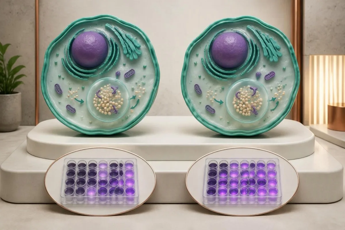
Pseudodeficiency is a biochemical testing issue, not an early stage of Pompe disease. A person with only pseudodeficiency variants does not need Pompe treatment because of 1 low activity result; the specialist must first verify that disease-causing variants or another explanation have not been missed.
The p.Gly576Ser and p.Glu689Lys variants are often found together on 1 gene copy and are particularly relevant in some East Asian ancestry populations. Ancestry changes how often laboratories encounter this pattern, but cannot establish or exclude pseudodeficiency in an individual; people from other backgrounds can carry these variants too.
Pseudodeficiency can remain low on repeat enzyme testing. The practical distinction is whether the complete biochemical and genetic evidence supports disease-causing GAA deficiency, not whether a second result falls below an arbitrary percentage; specialists may compare different assay substrates or cell preparations when the pattern remains ambiguous.
Pseudodeficiency variants can also coexist with a disease-causing variant, making a short report misleading if it lists everything under mutations. Our Fabry enzyme and genetics guide shows another situation where enzyme results need genetic context, although Fabry's X-linked inheritance differs from Pompe's 2-copy recessive mechanism.
GAA遺伝子検査では何を調べるべきですか?
GAA gene testing investigates whether disease-causing variants affect both copies of the GAA gene. Pompe disease is autosomal recessive, and the GAA gene is located on chromosome 17; interpretation requires variant classification, adequate test coverage and, when needed, confirmation that variants affect opposite gene copies.
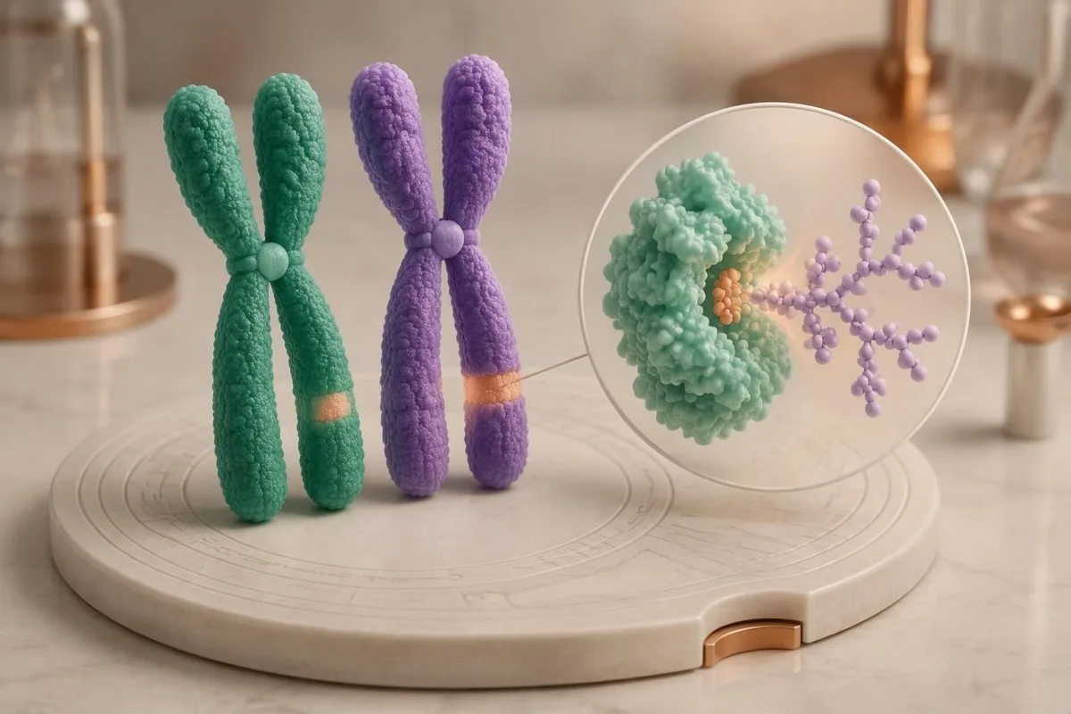
Two pathogenic or likely pathogenic GAA variants in trans support molecular confirmation of Pompe disease. In trans means the variants affect opposite gene copies; 2 variants in cis affect the same copy and do not, by themselves, demonstrate a recessive disease genotype.
Standard sequencing detects many small substitutions and insertions or deletions, but does not answer every possible genetic question. If only 1 disease-causing variant is found despite convincing enzyme deficiency, ask whether deletion/duplication analysis was included and whether poorly covered or deep intronic regions require further investigation.
A common later-onset-associated GAA variant is c.-32-13T>G, which affects splicing. Finding this variant on 1 copy does not diagnose Pompe disease, and finding it with another disease-causing variant does not precisely predict the age symptoms will begin; residual splicing and other biological factors influence presentation.
Parental testing can help establish phase when a laboratory detects 2 different variants but cannot determine their arrangement. Our guide to inherited disease genetic reports explains why inheritance and family testing matter, while the actual gene coverage and diagnostic criteria must remain specific to GAA.
病原性、不確実、陰性の結果はどのように解釈すべきですか?
A pathogenic result, a variant of uncertain significance and a negative result have different diagnostic meanings. Genetic laboratories commonly use 5 categories: pathogenic, likely pathogenic, uncertain significance, likely benign and benign; a VUS is not equivalent to a disease-causing GAA variant.
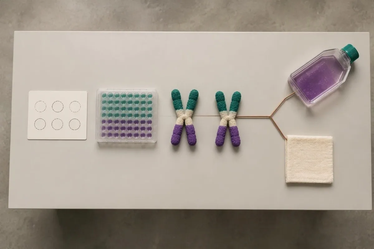
Richards et al.'s 2015 ACMG/AMP consensus established the widely used 5-category sequence-variant framework and states that a VUS should not guide clinical decision-making as though pathogenic. For Pompe evaluation, low enzyme activity can contribute evidence, but its value depends on whether pseudodeficiency and assay limitations have been addressed.
One pathogenic variant plus 1 VUS is an unresolved combination, not automatic confirmation or exclusion. Depending on the enzyme and clinical findings, specialists may investigate family segregation, RNA effects or variant-specific functional evidence; a diagnostic enzyme result may support disease even while the second molecular finding remains incompletely characterized.
A negative sequencing report means the test found no reportable variants within its scope. It does not prove that every GAA-related mechanism was examined; 1 negative limited test may warrant expanded testing when properly performed enzyme studies and the clinical pattern remain strongly suggestive.
カンテスティは AI血液検査の読み方プラットフォーム that can help separate a report's 5 classification terms from its numerical enzyme findings, without adjudicating an uncertain genotype. Our 技術的および臨床的基準 describe interpretation boundaries; our uncertain gene-result explanation provides a related example of why variants and diagnoses are not interchangeable.
GAAバリアントが1つある場合、保因者状態ですか、それとも疾患ですか?
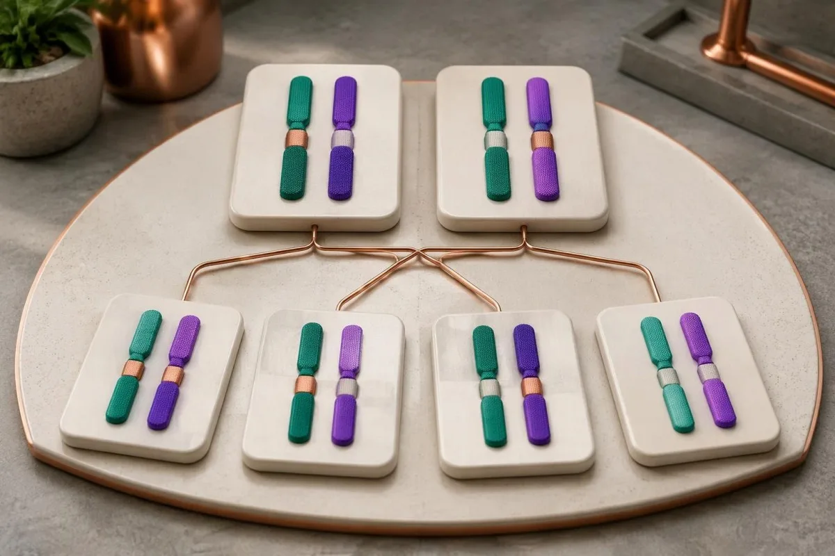
One confirmed disease-causing GAA variant usually indicates carrier status if a second disease-causing variant and diagnostic enzyme deficiency are absent. Carriers do not typically develop Pompe disease; when both parents are carriers, each pregnancy has a 25% affected, 50% carrier and 25% non-carrier probability.
Carrier enzyme values can overlap with values in unaffected people. GAA activity is therefore not a reliable standalone carrier test, and a carrier's activity should not be assumed to equal exactly 50% of a laboratory's normal mean; targeted genetic testing is generally more informative when a familial variant is known.
The 25% affected-child probability applies independently to each pregnancy when both parents carry disease-causing variants capable of producing Pompe disease together. Having 1 unaffected child does not reduce the next pregnancy's Mendelian probability, and pseudodeficiency variants should not be substituted for pathogenic variants in that calculation.
Family testing is most useful after the affected person's variants are clarified. If a symptomatic adult has only 1 identified pathogenic variant, relatives should not be reassured that the entire family question is settled until the specialist considers whether the second variant was missed by the original assay.
For a hypothetical couple, one partner carrying a pathogenic GAA variant and the other carrying only a pseudodeficiency variant does not create the same 25% Pompe risk as 2 pathogenic-variant carriers. Our carrier screening limitations guide explains why a report's coverage and residual risk deserve discussion before reproductive decisions.
ポンペ病の評価を裏付ける他の検査結果はどれですか?
Creatine kinase, AST, ALT and urinary glucose tetrasaccharide can support Pompe evaluation, but none confirms the diagnosis alone. Urinary glucose tetrasaccharide may be reported as Glc4 or within a Hex4 assay; these 2 report terms require method-specific interpretation rather than assumption that every laboratory measures exactly the same thing.
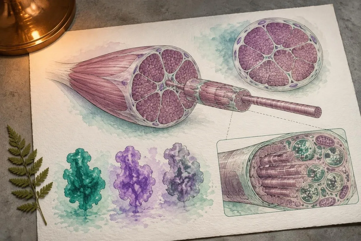
Normal CK does not exclude later-onset Pompe disease. In a hypothetical adult with progressive difficulty rising from a chair, CK of 140 U/L may fall within that laboratory's interval despite clinically meaningful weakness; the combination of symptoms and low confirmed GAA activity matters more than a reassuring CK flag.
AST and ALT can rise because of muscle involvement, not only liver disease. A hypothetical AST of 86 U/L with elevated CK and muscle weakness deserves a muscle-aware assessment; our aldolase and muscle marker guide explains why additional muscle enzymes may help characterize the pattern without identifying its cause.
Urinary Glc4 is a supportive glycogen-related biomarker, not a Pompe-specific diagnostic test. Reference limits vary with age and method, and elevation can occur in other circumstances; 1 elevated result should be interpreted alongside enzyme findings, genetics and the laboratory's creatinine-normalized units.
A falling CK or Glc4 after treatment does not automatically prove that strength or breathing has improved by the same percentage. Our discussion of AST interpretation beyond liver disease illustrates why 2 related markers can move together while still answering different questions about tissue injury and function.
ポンペ病検査に異常がある場合、なぜ呼吸機能を評価するのですか?
Pompe disease can weaken the diaphragm and other respiratory muscles, sometimes before obvious walking difficulty. Assessment may include seated and supine forced vital capacity, respiratory muscle pressures and sleep-related testing; a daytime oxygen saturation of 97% does not rule out overnight hypoventilation.
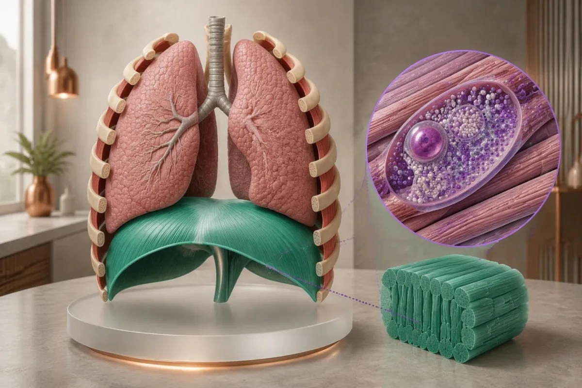
A position-related fall in vital capacity can suggest diaphragm weakness. For example, a hypothetical seated FVC of 3.0 L and supine FVC of 2.4 L represents a 20% decline; the specialist interprets that difference with technique, predicted values and symptoms, rather than treating it as a Pompe-specific cutoff.
Morning headaches, unrefreshing sleep and difficulty breathing when lying flat can justify evaluation for sleep-related hypoventilation. These symptoms do not identify Pompe disease on their own; our 息切れ検査ガイド explains why laboratory results cannot replace respiratory examination and physiological testing.
Arterial carbon dioxide is normally about 35–45 mmHg in adults, with context-dependent variation. A value above 45 mmHg indicates hypercapnia, but its urgency depends on pH, symptoms and the clinical setting; our blood gas interpretation guide explains why oxygen and carbon dioxide answer different questions.
Severe breathlessness, confusion, blue coloration or rapidly worsening weakness warrants emergency care, not an online interpretation session. A confirmed molecular result affecting 2 GAA copies does not measure current respiratory safety; clinicians need direct functional assessment to decide whether ventilation support or urgent monitoring is necessary.
新生児ポンペ病スクリーニング陽性の後はどうなりますか?
A positive newborn Pompe screen requires prompt specialist assessment, confirmatory enzyme testing and genetic evaluation. It does not mean every infant has Pompe disease; the immediate task is to distinguish pseudodeficiency or carrier findings from infantile disease and a later-onset-associated genotype affecting both GAA copies.

Burton et al.'s 2017 Pediatrics newborn-screening algorithms describe an initial evaluation integrating GAA confirmation, molecular testing and clinical assessment. If an infant has feeding difficulty, marked hypotonia or suspected cardiomyopathy, do not wait 2 weeks for a routine repeat appointment; the screening team should arrange urgent metabolic and cardiac evaluation.
Classic infantile Pompe disease commonly involves hypertrophic cardiomyopathy. A newborn's echocardiogram and electrocardiogram help assess risk that cannot be estimated from 1 enzyme number; absence of obvious heart symptoms at home does not replace the specialist examination after an abnormal screen.
CRIM status concerns whether cross-reactive GAA protein is present and helps specialists plan immune-risk management around enzyme replacement therapy. Less than 1% enzyme activity does not by itself establish CRIM-negative status, because an inactive protein may still be present; this is a protein-expression question, not just an activity threshold.
Later-onset-associated findings in a newborn require a monitoring plan, not an invented age-of-onset prediction. Even 2 classified disease-causing variants may not reveal when symptoms will emerge; our 小児向けAI安全性ガイダンス explains why pediatric results require age-specific context and clinician-led decisions.
専門医は、成人の検査結果に矛盾がある場合、どのように解決しますか?
Conflicting adult Pompe results are resolved by checking specimen quality, confirmatory enzyme methods, variant classification and clinical fit. One low screen plus 1 genetic finding rarely tells the whole story; specialists investigate whether the apparent conflict comes from pseudodeficiency, an incomplete molecular test or another cause of weakness.

Consider a hypothetical 52-year-old with mild fatigue, low DBS GAA activity and only the 2 commonly discussed pseudodeficiency variants. If specialist testing supports pseudodeficiency and no disease-causing genotype, the low screen does not explain the fatigue; other causes should be assessed rather than attaching a Pompe label to an incidental biochemical finding.
A second hypothetical adult has progressive hip weakness, a 20% positional FVC decline, confirmed cellular GAA deficiency and 2 pathogenic variants in trans. Together, those findings support Pompe disease; a normal routine glucose or modest CK result would not outweigh the converging biochemical, genetic and functional evidence.
Convincing enzyme deficiency with only 1 identified pathogenic variant deserves further investigation. The appropriate next step may be deletion/duplication analysis, RNA studies or another specialist assay—not automatically calling the person a carrier; the investigation should also check whether non-Pompe neuromuscular conditions better explain the examination.
Variant-related disease monitoring is not the same as predicting a person's future from their DNA. Our inherited neuromuscular risk guide offers a related family-testing perspective, but 2 conditions can differ substantially in inheritance, penetrance and treatment triggers.
ポンペ病専門医の受診時には何を持参すべきですか?
Bring the complete enzyme report, complete genetic report and a dated symptom history to a Pompe specialist appointment. Three report details are especially useful: specimen type, assay units and variant classification; screenshots showing only a red flag or the word mutation can omit information that changes the interpretation.

Ask whether confirmatory testing has already been completed and whether the laboratory measured GAA specifically in an appropriate cell preparation. If 2 variants were found, ask whether they are in trans; if only 1 was found, ask what variant types and regions the test could detect.
Do not try to raise GAA activity with supplements or diet before confirmation. No vitamin or food corrects inherited pathogenic variants affecting both GAA copies; nutrition may support general care after diagnosis, but cannot substitute for enzyme confirmation or specialist treatment planning.
カンテスティは AI搭載の血液検査解析ツール that can help organize report terminology and associated laboratory trends, not decide whether 2 uncertain variants establish Pompe disease. Our 当社の報告書の情報源確認ガイド explains why the original document remains the reference when an automated explanation and a specialist interpretation differ.
Genetic documents can contain information relevant to relatives as well as the tested person. Before sharing 1 family member's report, check consent and remove unnecessary identifiers; Kantesti's プライバシーとデータに関する情報を確認し、 describes our handling approach, while sharing results with relatives should follow a genetic counselor's guidance.
ポンペ病検査の解釈に関する研究論文と限界
Pompe test interpretation rests on disease-specific enzyme, genetic and clinical evidence—not on general laboratory publications or an AI explanation. Kantesti includes 3 directly relevant external sources below and 2 separate educational publications; the iron and coagulation publications are not evidence for diagnosing Pompe disease.
The 2006 Kishnani guideline provides disease-specific diagnostic and management context, while the 2017 Burton newborn-screening paper addresses confirmation after population screening. Their recommendations should be applied with the specialist laboratory's validated methods; neither supplies 1 universally transferable DBS enzyme cutoff for every instrument and population.
Richards et al.'s 2015 ACMG/AMP publication explains the 5-category variant framework used to distinguish pathogenic findings from uncertainty. Classification can change when new evidence becomes available, so ask the genetics service whether an unresolved result has a reanalysis pathway rather than assuming every VUS will eventually become pathogenic.
当社の教育用 鉄研究の出版物 addresses a different laboratory domain. The separate 凝固検査に関する文献 also covers unrelated markers; their 2 DOI records are provided for publication transparency, not as supporting citations for GAA deficiency or pseudodeficiency.
Kantestiの 医療コンテンツ基準 distinguish patient education from diagnosis and treatment instructions. The practical endpoint is a documented interpretation of 3 evidence streams—enzyme testing, genetic analysis and clinical assessment—with urgent care taking priority whenever infantile disease or respiratory compromise is suspected.
よくある質問
GAA酵素活性が低い場合、必ずポンペ病を意味しますか?
GAA酵素活性が低くても、必ずしもポンペ病を意味するわけではありません。スクリーニング検査で異常値が1回認められても、真の欠損、偽欠損、検体の劣化、または保因者に関連する活性との重複が反映されている可能性があります。確定には、適切な酵素アッセイ、GAA遺伝学的所見、および臨床的背景を専門医が解釈する必要があります。同じスクリーニングアッセイを繰り返すだけでは、偽欠損が再現されるだけで、問題が解決しない可能性があります。.
正常なGAA酵素レベルとはどのくらいですか?
There is no universal normal GAA enzyme level because specimen preparation, assay substrate and reporting units differ between laboratories. A value in nmol/h/mL cannot be directly compared with a value in nmol/h/mg protein. Classic infantile Pompe disease often has less than 1% of normal activity in diagnostic tissue assays, but this is not a universal newborn-screening cutoff. Use the reference information printed on the original report and the specialist laboratory's interpretation.
2回のGAA低値検査でも、なお偽欠損症の可能性はありますか?
GAA活性の低値を示す検査が2回あっても、特に同一または類似の測定法が用いられている場合は、偽欠損を反映している可能性があります。偽欠損バリアントは、ポンペ病(グリコーゲン蓄積症)そのものを引き起こすことなく、測定される活性を低下させます。専門医は、GAA遺伝子型全体、測定法の特性、および必要に応じて別の細胞検体や機能検査を用いて、このパターンを見分けます。したがって、低値が2回繰り返されたとしても、疾患が独立して2回確認されたことと同等ではありません。.
2つのGAA遺伝子バリアントがあれば、ポンペ病と確定できますか?
2つのGAAバリアントがあっても、自動的にポンペ病が確定するわけではありません。互いに反対側の遺伝子コピーに影響する、病原性または病原性の可能性が高いバリアントが2つある場合は、分子学的診断を支持しますが、シスにある2つのバリアントは片方のコピーにしか影響しません。意義不明のバリアントや偽欠損バリアントは、疾患原因であることが確立した所見として数えることはできません。分類または位相が未確定の場合、酵素学的および臨床的エビデンスは特に有用です。.
正常なCK値で遅発型ポンペ病を除外できますか?
Normal CK cannot rule out late-onset Pompe disease. A hypothetical CK of 140 U/L may fall within a laboratory's reference interval despite progressive proximal weakness or diaphragm involvement. CK measures muscle injury-related enzyme release, not lysosomal GAA function. Persistent compatible symptoms deserve specialist assessment even when routine muscle markers are reassuring.
新生児ポンペ病検査で陽性が出た場合、どの程度緊急ですか?
新生児ポンペ病の検査結果が陽性の場合は、スクリーニング担当チームまたは代謝疾患チームに速やかに連絡する必要があります。また、哺乳困難、著しい筋緊張低下、または心臓への影響が疑われる乳児は、緊急の評価が必要です。スクリーニングカード1枚の陽性だけでは診断は確定しませんが、古典的乳児型が疑われる場合は、数週間後の通常診療の予約まで待つべきではありません。専門医は、確定のための酵素検査、遺伝学的評価、心臓の評価を、多くの場合並行して手配します。重度の呼吸困難または反応性の低下がある場合は、救急医療が必要です。.
両親がともにポンペ病の保因者である場合、子どもが発症するリスクはどのくらいですか?
両親の双方が疾患の原因となるGAAバリアントを保有している場合、妊娠ごとに、罹患児となる確率は25%、保因児となる確率は50%、家族性バリアントのいずれも受け継がない児となる確率は25%です。これらの確率は、妊娠ごとに独立して適用されます。この計算では、偽欠損バリアントを病原性バリアントと同等に扱うことはできません。遺伝カウンセリングは、両親の所見の確認と検査の選択肢の説明に役立ちます。.
血液検査結果の見方を確認しましょう
検査報告書をアップロードすると、KantestiのAIが数秒で各バイオマーカーを平易な言葉で説明します。世界中で200万人以上が利用しています。.
参考文献
- Klein, T., Mitchell, S., & Weber, H. (2026). 鉄検査ガイド:TIBC、鉄飽和度、鉄結合能。. Kantesti AI医学研究。. 掲載日
- Klein, T., Mitchell, S., & Weber, H. (2026). aPTTの基準範囲:Dダイマー、プロテインC、血液凝固ガイド。. Kantesti AI医学研究。. 掲載日
- Kishnani PS et al. (2006). Pompe disease diagnosis and management guideline. Genetics in Medicine。. 掲載日 パブメッド
- Burton BK et al. (2017). The Initial Evaluation of Patients After Positive Newborn Screening: Recommended Algorithms Leading to a Confirmed Diagnosis of Pompe Disease. Pediatrics. 掲載日
- Richards Sら(2015年)。. 配列バリアントの解釈に関する基準およびガイドライン:米国臨床遺伝・ゲノム学会と分子病理学会による共同コンセンサス推奨。. Genetics in Medicine。. 掲載日 パブメッド
- 200万以上分析対象の検査
- 127+国
- 100+言語
医療チームについて


E-E-A-T 信頼性シグナル
経験
医師主導による、検査結果解釈ワークフローの臨床レビュー。.
専門知識
臨床的な文脈においてバイオマーカーがどのように振る舞うかに焦点を当てた検査医学。.
権威
Thomas Klein医師による医学的レビュー。Sarah Mitchell医師およびHans Weber教授の協力。.
信頼性
アラームを減らすための明確なフォローアップ経路を備えた、エビデンスに基づく解釈。.



医療免責事項
この記事は教育目的のみを対象としており、医療助言を構成するものではありません。診断や治療の判断を行う際は、必ず有資格の医療提供者にご相談ください。.
公開 · 編集チームにお問い合わせください
Kantesti Ltdが発行。イングランドおよびウェールズで登録、会社番号. 17090423, 、英国ロンドン。.
