
Ein reaktives Band weist auf eine Antikörperbindung hin – nicht auf eine Diagnose. Der vollständige Testalgorithmus, die Expositionsgeschichte und der zeitliche Verlauf der Symptome bestimmen, ob dieser Befund für eine Lyme-Borreliose spricht.

Wichtigste Erkenntnisse
- Einzelne Banden begründen keine Lyme-Borreliose; selbst eine relativ spezifische Bande muss im Rahmen eines validierten zweistufigen Testalgorithmus interpretiert werden.
- Traditionelle IgM-Kriterien erfordern mindestens 2 von 3 festgelegten Banden: etwa 23–25, 39 und 41 kDa.
- Traditionelle IgG-Kriterien erfordern mindestens 5 von 10 festgelegten Banden: 18, 23, 28, 30, 39, 41, 45, 58, 66 und 93 kDa.
- Die 30-Tage-Regel bedeutet, dass isoliertes IgM eine Lyme-Diagnose nicht stützen sollte, wenn die Symptome länger als 30 Tage bestanden haben.
- Ein insgesamt negatives Ergebnis kann mit reaktiven Banden einhergehen, weil das Labor ein validiertes Muster bewertet und nicht, ob irgendeine Bande erscheint.
- Modifizierte Zwei-Stufen-Testung verwendet 2 sequenzielle Enzymimmunoassays anstelle eines Immunoblots; möglicherweise wird keine Bandenliste bereitgestellt.
- Frühtestung kann negativ sein, bevor sich Antikörper entwickeln; ein typischer Erythema-migrans-Ausschlag bei entsprechender Exposition wird klinisch diagnostiziert.
- Persistierende Antikörper kann nach der Behandlung noch jahrelang nachweisbar bleiben und weder eine fortbestehende Infektion belegen noch den Behandlungserfolg messen.
Was bedeutet ein positives Lyme-Western-Blot-Band tatsächlich?
A positive Lyme-Western-Blot-Bande bedeutet, dass Antikörper an ein Testantigen gebunden haben; sie belegt für sich genommen keine Lyme-Borreliose. Bei der traditionellen US-amerikanischen Zwei-Stufen-Testung sind während der ersten 30 Symptomtage 2 von 3 festgelegten Banden für IgM erforderlich, für IgG hingegen 5 von 10; bei der modifizierten Testung werden stattdessen 2 Immunoassays verwendet.

Der Gesamtergebnis kombiniert den Screeningtest der ersten Stufe mit dem entsprechenden Ergebnis der zweiten Stufe, anstatt jeden sichtbaren Streifen als separate Diagnose zu behandeln. Ein Befund mit 1 reaktiven Bande kann daher korrekt als negativ eingestuft werden, und selbst ein vollständiger positiver Antikörperbefund muss im Hinblick auf Exposition, passende Befunde und eine frühere Lyme-Borreliose bewertet werden.
Die Identität der Bande beantwortet eine eng gefasste Frage: Welches Antigen hat unter den Bedingungen dieses Tests die Antikörperbindung ausgelöst? Sie beantwortet nicht die 3 Fragen, die Patienten üblicherweise beschäftigen – ob die Infektion jetzt vorliegt, ob sie die Symptome erklärt oder ob Antibiotika erforderlich sind; unser Leitfaden zu positiven Antikörperergebnissen unterscheidet diese Aspekte.
Kantesti ist ein KI-Bluttestanalysator die erklären können, warum ein Lyme-Befund reaktive Banden, aber eine negative Interpretation der Zwei-Stufen-Testung aufweist. Wir betrachten die abschließende Laborinterpretation, die Testmethode und die Symptomdauer als wesentlichen Kontext – nicht als Details, die durch das Zählen hervorgehobener Zeilen in einem hochgeladenen Befund außer Kraft gesetzt werden können.
Wie die traditionelle zweistufige Lyme-Testung das Ergebnis bestimmt
Standardmäßige Zwei-Stufen-Testung beginnt mit einem Enzymimmunoassay oder manchmal einem Immunfluoreszenztest und wird bei positivem oder grenzwertigem Ergebnis des ersten Tests mit einem Immunoblot fortgesetzt. Die 2 Stufen wirken zusammen; ein positives Screening allein ist kein bestätigter positiver serologischer Lyme-Befund.

A positiver oder grenzwertiger Test der ersten Stufe, gefolgt von negativen geeigneten Immunoblots wird nach dem Standardalgorithmus als negativ interpretiert. Der erste Screeningtest erfasst bewusst mehr mögliche Reaktionen, während die zweite Stufe unspezifische Reaktivität herausfiltert; Moore et al. beschrieben dieses Gleichgewicht und häufige Interpretationsfehler 2016 in Emerging Infectious Diseases.
Ein Index von 1.4 bei einem Screeningtest ist nicht mit 1.4 bei einem anderen gleichzusetzen, da sich Antigenzusammensetzung und Herstellergrenzwerte unterscheiden. Ebenso ist ein grenzwertiges Screening-Ergebnis die Aufforderung, den Algorithmus abzuschließen – keine Diagnose auf halbem Weg zwischen Infektion und keiner Infektion; qualitative vs. quantitative Ergebnisse erklärt, warum das Format des Befunds eine Rolle spielt.
Einen Immunoblot nach einem negativen Screeningtest anzufordern, ohne klinischen Grund, den Zeitpunkt oder die Eignung des Tests neu zu beurteilen, umgeht die Testsequenz, anhand derer die Leistungsfähigkeit geprüft wurde. Erfragen Sie 3 Angaben: das Screening-Ergebnis, die Interpretation der zweiten Stufe und ob beide Teil desselben validierten Testverfahrens sind; ein unvollständiger Befund sollte nicht allein anhand der Banden rekonstruiert werden.
Lyme-Western-Blot-Banden erklärt: IgM- und IgG-Kriterien
Nach den konventionellen US-amerikanischen Kriterien erfordert ein positiver IgM-Immunoblot mindestens 2 der Banden 23–25, 39 und 41 kDa. Ein positiver IgG-Immunoblot requires at least 5 of the 18, 23, 28, 30, 39, 41, 45, 58, 66 and 93 kDa bands.

Kilodaltons, abbreviated kDa, describe approximate protein molecular mass, not antibody concentration or disease severity. The approximately 23–25 kDa band commonly represents outer surface protein C, 39 kDa represents BmpA and 41 kDa represents flagellin; the numbers identify antigen regions, so a higher-numbered band does not mean a more advanced infection.
The IgG list's 58 kDa criterion excludes GroEL, and reports may use assay-specific names or approximate mass conventions rather than identical labels. Because not every commercial strip uses the same antigen preparation, our Leitfaden zu Bluttest-Markern should complement—not replace—the actual laboratory legend and the assay manufacturer's interpretation instructions.
Bands at 31 and 34 kDa are not part of the conventional CDC IgG counting criteria, even though they may appear on some reports. Their absence from that list does not make them meaningless biologically, but adding them to manufacture a 5-band positive result changes the validated rule; neither band darkness nor extra bands quantify bacterial burden.
Warum Band 41 und andere reaktive Banden falsch-positive Signale sein können
Der 41 kDa band reacts with flagellin, a protein associated with bacterial movement, and can cross-react with antibodies unrelated to Lyme disease. A single reactive 41 kDa band is therefore insufficient to diagnose Lyme disease, even when a patient has fatigue, aching or a remembered tick bite.
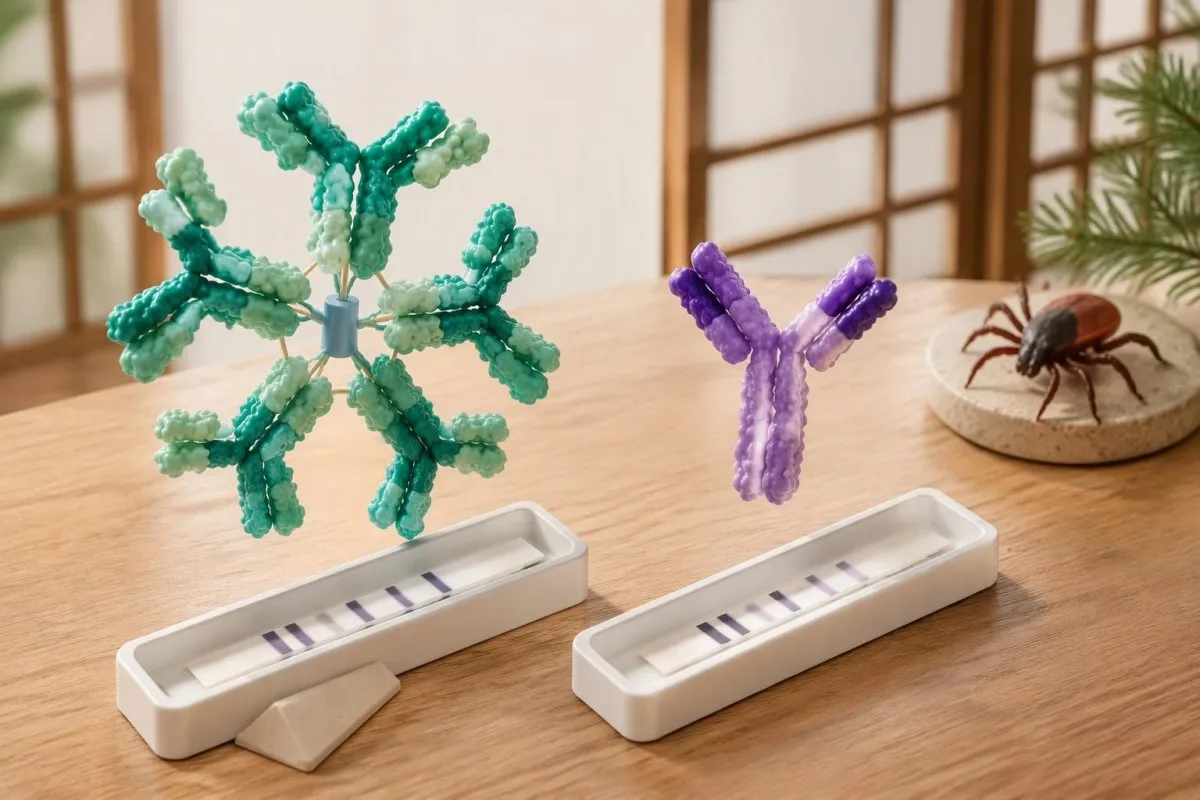
Moore et al. (2016) cited a study in which 41 kDa reactivity occurred in 43% of healthy controls, illustrating why this band cannot stand alone. That percentage is not a universal false-positive rate for every modern assay; it is evidence that recognizable antigen binding can occur in people without the clinical condition being investigated.
Epstein–Barr virus, syphilis, relapsing-fever Borrelia and some autoimmune conditions can contribute to false-positive Lyme serology, although the mechanism and affected assay vary. A recent mononucleosis-like illness should prompt attention to the complete EBV antibody pattern, not the assumption that 1 Lyme band proves a simultaneous tick-borne infection.
Cross-reactivity is not a reason to order every alternative antibody panel: 1 unexplained band usually does not justify broad autoimmune screening. When examination findings genuinely suggest an immune disorder, Interpretation von Komplement und ANA belongs to a separate clinical question; repeated low-probability testing can generate additional misleading positives rather than a clearer answer.
Lyme-IgM vs. IgG: Warum die 30-Tage-Symptomregel wichtig ist
Lyme IgM should not be used to support a diagnosis when symptoms have lasted more than 30 days. IgM is most useful early, whereas an appropriate IgG response carries greater diagnostic weight later; the clock starts with symptoms of the suspected illness, not the testing date or remembered tick removal.
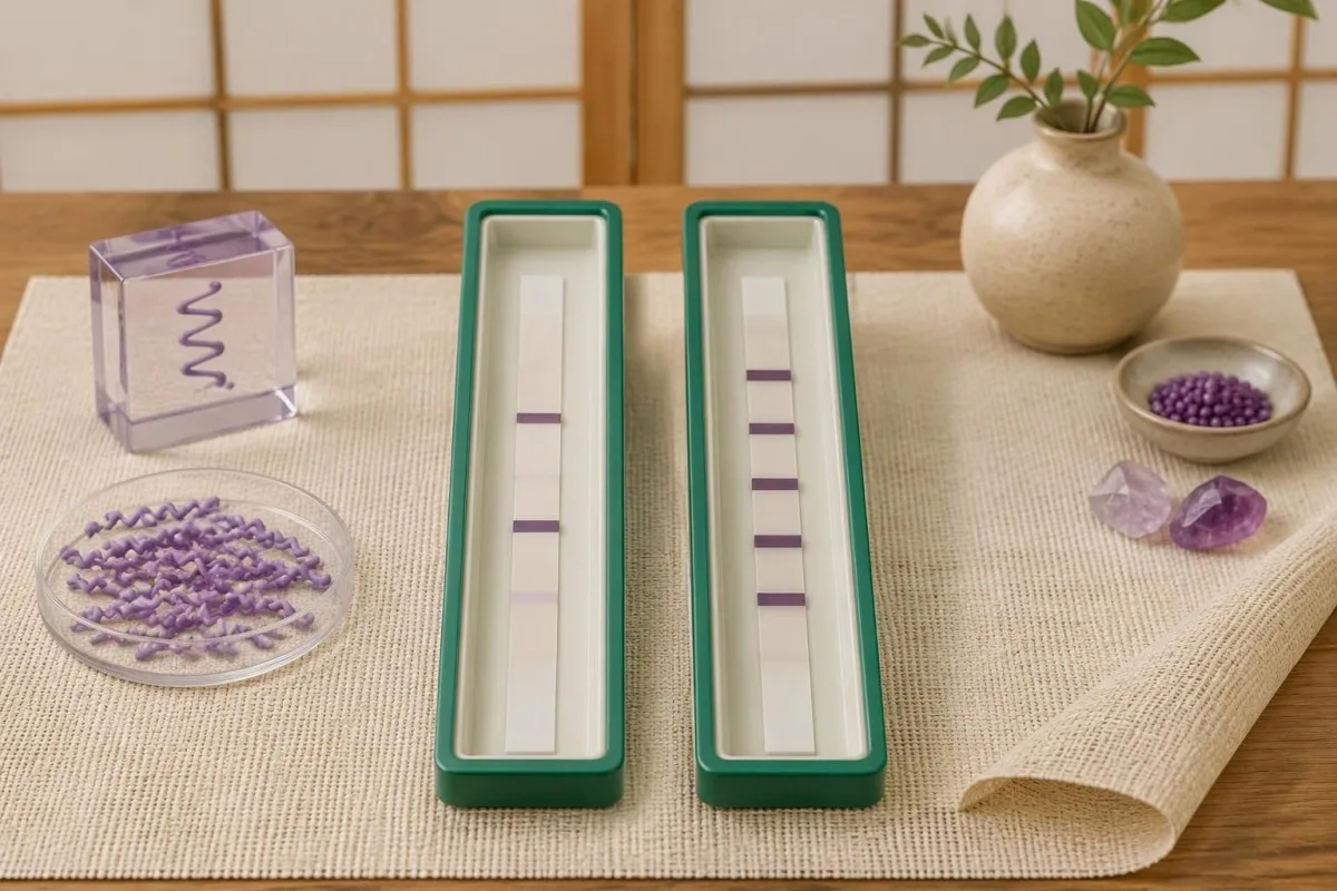
Der 30-day restriction reduces misclassification from persistent or nonspecific IgM responses when later disease should usually have an established IgG response. It is an interpretation rule, not a biological switch that flips overnight: antibody maturation varies, and early treatment may alter seroconversion, but those exceptions do not make isolated late IgM reliable evidence.
For a hypothetical patient tested on day 75 of continuous symptoms, IgM bands 23 and 41 with negative IgG should not be used as proof of Lyme disease. By contrast, the same IgM pattern on day 12, following a reactive first-tier assay and compatible exposure, can support an early diagnosis; the numerical band count is identical, but its evidential value changes.
A positive IgM result does not automatically mean a new infection, a misconception also addressed in our Mycoplasma IgM interpretation guide. If symptoms began 8 weeks ago but one symptom appeared yesterday, clinicians need to decide whether this is a genuinely new illness or another feature of the original episode—not simply restart the IgM clock.
Positive Lyme-Banden, aber insgesamt negatives Ergebnis: Beispiele
Positive Lyme bands but negative overall usually means the report did not meet the required pattern within the testing algorithm. For example, 3 designated IgG bands are below the conventional 5-of-10 threshold; that result is negative, not a smaller positive, and it does not measure how much infection is present.
Consider a clearly hypothetical 42-year-old with 10 weeks of fatigue, a reactive screening assay and IgG bands 18, 41 and 58. The IgG immunoblot is negative because only 3 qualifying bands are present; if the examination lacks objective Lyme findings, the next step is a symptom-focused assessment rather than antibiotics prescribed to treat the band's molecular masses.
A second hypothetical patient has a reactive screen, IgM bands 23 and 39, and compatible symptoms beginning 14 days earlier. This can meet the traditional early serologic criteria, but an atypical exposure history or a stronger alternative diagnosis still affects the clinical conclusion; Kantesti AI can explain the 2-of-3 rule without converting it into an independent diagnosis.
A third report lists 5 IgG bands but provides no first-tier result; the missing screening stage prevents straightforward confirmation of a standard 2-tier outcome. Confirmation requirements are assay-specific, as our HSV IgG confirmation guide also illustrates, so request the complete laboratory report rather than borrowing a cutoff or confirmation rule from another infection.
Wie sich die modifizierte zweistufige Testung vom Western Blot unterscheidet
Modified two-tier testing, or MTTT, uses 2 sequential enzyme immunoassays instead of an enzyme immunoassay followed by an immunoblot. A report from this pathway may have no bands at all; its overall interpretation depends on the particular assay combination and its cleared instructions, not the conventional immunoblot band-count criteria.
Der 2019 CDC recommendation accepted modified algorithms using FDA-cleared assays for that purpose, following clearance of relevant test combinations (Mead et al., 2019). The change was not permission to combine any 2 tests: the evaluated pairing matters, and some combinations improve early sensitivity while preserving high specificity compared with standard two-tier testing.
Some modified algorithms measure total antibodies, while others report separate IgM and IgG components; consequently, the familiar 30-day IgM restriction cannot be mechanically applied to a total-antibody result. Kantesti is an AI-Bluttest-Auswertungsplattform that can help distinguish a 2-immunoassay report from an immunoblot report before discussing what its positive or negative designation means.
A second immunoassay is not simply a repeat of the same screening test, and an immunoblot is not routinely needed after a completed validated MTTT result. Our Leitfaden zur Bestätigung von Chagas illustrates the broader principle of complementary assays, but its 2-test rules are different; ask your laboratory which Lyme algorithm produced the final interpretation.
Kann ein Lyme-Test trotz Infektion im Frühstadium negativ sein?
Lyme antibody testing can be negative early because antibodies have not yet reached detectable levels. A typical erythema migrans rash with an appropriate exposure history is diagnosed clinically rather than by waiting for a positive immunoblot; a negative result during the first 1–2 weeks does not safely exclude that presentation.

Across early-infection settings, conventional 2-tier sensitivity has been reported at approximately 30–40%, while specificity remains high when testing and interpretation are appropriate (Moore et al., 2016). These are population estimates, not a personal probability: sensitivity changes with the clinical manifestation, time since onset and whether the patient has already received antibiotics.
For a rash suggestive of but not typical for erythema migrans, the 2020 IDSA/AAN/ACR guideline supports acute serology and, if initially negative, a convalescent sample at least 2–3 weeks later (Lantos et al., 2021). The same guideline recommends clinical diagnosis for a typical lesion; a rash need not have textbook concentric rings to warrant assessment.
Repeating a test 24 hours later usually offers little advantage when the limitation is antibody development rather than a processing error. Our spotted fever testing guide explains a related timing problem, but suspected spotted fever has its own urgent treatment rules; do not delay care for an expanding rash or significant illness while waiting for more bands.
Bedeutet ein positives Lyme-IgG eine aktive Infektion oder ein Versagen der Behandlung?
Positive Lyme IgG cannot distinguish active infection from a previous infection that has resolved. Lyme antibodies can remain detectable for years or decades, and repeat immunoblot testing is not a test of cure; neither a persistent 5-band pattern nor changes in band intensity establish that treatment failed.
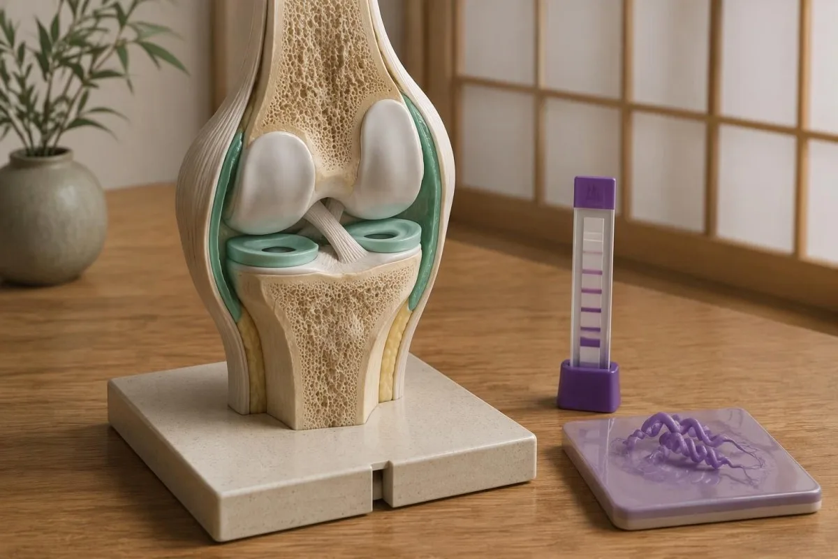
Der 2020 IDSA/AAN/ACR guideline describes persistence of both IgM and IgG responses after infection, which complicates interpretation in previously affected patients (Lantos et al., 2021). A positive result obtained 4 years after treatment must therefore be compared with the clinical history, not interpreted as a laboratory photograph of living bacteria in the body.
Reinfection can occur because previous Lyme disease does not provide reliable lifelong protection, but an old positive antibody result cannot prove a new episode. For a hypothetical person with positive IgG from 2019 and a new expanding rash after recent tick exposure, the new clinical findings matter more than whether a sixth band appears; CMV-Antikörpermustern illustrate why exposure history and current illness are separate questions.
Persistent fatigue, pain or cognitive symptoms deserve evaluation even when no new laboratory evidence appears. The guideline recommends against additional antibiotics for persistent nonspecific symptoms after recommended treatment when there is no objective evidence of reinfection or treatment failure; 1 positive antibody panel cannot identify the cause of those symptoms or justify an open-ended antibiotic course.
Was IgG bei Verdacht auf Lyme-Arthritis oder eine neurologische Erkrankung bedeutet
Lyme arthritis usually produces a strong IgG response, so isolated IgM is poor evidence for that diagnosis. Conventional two-tier serology has approximately 95–100% sensitivity in Lyme arthritis, according to the 2020 IDSA/AAN/ACR guideline; a negative appropriate IgG result should prompt serious consideration of other causes of joint swelling.
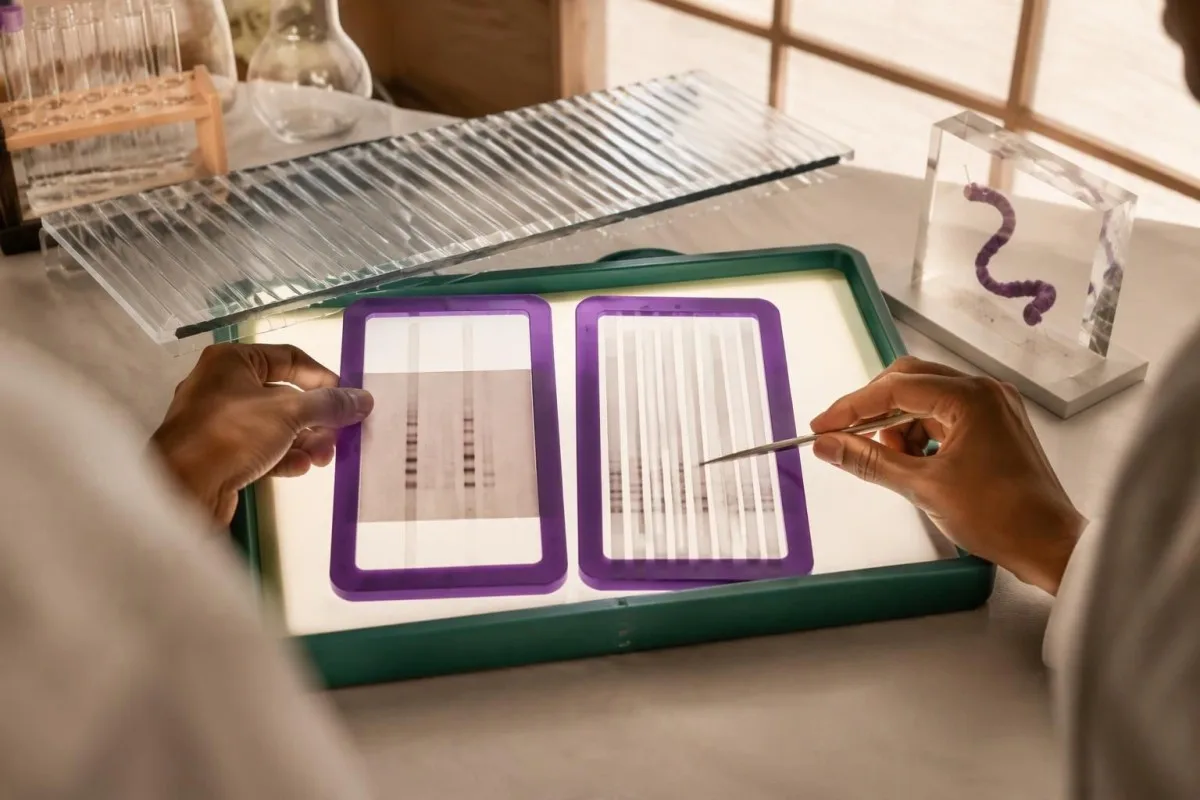
A hypothetical patient with a markedly swollen knee and only 2 IgM bands after 3 months of symptoms does not have the expected serologic pattern for Lyme arthritis. The clinician should reassess alternative causes and assay reliability rather than call this late Lyme based on IgM alone; when indicated, joint fluid examination answers additional questions that the antibody test cannot.
Suspected central nervous system Lyme disease requires clinical assessment and, when cerebrospinal fluid testing is undertaken, simultaneous CSF and serum for a validated antibody index. Testing CSF antibody alone or running a standalone CSF immunoblot is not the preferred approach; 1 elevated index may also persist after treatment, so it must be interpreted alongside the neurological findings and CSF cell response.
Numb feet or tingling do not become Lyme-specific because 41 kDa is reactive; neuropathy has metabolic, mechanical and immune causes. Our anti-MAG neuropathy guide explains one targeted alternative, while immunoglobulin panel interpretation clarifies that total serum IgG is different from the 5-of-10 Lyme-specific IgG immunoblot criterion.
Warum Labore und Länder unterschiedliche Lyme-Kriterien verwenden können
Lyme immunoblot criteria are assay- and region-dependent, so conventional US 5-of-10 IgG counting rules should not automatically be imposed on every European report. European assays may use different Borrelia antigens and recombinant line blots because the species causing human Lyme borreliosis differ across regions.
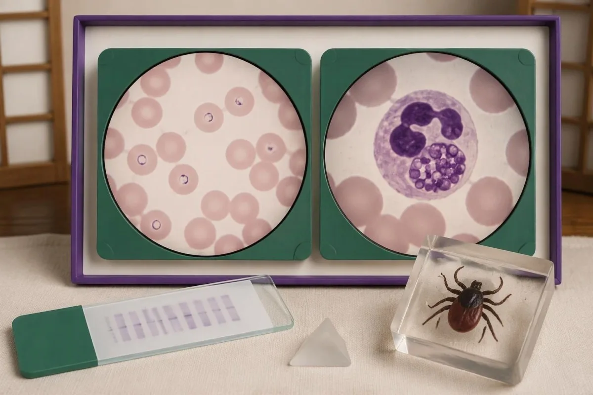
B. burgdorferi sensu stricto predominates in North America, while European disease also commonly involves B. afzelii and B. garinii. A report using VlsE, OspC or other recombinant antigen labels may therefore not resemble a 10-position conventional US blot; the clinically useful question is whether the assay was validated for the exposure setting and interpreted according to its instructions.
Different laboratory criteria are not automatically inferior, but claims that 1 or 2 bands prove disease require evidence for that assay's complete diagnostic performance. When reading a report described by Kantesti, consult the laboratory's interpretation and our medizinischer Validierungsrahmen separately; general AI assessment or a software benchmark is not proof that a particular Lyme assay has validated sensitivity and specificity.
Record 3 details before comparing reports from different laboratories: assay manufacturer, algorithm type and the country where exposure occurred. Our Leitfaden zur Überprüfung der AI-Berichtsquelle explains why preserving source wording matters; apparent conversion from negative to positive may reflect a changed method or cutoff rather than a newly acquired infection or worsening illness.
Weisen Lyme-Banden auf Babesiose, Anaplasmose oder andere Koinfektionen hin?
Lyme Western blot bands do not diagnose tick-borne coinfections. Babesia, Anaplasma and Ehrlichia require their own clinical assessment and tests; 1 positive Lyme antibody result neither confirms nor excludes these infections, even when an exposure setting allows more than one tick-borne organism to be transmitted.

Babesiose deserves consideration when relevant exposure accompanies fever and evidence such as anemia, low platelets or hemolysis, especially if fever persists during Lyme-directed treatment. A Babesia antibody titer of 1:256 alone does not establish current infection; our Babesia test interpretation guide explains why a cellular sample examination or PCR may be needed to demonstrate active infection.
Anaplasmose can produce fever, leukopenia, thrombocytopenia and elevated liver enzymes, findings that should not be explained away as extra Lyme bands. PCR is most useful during the first week of illness and becomes less sensitive after appropriate antibiotics; our Anaplasma sample timing guide separates this early direct-detection window from later antibody interpretation.
Ehrlichiosis follows a different organism-specific testing pathway, and a negative early antibody result is not a safe reason to dismiss significant febrile illness. Our Ehrlichia PCR and titer guide explains why 2 samples collected at different times may help establish seroconversion; targeted testing should follow symptoms, geography and laboratory abnormalities rather than an indiscriminate panel ordered for isolated fatigue.
Was sollten Sie mit einem unklaren Lyme-Testbericht tun?
For an unclear Lyme report, obtain the complete 2-tier interpretation, write down symptom onset and discuss exposure plus objective findings with a clinician. Faint or isolated bands do not determine urgency; fainting, chest pain, breathlessness, a markedly slow pulse or new neurological deficits require prompt assessment regardless of the antibody result.

Lyme carditis can cause heart conduction block, so fainting or palpitations after compatible exposure should not wait for an immunoblot to become positive. The 2020 IDSA/AAN/ACR guideline identifies a PR interval greater than 300 milliseconds, other arrhythmias or features of myopericarditis as reasons for hospital admission with monitoring; that interval is measured on an ECG, not inferred from band intensity.
New facial weakness warrants urgent assessment, and sudden facial droop with arm weakness or speech difficulty needs emergency evaluation for stroke rather than attribution to Lyme. Our Ratgeber zu Warnzeichen eines Schlaganfalls explains that distinction; for a routine appointment, bring 4 dates—possible exposure, first symptom, sample collection and antibiotic initiation—because each changes a different part of the reasoning.
Kantesti ist ein KI-gestütztes Analyse-Tool für Bluttests that can help organize the 2-tier result, reported bands and missing clinical context into questions for your appointment. Our KI-Technologie-Leitfaden describes the explanation workflow, but our AI cannot examine a rash, measure an ECG interval, confirm Lyme disease independently or decide whether you should start, stop or extend antibiotics.
Forschungspublikationen, Grenzen der Evidenz und sichere Interpretation
The clinical interpretation in this article rests on 3 directly relevant sources: a laboratory-diagnosis review, the CDC's modified two-tier recommendation and the IDSA/AAN/ACR Lyme guideline. The 2 additional Figshare publications listed below concern other health topics and must not be treated as validation of Lyme band interpretation or Lyme diagnosis.
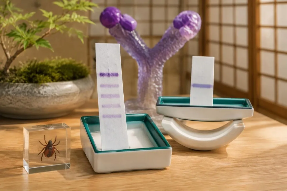
Moore et al. (2016) explains why individual bands and incomplete testing sequences create diagnostic errors; Mead et al. (2019) establishes the CDC recommendation for appropriately cleared modified algorithms. Lantos et al. (2021), publishing the 2020 guideline, connects testing to clinical manifestations, including early rash, arthritis and neurological disease; these sources support different parts of the reasoning rather than one universal band shortcut.
The 2 Figshare records address women's hormonal health and multilingual AI-assisted hantavirus triage, respectively, not Lyme immunoblot performance. A deployment involving 50,000 interpreted reports, as described in the hantavirus publication's title, cannot establish sensitivity, specificity or patient benefit for Lyme diagnosis; those claims would require Lyme-specific methods, reference standards and evaluation populations.
Die verantwortungsvollen KI-Prinzipien place report explanation alongside—not above—the laboratory and clinician, including when IgM appears after 30 days of symptoms. For symptoms linked to cycle changes or menopausal transition, our hormonal symptom guide addresses a separate assessment pathway; symptom overlap is a reason to investigate carefully, not evidence that a reactive Lyme band explains everything.
Häufig gestellte Fragen
Kann ein einzelnes positives Band im Lyme-Western-Blot bedeuten, dass ich an Lyme-Borreliose erkrankt bin?
Ein einzelnes reaktives Band im Lyme-Western-Blot begründet keine Lyme-Borreliose. Nach den in den USA üblichen Kriterien sind innerhalb der ersten 30 Tage nach Symptombeginn mindestens 2 von 3 festgelegten IgM-Banden oder 5 von 10 festgelegten IgG-Banden erforderlich, und zwar im Rahmen des geeigneten zweistufigen Testverfahrens. Ein einzelnes Band kann je nach Situation auf Kreuzreaktivität oder eine noch unvollständige frühe Antikörperantwort zurückzuführen sein. Ein typischer Erythema-migrans-Ausschlag nach einer entsprechenden Exposition kann trotz negativer früher Serologie weiterhin eine klinische Diagnose stützen.
Warum sind einige Lyme-Banden positiv, der Gesamttest jedoch negativ?
Ein Lyme-Test kann reaktive Banden aufweisen und dennoch negativ bleiben, weil der Immunoblot nicht dem validierten Muster entspricht, das der Testalgorithmus voraussetzt. Beispielsweise liegen 3 qualifizierende IgG-Banden unter dem herkömmlichen Schwellenwert von 5 von 10 IgG-Banden. Ein positiver oder grenzwertiger Suchtest der ersten Stufe, auf den negative geeignete Immunoblots folgen, wird bei der standardmäßigen zweistufigen Testung als negativ gewertet. Wenn die Symptome erst seit Kurzem bestehen, kann eine Ärztin oder ein Arzt eine Wiederholung des Tests in einem angemessenen Abstand erwägen, statt das unvollständige Muster als positiv einzustufen.
Was bedeutet ein positives 41-kDa-Lyme-Band?
Das 41-kDa-Lyme-Band weist gewöhnlich auf eine Antikörperbindung an Flagellin hin, ein mit der bakteriellen Fortbewegung zusammenhängendes Protein, das mit Antikörpern kreuzreagieren kann, die nicht mit Lyme-Borreliose zusammenhängen. Es ist nur eines der festgelegten Banden, die bei den herkömmlichen Immunoblot-Kriterien verwendet werden, und kann allein keine Lyme-Borreliose diagnostizieren. Moore et al. (2016) verwiesen auf eine Studie, in der dieses Band bei 43% der gesunden Kontrollpersonen gefunden wurde, wobei dieser Prozentsatz nicht für alle Assays allgemein gilt. Die vollständige Testsequenz und das klinische Erscheinungsbild bestimmen, ob das Ergebnis für eine Lyme-Borreliose spricht.
Sind positives Lyme-IgM und negatives IgG ein Beweis für eine kürzlich erfolgte Infektion?
Eine Lyme-IgM-Positivität bei negativem IgG kann eine frühe Infektion nur dann stützen, wenn sie mit dem geeigneten Testalgorithmus, dem klinischen Bild und dem Zeitpunkt vereinbar ist. Ein isoliertes IgM sollte nicht zur Stützung der Diagnose einer Lyme-Borreliose herangezogen werden, wenn die Symptome länger als 30 Tage bestanden haben, da persistierendes oder unspezifisches IgM die Interpretation in die Irre führen kann. So haben beispielsweise 2 qualifizierende IgM-Banden an Tag 14 eine andere Bedeutung als dasselbe Muster nach 3 Monaten anhaltender Symptome. Die Zeitregel bezieht sich auf den Symptombeginn, nicht auf den Tag der Entnahme der Laborprobe.
Kann ich einen positiven Lyme-Test haben, ohne dass im Western Blot Banden nachweisbar sind?
Ja: Ein validierter modifizierter Lyme-Testalgorithmus mit zwei Stufen verwendet zwei aufeinanderfolgende Enzymimmunoassays und sieht unter Umständen gar keinen Western Blot vor. Die CDC empfahlen 2019 entsprechend von der FDA zugelassene modifizierte Algorithmen. Einige Testkombinationen messen die Gesamtantikörper, während andere getrennte IgM- und IgG-Ergebnisse liefern. Daher gelten die herkömmlichen Regeln zur Anzahl der Banden nicht für jeden Befund. Fragen Sie das Labor, welcher Algorithmus und welche Testkombination der abschließenden Beurteilung zugrunde liegen.
Sollten die Banden im Lyme-Western-Blot nach einer Antibiotikabehandlung verschwinden?
Lyme-Western-Blot-Banden müssen nach erfolgreicher Behandlung nicht verschwinden, da Antikörper jahrelang oder jahrzehntelang bestehen bleiben können. Ein persistierendes IgG-Muster mit 5 Banden beweist keine fortbestehende Infektion, und wiederholte Bandentests sind kein validierter Heilungsnachweis. Das Ansprechen auf die Behandlung wird anhand des klinischen Verlaufs und relevanter objektiver Befunde beurteilt, nicht daran, ob ein Antikörperpanel negativ wird. Neue Symptome oder ein weiterer Zeckenstich erfordern eine erneute klinische Beurteilung, da alte Antikörper keine Reinfektion belegen können.
Lassen Sie sich Ihre Blutwerte erklären
Laden Sie Ihren Laborbericht hoch und die KI von Kantesti erklärt jeden Biomarker innerhalb von Sekunden in einfacher Sprache. Weltweit von mehr als 2 Millionen Menschen genutzt.
Referenzen
- Klein, T., Mitchell, S. & Weber, H. (2026). Ratgeber zur Frauengesundheit: Eisprung, Menopause und hormonelle Symptome. Kantesti KI-Medizinrecherche. DOI
- Klein, T., Mitchell, S. & Weber, H. (2026). Mehrsprachige, KI-gestützte klinische Entscheidungshilfe für die frühe Triage bei Hantavirus: Design, technische Validierung und realer Einsatz bei 50.000 interpretierten Blutbefunden. Kantesti KI-Medizinrecherche. DOI
- Moore A et al. (2016). Current Guidelines, Common Clinical Pitfalls, and Future Directions for Laboratory Diagnosis of Lyme Disease, United States. Emerging Infectious Diseases. DOI PubMed
- Mead P et al. (2019). Updated CDC Recommendation for Serologic Diagnosis of Lyme Disease. MMWR Morbidity and Mortality Weekly Report. DOI
- Lantos PM et al. (2021). Clinical Practice Guidelines by the Infectious Diseases Society of America (IDSA), American Academy of Neurology (AAN), and American College of Rheumatology (ACR): 2020 Guidelines for the Prevention, Diagnosis and Treatment of Lyme Disease. Clinical Infectious Diseases. DOI
- 2M+Analysierte Tests
- 127+Länder
- 100+Sprachen
Über das medizinische Team


E-E-A-T Vertrauenssignale
Erfahrung
Ärztlich geleitete klinische Überprüfung von Labor-Interpretations-Workflows.
Sachverstand
Fokus der Labormedizin darauf, wie Biomarker sich im klinischen Kontext verhalten.
Autorität
Medizinisch geprüft von Dr. Thomas Klein, mit Beiträgen von Dr. Sarah Mitchell und Prof. Dr. Hans Weber.
Vertrauenswürdigkeit
Evidenzbasierte Interpretation mit klaren nächsten Schritten zur Reduzierung von Alarm.





Medizinischer Haftungsausschluss
Dieser Artikel dient nur zu Bildungszwecken und stellt keine medizinische Beratung dar. Wende dich für Diagnose- und Behandlungsentscheidungen immer an eine qualifizierte medizinische Fachkraft.
Veröffentlicht · Kontaktieren Sie das Redaktionsteam
Veröffentlicht von Kantesti Ltd, eingetragen in England und Wales, Firmennr. 17090423, London, Vereinigtes Königreich.
