
Реактивная полоса указывает на связывание антител, а не на диагноз. Полный алгоритм тестирования, история контактов и динамика симптомов определяют, подтверждает ли этот результат болезнь Лайма.

Основные выводы
- Отдельные полосы не позволяют установить болезнь Лайма; даже относительно специфичную полосу необходимо интерпретировать в рамках валидированного двухэтапного алгоритма тестирования.
- Традиционные критерии IgM требуют наличия как минимум 2 из 3 указанных полос: приблизительно 23–25, 39 и 41 кДа.
- Традиционные критерии IgG требуют наличия как минимум 5 из 10 указанных полос: 18, 23, 28, 30, 39, 41, 45, 58, 66 и 93 кДа.
- Правило 30 дней означает, что изолированный IgM не должен служить основанием для диагноза болезни Лайма, если симптомы сохраняются более 30 дней.
- Отрицательный общий результат могут сочетаться с реактивными полосами, поскольку лаборатория оценивает валидированный паттерн, а не наличие любой полосы.
- Модифицированное двухэтапное тестирование использует 2 последовательных иммуноферментных анализа вместо иммуноблота; список полос может не предоставляться.
- Раннее тестирование может быть отрицательным до выработки антител; типичная мигрирующая эритема при соответствующих обстоятельствах заражения диагностируется клинически.
- Сохраняющиеся антитела может сохраняться в крови годами после лечения и не позволяет установить наличие продолжающейся инфекции или оценить излечение.
Что на самом деле означает положительная полоса в иммуноблоте на болезнь Лайма?
A положительная полоса в иммуноблоте на болезнь Лайма означает, что антитела связались с тестовым антигеном; сама по себе она не подтверждает болезнь Лайма. При традиционном двухэтапном тестировании в США для IgM требуются 2 из 3 указанных полос в течение первых 30 дней симптомов, а для IgG — 5 из 10; при модифицированном тестировании вместо этого используют 2 иммуноанализа.

The итоговый результат объединяет скрининговый анализ первого этапа с соответствующим результатом второго этапа, а не рассматривает каждую видимую полосу как отдельный диагноз. Поэтому в заключении с 1 реактивной полосой может быть правильно указан отрицательный результат, а даже положительный результат полного анализа на антитела следует оценивать с учетом контакта с возбудителем, совместимых клинических проявлений и перенесенной ранее болезни Лайма.
Идентификация полосы отвечает на узкий вопрос: какой антиген привлек антитела при условиях проведения этого анализа? Она не отвечает на 3 вопроса, которые обычно волнуют пациентов: есть ли инфекция сейчас, объясняет ли она их симптомы и нужны ли антибиотики; наше руководство по положительных результатов на антитела помогает разобраться в этих вопросах.
Кантести — это Анализатор анализа крови на основе искусственного интеллекта которая может помочь объяснить, почему в заключении по болезни Лайма указаны реактивные полосы, но интерпретация двухэтапного тестирования отрицательная. Мы считаем итоговую интерпретацию лаборатории, метод анализа и длительность симптомов необходимым контекстом — это не детали, которые можно игнорировать, просто подсчитав выделенные строки в загруженном заключении.
Как традиционное двухэтапное тестирование на болезнь Лайма определяет результат
Стандартное двухэтапное тестирование начинается с иммуноферментного анализа, а иногда — с реакции иммунофлуоресценции, после чего при положительном или сомнительном результате первого теста проводят иммуноблот. Эти 2 этапа дополняют друг друга; положительный результат одного лишь скринингового теста не является подтвержденным положительным результатом серологического исследования на болезнь Лайма.

A положительный или сомнительный результат теста первого этапа с последующими отрицательными результатами соответствующих иммуноблотов интерпретируется как отрицательный в соответствии со стандартным алгоритмом. Первичный скрининг намеренно выявляет больше возможных реакций, а второй этап отфильтровывает неспецифическую реактивность; Moore et al. описали этот баланс и распространенные ошибки интерпретации в журнале Emerging Infectious Diseases в 2016 году.
Индекс 1,4 в одном скрининговом анализе нельзя приравнивать к 1,4 в другом, поскольку состав антигенов и пороговые значения, установленные производителями, различаются. Аналогично, сомнительный результат скрининга означает, что нужно завершить алгоритм, а не ставить диагноз, промежуточный между наличием и отсутствием инфекции; качественные против количественных результатов объясняет, почему важен формат заключения.
Проведение иммуноблота после отрицательного результата скрининга без клинических оснований пересматривать сроки тестирования или пригодность анализа нарушает последовательность, для которой была оценена эффективность метода. Запросите 3 сведения: результат скрининга, интерпретацию второго этапа и информацию о том, относятся ли оба анализа к одному валидированному алгоритму тестирования; неполное заключение не следует восстанавливать только по полосам.
Полосы иммуноблота на болезнь Лайма: критерии IgM и IgG
Согласно традиционным критериям США, для положительного иммуноблота IgM требуется наличие как минимум 2 из полос с массой 23–25, 39 и 41 кДа. Для положительного иммуноблота IgG requires at least 5 of the 18, 23, 28, 30, 39, 41, 45, 58, 66 and 93 kDa bands.
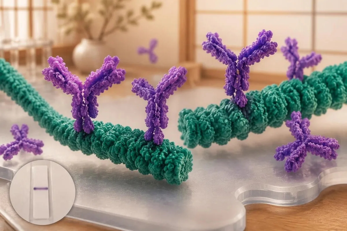
Kilodaltons, abbreviated kDa, describe approximate protein molecular mass, not antibody concentration or disease severity. The approximately 23–25 kDa band commonly represents outer surface protein C, 39 kDa represents BmpA and 41 kDa represents flagellin; the numbers identify antigen regions, so a higher-numbered band does not mean a more advanced infection.
The IgG list's 58 kDa criterion excludes GroEL, and reports may use assay-specific names or approximate mass conventions rather than identical labels. Because not every commercial strip uses the same antigen preparation, our руководство по маркерам анализов крови should complement—not replace—the actual laboratory legend and the assay manufacturer's interpretation instructions.
Bands at 31 and 34 kDa are not part of the conventional CDC IgG counting criteria, even though they may appear on some reports. Their absence from that list does not make them meaningless biologically, but adding them to manufacture a 5-band positive result changes the validated rule; neither band darkness nor extra bands quantify bacterial burden.
Почему полоса 41 и другие реактивные полосы могут давать ложноположительные сигналы
The 41 kDa band reacts with flagellin, a protein associated with bacterial movement, and can cross-react with antibodies unrelated to Lyme disease. A single reactive 41 kDa band is therefore insufficient to diagnose Lyme disease, even when a patient has fatigue, aching or a remembered tick bite.
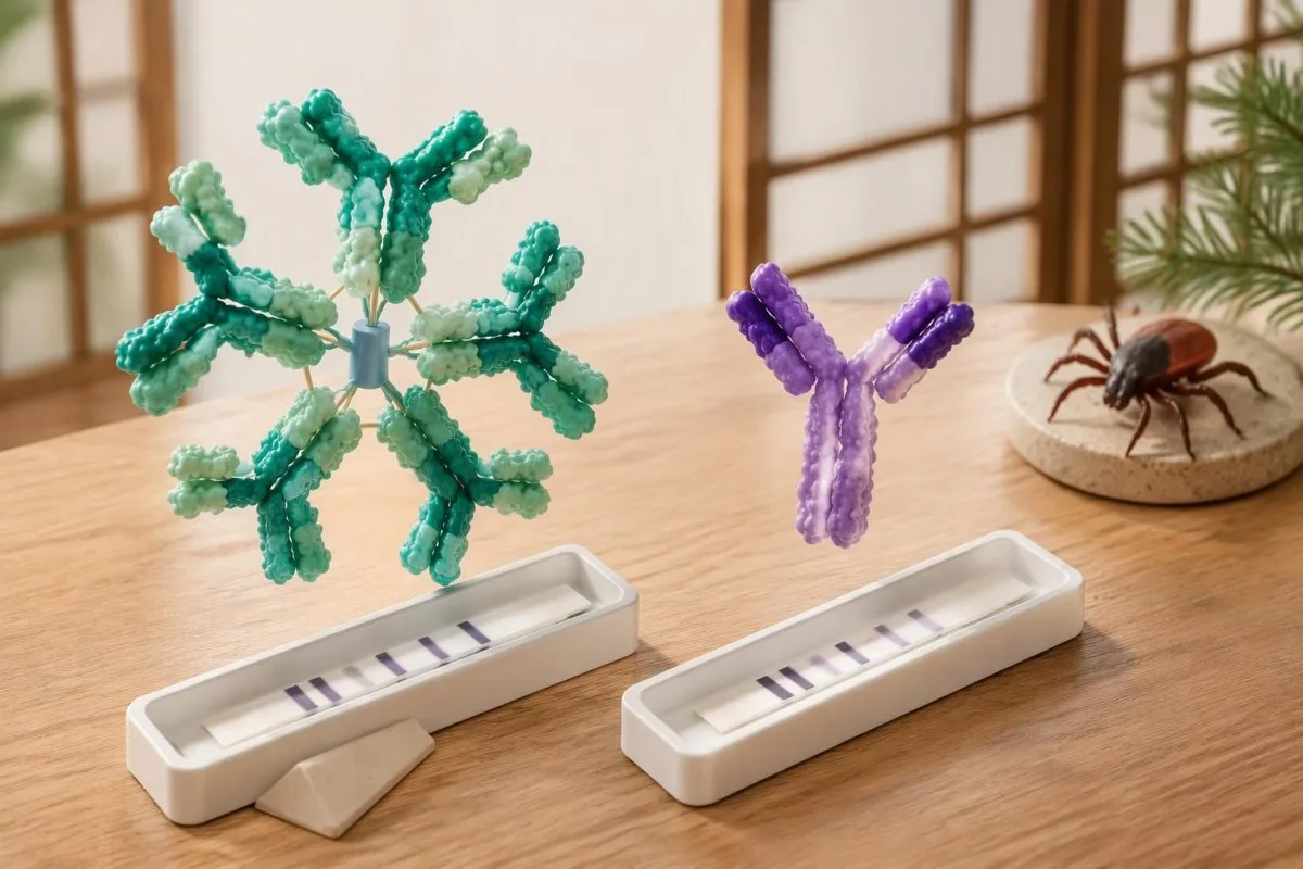
Moore et al. (2016) cited a study in which 41 kDa reactivity occurred in 43% of healthy controls, illustrating why this band cannot stand alone. That percentage is not a universal false-positive rate for every modern assay; it is evidence that recognizable antigen binding can occur in people without the clinical condition being investigated.
Epstein–Barr virus, syphilis, relapsing-fever Borrelia and some autoimmune conditions can contribute to false-positive Lyme serology, although the mechanism and affected assay vary. A recent mononucleosis-like illness should prompt attention to the complete EBV antibody pattern, not the assumption that 1 Lyme band proves a simultaneous tick-borne infection.
Cross-reactivity is not a reason to order every alternative antibody panel: 1 unexplained band usually does not justify broad autoimmune screening. When examination findings genuinely suggest an immune disorder, интерпретации показателей комплемента и ANA belongs to a separate clinical question; repeated low-probability testing can generate additional misleading positives rather than a clearer answer.
IgM и IgG при болезни Лайма: почему важно правило 30 дней
Lyme IgM should not be used to support a diagnosis when symptoms have lasted more than 30 days. IgM is most useful early, whereas an appropriate IgG response carries greater diagnostic weight later; the clock starts with symptoms of the suspected illness, not the testing date or remembered tick removal.
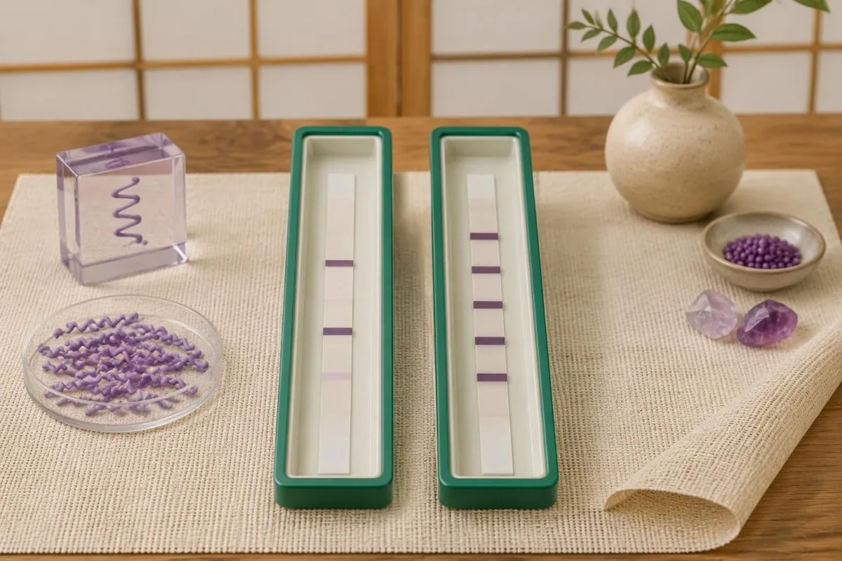
The 30-day restriction reduces misclassification from persistent or nonspecific IgM responses when later disease should usually have an established IgG response. It is an interpretation rule, not a biological switch that flips overnight: antibody maturation varies, and early treatment may alter seroconversion, but those exceptions do not make isolated late IgM reliable evidence.
For a hypothetical patient tested on day 75 of continuous symptoms, IgM bands 23 and 41 with negative IgG should not be used as proof of Lyme disease. By contrast, the same IgM pattern on day 12, following a reactive first-tier assay and compatible exposure, can support an early diagnosis; the numerical band count is identical, but its evidential value changes.
A positive IgM result does not automatically mean a new infection, a misconception also addressed in our Mycoplasma IgM interpretation guide. If symptoms began 8 weeks ago but one symptom appeared yesterday, clinicians need to decide whether this is a genuinely new illness or another feature of the original episode—not simply restart the IgM clock.
Положительные полосы при болезни Лайма, но отрицательный общий результат: примеры с разбором
Positive Lyme bands but negative overall usually means the report did not meet the required pattern within the testing algorithm. For example, 3 designated IgG bands are below the conventional 5-of-10 threshold; that result is negative, not a smaller positive, and it does not measure how much infection is present.
Consider a clearly hypothetical 42-year-old with 10 weeks of fatigue, a reactive screening assay and IgG bands 18, 41 and 58. The IgG immunoblot is negative because only 3 qualifying bands are present; if the examination lacks objective Lyme findings, the next step is a symptom-focused assessment rather than antibiotics prescribed to treat the band's molecular masses.
A second hypothetical patient has a reactive screen, IgM bands 23 and 39, and compatible symptoms beginning 14 days earlier. This can meet the traditional early serologic criteria, but an atypical exposure history or a stronger alternative diagnosis still affects the clinical conclusion; Kantesti AI can explain the 2-of-3 rule without converting it into an independent diagnosis.
A third report lists 5 IgG bands but provides no first-tier result; the missing screening stage prevents straightforward confirmation of a standard 2-tier outcome. Confirmation requirements are assay-specific, as our HSV IgG confirmation guide also illustrates, so request the complete laboratory report rather than borrowing a cutoff or confirmation rule from another infection.
Чем модифицированное двухэтапное тестирование отличается от иммуноблота
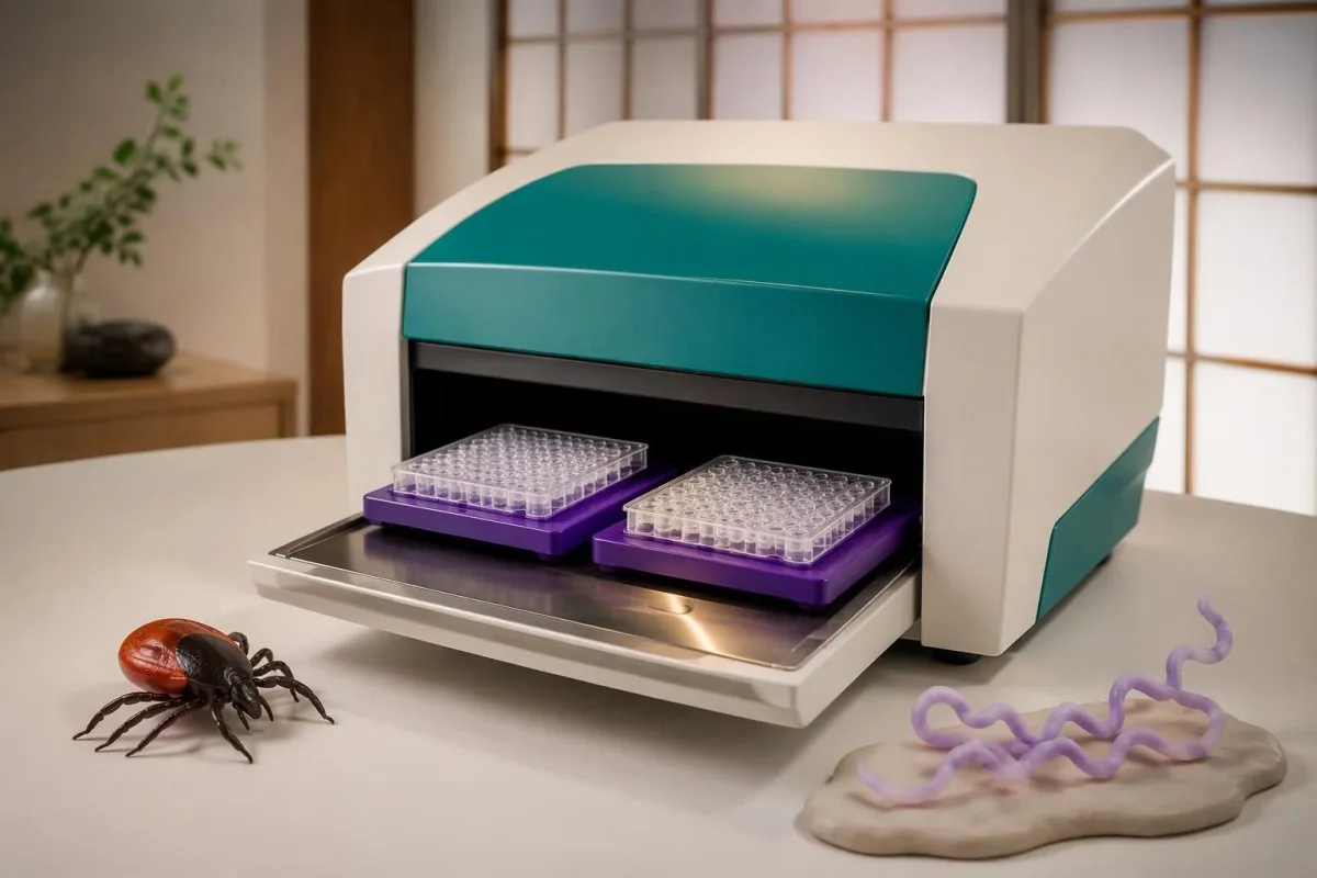
Modified two-tier testing, or MTTT, uses 2 sequential enzyme immunoassays instead of an enzyme immunoassay followed by an immunoblot. A report from this pathway may have no bands at all; its overall interpretation depends on the particular assay combination and its cleared instructions, not the conventional immunoblot band-count criteria.

The 2019 CDC recommendation accepted modified algorithms using FDA-cleared assays for that purpose, following clearance of relevant test combinations (Mead et al., 2019). The change was not permission to combine any 2 tests: the evaluated pairing matters, and some combinations improve early sensitivity while preserving high specificity compared with standard two-tier testing.
Some modified algorithms measure total antibodies, while others report separate IgM and IgG components; consequently, the familiar 30-day IgM restriction cannot be mechanically applied to a total-antibody result. Kantesti is an Платформа для расшифровки анализа крови AI that can help distinguish a 2-immunoassay report from an immunoblot report before discussing what its positive or negative designation means.
A second immunoassay is not simply a repeat of the same screening test, and an immunoblot is not routinely needed after a completed validated MTTT result. Our руководство по подтверждению болезни Шагаса illustrates the broader principle of complementary assays, but its 2-test rules are different; ask your laboratory which Lyme algorithm produced the final interpretation.
Может ли тест на болезнь Лайма быть отрицательным на ранней стадии, несмотря на инфекцию?
Lyme antibody testing can be negative early because antibodies have not yet reached detectable levels. A typical erythema migrans rash with an appropriate exposure history is diagnosed clinically rather than by waiting for a positive immunoblot; a negative result during the first 1–2 weeks does not safely exclude that presentation.
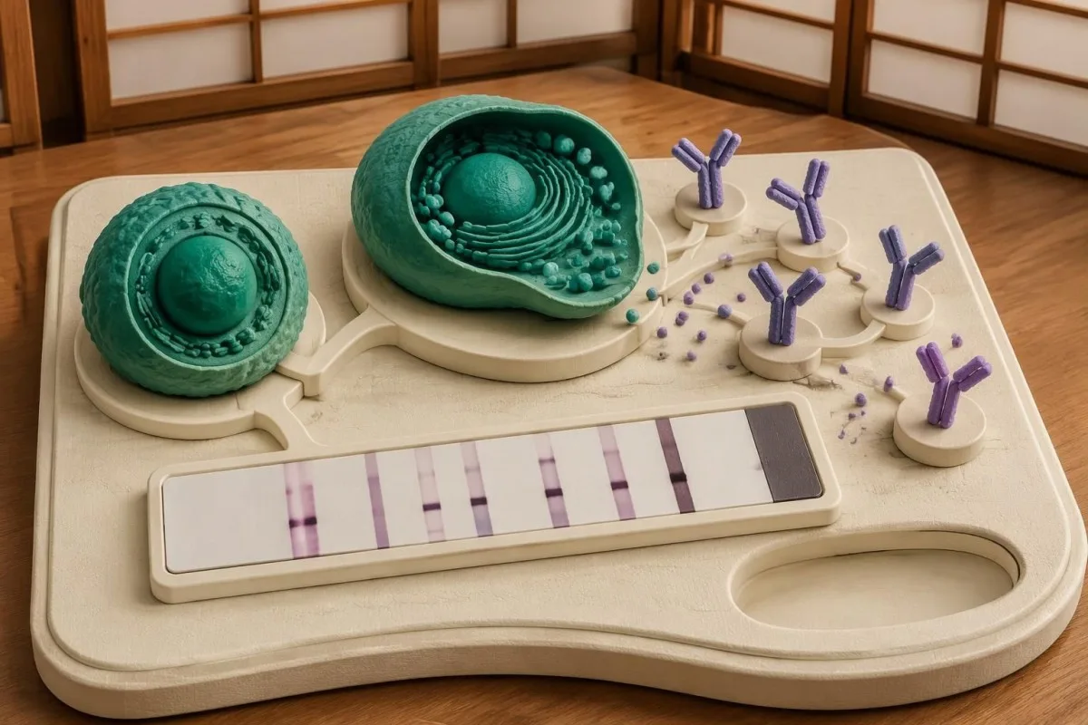
Across early-infection settings, conventional 2-tier sensitivity has been reported at approximately 30–40%, while specificity remains high when testing and interpretation are appropriate (Moore et al., 2016). These are population estimates, not a personal probability: sensitivity changes with the clinical manifestation, time since onset and whether the patient has already received antibiotics.
For a rash suggestive of but not typical for erythema migrans, the 2020 IDSA/AAN/ACR guideline supports acute serology and, if initially negative, a convalescent sample at least 2–3 weeks later (Lantos et al., 2021). The same guideline recommends clinical diagnosis for a typical lesion; a rash need not have textbook concentric rings to warrant assessment.
Repeating a test 24 hours later usually offers little advantage when the limitation is antibody development rather than a processing error. Our spotted fever testing guide explains a related timing problem, but suspected spotted fever has its own urgent treatment rules; do not delay care for an expanding rash or significant illness while waiting for more bands.
Означает ли положительный IgG к возбудителю болезни Лайма активную инфекцию или неэффективность лечения?
Positive Lyme IgG cannot distinguish active infection from a previous infection that has resolved. Lyme antibodies can remain detectable for years or decades, and repeat immunoblot testing is not a test of cure; neither a persistent 5-band pattern nor changes in band intensity establish that treatment failed.
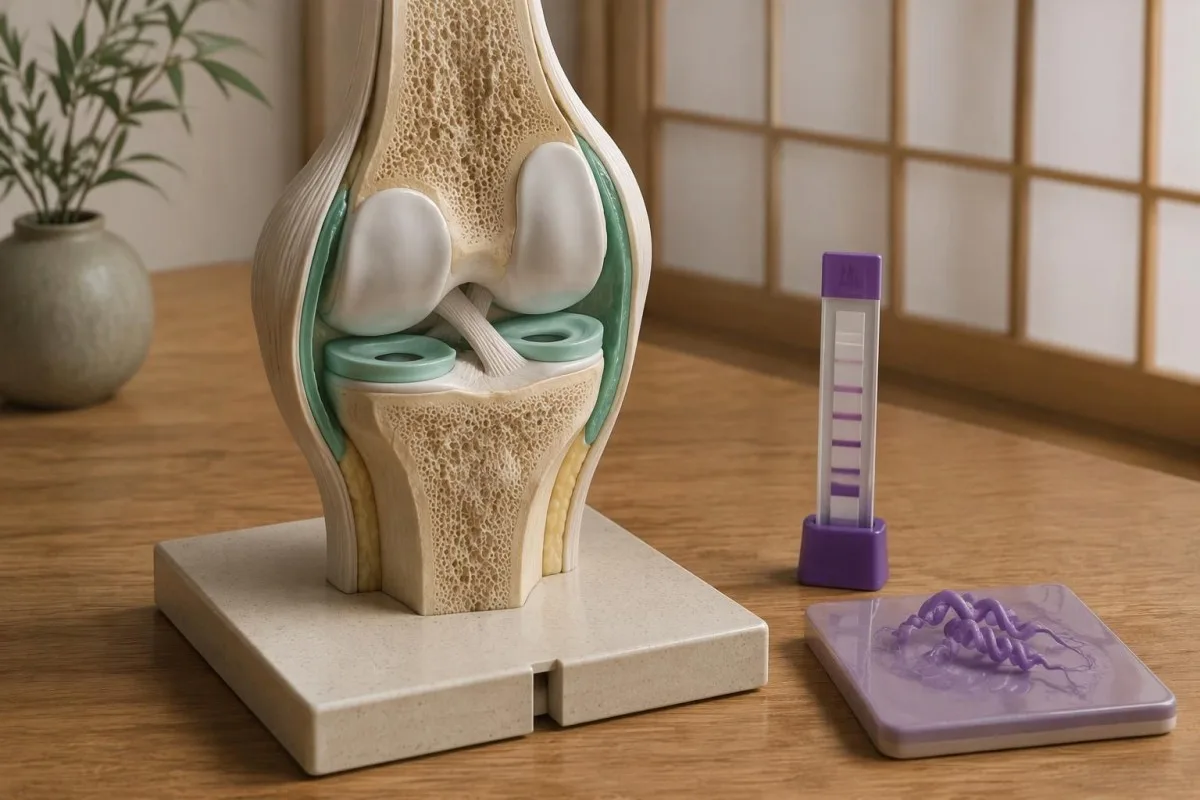
The 2020 IDSA/AAN/ACR guideline describes persistence of both IgM and IgG responses after infection, which complicates interpretation in previously affected patients (Lantos et al., 2021). A positive result obtained 4 years after treatment must therefore be compared with the clinical history, not interpreted as a laboratory photograph of living bacteria in the body.
Reinfection can occur because previous Lyme disease does not provide reliable lifelong protection, but an old positive antibody result cannot prove a new episode. For a hypothetical person with positive IgG from 2019 and a new expanding rash after recent tick exposure, the new clinical findings matter more than whether a sixth band appears; паттернов антител к ЦМВ illustrate why exposure history and current illness are separate questions.
Persistent fatigue, pain or cognitive symptoms deserve evaluation even when no new laboratory evidence appears. The guideline recommends against additional antibiotics for persistent nonspecific symptoms after recommended treatment when there is no objective evidence of reinfection or treatment failure; 1 positive antibody panel cannot identify the cause of those symptoms or justify an open-ended antibiotic course.
Что означает IgG при подозрении на лайм-артрит или неврологическое заболевание
Lyme arthritis usually produces a strong IgG response, so isolated IgM is poor evidence for that diagnosis. Conventional two-tier serology has approximately 95–100% sensitivity in Lyme arthritis, according to the 2020 IDSA/AAN/ACR guideline; a negative appropriate IgG result should prompt serious consideration of other causes of joint swelling.

A hypothetical patient with a markedly swollen knee and only 2 IgM bands after 3 months of symptoms does not have the expected serologic pattern for Lyme arthritis. The clinician should reassess alternative causes and assay reliability rather than call this late Lyme based on IgM alone; when indicated, joint fluid examination answers additional questions that the antibody test cannot.
Suspected central nervous system Lyme disease requires clinical assessment and, when cerebrospinal fluid testing is undertaken, simultaneous CSF and serum for a validated antibody index. Testing CSF antibody alone or running a standalone CSF immunoblot is not the preferred approach; 1 elevated index may also persist after treatment, so it must be interpreted alongside the neurological findings and CSF cell response.
Numb feet or tingling do not become Lyme-specific because 41 kDa is reactive; neuropathy has metabolic, mechanical and immune causes. Our anti-MAG neuropathy guide explains one targeted alternative, while immunoglobulin panel interpretation clarifies that total serum IgG is different from the 5-of-10 Lyme-specific IgG immunoblot criterion.
Почему лаборатории и страны могут использовать разные критерии диагностики болезни Лайма
Lyme immunoblot criteria are assay- and region-dependent, so conventional US 5-of-10 IgG counting rules should not automatically be imposed on every European report. European assays may use different Borrelia antigens and recombinant line blots because the species causing human Lyme borreliosis differ across regions.
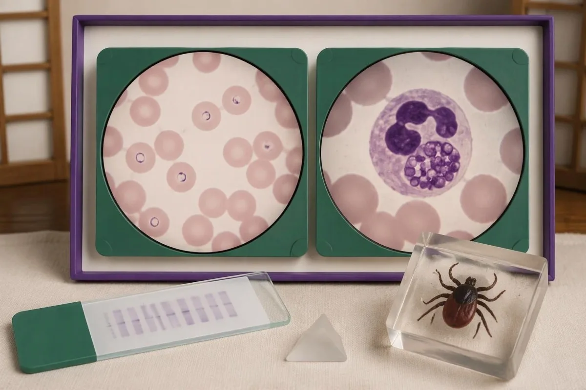
B. burgdorferi sensu stricto predominates in North America, while European disease also commonly involves B. afzelii and B. garinii. A report using VlsE, OspC or other recombinant antigen labels may therefore not resemble a 10-position conventional US blot; the clinically useful question is whether the assay was validated for the exposure setting and interpreted according to its instructions.
Different laboratory criteria are not automatically inferior, but claims that 1 or 2 bands prove disease require evidence for that assay's complete diagnostic performance. When reading a report described by Kantesti, consult the laboratory's interpretation and our медицинская валидационная платформа separately; general AI assessment or a software benchmark is not proof that a particular Lyme assay has validated sensitivity and specificity.
Record 3 details before comparing reports from different laboratories: assay manufacturer, algorithm type and the country where exposure occurred. Our руководство по проверке источника отчетов ИИ explains why preserving source wording matters; apparent conversion from negative to positive may reflect a changed method or cutoff rather than a newly acquired infection or worsening illness.
Позволяют ли полосы при болезни Лайма выявить бабезиоз, анаплазмоз или другие коинфекции?
Lyme Western blot bands do not diagnose tick-borne coinfections. Babesia, Anaplasma and Ehrlichia require their own clinical assessment and tests; 1 positive Lyme antibody result neither confirms nor excludes these infections, even when an exposure setting allows more than one tick-borne organism to be transmitted.

Бабезиоз deserves consideration when relevant exposure accompanies fever and evidence such as anemia, low platelets or hemolysis, especially if fever persists during Lyme-directed treatment. A Babesia antibody titer of 1:256 alone does not establish current infection; our Babesia test interpretation guide explains why a cellular sample examination or PCR may be needed to demonstrate active infection.
Анаплазмоз can produce fever, leukopenia, thrombocytopenia and elevated liver enzymes, findings that should not be explained away as extra Lyme bands. PCR is most useful during the first week of illness and becomes less sensitive after appropriate antibiotics; our Anaplasma sample timing guide separates this early direct-detection window from later antibody interpretation.
Ehrlichiosis follows a different organism-specific testing pathway, and a negative early antibody result is not a safe reason to dismiss significant febrile illness. Our Ehrlichia PCR and titer guide explains why 2 samples collected at different times may help establish seroconversion; targeted testing should follow symptoms, geography and laboratory abnormalities rather than an indiscriminate panel ordered for isolated fatigue.
Что делать, если заключение теста на болезнь Лайма неясно?
For an unclear Lyme report, obtain the complete 2-tier interpretation, write down symptom onset and discuss exposure plus objective findings with a clinician. Faint or isolated bands do not determine urgency; fainting, chest pain, breathlessness, a markedly slow pulse or new neurological deficits require prompt assessment regardless of the antibody result.

Lyme carditis can cause heart conduction block, so fainting or palpitations after compatible exposure should not wait for an immunoblot to become positive. The 2020 IDSA/AAN/ACR guideline identifies a PR interval greater than 300 milliseconds, other arrhythmias or features of myopericarditis as reasons for hospital admission with monitoring; that interval is measured on an ECG, not inferred from band intensity.
New facial weakness warrants urgent assessment, and sudden facial droop with arm weakness or speech difficulty needs emergency evaluation for stroke rather than attribution to Lyme. Our руководства по предупреждающим признакам инсульта explains that distinction; for a routine appointment, bring 4 dates—possible exposure, first symptom, sample collection and antibiotic initiation—because each changes a different part of the reasoning.
Кантести — это Инструмент анализа результатов анализа крови с ИИ that can help organize the 2-tier result, reported bands and missing clinical context into questions for your appointment. Our Руководство по технологии ИИ describes the explanation workflow, but our AI cannot examine a rash, measure an ECG interval, confirm Lyme disease independently or decide whether you should start, stop or extend antibiotics.
Научные публикации, границы доказательств и безопасная интерпретация
The clinical interpretation in this article rests on 3 directly relevant sources: a laboratory-diagnosis review, the CDC's modified two-tier recommendation and the IDSA/AAN/ACR Lyme guideline. The 2 additional Figshare publications listed below concern other health topics and must not be treated as validation of Lyme band interpretation or Lyme diagnosis.
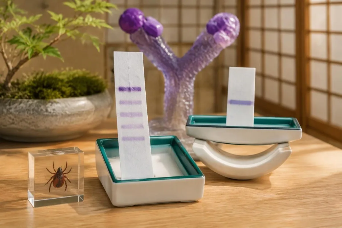
Moore et al. (2016) explains why individual bands and incomplete testing sequences create diagnostic errors; Mead et al. (2019) establishes the CDC recommendation for appropriately cleared modified algorithms. Lantos et al. (2021), publishing the 2020 guideline, connects testing to clinical manifestations, including early rash, arthritis and neurological disease; these sources support different parts of the reasoning rather than one universal band shortcut.
The 2 Figshare records address women's hormonal health and multilingual AI-assisted hantavirus triage, respectively, not Lyme immunoblot performance. A deployment involving 50,000 interpreted reports, as described in the hantavirus publication's title, cannot establish sensitivity, specificity or patient benefit for Lyme diagnosis; those claims would require Lyme-specific methods, reference standards and evaluation populations.
Материалы Kantesti принципы ответственного применения ИИ place report explanation alongside—not above—the laboratory and clinician, including when IgM appears after 30 days of symptoms. For symptoms linked to cycle changes or menopausal transition, our hormonal symptom guide addresses a separate assessment pathway; symptom overlap is a reason to investigate carefully, not evidence that a reactive Lyme band explains everything.
Часто задаваемые вопросы
Может ли одна положительная полоса в иммуноблоте на болезнь Лайма означать, что у меня болезнь Лайма?
Одна реактивная полоса при иммуноблоттинге на болезнь Лайма не подтверждает диагноз. Согласно общепринятым критериям США, в течение первых 30 дней с момента появления симптомов должны выявляться не менее 2 из 3 установленных полос IgM или 5 из 10 установленных полос IgG в рамках соответствующего двухэтапного тестирования. Одна полоса может указывать на перекрёстную реактивность или неполный ранний антительный ответ — в зависимости от ситуации. Типичная мигрирующая эритема после соответствующего контакта всё ещё может служить основанием для клинического диагноза, даже если ранние серологические тесты отрицательны.
Почему некоторые полосы в тесте на болезнь Лайма положительные, а общий результат отрицательный?
Тест на болезнь Лайма может выявить реактивные полосы, но результат при этом останется отрицательным, поскольку иммуноблот не соответствует валидированному профилю, предусмотренному алгоритмом тестирования. Например, 3 соответствующие критериям полосы IgG не достигают общепринятого порога в 5 из 10 полос IgG. Положительный или сомнительный результат скринингового теста первого этапа с последующими отрицательными результатами соответствующих иммуноблотов трактуется как отрицательный при стандартном двухэтапном тестировании. Если симптомы появились совсем недавно, врач может рассмотреть возможность повторного тестирования через подходящий интервал, а не трактовать неполный профиль как положительный.
Что означает положительная полоса 41 кДа при тесте на болезнь Лайма?
Полоса 41 кДа при болезни Лайма обычно отражает связывание антител с флагеллином — белком, связанным с подвижностью бактерий, который может давать перекрёстные реакции с антителами, не связанными с болезнью Лайма. Это лишь одна из установленных полос, используемых в стандартных критериях иммуноблота, и сама по себе она не позволяет диагностировать болезнь Лайма. Moore et al. (2016) сослались на исследование, в котором эту полосу выявили у 43% здоровых участников контрольной группы, однако эта доля не является универсальной для всех анализов. Полная последовательность тестирования и клиническая картина определяют, свидетельствует ли результат в пользу болезни Лайма.
Являются ли положительный результат IgM на болезнь Лайма и отрицательный результат IgG доказательством недавнего заражения?
Положительный результат IgM на боррелиоз при отрицательном IgG может свидетельствовать о ранней инфекции только при соответствии установленному алгоритму тестирования, клинической картине и срокам. Изолированный IgM не следует использовать для подтверждения болезни Лайма, если симптомы сохраняются более 30 дней, поскольку персистирующий или неспецифический IgM может привести к неверной интерпретации. Например, 2 диагностически значимые полосы IgM на 14-й день имеют иное значение, чем такая же картина после 3 месяцев непрерывных симптомов. Срок отсчитывается от появления симптомов, а не от дня взятия лабораторного образца.
Может ли тест на болезнь Лайма быть положительным, если в иммуноблоте нет ни одной полосы?
Да: валидированный модифицированный двухэтапный алгоритм диагностики болезни Лайма включает два последовательных иммуноферментных анализа и может вообще не предусматривать иммуноблоттинг. В 2019 году CDC рекомендовал соответствующим образом модифицированные алгоритмы, одобренные FDA. Некоторые комбинации анализов определяют общий уровень антител, тогда как другие дают отдельные результаты для IgM и IgG, поэтому обычные правила подсчёта полос не применимы ко всем заключениям. Уточните в лаборатории, какой алгоритм и комбинация анализов использовались для получения итогового заключения.
Должны ли полосы иммуноблота на болезнь Лайма исчезать после курса антибиотиков?
Полосы на иммуноблоте при болезни Лайма не обязательно должны исчезнуть после успешного лечения, поскольку антитела могут сохраняться годами или десятилетиями. Стойкий профиль IgG из 5 полос не доказывает продолжающуюся инфекцию, а повторное исследование полос не является валидированным методом подтверждения излечения. Ответ на лечение оценивают по клиническому течению и соответствующим объективным данным, а не по тому, стал ли анализ на антитела отрицательным. При появлении новых симптомов или повторном контакте с клещом необходима новая клиническая оценка, поскольку старые антитела не позволяют установить повторное заражение.
Получите объяснение результатов анализа крови
Загрузите отчёт лаборатории, и ИИ Kantesti за считаные секунды объяснит каждый биомаркер простым языком. Им пользуются более 2 миллионов человек по всему миру.
Ссылки
- Кляйн, Т., Митчелл, С., и Вебер, Х. (2026). Руководство по женскому здоровью: овуляция, менопауза и гормональные симптомы. Kantesti AI Медицинские исследования. DOI
- Кляйн, Т., Митчелл, С., и Вебер, Х. (2026). Мультиязычная клиническая поддержка принятия решений с помощью ИИ для ранней сортировки пациентов с хантавирусом: проектирование, инженерная валидация и внедрение в реальных условиях при интерпретации 50,000 отчётов об анализах крови. Kantesti AI Медицинские исследования. DOI
- Moore A et al. (2016). Current Guidelines, Common Clinical Pitfalls, and Future Directions for Laboratory Diagnosis of Lyme Disease, United States. Emerging Infectious Diseases. DOI PubMed
- Mead P et al. (2019). Updated CDC Recommendation for Serologic Diagnosis of Lyme Disease. Еженедельный отчёт MMWR о заболеваемости и смертности. DOI
- Lantos PM et al. (2021). Clinical Practice Guidelines by the Infectious Diseases Society of America (IDSA), American Academy of Neurology (AAN), and American College of Rheumatology (ACR): 2020 Guidelines for the Prevention, Diagnosis and Treatment of Lyme Disease. Clinical Infectious Diseases. DOI
- 2М+Проанализированные исследования
- 127+Страны
- 100+Языки
О медицинской команде


Сигналы доверия E-E-A-T
Опыт
Клинический обзор рабочих процессов интерпретации лабораторных данных под руководством врача.
Экспертиза
Акцент на лабораторной медицине: как биомаркеры ведут себя в клиническом контексте.
Авторитетность
Медицинскую проверку провёл доктор Томас Кляйн при участии доктора Сары Митчелл и профессора, доктора Ганса Вебера.
Доверие
Интерпретация на основе доказательств с понятными маршрутами дальнейших действий, чтобы снизить тревожность.





Медицинская оговорка
Эта статья предназначена только для образовательных целей и не является медицинской консультацией. Всегда консультируйтесь с квалифицированным специалистом здравоохранения по вопросам диагностики и решений по лечению.
Опубликовано · Связаться с редакцией
Издатель — Kantesti Ltd, зарегистрированная в Англии и Уэльсе компания, рег. №. 17090423, Лондон, Великобритания.
