
یک باند واکنشی نشاندهندهٔ اتصال آنتیبادی است، نه تشخیص بیماری. الگوریتم کامل آزمایش، سابقهٔ مواجهه و خط زمانی علائم تعیین میکنند که آیا این یافته مؤید بیماری لایم است یا خیر.

نکات کلیدی
- باندهای منفرد بیماری لایم را اثبات نمیکنند؛ حتی باندی با ویژگی نسبتاً بالا نیز باید در چارچوب الگوریتم معتبر آزمایش دومرحلهای تفسیر شود.
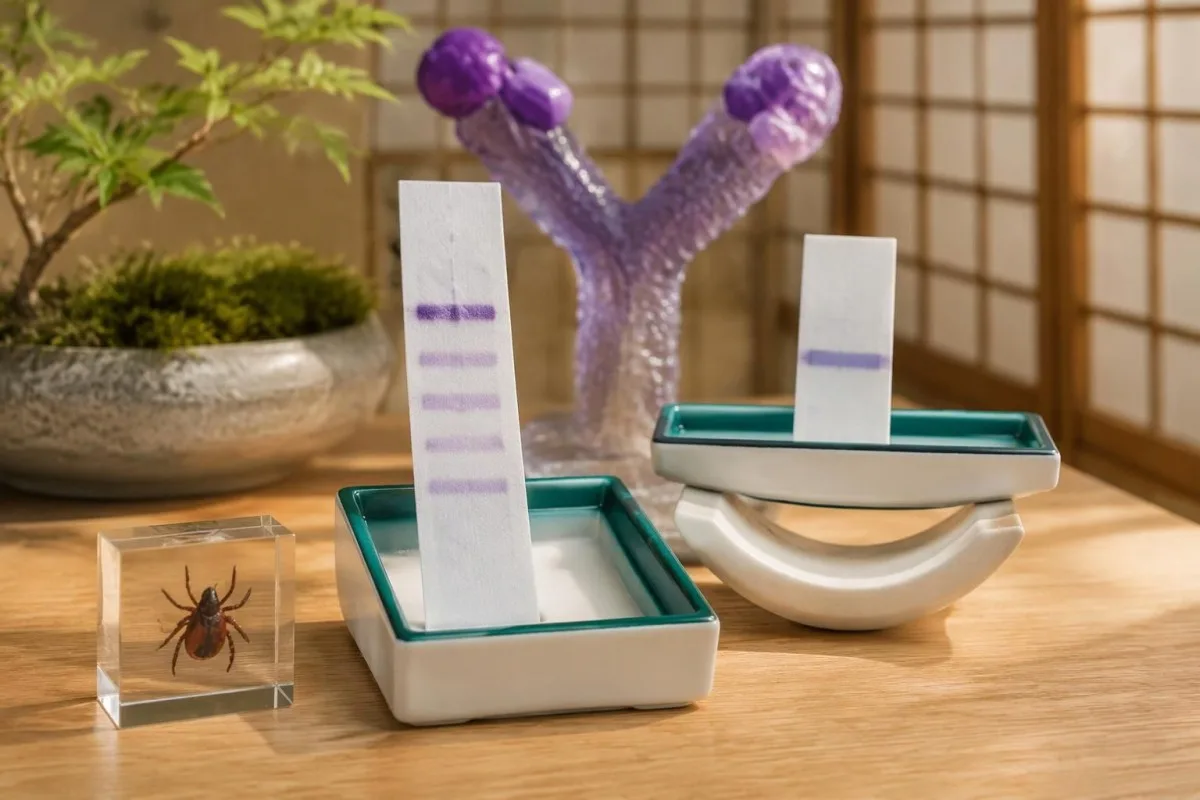
- معیارهای سنتی IgM دستکم ۲ مورد از ۳ باند تعیینشده را لازم دارند: حدود ۲۳–۲۵، ۳۹ و ۴۱ کیلودالتون.
- معیارهای سنتی IgG دستکم ۵ مورد از ۱۰ باند تعیینشده را لازم دارند: ۱۸، ۲۳، ۲۸، ۳۰، ۳۹، ۴۱، ۴۵، ۵۸، ۶۶ و ۹۳ کیلودالتون.
- قاعدهٔ ۳۰ روزه یعنی وقتی علائم بیش از ۳۰ روز ادامه داشتهاند، IgM منفرد نباید مبنای تشخیص لایم باشد.
- نتیجهٔ کلی منفی میتواند همراه با باندهای واکنشی وجود داشته باشد، زیرا آزمایشگاه یک الگوی اعتبارسنجیشده را ارزیابی میکند، نه اینکه آیا باندی ظاهر میشود یا خیر.
- آزمایش اصلاحشده دومرحلهای بهجای ایمونوبلات، از ۲ ایمونواسی متوالی استفاده میکند؛ ممکن است فهرستی از باندها ارائه نکند.
- تست زودهنگام ممکن است پیش از ایجاد آنتیبادیها منفی باشد؛ بثورات معمول اریتم مهاجر در شرایط مواجهه مناسب، بهصورت بالینی تشخیص داده میشود.
- آنتیبادیهای پایدار ممکن است سالها پس از درمان همچنان قابلتشخیص باشد و نمیتواند عفونت فعال را اثبات کند یا میزان بهبودی را بسنجد.
باند مثبت وسترن بلات لایم در واقع چه معنایی دارد؟
A باند وسترن بلات مثبت برای لایم یعنی آنتیبادیها به یک آنتیژن آزمایش متصل شدهاند؛ این موضوع بهتنهایی بیماری لایم را اثبات نمیکند. در آزمایش سنتی دومرحلهای آمریکا، IgM در ۳۰ روز نخست علائم به ۲ مورد از ۳ باند مشخصشده نیاز دارد، درحالیکه IgG به ۵ مورد از ۱۰ باند نیاز دارد؛ در آزمایش اصلاحشده، بهجای آن از ۲ ایمونواسی استفاده میشود.

این نتیجه کلی آزمون غربالگری مرحله اول را با نتیجه مناسب مرحله دوم ترکیب میکند، نه اینکه هر نوار قابلمشاهده را تشخیصی جداگانه تلقی کند. بنابراین، گزارشی که ۱ باند واکنشی نشان میدهد میتواند بهدرستی نتیجه را منفی اعلام کند؛ حتی نتیجه مثبت کامل آنتیبادی نیز باید با توجه به مواجهه، یافتههای سازگار و سابقه قبلی بیماری لایم ارزیابی شود.
هویت باند به پرسشی محدود پاسخ میدهد: تحت شرایط این آزمون، کدام آنتیژن باعث اتصال آنتیبادی شده است؟ این موضوع به ۳ پرسشی که معمولاً برای بیماران مهم است پاسخ نمیدهد—اینکه آیا اکنون عفونتی وجود دارد، آیا آن عفونت علائمشان را توضیح میدهد یا آیا به آنتیبیوتیک نیاز دارند؛ راهنمای ما درباره نتایج مثبت آنتیبادی این مسائل را از هم تفکیک میکند.
کانتستی یک آنالایزر آزمایش خون هوش مصنوعی که میتواند توضیح دهد چرا گزارش لایم حاوی باندهای واکنشی است اما تفسیر دومرحلهای آن منفی است. تفسیر نهایی آزمایشگاه، روش آزمون و مدت علائم را زمینهای ضروری میدانیم—نه جزئیاتی که بتوان با شمردن خطوط برجستهشده در یک گزارش بارگذاریشده نادیده گرفت.
آزمایش دومرحلهای سنتی لایم چگونه نتیجه را تعیین میکند
آزمایش استاندارد دومرحلهای با ایمونواسی آنزیمی، یا گاهی آزمون ایمونوفلورسانس، آغاز میشود و اگر نتیجه آزمایش نخست مثبت یا مبهم باشد، ایمونوبلات انجام میشود. این ۲ مرحله در کنار هم عمل میکنند؛ نتیجه مثبت غربالگری بهتنهایی نتیجه مثبت تأییدشده سرولوژی لایم نیست.

A نتیجه مثبت یا مبهم آزمایش مرحله اول و سپس ایمونوبلاتهای مناسب با نتیجه منفی طبق الگوریتم استاندارد، منفی تفسیر میشود. آزمون غربالگری اولیه عمداً پاسخهای احتمالی بیشتری را شناسایی میکند، درحالیکه مرحله دوم واکنشپذیری غیراختصاصی را فیلتر میکند؛ مور و همکاران در سال ۲۰۱۶ این موازنه و خطاهای رایج در تفسیر آن را در مجله Emerging Infectious Diseases شرح دادند.
شاخص ۱٫۴ در یک آزمون غربالگری با ۱٫۴ در آزمونی دیگر قابلجایگزینی نیست، زیرا ترکیب آنتیژن و حدود برش سازنده متفاوت است. همچنین، نتیجه مبهم غربالگری یعنی باید الگوریتم کامل شود—نه اینکه تشخیصی بینابینی میان عفونت و نبود عفونت باشد؛ نتایج کیفی در مقابل کمی توضیح میدهد چرا قالب گزارشها اهمیت دارد.
انجام ایمونوبلات پس از نتیجه منفی غربالگری، بدون دلیل بالینی برای بازنگری در زمانبندی یا مناسببودن آزمون، توالیای را دور میزند که عملکرد آن ارزیابی شده است. ۳ مورد را درخواست کنید: نتیجه غربالگری، تفسیر مرحله دوم و اینکه آیا هر دو به یک مسیر آزمایش معتبر تعلق دارند؛ گزارش ناقص را نباید صرفاً بر اساس باندها بازسازی کرد.
توضیح باندهای وسترن بلات لایم: معیارهای IgM و IgG
بر اساس معیارهای متعارف آمریکا، یک ایمونوبلات IgM مثبت دستکم به ۲ مورد از باندهای ۲۳–۲۵، ۳۹ و ۴۱ kDa نیاز دارد. یک ایمونوبلات IgG مثبت requires at least 5 of the 18, 23, 28, 30, 39, 41, 45, 58, 66 and 93 kDa bands.
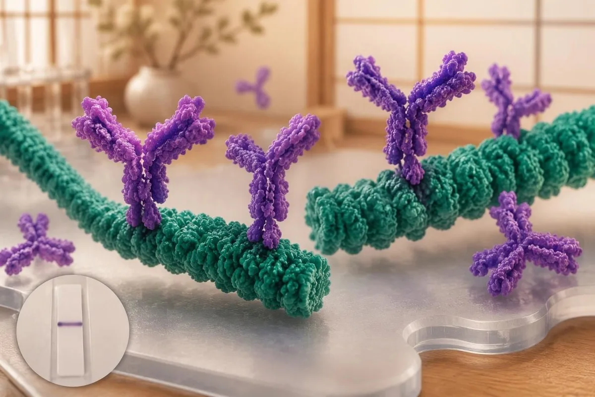
Kilodaltons, abbreviated kDa, describe approximate protein molecular mass, not antibody concentration or disease severity. The approximately 23–25 kDa band commonly represents outer surface protein C, 39 kDa represents BmpA and 41 kDa represents flagellin; the numbers identify antigen regions, so a higher-numbered band does not mean a more advanced infection.
The IgG list's 58 kDa criterion excludes GroEL, and reports may use assay-specific names or approximate mass conventions rather than identical labels. Because not every commercial strip uses the same antigen preparation, our راهنمای نشانگر تست خون ما should complement—not replace—the actual laboratory legend and the assay manufacturer's interpretation instructions.
Bands at 31 and 34 kDa are not part of the conventional CDC IgG counting criteria, even though they may appear on some reports. Their absence from that list does not make them meaningless biologically, but adding them to manufacture a 5-band positive result changes the validated rule; neither band darkness nor extra bands quantify bacterial burden.
چرا باند ۴۱ و دیگر باندهای واکنشی میتوانند سیگنالهای کاذب باشند
این 41 kDa band reacts with flagellin, a protein associated with bacterial movement, and can cross-react with antibodies unrelated to Lyme disease. A single reactive 41 kDa band is therefore insufficient to diagnose Lyme disease, even when a patient has fatigue, aching or a remembered tick bite.
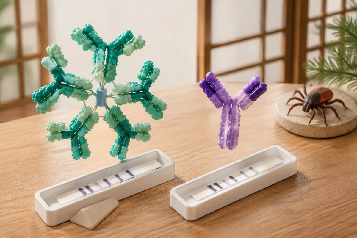
Moore et al. (2016) cited a study in which 41 kDa reactivity occurred in 43% of healthy controls, illustrating why this band cannot stand alone. That percentage is not a universal false-positive rate for every modern assay; it is evidence that recognizable antigen binding can occur in people without the clinical condition being investigated.
Epstein–Barr virus, syphilis, relapsing-fever Borrelia and some autoimmune conditions can contribute to false-positive Lyme serology, although the mechanism and affected assay vary. A recent mononucleosis-like illness should prompt attention to the complete EBV antibody pattern, not the assumption that 1 Lyme band proves a simultaneous tick-borne infection.
Cross-reactivity is not a reason to order every alternative antibody panel: 1 unexplained band usually does not justify broad autoimmune screening. When examination findings genuinely suggest an immune disorder, تفسیر کمپلمان و ANA belongs to a separate clinical question; repeated low-probability testing can generate additional misleading positives rather than a clearer answer.
IgM در برابر IgG لایم: چرا قاعدهٔ ۳۰ روزهٔ علائم اهمیت دارد
Lyme IgM should not be used to support a diagnosis when symptoms have lasted more than 30 days. IgM is most useful early, whereas an appropriate IgG response carries greater diagnostic weight later; the clock starts with symptoms of the suspected illness, not the testing date or remembered tick removal.
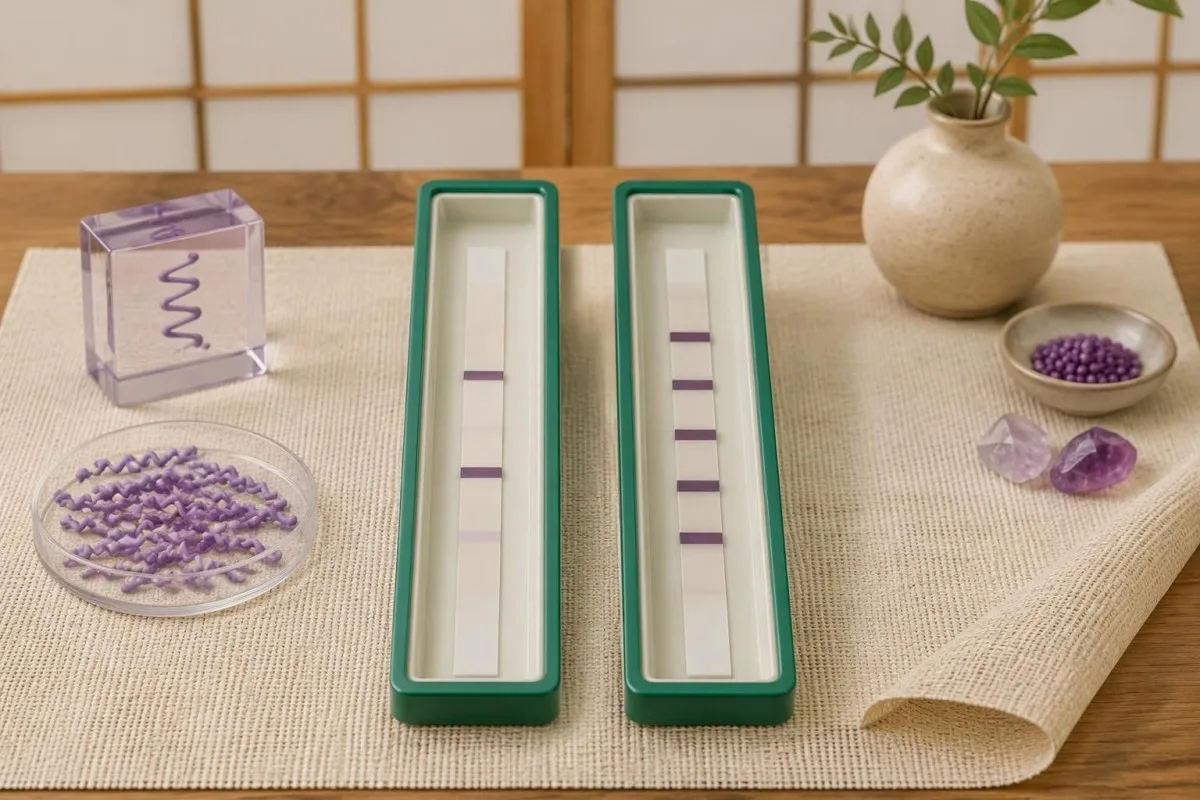
این 30-day restriction reduces misclassification from persistent or nonspecific IgM responses when later disease should usually have an established IgG response. It is an interpretation rule, not a biological switch that flips overnight: antibody maturation varies, and early treatment may alter seroconversion, but those exceptions do not make isolated late IgM reliable evidence.
For a hypothetical patient tested on day 75 of continuous symptoms, IgM bands 23 and 41 with negative IgG should not be used as proof of Lyme disease. By contrast, the same IgM pattern on day 12, following a reactive first-tier assay and compatible exposure, can support an early diagnosis; the numerical band count is identical, but its evidential value changes.
A positive IgM result does not automatically mean a new infection, a misconception also addressed in our Mycoplasma IgM interpretation guide. If symptoms began 8 weeks ago but one symptom appeared yesterday, clinicians need to decide whether this is a genuinely new illness or another feature of the original episode—not simply restart the IgM clock.
باندهای مثبت لایم اما نتیجهٔ کلی منفی: نمونههای حلشده
Positive Lyme bands but negative overall usually means the report did not meet the required pattern within the testing algorithm. For example, 3 designated IgG bands are below the conventional 5-of-10 threshold; that result is negative, not a smaller positive, and it does not measure how much infection is present.
Consider a clearly hypothetical 42-year-old with 10 weeks of fatigue, a reactive screening assay and IgG bands 18, 41 and 58. The IgG immunoblot is negative because only 3 qualifying bands are present; if the examination lacks objective Lyme findings, the next step is a symptom-focused assessment rather than antibiotics prescribed to treat the band's molecular masses.
A second hypothetical patient has a reactive screen, IgM bands 23 and 39, and compatible symptoms beginning 14 days earlier. This can meet the traditional early serologic criteria, but an atypical exposure history or a stronger alternative diagnosis still affects the clinical conclusion; Kantesti AI can explain the 2-of-3 rule without converting it into an independent diagnosis.
A third report lists 5 IgG bands but provides no first-tier result; the missing screening stage prevents straightforward confirmation of a standard 2-tier outcome. Confirmation requirements are assay-specific, as our HSV IgG confirmation guide also illustrates, so request the complete laboratory report rather than borrowing a cutoff or confirmation rule from another infection.
تفاوت آزمایش دومرحلهای اصلاحشده با وسترن بلات
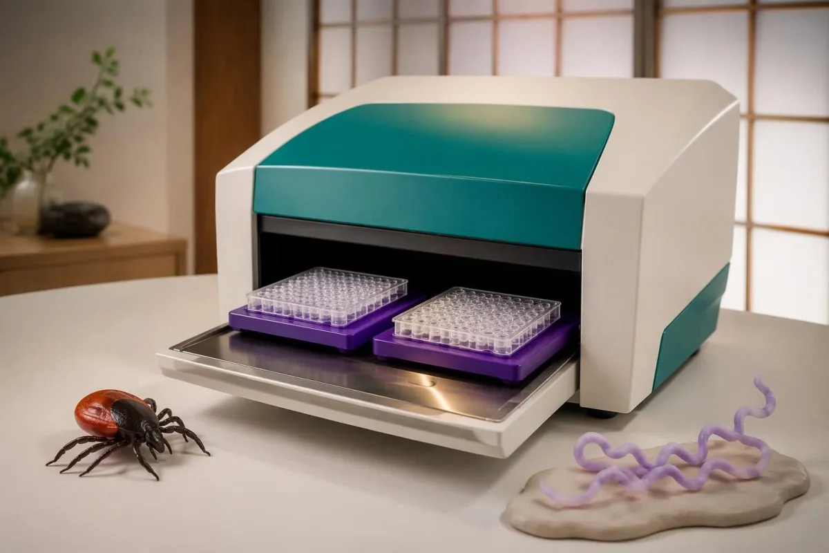
Modified two-tier testing, or MTTT, uses 2 sequential enzyme immunoassays instead of an enzyme immunoassay followed by an immunoblot. A report from this pathway may have no bands at all; its overall interpretation depends on the particular assay combination and its cleared instructions, not the conventional immunoblot band-count criteria.
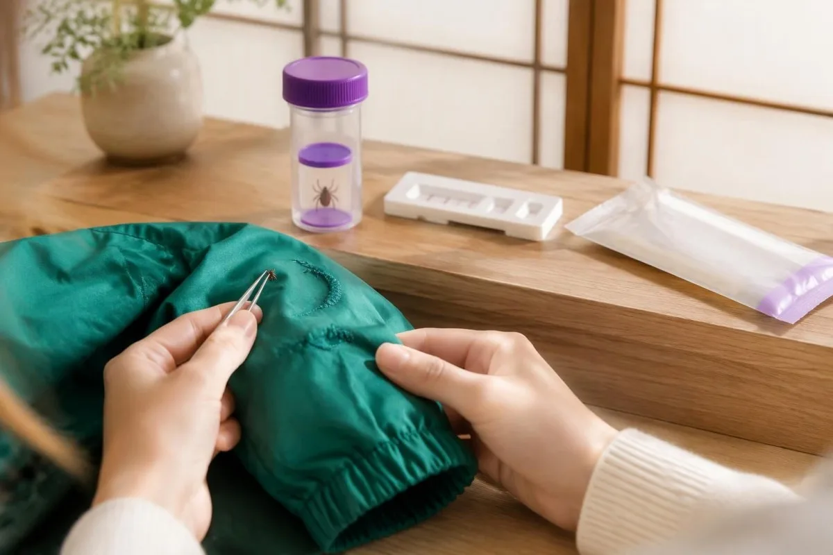
این 2019 CDC recommendation accepted modified algorithms using FDA-cleared assays for that purpose, following clearance of relevant test combinations (Mead et al., 2019). The change was not permission to combine any 2 tests: the evaluated pairing matters, and some combinations improve early sensitivity while preserving high specificity compared with standard two-tier testing.
Some modified algorithms measure total antibodies, while others report separate IgM and IgG components; consequently, the familiar 30-day IgM restriction cannot be mechanically applied to a total-antibody result. Kantesti is an پلتفرم تفسیر آزمایش خون AI that can help distinguish a 2-immunoassay report from an immunoblot report before discussing what its positive or negative designation means.
A second immunoassay is not simply a repeat of the same screening test, and an immunoblot is not routinely needed after a completed validated MTTT result. Our راهنمای تأیید شاگاس illustrates the broader principle of complementary assays, but its 2-test rules are different; ask your laboratory which Lyme algorithm produced the final interpretation.
آیا آزمایش لایم ممکن است با وجود عفونت، در مراحل اولیه منفی باشد؟
Lyme antibody testing can be negative early because antibodies have not yet reached detectable levels. A typical erythema migrans rash with an appropriate exposure history is diagnosed clinically rather than by waiting for a positive immunoblot; a negative result during the first 1–2 weeks does not safely exclude that presentation.

Across early-infection settings, conventional 2-tier sensitivity has been reported at approximately 30–40%, while specificity remains high when testing and interpretation are appropriate (Moore et al., 2016). These are population estimates, not a personal probability: sensitivity changes with the clinical manifestation, time since onset and whether the patient has already received antibiotics.
For a rash suggestive of but not typical for erythema migrans, the 2020 IDSA/AAN/ACR guideline supports acute serology and, if initially negative, a convalescent sample at least 2–3 weeks later (Lantos et al., 2021). The same guideline recommends clinical diagnosis for a typical lesion; a rash need not have textbook concentric rings to warrant assessment.
Repeating a test 24 hours later usually offers little advantage when the limitation is antibody development rather than a processing error. Our spotted fever testing guide explains a related timing problem, but suspected spotted fever has its own urgent treatment rules; do not delay care for an expanding rash or significant illness while waiting for more bands.
آیا IgG مثبت لایم به معنای عفونت فعال یا شکست درمان است؟
Positive Lyme IgG cannot distinguish active infection from a previous infection that has resolved. Lyme antibodies can remain detectable for years or decades, and repeat immunoblot testing is not a test of cure; neither a persistent 5-band pattern nor changes in band intensity establish that treatment failed.
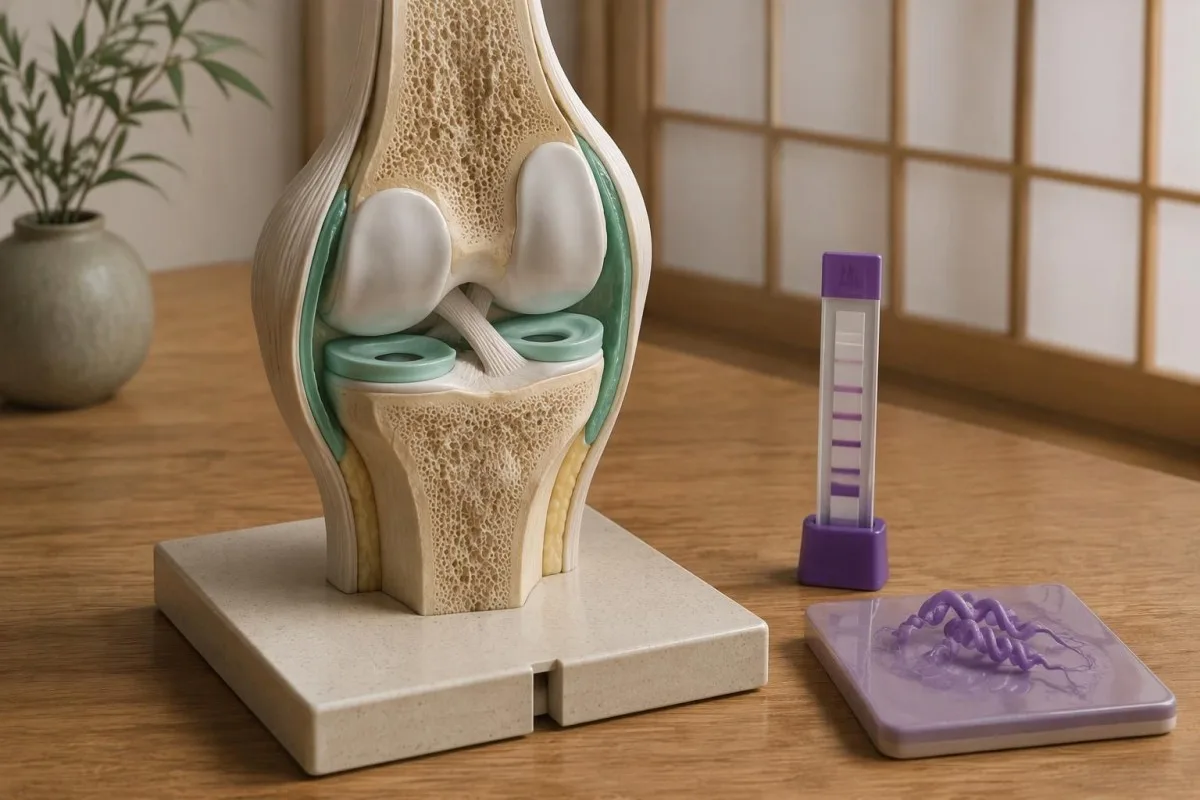
این 2020 IDSA/AAN/ACR guideline describes persistence of both IgM and IgG responses after infection, which complicates interpretation in previously affected patients (Lantos et al., 2021). A positive result obtained 4 years after treatment must therefore be compared with the clinical history, not interpreted as a laboratory photograph of living bacteria in the body.
Reinfection can occur because previous Lyme disease does not provide reliable lifelong protection, but an old positive antibody result cannot prove a new episode. For a hypothetical person with positive IgG from 2019 and a new expanding rash after recent tick exposure, the new clinical findings matter more than whether a sixth band appears; الگوهای آنتیبادی CMV illustrate why exposure history and current illness are separate questions.
Persistent fatigue, pain or cognitive symptoms deserve evaluation even when no new laboratory evidence appears. The guideline recommends against additional antibiotics for persistent nonspecific symptoms after recommended treatment when there is no objective evidence of reinfection or treatment failure; 1 positive antibody panel cannot identify the cause of those symptoms or justify an open-ended antibiotic course.
معنای IgG در آرتریت لایمی یا بیماری عصبیِ مشکوک
Lyme arthritis usually produces a strong IgG response, so isolated IgM is poor evidence for that diagnosis. Conventional two-tier serology has approximately 95–100% sensitivity in Lyme arthritis, according to the 2020 IDSA/AAN/ACR guideline; a negative appropriate IgG result should prompt serious consideration of other causes of joint swelling.

A hypothetical patient with a markedly swollen knee and only 2 IgM bands after 3 months of symptoms does not have the expected serologic pattern for Lyme arthritis. The clinician should reassess alternative causes and assay reliability rather than call this late Lyme based on IgM alone; when indicated, joint fluid examination answers additional questions that the antibody test cannot.
Suspected central nervous system Lyme disease requires clinical assessment and, when cerebrospinal fluid testing is undertaken, simultaneous CSF and serum for a validated antibody index. Testing CSF antibody alone or running a standalone CSF immunoblot is not the preferred approach; 1 elevated index may also persist after treatment, so it must be interpreted alongside the neurological findings and CSF cell response.
Numb feet or tingling do not become Lyme-specific because 41 kDa is reactive; neuropathy has metabolic, mechanical and immune causes. Our anti-MAG neuropathy guide explains one targeted alternative, while immunoglobulin panel interpretation clarifies that total serum IgG is different from the 5-of-10 Lyme-specific IgG immunoblot criterion.
چرا آزمایشگاهها و کشورها ممکن است از معیارهای متفاوتی برای لایم استفاده کنند
Lyme immunoblot criteria are assay- and region-dependent, so conventional US 5-of-10 IgG counting rules should not automatically be imposed on every European report. European assays may use different Borrelia antigens and recombinant line blots because the species causing human Lyme borreliosis differ across regions.
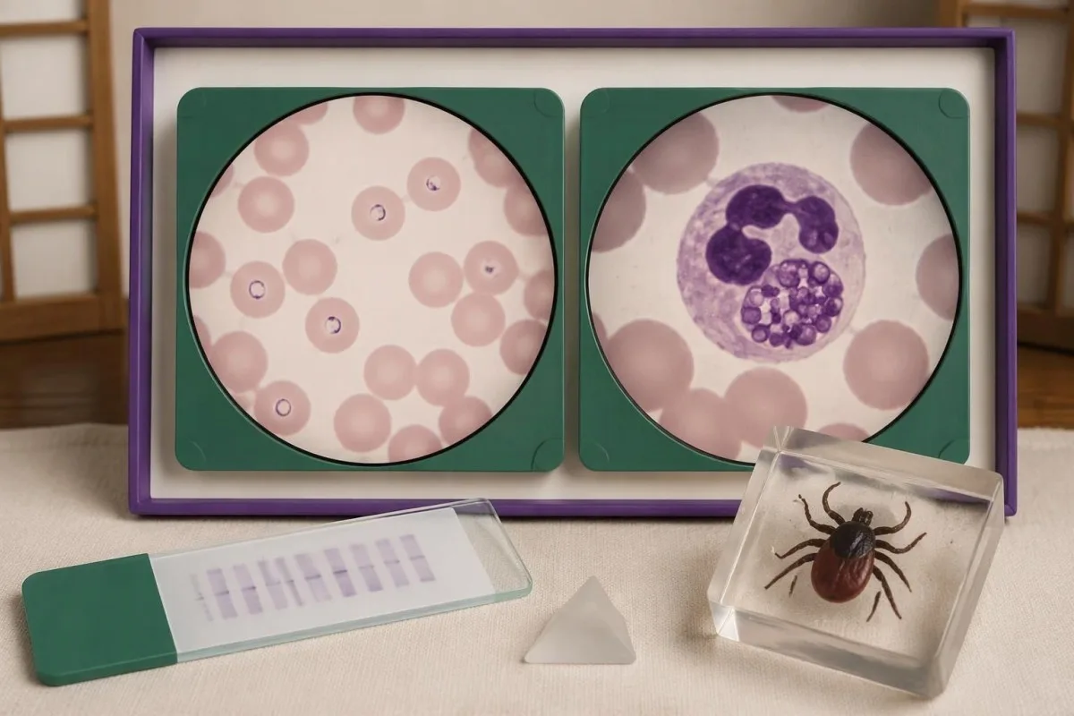
B. burgdorferi sensu stricto predominates in North America, while European disease also commonly involves B. afzelii and B. garinii. A report using VlsE, OspC or other recombinant antigen labels may therefore not resemble a 10-position conventional US blot; the clinically useful question is whether the assay was validated for the exposure setting and interpreted according to its instructions.
Different laboratory criteria are not automatically inferior, but claims that 1 or 2 bands prove disease require evidence for that assay's complete diagnostic performance. When reading a report described by Kantesti, consult the laboratory's interpretation and our اعتبارسنجی پزشکی ما separately; general AI assessment or a software benchmark is not proof that a particular Lyme assay has validated sensitivity and specificity.
Record 3 details before comparing reports from different laboratories: assay manufacturer, algorithm type and the country where exposure occurred. Our راهنمای بررسی منابع گزارش هوش مصنوعی ما explains why preserving source wording matters; apparent conversion from negative to positive may reflect a changed method or cutoff rather than a newly acquired infection or worsening illness.
آیا باندهای لایم بابزیا، آناپلاسما یا عفونتهای همزمان دیگر را شناسایی میکنند؟
Lyme Western blot bands do not diagnose tick-borne coinfections. Babesia, Anaplasma and Ehrlichia require their own clinical assessment and tests; 1 positive Lyme antibody result neither confirms nor excludes these infections, even when an exposure setting allows more than one tick-borne organism to be transmitted.

بابزیوز deserves consideration when relevant exposure accompanies fever and evidence such as anemia, low platelets or hemolysis, especially if fever persists during Lyme-directed treatment. A Babesia antibody titer of 1:256 alone does not establish current infection; our Babesia test interpretation guide explains why a cellular sample examination or PCR may be needed to demonstrate active infection.
آناپلاسموز can produce fever, leukopenia, thrombocytopenia and elevated liver enzymes, findings that should not be explained away as extra Lyme bands. PCR is most useful during the first week of illness and becomes less sensitive after appropriate antibiotics; our Anaplasma sample timing guide separates this early direct-detection window from later antibody interpretation.
Ehrlichiosis follows a different organism-specific testing pathway, and a negative early antibody result is not a safe reason to dismiss significant febrile illness. Our Ehrlichia PCR and titer guide explains why 2 samples collected at different times may help establish seroconversion; targeted testing should follow symptoms, geography and laboratory abnormalities rather than an indiscriminate panel ordered for isolated fatigue.
با گزارش آزمایش لایمِ نامشخص چه باید کرد؟
For an unclear Lyme report, obtain the complete 2-tier interpretation, write down symptom onset and discuss exposure plus objective findings with a clinician. Faint or isolated bands do not determine urgency; fainting, chest pain, breathlessness, a markedly slow pulse or new neurological deficits require prompt assessment regardless of the antibody result.

Lyme carditis can cause heart conduction block, so fainting or palpitations after compatible exposure should not wait for an immunoblot to become positive. The 2020 IDSA/AAN/ACR guideline identifies a PR interval greater than 300 milliseconds, other arrhythmias or features of myopericarditis as reasons for hospital admission with monitoring; that interval is measured on an ECG, not inferred from band intensity.
New facial weakness warrants urgent assessment, and sudden facial droop with arm weakness or speech difficulty needs emergency evaluation for stroke rather than attribution to Lyme. Our راهنمای علائم هشداردهنده سکته مغزی explains that distinction; for a routine appointment, bring 4 dates—possible exposure, first symptom, sample collection and antibiotic initiation—because each changes a different part of the reasoning.
کانتستی یک ابزار تحلیل آزمایش خون مبتنی بر AI that can help organize the 2-tier result, reported bands and missing clinical context into questions for your appointment. Our راهنمای فناوری هوش مصنوعی describes the explanation workflow, but our AI cannot examine a rash, measure an ECG interval, confirm Lyme disease independently or decide whether you should start, stop or extend antibiotics.
انتشارات پژوهشی، حدود شواهد و تفسیر ایمن
The clinical interpretation in this article rests on 3 directly relevant sources: a laboratory-diagnosis review, the CDC's modified two-tier recommendation and the IDSA/AAN/ACR Lyme guideline. The 2 additional Figshare publications listed below concern other health topics and must not be treated as validation of Lyme band interpretation or Lyme diagnosis.
Moore et al. (2016) explains why individual bands and incomplete testing sequences create diagnostic errors; Mead et al. (2019) establishes the CDC recommendation for appropriately cleared modified algorithms. Lantos et al. (2021), publishing the 2020 guideline, connects testing to clinical manifestations, including early rash, arthritis and neurological disease; these sources support different parts of the reasoning rather than one universal band shortcut.
The 2 Figshare records address women's hormonal health and multilingual AI-assisted hantavirus triage, respectively, not Lyme immunoblot performance. A deployment involving 50,000 interpreted reports, as described in the hantavirus publication's title, cannot establish sensitivity, specificity or patient benefit for Lyme diagnosis; those claims would require Lyme-specific methods, reference standards and evaluation populations.
مطالب Kantesti هوش مصنوعی مسئولانه ما place report explanation alongside—not above—the laboratory and clinician, including when IgM appears after 30 days of symptoms. For symptoms linked to cycle changes or menopausal transition, our hormonal symptom guide addresses a separate assessment pathway; symptom overlap is a reason to investigate carefully, not evidence that a reactive Lyme band explains everything.
سوالات متداول
آیا وجود یک باند مثبت در وسترن بلات لایم به این معناست که به بیماری لایم مبتلا هستم؟
وجود یک باند واکنشی در وسترن بلات لایم، ابتلا به بیماری لایم را اثبات نمیکند. طبق معیارهای رایج در ایالات متحده، در ۳۰ روز نخست بروز علائم باید دستکم ۲ مورد از ۳ باند IgM مشخصشده، یا ۵ مورد از ۱۰ باند IgG مشخصشده، در چارچوب مسیر مناسب آزمایش دومرحلهای وجود داشته باشد. بسته به شرایط، وجود یک باند منفرد ممکن است ناشی از واکنش متقاطع یا پاسخ آنتیبادی زودرسِ ناکامل باشد. پس از مواجهه مناسب، بروز بثورات معمول اریتما میگرانس همچنان میتواند با وجود منفی بودن سرولوژی در مراحل اولیه، از تشخیص بالینی حمایت کند.
چرا بعضی از باندهای آزمایش لایم مثبت هستند، اما نتیجهٔ کلی آزمایش منفی است؟
آزمایش لایم ممکن است حاوی باندهای واکنشی باشد، اما همچنان منفی باقی بماند؛ زیرا ایمونوبلات با الگوی اعتبارسنجیشدهای که الگوریتم آزمایش لازم میداند، مطابقت نداشته است. برای مثال، ۳ باند IgG واجد شرایط، کمتر از آستانهٔ متعارف ۵ باند از ۱۰ باند IgG است. در آزمایش استاندارد دومرحلهای، نتیجهٔ مثبت یا مرزی غربالگری مرحلهٔ اول که پس از آن ایمونوبلاتهای مناسب منفی باشند، منفی تفسیر میشود. اگر علائم بسیار جدید باشند، پزشک ممکن است تکرار آزمایش در فاصلهٔ زمانی مناسب را در نظر بگیرد، بهجای آنکه الگوی ناقص را مثبت تلقی کند.
باند مثبت ۴۱ کیلودالتونی در آزمایش لایم به چه معناست؟
باند ۴۱ کیلودالتونی لایم معمولاً نشاندهندهٔ اتصال آنتیبادی به فلاژلین است؛ پروتئینی مرتبط با حرکت باکتری که ممکن است با آنتیبادیهای نامرتبط با بیماری لایم واکنش متقاطع داشته باشد. این باند فقط یکی از باندهای تعیینشدهای است که در معیارهای متداول ایمونوبلات بهکار میروند و بهتنهایی نمیتواند بیماری لایم را تشخیص دهد. Moore و همکاران (۲۰۱۶) به مطالعهای اشاره کردند که این باند را در 43% افراد سالمِ گروه کنترل یافت؛ هرچند این درصد در همهٔ آزمونها نرخ ثابتی نیست. توالی کامل آزمایشها و تظاهرات بالینی تعیین میکنند که آیا نتیجه از تشخیص بیماری لایم حمایت میکند یا خیر.
آیا مثبت بودن IgM و منفی بودن IgG در آزمایش لایم، اثباتکنندهٔ عفونت اخیر است؟
مثبت بودن IgM لایم همراه با IgG منفی، تنها زمانی میتواند مؤید عفونت زودرس باشد که با الگوریتم آزمایش مناسب، تظاهرات بالینی و زمانبندی سازگار باشد. وقتی علائم بیش از ۳۰ روز ادامه داشتهاند، نباید از IgM منفرد برای تأیید بیماری لایم استفاده کرد، زیرا IgM پایدار یا غیراختصاصی میتواند به تفسیر نادرست منجر شود. برای مثال، وجود ۲ باند واجد شرایط IgM در روز ۱۴، معنایی متفاوت از مشاهده همان الگو پس از ۳ ماه علائم مداوم دارد. قاعده زمانبندی به زمان شروع علائم اشاره دارد، نه روزی که نمونه آزمایشگاهی جمعآوری شده است.
آیا ممکن است آزمایش لایم من مثبت باشد، اما هیچ باندی در وسترن بلات دیده نشود؟
بله: در یک الگوریتم اصلاحشده و معتبرِ دومرحلهای برای بیماری لایم، از دو سنجش ایمونولوژیک آنزیمیِ متوالی استفاده میشود و ممکن است اصلاً وسترن بلات انجام نشود. CDC در سال ۲۰۱۹ الگوریتمهای اصلاحشدهای را توصیه کرد که مجوزهای لازم FDA را دریافت کردهاند. برخی ترکیبهای آزمایش، آنتیبادیهای تام را اندازهگیری میکنند، درحالیکه برخی دیگر نتایج IgM و IgG را جداگانه ارائه میدهند؛ بنابراین، قواعد متعارفِ شمارش باندها برای همه گزارشها کاربرد ندارند. از آزمایشگاه بپرسید که تفسیر نهایی بر اساس کدام الگوریتم و ترکیب آزمایشی انجام شده است.
آیا باندهای وسترن بلات لایم باید پس از مصرف آنتیبیوتیک ناپدید شوند؟
لازم نیست باندهای وسترن بلات لایم پس از درمان موفق ناپدید شوند، زیرا پادتنها ممکن است سالها یا حتی دههها باقی بمانند. باقی ماندن الگوی ۵ باندی IgG، عفونت فعال را اثبات نمیکند و تکرار آزمایش باندها، روش تأییدشدهای برای اطمینان از درمان نیست. پاسخ به درمان بر اساس روند بالینی بیماری و یافتههای عینی مرتبط ارزیابی میشود، نه با الزام به منفی شدن پنل پادتنها. بروز علائم جدید یا مواجهه دوباره با کنه، به ارزیابی بالینی جدید نیاز دارد، زیرا پادتنهای قدیمی نمیتوانند عفونت مجدد را اثبات کنند.
چگونه نتایج آزمایش خون را بخوانیم
گزارش آزمایشگاهی خود را بارگذاری کنید تا هوش مصنوعی Kantesti ظرف چند ثانیه هر زیستنشانگر را به زبان ساده توضیح دهد. بیش از 2 میلیون نفر در سراسر جهان از آن استفاده کردهاند.
منابع
- Klein, T., Mitchell, S., & Weber, H. (2026). راهنمای سلامت زنان: تخمکگذاری، یائسگی و علائم هورمونی. پژوهش پزشکی هوش مصنوعی Kantesti. DOI
- Klein, T., Mitchell, S., & Weber, H. (2026). پشتیبانی چندزبانه از تصمیمگیری بالینی با کمک هوش مصنوعی برای تریاژ زودهنگام هانتاویروس: طراحی، اعتبارسنجی مهندسی و پیادهسازی در دنیای واقعی در 50,000 گزارش تفسیر آزمایش خون. پژوهش پزشکی هوش مصنوعی Kantesti. DOI
- Moore A et al. (2016). Current Guidelines, Common Clinical Pitfalls, and Future Directions for Laboratory Diagnosis of Lyme Disease, United States. Emerging Infectious Diseases. DOI پابمد
- Mead P et al. (2019). Updated CDC Recommendation for Serologic Diagnosis of Lyme Disease. گزارش هفتگی بیماریها و مرگومیر MMWR. DOI
- Lantos PM et al. (2021). Clinical Practice Guidelines by the Infectious Diseases Society of America (IDSA), American Academy of Neurology (AAN), and American College of Rheumatology (ACR): 2020 Guidelines for the Prevention, Diagnosis and Treatment of Lyme Disease. بیماریهای عفونی بالینی. DOI
- ۲ میلیون+آزمایشهای تجزیهوتحلیلشده
- 127+کشورها
- 100+زبانها
درباره تیم پزشکی


سیگنالهای اعتماد E-E-A-T
تجربه
بازبینی بالینی مبتنی بر نظر پزشک از فرایندهای تفسیر آزمایشگاه.
تخصص
تمرکز بر پزشکی آزمایشگاهی و اینکه نشانگرهای زیستی در زمینه بالینی چگونه رفتار میکنند.
اقتدارگرایی
با بازبینی پزشکی دکتر توماس کلاین، با مشارکت دکتر سارا میچل و پروفسور دکتر هانس وبر.
قابل اعتماد بودن
تفسیر مبتنی بر شواهد با مسیرهای پیگیری روشن برای کاهش هشدارها.





سلب مسئولیت پزشکی
این مقاله فقط برای اهداف آموزشی است و توصیه پزشکی محسوب نمیشود. برای تصمیمهای مربوط به تشخیص و درمان، همیشه با یک ارائهدهنده مراقبتهای بهداشتی واجد شرایط مشورت کنید.
منتشر شد · با تیم تحریریه تماس بگیرید
منتشرشده توسط Kantesti Ltd، ثبتشده در انگلستان و ولز، شماره شرکت. 17090423, ، لندن، بریتانیا.
