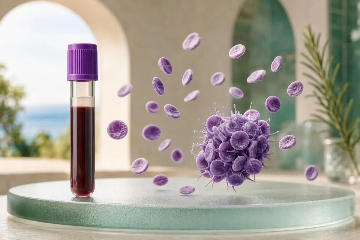
ภาวะเกล็ดเลือดต่ำอาจเกิดจากข้อผิดพลาดในการวัดจากหลอดเก็บสิ่งส่งตรวจได้เช่นกัน ไม่ใช่ปัญหาภายในร่างกายของคุณ นี่คือวิธีการวินิจฉัยภาวะเกล็ดเลือดต่ำเทียมจาก EDTA ก่อนที่จะมีใครวินิจฉัยว่าคุณมีภาวะเกล็ดเลือดต่ำ.


- ภาวะเกล็ดเลือดต่ำเทียม คือจำนวนเกล็ดเลือดต่ำกว่าความเป็นจริงที่เกิดจากการจับตัวเป็นก้อนของเกล็ดเลือดหลังการเก็บสิ่งส่งตรวจ ซึ่งส่วนใหญ่มักเกิดขึ้นในหลอด EDTA.
- ช่วงเกล็ดเลือดโดยทั่วไป สำหรับผู้ใหญ่ส่วนใหญ่ที่ไม่ตั้งครรภ์ จะอยู่ที่ประมาณ 150-450 × 10⁹/L แม้ว่าห้องปฏิบัติการจะกำหนดช่วงของตนเองก็ตาม.
- เกล็ดเลือดจับตัวเป็นก้อนบนสเมียร์ อาจทำให้เครื่องวิเคราะห์อัตโนมัติรายงานจำนวนเกล็ดเลือดต่ำกว่าความเป็นจริง เนื่องจากกลุ่มก้อนไม่ได้ถูกนับเป็นเซลล์เดี่ยวๆ.
- การเจาะเลือดใหม่ด้วยสารซิเตรต มักจะแก้ไขปัญหาการจับตัวเป็นก้อนของเกล็ดเลือดใน EDTA ได้ โดยทั่วไปห้องปฏิบัติการจะใช้การแก้ไขค่าความเจือจาง 1.1 กับจำนวนเกล็ดเลือดในสารซิเตรต.
- ไม่มีอาการเลือดออก ร่วมกับจำนวนเกล็ดเลือดต่ำผิดปกติเพียงอย่างเดียว ควรได้รับการตรวจสอบสเมียร์ก่อนตัดสินใจให้การรักษาหรือส่งต่อไปยังผู้เชี่ยวชาญโดยด่วน.
- ภาวะเกล็ดเลือดต่ำที่แท้จริงอาจเกิดขึ้นร่วมด้วย กับการจับกลุ่มของเกล็ดเลือดในหลอด EDTA ดังนั้นผลการตรวจในหลอดโซเดียมซิเตรตที่ปกติจะช่วยคลายความกังวลได้ แต่ต้องสอดคล้องกับภาพรวมทางคลินิก.
- ต้องประเมินอย่างเร่งด่วน ยังคงจำเป็นสำหรับการตกเลือดที่ควบคุมไม่ได้, จุดเลือดออกใต้ผิวหนัง (petechiae) ที่กระจายทั่ว, อาการปวดศีรษะอย่างรุนแรง, สับสน, หรือมีจำนวนเกล็ดเลือดต่ำกว่า 10 × 10⁹/L.
ความหมายที่แท้จริงของการจับตัวเป็นก้อนของเกล็ดเลือดใน EDTA
การจับกลุ่มของเกล็ดเลือดในหลอด EDTA เป็นสิ่งประดิษฐ์ในหลอดทดลอง (in-vitro laboratory artifact) ซึ่งเกล็ดเลือดจะจับตัวกันในหลอดเก็บเลือด ทำให้จำนวนเกล็ดเลือดที่นับโดยเครื่องอัตโนมัติมีค่าต่ำกว่าความเป็นจริง (spuriously low). สิ่งนี้ไม่ได้หมายความว่าเกล็ดเลือดกำลังถูกทำลายในร่างกาย และโดยทั่วไปก็ไม่ได้ก่อให้เกิดความเสี่ยงต่อการตกเลือด.
EDTA คือสารเติมแต่งในหลอดสีม่วง (lavender-top tube) ที่ใช้สำหรับการตรวจนับเม็ดเลือดสมบูรณ์ (full blood count) ส่วนใหญ่ เนื่องจากมันจะจับกับแคลเซียมและป้องกันการแข็งตัวของเลือด ในประชากรกลุ่มเล็กๆ การกำจัดแคลเซียมจะเผยให้เห็นเป้าหมายบนผิวเกล็ดเลือดที่ทำให้แอนติบอดีที่มีอยู่ตามธรรมชาติสามารถเชื่อมเกล็ดเลือดเข้าด้วยกัน เครื่องวิเคราะห์อาจนับกลุ่มเกล็ดเลือดเป็นอนุภาคขนาดใหญ่หนึ่งอนุภาค, เหตุการณ์ขนาดเท่าเม็ดเลือดขาว, หรือไม่นับเลย ดูภาพรวมของเราเกี่ยวกับ สิ่งที่ CBC ประกอบด้วย.
ในการทำงานทางคลินิกของฉัน รายงานที่พบบ่อยคือจำนวนเกล็ดเลือด 48 × 10⁹/L ในผู้ป่วยที่รู้สึกปกติทุกประการ, ไม่มีการช้ำ, และมีระดับฮีโมโกลบินและจำนวนเม็ดเลือดขาวปกติ ความแตกต่างนี้ไม่ใช่ข้อพิสูจน์ แต่เป็นช่วงเวลาที่ต้องหยุดคิด Kantesti คือ เครื่องวิเคราะห์ผลเลือด AI ที่แจ้งเตือนจำนวนเกล็ดเลือดต่ำที่แยกออกมาพร้อมกับข้อสังเกตของห้องปฏิบัติการ เช่น กลุ่มเกล็ดเลือด (platelet aggregates) แทนที่จะถือว่าตัวเลขนั้นเป็นการวินิจฉัย.
ภาวะเกล็ดเลือดต่ำเทียมที่ขึ้นกับ EDTA (EDTA-dependent pseudothrombocytopenia) มีอุบัติการณ์ประมาณ 0.03-0.27% ของตัวอย่างผู้ป่วยนอกที่มารับการตรวจตามปกติ โดยมีอัตราสูงขึ้นในกลุ่มตัวอย่างที่ส่งมาตรวจเนื่องจากมีจำนวนเกล็ดเลือดต่ำอยู่แล้ว Lippi และคณะอธิบายว่าเป็นปรากฏการณ์ก่อนการวิเคราะห์ (pre-analytical phenomenon) ที่ไม่เป็นอันตรายต่อคลินิก แต่มีผลกระทบที่อาจเกิดขึ้นได้ เพราะสามารถกระตุ้นให้เกิดการเข้ารับการรักษาในโรงพยาบาลโดยไม่จำเป็น, การให้เลือด/เกล็ดเลือด, หรือการรักษาด้วยสเตียรอยด์ (Lippi et al., 2012).
เครื่องวิเคราะห์เกล็ดเลือดสามารถนับจำนวนตัวอย่างที่จับตัวเป็นก้อนต่ำกว่าความเป็นจริงได้อย่างไร
การนับเกล็ดเลือดด้วยเครื่องอัตโนมัติมีความน่าเชื่อถือสำหรับเกล็ดเลือดที่กระจายตัว แต่กลุ่มเกล็ดเลือดจะอยู่นอกช่วงขนาดและรูปแบบสัญญาณที่เครื่องวิเคราะห์คาดหวัง. ดังนั้นจำนวนเกล็ดเลือดจึงอาจลดลงจาก 220 × 10⁹/L ที่แท้จริงเป็น 40 × 10⁹/L ที่เห็นได้ โดยไม่มีการสูญเสียเกล็ดเลือดจริง.
เครื่องวิเคราะห์แบบอิมพีแดนซ์ (impedance analyzers) จำแนกอนุภาคตามปริมาตรเป็นหลัก ในขณะที่ระบบแสง (optical systems) จะเพิ่มข้อมูลการกระเจิงแสง เกล็ดเลือดปกติมีขนาดประมาณ 2-3 ไมโครเมตร แต่กลุ่มเกล็ดเลือดอาจทับซ้อนกับส่วนของเศษเม็ดเลือดแดงหรือเม็ดเลือดขาว นี่คือเหตุผลว่าทำไมฮิสโตแกรมของเกล็ดเลือด (platelet histogram) อาจผิดปกติ และทำไมห้องปฏิบัติการอาจเพิ่มข้อความว่า “พบกลุ่มเกล็ดเลือด” หรือ “ผลการนับไม่น่าเชื่อถือ”
การแจ้งเตือนเกล็ดเลือดของเครื่องวิเคราะห์เป็นตัวกระตุ้นให้ทำการตรวจสอบโดยมนุษย์ ไม่ใช่การวินิจฉัยสุดท้าย การประเมินด้วยตาเปล่าจากสเมียร์เลือดรอบนอกที่เตรียมไว้อย่างดีอาจมีประโยชน์มากกว่าการนำหลอด EDTA เดิมไปตรวจซ้ำ โดยเฉพาะอย่างยิ่งหากตัวอย่างถูกทิ้งไว้ 2 ชั่วโมงก่อนการวิเคราะห์ สำหรับเกณฑ์เชิงปฏิบัติก่อนการทำหัตถการ คู่มือของเราเกี่ยวกับ จำนวนเกล็ดเลือดก่อนการถอนฟัน อธิบายว่าทำไมผลการตรวจที่ได้รับการยืนยันจึงมีความสำคัญ.
เกล็ดเลือดขนาดใหญ่มักก่อให้เกิดปัญหาที่แตกต่างกัน ซึ่งบางครั้งก็ทับซ้อนกัน เกล็ดเลือดเดี่ยวที่มีขนาดใหญ่มากอาจถูกเครื่องวิเคราะห์แบบอิมพีแดนซ์มองข้ามไป แม้จะไม่มีกลุ่มเกล็ดเลือดที่มองเห็นได้ ซึ่งมักจะทำให้ค่าเฉลี่ยปริมาตรเกล็ดเลือด (MPV) สูงขึ้น ค่า MPV ที่สูงไม่สามารถใช้แทนที่ภาวะเกล็ดเลือดต่ำเทียมได้ การทบทวนของเราเกี่ยวกับ สาเหตุของเกล็ดเลือดขนาดใหญ่ แยกแยะสองผลการตรวจนี้ออกจากกัน.
ลักษณะของเกล็ดเลือดที่จับตัวเป็นก้อนบนสเมียร์
กลุ่มเกล็ดเลือด (platelet clumps) บนสเมียร์จะปรากฏเป็นกลุ่มของส่วนประกอบที่เป็นเม็ดสีม่วงเล็กๆ ที่มีลักษณะไม่สม่ำเสมอเหมือนพวงองุ่น มักกระจุกตัวอยู่ที่ขอบที่บางเฉียบของสไลด์. ตำแหน่งของกลุ่มเกล็ดเลือดมีความสำคัญ เพราะการนับเฉพาะบริเวณกลางของสเมียร์อาจพลาดลักษณะที่อธิบายผลการตรวจที่ต่ำ.
นักพยาธิวิทยาที่ได้รับการฝึกฝนจะตรวจสอบส่วนลำตัวและหางของฟิล์มภายใต้กำลังขยายสูง ไม่ใช่เพียงแค่ฟิลด์ที่เป็นตัวแทนเพียงฟิลด์เดียว Platelet satellitism เป็นรูปแบบที่เกี่ยวข้องกับ EDTA ที่พบได้น้อยกว่า ซึ่งเกล็ดเลือดจะล้อมรอบนิวโทรฟิลเหมือนดอกกุหลาบ ซึ่งอาจทำให้จำนวนเกล็ดเลือดที่รายงานต่ำลงได้เช่นกัน ทั้งสองผลการตรวจเป็นปรากฏการณ์ของตัวอย่าง แม้ว่า satellitism และกลุ่มเกล็ดเลือดขนาดใหญ่อาจเกิดขึ้นร่วมกันได้เป็นครั้งคราว.
รายละเอียดที่เป็นประโยชน์อย่างหนึ่งคือการจับกลุ่มในหลอด EDTA มักจะเพิ่มขึ้นตามเวลา ตัวอย่างที่วัดได้ 118 × 10⁹/L หลังเก็บตัวอย่าง 10 นาที อาจกลายเป็น 42 × 10⁹/L ที่ 90 นาที เนื่องจากมีกลุ่มเกล็ดเลือดก่อตัวมากขึ้น ห้องปฏิบัติการมีขั้นตอนการทำงานที่แตกต่างกัน ดังนั้นจำนวนเกล็ดเลือดเริ่มต้นที่ปกติจึงไม่สามารถตัดภาวะประดิษฐ์นี้ออกได้อย่างสมบูรณ์ หากพบการแจ้งเตือนของเครื่องวิเคราะห์ในภายหลัง.
เมื่อผม, Thomas Klein, MD, เห็นผลจำนวนเกล็ดเลือดต่ำร่วมกับข้อความอธิบายลักษณะจากสไลด์ แต่ไม่มีภาวะเลือดออกทางคลินิก ผมจะสอบถามการแปลผลสไลด์จริงก่อนที่จะพูดคุยเกี่ยวกับภาวะเกล็ดเลือดต่ำจากภูมิคุ้มกัน การใช้เหตุผลแบบเน้นกล้องจุลทรรศน์เป็นหลักก็มีประโยชน์เช่นกันเมื่อรายงานระบุว่า มีเซลล์เม็ดเลือดแดงแตก (schistocytes) ในฟิล์มเลือด, แม้ว่าเซลล์เม็ดเลือดแดงแตก (schistocytes) จะต้องการบริบททางคลินิกที่เร่งด่วนกว่ามาก.
เหตุใด EDTA จึงทำให้เกล็ดเลือดจับตัวเป็นก้อนในบางคน
EDTA สามารถกระตุ้นการจับตัวเป็นก้อนของเกล็ดเลือดได้โดยการจับแคลเซียมและเปลี่ยนแปลงรูปร่างของไกลโคโปรตีนบนเยื่อหุ้มเกล็ดเลือด ทำให้แอนติบอดีสามารถจับและเชื่อมเกล็ดเลือดที่อยู่ติดกันได้. ปฏิกิริยานี้เกิดขึ้นหลังการเก็บตัวอย่าง ไม่ใช่เพราะ EDTA ทำอันตรายต่อเกล็ดเลือดที่ไหลเวียนอยู่ภายในร่างกาย.
กลไกที่ได้รับการสนับสนุนมากที่สุดเกี่ยวข้องกับแอนติบอดี ซึ่งมักจะเป็น IgG, IgM หรือ IgA ที่รับรู้ตำแหน่งที่ซ่อนอยู่บนคอมเพล็กซ์ GPIIb/IIIa ของเกล็ดเลือดหลังจากการเปลี่ยนแปลงโครงสร้างที่ขึ้นกับแคลเซียม ปฏิกิริยานี้ขึ้นอยู่กับอุณหภูมิและเวลาในผู้ป่วยบางราย ซึ่งอธิบายได้ว่าทำไมตัวอย่างที่อุ่นขึ้นจึงสามารถช่วยปรับปรุงการนับจำนวนได้เป็นครั้งคราว แต่ก็ไม่ใช่การแก้ไขที่ใช้ได้กับทุกคน นี่เป็นปัญหาความเข้ากันได้ของห้องปฏิบัติการ ไม่ใช่การแพ้ EDTA.
ภาวะเกล็ดเลือดต่ำเทียม (Pseudothrombocytopenia) สามารถเกิดขึ้นได้ในคนปกติ และเคยถูกรายงานร่วมกับโรคภูมิต้านตนเอง การติดเชื้อ มะเร็ง และการตั้งครรภ์ แต่ความสัมพันธ์เหล่านั้นไม่ได้บ่งชี้ถึงสาเหตุในแต่ละบุคคล การตรวจ ANA ให้ผลบวก หรือการเจ็บป่วยจากไวรัสเมื่อเร็วๆ นี้ ไม่ควรนำมาใช้เพื่ออธิบายจำนวนเกล็ดเลือด จนกว่าจะมีการแยกแยะสิ่งแปลกปลอมออกไปแล้ว Kantesti เป็น แพลตฟอร์มการแปลผลตรวจเลือดของ AI ที่อ่านผลเกล็ดเลือดพร้อมกับประเภทหลอดทดลอง สัญญาณของเครื่องวิเคราะห์ และส่วนที่เหลือของ CBC แทนที่จะพิจารณาแยกต่างหาก.
ปรากฏการณ์นี้อาจคงอยู่เป็นเวลาหลายปี แต่ก็อาจแตกต่างกันไปในแต่ละครั้งที่เก็บตัวอย่าง ผู้ป่วยอาจมีจำนวนเกล็ดเลือด 35 × 10⁹/L ในหลอด EDTA ในวันจันทร์ และ 230 × 10⁹/L ในหลอดซิเตรตในตอนเช้าวันเดียวกัน จากนั้นจึงแสดงการจับตัวเป็นก้อนบางส่วนอีกครั้งในอีกหลายเดือนต่อมา การบันทึกหลอดที่ต้องการในบันทึกของห้องปฏิบัติการสามารถป้องกันการแจ้งเตือนซ้ำๆ ได้.
การเจาะเลือดใหม่ด้วยสารซิเตรตยืนยันภาวะเกล็ดเลือดต่ำเทียมได้อย่างไร
จำนวนเกล็ดเลือดที่กลับมาเป็นปกติในหลอดโซเดียมซิเตรต ในขณะที่หลอด EDTA มีการรวมตัวกันอย่างหนาแน่น สนับสนุนอย่างยิ่งว่าเกิดภาวะเกล็ดเลือดต่ำเทียมที่ขึ้นกับ EDTA. การนับจำนวนในหลอดซิเตรตต้องการการปรับค่าเนื่องจากหลอดมีสารกันเลือดแข็งชนิดของเหลว.
หลอดโซเดียมซิเตรตสีฟ้าโดยทั่วไปจะบรรจุในอัตราส่วนเลือดต่อสารกันเลือดแข็ง 9:1 ห้องปฏิบัติการหลายแห่งจะคูณจำนวนเกล็ดเลือดที่วัดได้ในหลอดซิเตรตด้วย 1.1 เพื่อปรับค่าเนื่องจากการเจือจาง แม้ว่าวิธีที่ได้รับการตรวจสอบแล้วของห้องปฏิบัติการในพื้นที่จะมีผลบังคับใช้ก่อน ตัวอย่างเช่น ผลดิบจากหลอดซิเตรต 182 × 10⁹/L จะกลายเป็นประมาณ 200 × 10⁹/L หลังจากการปรับค่า.
การเจาะเลือดซ้ำจะต้องกรอกหลอดให้ถูกต้องและผสมเบาๆ หลอดซิเตรตที่กรอกน้อยเกินไปจะเปลี่ยนอัตราส่วนสารกันเลือดแข็งและอาจทำให้เกิดผลลัพธ์ที่เข้าใจผิดได้ ซึ่งเป็นเหตุผลหนึ่งที่นักวิทยาศาสตร์ห้องปฏิบัติการอาจปฏิเสธการเจาะซ้ำที่ดูเหมือนง่าย การอธิบายของเราเกี่ยวกับ สีของหลอดเก็บสิ่งส่งตรวจทางเลือด ช่วยให้ผู้ป่วยเข้าใจว่าเหตุใดการเลือกหลอดจึงเป็นส่วนหนึ่งของวิธีการทดสอบ.
หลอดแมกนีเซียมซัลเฟต หรือ กรด-ซิเตรต-เดกซ์โทรส อาจมีประโยชน์เมื่อการจับตัวเป็นก้อนยังคงเกิดขึ้นในหลอดซิเตรต แต่นี่มักจะเป็นการตัดสินใจโดยห้องปฏิบัติการ Nagler และคณะ ได้บันทึกกรณีที่การรับรู้ภาวะเกล็ดเลือดต่ำเทียมที่ขึ้นกับ EDTA ช่วยหลีกเลี่ยงการตรวจวินิจฉัยที่ไม่จำเป็นหลังจากที่การทดสอบด้วยหลอดซิเตรตแสดงจำนวนที่เพียงพอ (Nagler et al., 2014).
เมื่อใดที่จำนวนเกล็ดเลือดในสารซิเตรตไม่สามารถคลี่คลายข้อสงสัยได้
การเจาะเลือดซ้ำด้วยหลอดซิเตรตไม่สามารถตัดภาวะเกล็ดเลือดต่ำที่แท้จริงออกไปได้ เมื่อจำนวนที่ปรับค่าแล้วยังคงต่ำ การจับตัวเป็นก้อนเกิดขึ้นในสารกันเลือดแข็งมากกว่าหนึ่งชนิด หรือมีอาการบ่งชี้ถึงภาวะเลือดออก. ผู้ป่วยบางรายมีการรวมตัวของเกล็ดเลือดในสารกันเลือดแข็งหลายชนิด และบางรายมีจำนวนเกล็ดเลือดต่ำจริงควบคู่ไปกับสิ่งแปลกปลอม.
หากจำนวนเกล็ดเลือดที่ปรับค่าแล้วในหลอดซิเตรตเท่ากับ 82 × 10⁹/L และสไลด์ไม่มีการจับตัวเป็นก้อน นั่นคือภาวะเกล็ดเลือดต่ำที่แท้จริงจนกว่าจะพิสูจน์ได้เป็นอย่างอื่น หากหลอด EDTA คือ 25 × 10⁹/L และหลอดซิเตรตคือ 86 × 10⁹/L พร้อมกับการรวมตัวกันที่หลงเหลืออยู่ อาจมีทั้งสิ่งแปลกปลอมและการลดลงจริงเกิดขึ้น ทีมแพทย์ควรใช้ค่าประมาณที่ดีที่สุดที่ผ่านการตรวจสอบแล้ว ไม่ใช่เพียงแค่เลือกตัวเลขที่น่าพึงพอใจกว่า.
การแข็งตัวของเลือดในสิ่งส่งตรวจสามารถทำให้ CBC ไม่ถูกต้องได้ เนื่องจากเกล็ดเลือดถูกใช้ไปภายในลิ่มเลือดขนาดเล็ก แตกต่างจากภาวะเกล็ดเลือดต่ำเทียมจาก EDTA สิ่งส่งตรวจที่มีการแข็งตัวบางส่วนอาจส่งผลต่อเซลล์เม็ดเลือดขาวและค่าดัชนีของเซลล์เม็ดเลือดแดงได้เช่นกัน อ่าน คู่มือการเจาะเลือดซ้ำหลังปัญหาตัวอย่าง สำหรับความแตกต่างที่ใช้ได้จริงระหว่างสิ่งแปลกปลอมในการเก็บตัวอย่าง.
การเก็บตัวอย่างจากการเจาะนิ้วไม่ได้ปลอดภัยกว่าโดยอัตโนมัติ การเก็บเลือดช้า การบีบ หรือการผสมที่ล่าช้า สามารถส่งเสริมการจับกลุ่มเฉพาะที่และส่งผลต่อจำนวนเกล็ดเลือด โดยเฉพาะอย่างยิ่งเมื่อค่าที่รายงานต่ำกว่า 100 × 10⁹/ลิตร การเก็บเลือดดำใหม่พร้อมการดำเนินการทันที มักจะเป็นวิธีการยืนยันที่ถูกต้องกว่า.
ช่วงของจำนวนเกล็ดเลือด ความเสี่ยงต่อเลือดออก และกับดักของสิ่งแปลกปลอม
ห้องปฏิบัติการส่วนใหญ่สำหรับผู้ใหญ่ใช้ช่วงอ้างอิงจำนวนเกล็ดเลือดใกล้เคียง 150-450 × 10⁹/ลิตร แต่ความเสี่ยงต่อการตกเลือดขึ้นอยู่กับจำนวนที่ยืนยันแล้ว สาเหตุ การทำงานของเกล็ดเลือด และยาที่ใช้. ผล EDTA ที่ต่ำเกินจริงจะต้องไม่ถูกนำมาใช้เพียงอย่างเดียวในการปฏิเสธหัตถการหรือสั่งการรักษา.
การตกเลือดครั้งใหญ่ที่เกิดขึ้นเองจะมีความเป็นไปได้มากขึ้นเมื่อจำนวนเกล็ดเลือดที่แท้จริงลดลงต่ำกว่าประมาณ 10 × 10⁹/ลิตร ในขณะที่หลายคนที่มีจำนวนคงที่สูงกว่า 50 × 10⁹/ลิตร มีเลือดออกเองน้อยมาก เหล่านี้เป็นแนวทางทางคลินิกที่กว้างๆ ไม่ใช่การอนุญาตให้ละเลยอาการ ยาต้านการแข็งตัวของเลือด แอสไพริน โรคตับ ไตวาย และความผิดปกติของเกล็ดเลือด สามารถเปลี่ยนแปลงความเสี่ยงได้ที่ตัวเลขใดก็ได้.
จำนวนเกล็ดเลือด 90 × 10⁹/ลิตร สมควรได้รับการจัดการที่แตกต่างกันในบุคคลที่มีสุขภาพดีพร้อมผลการตรวจซิเตรตปกติ มากกว่าในผู้ที่มีอาการถ่ายดำ (melena) ใหม่และมีค่า INR สูง นั่นคือเหตุผลที่การตีความจำนวนเกล็ดเลือดควรอยู่ควบคู่กับการตรวจการแข็งตัวของเลือด; aPTT และการตรวจการแข็งตัวของเลือด อธิบายว่าการตรวจคัดกรองการแข็งตัวของเลือดปกติสามารถและไม่สามารถตัดประเด็นใดออกไปได้.
ห้องปฏิบัติการอาจใช้ 140 × 10⁹/ลิตร แทน 150 × 10⁹/ลิตร เป็นขีดจำกัดอ้างอิงต่ำสุด โดยเฉพาะอย่างยิ่งกับช่วงที่ได้จากท้องถิ่น ช่วงอ้างอิงอธิบายถึงประชากรเปรียบเทียบ 90% มันไม่ได้กำหนดเกณฑ์การรักษาที่เป็นสากล ความแตกต่างนี้ช่วยลดความกังวลที่ไม่จำเป็นได้อย่างน่าทึ่ง.
รูปแบบ CBC ที่บ่งชี้ถึงสิ่งแปลกปลอมที่น่าจะเป็นไปได้มากขึ้น
จำนวนเกล็ดเลือดต่ำผิดปกติที่แยกได้พร้อมฮีโมโกลบินปกติ, เซลล์เม็ดเลือดขาวปกติ, และข้อความเกี่ยวกับการจับกลุ่ม คือรูปแบบ CBC ที่บ่งชี้ถึงภาวะเกล็ดเลือดต่ำเทียมมากที่สุด. รูปแบบนี้บ่งชี้มากกว่าการวินิจฉัย เนื่องจากภาวะเกล็ดเลือดต่ำที่เกิดจากภูมิคุ้มกันหรือยาในระยะเริ่มต้นก็อาจแยกได้เช่นกัน.
โรคไขกระดูกที่แท้จริงมักส่งผลต่อสายเซลล์เม็ดเลือดมากกว่าหนึ่งสาย แม้ว่าจะมีข้อยกเว้นก็ตาม เกล็ดเลือดต่ำร่วมกับฮีโมโกลบินลดลง, เซลล์เม็ดเลือดขาวผิดปกติ, ตัวอ่อน (blasts), หรืออาการทางร่างกายที่คงอยู่ เป็นสัญญาณที่ต้องได้รับการทบทวนโดยแพทย์อย่างเร่งด่วนและมักจะมีการตรวจฟิล์มอย่างเป็นทางการ บทความของเราเกี่ยวกับ เส้นทางการตรวจ CBC สำหรับโรคเลือด อธิบายว่าเหตุใดกลุ่มอาการเหล่านี้จึงเปลี่ยนแปลงความเร่งด่วน.
อัตราส่วนเกล็ดเลือดต่อลิมโฟไซต์ (PLR) อาจไม่มีความหมายเมื่อตัวหารถูกต้อง แต่ตัวเศษของเกล็ดเลือดลดลงอย่างผิดปกติ PLR 60 ที่คำนวณจากจำนวนเกล็ดเลือดที่จับกลุ่ม EDTA จะต้องไม่ถูกตีความว่าเป็นสัญญาณของการอักเสบหรือโรคหัวใจและหลอดเลือด นี่เป็นข้อผิดพลาดรองที่เงียบแต่พบบ่อยในรายงานสุขภาพอัตโนมัติ; ดูคำอธิบายของเราเกี่ยวกับ ผล PLR สูง.
AI ตรวจจับความไม่สอดคล้องกันระหว่างจำนวนเกล็ดเลือดต่ำและค่า CBC ข้างเคียงปกติ จากนั้นจึงนำผู้อ่านไปยืนยันข้อความในตัวอย่าง เครื่องมือวิเคราะห์ผลตรวจเลือดที่ขับเคลื่อนด้วย AI ไม่ควรแปลงค่าเกล็ดเลือดเดี่ยวที่ไม่ได้รับการยืนยันให้เป็นชื่อโรค การยับยั้งทางคลินิกนี้สำคัญกว่าความเร็ว.
การตั้งครรภ์ การดูแลในโรงพยาบาล และสถานการณ์ที่มีความสำคัญสูงอื่นๆ
การจับกลุ่มเกล็ดเลือด EDTA อาจเกิดขึ้นระหว่างการตั้งครรภ์และการดูแลในโรงพยาบาล แต่ต้องไม่สันนิษฐานเมื่อภาวะเกล็ดเลือดต่ำที่แท้จริงอาจเปลี่ยนแปลงการจัดการเร่งด่วนได้. สาเหตุที่จำเพาะต่อการตั้งครรภ์ เช่น ภาวะเกล็ดเลือดต่ำขณะตั้งครรภ์, pre-eclampsia, HELLP syndrome, และ immune thrombocytopenia จำเป็นต้องมีการนับยืนยัน.
จำนวนเกล็ดเลือดมักลดลงในระหว่างการตั้งครรภ์ที่ไม่มีภาวะแทรกซ้อน และบางห้องปฏิบัติการยอมรับขีดจำกัดอ้างอิงที่ต่ำกว่าตามไตรมาส การนับที่ต่ำกว่า 100 × 10⁹/L ไม่ใช่เรื่องปกติสำหรับภาวะเกล็ดเลือดต่ำขณะตั้งครรภ์ที่ไม่มีภาวะแทรกซ้อน และจำเป็นต้องมีการประเมินทางคลินิกอย่างรอบคอบ แม้ว่าจะเห็นการเกาะกลุ่มของ EDTA ก็ตาม คู่มืออ้างอิงเกล็ดเลือดตามไตรมาสของเรา ในระหว่างการตั้งครรภ์ ให้บริบทโดยไม่จำเป็นต้องแทนที่การทบทวนของแพทย์สูติกรรม.
ในโรงพยาบาล การลดลงอย่างกะทันหันมากกว่า 50% จากค่าตั้งต้นอาจทำให้เกิดคำถามเกี่ยวกับภาวะติดเชื้อ, การได้รับยา, ภาวะเกล็ดเลือดต่ำที่เกิดจากเฮปาริน, การเจือจาง, หรือการใช้ในอุปกรณ์. สิ่งแปลกปลอมจาก EDTA สามารถเลียนแบบการลดลงอย่างมาก โดยเฉพาะเมื่อผลลัพธ์ก่อนหน้านี้มาจากวิธีการห้องปฏิบัติการที่แตกต่างกัน การเปรียบเทียบผลการทดสอบด้วยซิเตรตที่ได้รับการยืนยันกับค่าตั้งต้นของผู้ป่วยมักจะมีประโยชน์มากกว่าการทำซ้ำ CBC ด้วย EDTA ตามปกติ.
ก่อนการให้ยาชาทางช่องไขสันหลัง, การผ่าตัด, การให้เคมีบำบัด, หรือการให้เลือด, ให้สอบถามว่าห้องปฏิบัติการได้ยืนยันค่าบนแผ่นสเมียร์หรือสารกันเลือดแข็งทางเลือกแล้วหรือไม่ ฉันเคยเห็นการรักษาที่ล่าช้าที่หลีกเลี่ยงได้ด้วยการโทรศัพท์ครั้งเดียวนั้น ในทางตรงกันข้าม ควรประเมินอาการที่กำลังเกิดขึ้นหรือผู้ป่วยที่ไม่คงที่ในขณะที่การยืนยันกำลังดำเนินการอยู่.
ยาและการวินิจฉัยที่ไม่ควรถูกตำหนิเร็วเกินไป
การเกาะกลุ่มของเกล็ดเลือด EDTA ควรถูกตัดออกก่อนที่จะระบุว่าจำนวนเกล็ดเลือดต่ำที่แยกได้เกิดจากยา, โรคภูมิต้านตนเอง, หรือ immune thrombocytopenia. การหยุดยาที่มีประโยชน์โดยอาศัยการนับที่ไม่ได้รับการยืนยัน อาจทำให้เกิดอันตรายที่ไม่จำเป็น.
เฮปาริน, ผลิตภัณฑ์ที่มีควินิน, trimethoprim-sulfamethoxazole, valproate, linezolid, เคมีบำบัด, และการรักษาทางภูมิคุ้มกันบางชนิดสามารถทำให้เกิดภาวะเกล็ดเลือดต่ำจริง ความสัมพันธ์ของเวลาเป็นสิ่งสำคัญ: การลดลงที่เกี่ยวข้องกับยาจริงมักจะคงอยู่ในการเก็บตัวอย่างด้วยซิเตรตที่ถูกต้อง และไม่หายไปเมื่อเปลี่ยนจาก EDTA ประวัติการใช้ยาควรครอบคลุมการฉีด, ผลิตภัณฑ์ที่ซื้อได้เอง, และการเตรียมสมุนไพร.
Immune thrombocytopenia เป็นการวินิจฉัยโดยการตัดสาเหตุอื่นออก ไม่ใช่สัญญาณจากห้องปฏิบัติการ อาจแสดงด้วยเกล็ดเลือดต่ำกว่า 100 × 10⁹/L แต่แผ่นสเมียร์ EDTA ที่เกาะกลุ่มทำให้การนับเบื้องต้นไม่สามารถตีความได้ Lippi และคณะ (2012) ให้ข้อควรระวังเป็นพิเศษว่าการรับรู้ pseudothrombocytopenia ช่วยป้องกันการรักษาที่ไม่เหมาะสม เช่น corticosteroids และ splenectomy.
หากรายงานดูเหมือนจะขัดแย้งกับผลลัพธ์ก่อนหน้าของคุณ ให้เก็บ PDF ต้นฉบับไว้ และบันทึกวันที่เก็บ, ความเห็นเกี่ยวกับหลอด, ความเจ็บป่วยเฉียบพลัน, และยาใหม่ๆ คู่มือของเราสำหรับ การเปลี่ยนแปลงที่มีนัยสำคัญระหว่างการตรวจเลือด อธิบายว่าเหตุใดการเปลี่ยนแปลงของตัวเลขจึงไม่ถือเป็นการเปลี่ยนแปลงทางชีววิทยาโดยอัตโนมัติ.
สิ่งที่ควรถามห้องปฏิบัติการหรือแพทย์ของคุณถัดไป
สำหรับจำนวนเกล็ดเลือดต่ำที่ไม่คาดคิด ให้สอบถามว่าพบการเกาะกลุ่มของเกล็ดเลือดหรือไม่, ได้มีการทบทวนแผ่นสเมียร์รอบนอกหรือไม่, และสามารถทำการนับด้วยสารกันเลือดแข็งทางเลือกได้หรือไม่. คำถามสามข้อนี้มักจะช่วยให้ขั้นตอนต่อไปชัดเจนเร็วกว่าการค้นหาอาการทางออนไลน์.
คำขอที่ปฏิบัติได้จริงคือ: “ห้องปฏิบัติการสามารถทบทวนฟิล์มเลือดเพื่อหาการเกาะกลุ่มของเกล็ดเลือด และหากจำเป็น ให้ทำการนับเกล็ดเลือดซ้ำด้วยซิเตรตได้หรือไม่?” นี่ไม่ใช่การเรียกร้องการวินิจฉัยเฉพาะเจาะจง; เป็นการขอให้ทำการวัดค่าให้ถูกต้อง หากตัวอย่างถูกเก็บที่อื่น ผู้ให้บริการทางคลินิกอาจจำเป็นต้องสั่งการเจาะเลือดใหม่ แทนที่จะเพิ่มการทดสอบลงในหลอดเดิม.
ขอ ฉบับแก้ไข ผลการทดสอบด้วยซิเตรต และว่าการเกาะกลุ่มยังคงมีอยู่ในหลอดนั้นหรือไม่ ผลดิบ 145 × 10⁹/L อาจดูเหมือนค่าปกติจนกระทั่งการแก้ไขการเจือจางให้ประมาณ 160 × 10⁹/L นอกจากนี้ ให้สอบถามว่าห้องปฏิบัติการใช้การนับเกล็ดเลือดด้วยแสง หรือการเรืองแสง หรือไม่ เนื่องจากวิธีการเหล่านี้บางครั้งสามารถปรับปรุงการนับในตัวอย่างที่ซับซ้อนได้.
เวิร์กโฟลว์การตีความของ Kantesti ในปี 2026 สนับสนุนให้ผู้ใช้เปรียบเทียบผลลัพธ์เชิงตัวเลขกับคำบรรยายของห้องปฏิบัติการและช่องข้อมูลธงก่อนดำเนินการตามการแจ้งเตือน สำหรับกรอบการทำงานด้านความปลอดภัยที่กว้างขึ้น, ของเรา เช็กลิสต์ความแม่นยำของรายงาน AI ครอบคลุมการถอดความ, หน่วย, ช่วงอ้างอิง, และการยืนยันผล.
สิ่งที่ไม่ควรทำระหว่างรอการยืนยัน
อย่าเริ่มอาหารเสริม, หยุดยาตามใบสั่งแพทย์, หรือสันนิษฐานว่าคุณมีความผิดปกติในการเลือดเพียงเพราะการนับเกล็ดเลือด EDTA ครั้งเดียวต่ำ. ในขณะเดียวกัน อย่าเพิกเฉยต่อการเลือดออกที่กำลังเกิดขึ้นเพียงเพราะสงสัยว่ามีการเกาะกลุ่ม.
ไม่มีอาหาร, วิตามิน, หรือการรักษาที่บ้านที่สามารถแก้ไข EDTA-dependent pseudothrombocytopenia ได้ เพราะเกล็ดเลือดกำลังเกาะกลุ่มในหลอด ไม่ได้ล้มเหลวภายในร่างกาย การขาดโฟเลต, วิตามิน B12, และธาตุเหล็ก สามารถส่งผลต่อการสร้างเลือดในสภาวะอื่น ๆ แต่ผลิตภัณฑ์เสริมอาหารไม่สามารถยืนยันการนับที่น่าสงสัยได้ การรักษาความผิดปกติที่เกิดจากสิ่งแปลกปลอมเกี่ยวข้องกับการเก็บตัวอย่างและการวิเคราะห์.
หลีกเลี่ยงการใช้ยาแอสไพริน, ไอบูโพรเฟน, นาโพรเซนที่ไม่จำเป็นและการดื่มแอลกอฮอล์ปริมาณมาก จนกว่าแพทย์จะยืนยันผลการนับค่าที่ต่ำมาก โดยเฉพาะอย่างยิ่งหากคุณมีรอยช้ำง่าย อย่าหยุดยาต้านการแข็งตัวของเลือด, ยาต้านเกล็ดเลือด, ยารักษาอาการชัก, หรือการรักษามะเร็ง โดยไม่ได้รับคำแนะนำจากแพทย์ผู้สั่งยา เนื่องจากข้อบ่งชี้สำหรับยาเหล่านั้นอาจมีความเสี่ยงในทันทีมากกว่าผลลัพธ์ที่ยังไม่ได้รับการยืนยัน.
การรักษาบันทึกผลลัพธ์ให้กระชับสามารถเปลี่ยนแปลงการนัดหมายได้ คู่มือติดตามผลการตรวจทางห้องปฏิบัติการของเรา แนะนำให้บันทึกเวลาที่เจาะเลือด, การเจ็บป่วยล่าสุด, ยา, ข้อสังเกตของตัวอย่าง, และว่าผลลัพธ์มาจากหลอด EDTA หรือ citrate หรือไม่.
สัญญาณอันตรายที่ต้องการการประเมินทางการแพทย์อย่างเร่งด่วน
ขอรับการประเมินทางการแพทย์อย่างเร่งด่วนหากมีอาการเลือดออกไม่หยุด, อุจจาระสีดำ, อาเจียนเป็นเลือด, ปวดศีรษะรุนแรงที่ไม่เคยเป็นมาก่อน, สับสน, เป็นลม, หรือมีผื่นจุดเล็กๆ ทั่วร่างกาย แม้ว่าจะสงสัยว่ามีลิ่มเลือด EDTA ก็ตาม. ความผิดพลาดทางห้องปฏิบัติการและภาวะฉุกเฉินที่แท้จริงสามารถเกิดขึ้นพร้อมกันได้.
การนับจำนวนเกล็ดเลือดจริงต่ำกว่า 10 × 10⁹/L โดยทั่วไปถือว่าเป็นผลที่เร่งด่วน เนื่องจากความเสี่ยงต่อการมีเลือดออกที่เยื่อบุเมือกและภายในอย่างกะทันหันเพิ่มขึ้นอย่างมาก การนับค่าต่ำกว่า 20 × 10⁹/L ร่วมกับอาการเลือดออกใหม่, มีไข้, ความดันโลหิตสูงรุนแรง, ภาวะแทรกซ้อนจากการตั้งครรภ์, หรือการสัมผัสฮีพารินเมื่อเร็วๆ นี้ ก็สมควรได้รับการประเมินจากแพทย์ในวันเดียวกัน คำแนะนำจากหน่วยฉุกเฉินในท้องถิ่นควรมีความสำคัญเหนือกว่าการตีความออนไลน์ใดๆ.
อาการทางระบบประสาทใหม่มีความสำคัญอย่างยิ่ง เนื่องจากเลือดออกในสมองนั้นพบได้น้อยแต่ร้ายแรง อย่าขับรถด้วยตนเองหากคุณมีอาการปวดศีรษะรุนแรงร่วมกับอาการอ่อนแรง, การเปลี่ยนแปลงของการพูด, หมดสติ, หรือสับสน ในเด็ก, ผื่นที่ไม่จางเมื่อกดร่วมกับมีไข้ จำเป็นต้องได้รับการประเมินอย่างเร่งด่วนโดยไม่คำนึงถึงผลการนับเกล็ดเลือด.
Thomas Klein, MD, แนะนำให้ผู้ป่วยแยกคำถาม “ผลการนับนี้ถูกต้องหรือไม่?” ออกจาก “ผู้ป่วยปลอดภัยในวันนี้หรือไม่?” คำถามแรกมักต้องการการตรวจสเมียร์และการเจาะเลือดซ้ำด้วยหลอด citrate ส่วนคำถามที่สองขึ้นอยู่กับอาการ, ผลการตรวจร่างกาย, และภาพรวมทางคลินิกทั้งหมด คู่มือขอความเห็นที่สองจากการตรวจเลือด สามารถช่วยเตรียมการอภิปรายที่มุ่งเน้นหลังจากจัดการกับปัญหาส่วนเร่งด่วนแล้ว.
Kantesti แปลผลจำนวนเกล็ดเลือดที่ถูกตั้งค่าสถานะอย่างปลอดภัยได้อย่างไร
Kantesti AI จัดการกับผลการนับเกล็ดเลือดต่ำที่มีข้อสังเกตเกี่ยวกับลิ่มเลือด EDTA เป็นสัญญาณยืนยัน ไม่ใช่หลักฐานของภาวะเกล็ดเลือดต่ำ. การตีความของเราให้ความสำคัญกับแฟล็กของเครื่องวิเคราะห์, บรรทัด CBC อื่นๆ, ค่าก่อนหน้า, และอาการที่รายงาน มาก่อนป้ายกำกับสีแดงหรือเขียวแบบง่ายๆ.
คันเตสตีเป็น บริการตีความผลการทดสอบของ AI ออกแบบมาเพื่ออธิบายข้อมูลทางห้องปฏิบัติการด้วยภาษาที่เข้าใจง่าย ขณะเดียวกันก็รักษาขอบเขตของรายงานต้นฉบับไว้ หาก PDF ที่อัปโหลดระบุว่า “ลิ่มเกล็ดเลือด” ระบบของเราควรแจ้งให้ผู้ใช้ทราบว่าตัวเลขอาจต่ำกว่าความเป็นจริง และห้องปฏิบัติการหรือแพทย์สามารถยืนยันได้ด้วยการตรวจสอบฟิล์มหรือการเจาะเลือดด้วยหลอด citrate.
การรู้จำอักขระด้วยแสง (OCR) อาจอ่านจุดทศนิยม, หน่วย, หรือหมายเหตุท้ายข้อความของห้องปฏิบัติการผิดพลาด นั่นคือเหตุผลที่ผู้ป่วยควรตรวจสอบภาพตัวอย่างก่อนที่จะเชื่อถือการตีความดิจิทัลใดๆ โดยเฉพาะอย่างยิ่งรอบๆ ค่าเช่น 18 เทียบกับ 180 × 10⁹/L เช็กลิสต์คุณภาพการอัปโหลด PDF อธิบายการตรวจสอบเพียงไม่กี่อย่างที่ป้องกันข้อผิดพลาดที่หลีกเลี่ยงได้ส่วนใหญ่.
การกำกับดูแลทางคลินิกยังคงเป็นงานของมนุษย์ วิธีการของ Kantesti ได้รับการตรวจสอบเทียบกับขั้นตอนการทำงานที่มุ่งเน้นความปลอดภัย ผู้อ่านที่ต้องการทำความเข้าใจแนวทางของเราสามารถตรวจสอบ การตรวจสอบความถูกต้องเชิงเทคนิคและการกำกับดูแลทางคลินิก. AI สามารถจัดระเบียบคำถามได้อย่างมีประสิทธิภาพ แต่ไม่สามารถตรวจสอบสเมียร์จริงหรือประเมินการเลือดออกที่กำลังเกิดขึ้นได้.
แผนการติดตามผลที่ใช้ได้จริงหลังจากการจับตัวเป็นก้อนของเกล็ดเลือดใน EDTA
แผนการติดตามผลโดยทั่วไปคือการบันทึกความผิดพลาดของ EDTA, ได้รับการนับเกล็ดเลือดที่ยืนยันจากหลอดอื่น, และใช้ผลลัพธ์นั้นสำหรับการตัดสินใจทางคลินิกในอนาคต. คนส่วนใหญ่ไม่จำเป็นต้องได้รับการรักษาสำหรับความผิดพลาดนั้นเอง เมื่อห้องปฏิบัติการได้ยืนยันรูปแบบนั้นแล้ว.
ขอให้บันทึกของห้องปฏิบัติการระบุว่า “ลิ่มเกล็ดเลือดที่ขึ้นกับ EDTA” หรือคำที่เทียบเท่า หากการยืนยันชัดเจน ในการเจาะเลือดครั้งต่อไป ทีมพยาบาลอาจเก็บหลอด citrate ควบคู่ไปกับหลอด EDTA CBC ตามปกติ ซึ่งช่วยประหยัดการนัดหมายครั้งที่สอง สิ่งนี้มีประโยชน์อย่างยิ่งก่อนการผ่าตัด, ระหว่างตั้งครรภ์, หรือเมื่อโปรโตคอลการรักษามีเกณฑ์เกล็ดเลือด.
ค่าพื้นฐานที่ได้รับการยืนยันมีค่ามากกว่าผลลัพธ์ที่ต่ำที่ไม่ได้รับการยืนยันซ้ำๆ หากค่า citrate ที่แก้ไขของคุณคงที่ที่ 205 × 10⁹/L ตลอด 12 เดือน ค่า EDTA ในอนาคตที่ 55 × 10⁹/L พร้อมลิ่มเลือดจะน่าตกใจน้อยกว่าตัวเลขเดียวกันในผู้ที่ไม่มีเอกสารก่อนหน้า แนวโน้มมีประโยชน์ก็ต่อเมื่อวิธีการทดสอบสามารถเปรียบเทียบกันได้.
Kantesti ได้สนับสนุนผู้ใช้มากกว่า 2 ล้านคนใน 127+ ประเทศ ด้วยคำอธิบายผลการตรวจทางห้องปฏิบัติการหลายภาษา แต่ผลลัพธ์ยังคงอยู่ในความสัมพันธ์ในการดูแลกับแพทย์ของคุณ ผู้ตรวจสอบทางการแพทย์ของเราและ คณะกรรมการที่ปรึกษาทางการแพทย์ เน้นกฎสุดท้ายเดียวกัน: ยืนยันผลการนับเกล็ดเลือดก่อน จากนั้นจึงตัดสินใจว่าต้องรักษาอะไรหรือไม่.
สิ่งพิมพ์งานวิจัย
ณ วันที่ 29 สิงหาคม 2569 เอกสารงานวิจัย 2 ฉบับที่ระบุไว้ด้านล่างนี้เป็นแหล่งข้อมูลการศึกษาเพิ่มเติม ไม่ได้ใช้เพื่อวินิจฉัยภาวะเกล็ดเลือดต่ำเทียมที่ขึ้นกับ EDTA การตรวจสไลด์ในห้องปฏิบัติการและการนับจำนวนเกล็ดเลือดด้วยสารกันเลือดแข็งทางเลือกยังคงเป็นเครื่องมือยืนยันที่เกี่ยวข้อง.
คำถามที่พบบ่อย
การเกาะกลุ่มของเกล็ดเลือดที่เกิดจาก EDTA ทำให้จำนวนเกล็ดเลือดต่ำมากได้หรือไม่
ใช่ ภาวะเกล็ดเลือดจับกลุ่มในหลอด EDTA สามารถทำให้ผลนับจำนวนเกล็ดเลือดต่ำกว่าความเป็นจริงอย่างมาก บางครั้งต่ำกว่า 50 × 10⁹/L ทั้งที่จำนวนเกล็ดเลือดที่ไหลเวียนจริงของบุคคลนั้นอยู่ในเกณฑ์ปกติ เครื่องวิเคราะห์นับจำนวนเกล็ดเลือดที่จับกลุ่มกันต่ำเกินไป เพราะไม่สามารถแยกแยะเกล็ดเลือดแต่ละตัวภายในกลุ่มได้อย่างน่าเชื่อถือ การตรวจสเมียร์จากเลือดส่วนปลายที่แสดงกลุ่มเกล็ดเลือดและผลนับจำนวนเกล็ดเลือดในหลอด citrate ที่ได้รับการแก้ไขแล้วอยู่ในเกณฑ์ปกติ เป็นหลักฐานสนับสนุนภาวะเกล็ดเลือดต่ำเทียมได้อย่างชัดเจน ควรมีการทบทวนผลการตรวจอย่างเร่งด่วนหากมีภาวะเลือดออกที่กำลังดำเนินอยู่ หรือผลการนับจำนวนเกล็ดเลือดในหลอด citrate ยังคงต่ำ.
ยืนยันภาวะเกล็ดเลือดปลอมได้อย่างไร
ภาวะเกล็ดเลือดต่ำเทียมมักได้รับการยืนยันโดยการตรวจแผ่นสเมียร์ของเลือดส่วนปลายเพื่อหาการจับกลุ่มของเกล็ดเลือด และทำการนับจำนวนเกล็ดเลือดซ้ำในหลอดที่มีสารกันเลือดแข็งโซเดียมซิเตรต หลอดซิเตรตมีสารกันเลือดแข็งชนิดของเหลว ดังนั้นหลายห้องปฏิบัติการจะคูณจำนวนเกล็ดเลือดที่วัดได้ด้วย 1.1 เพื่อปรับค่าจากการเจือจาง ตัวอย่างเช่น จำนวนเกล็ดเลือดดิบที่วัดได้จากหลอดซิเตรต 180 × 10⁹/L โดยทั่วไปจะรายงานเป็นประมาณ 198 × 10⁹/L หลังจากการปรับค่า การปรับค่าที่ผ่านการตรวจสอบและความเห็นจากการตรวจแผ่นสเมียร์ของห้องปฏิบัติการในพื้นที่ควรเป็นแนวทางในการแปลผลลัพธ์สุดท้าย.
การเกาะกลุ่มของเกล็ดเลือดในหลอด EDTA บ่งบอกว่าฉันเป็นโรคเลือดออกง่ายผิดปกติหรือไม่
การเกาะกลุ่มของเกล็ดเลือดในหลอดทดลองด้วย EDTA เพียงอย่างเดียวเป็นสิ่งประดิษฐ์ที่เกิดจากการเก็บตัวอย่าง และไม่ได้หมายความว่าคุณมีความผิดปกติของการมีเลือดออก หรือเกล็ดเลือดของคุณมีการเกาะกลุ่มในกระแสเลือดของคุณ ผู้ที่ได้รับผลกระทบส่วนใหญ่ไม่มีอาการเลือดออก และมีจำนวนเกล็ดเลือดที่ได้รับการยืนยันอยู่ในช่วงปกติของผู้ใหญ่ประมาณ 150-450 × 10⁹/L แพทย์ควรยังคงตรวจสอบอาการเลือดออก การทดสอบการแข็งตัวของเลือดที่ผิดปกติ หรือจำนวนเกล็ดเลือดในหลอดทดลองซิเตรตที่ต่ำอย่างต่อเนื่อง สิ่งประดิษฐ์นี้ไม่ได้ป้องกันบุคคลจากความเสี่ยงเลือดออกที่ไม่เกี่ยวข้องซึ่งเกิดจากยาหรือภาวะทางการแพทย์.
การเกาะกลุ่มของเกล็ดเลือดสามารถเกิดขึ้นในสารละลายโซเดียมซิเตรตได้หรือไม่
ใช่ แม้ว่าโซเดียมซิเตรตมักจะป้องกันการจับตัวเป็นก้อนที่เกี่ยวข้องกับ EDTA แต่การเกาะกลุ่มของเกล็ดเลือดก็ยังสามารถเกิดขึ้นได้เป็นครั้งคราวในซิเตรต หรือเกิดขึ้นกับสารกันเลือดแข็งหลายชนิด ในสถานการณ์นั้น ห้องปฏิบัติการอาจใช้การนับเกล็ดเลือดด้วยวิธี optical หรือ fluorescence, การประมวลผลด้วยความร้อนอย่างรวดเร็ว, แมกนีเซียมซัลเฟต, หรือ acid-citrate-dextrose ขึ้นอยู่กับโปรโตคอลที่ได้รับการตรวจสอบความถูกต้อง การนับจำนวนเกล็ดเลือดในซิเตรตที่แก้ไขแล้วต่ำกว่า 150 × 10⁹/L โดยไม่มีการจับตัวเป็นก้อน ควรได้รับการพิจารณาว่าอาจเป็นภาวะเกล็ดเลือดต่ำจริง การจับตัวเป็นก้อนของสารกันเลือดแข็งหลายชนิดนั้นไม่พบได้บ่อยและจำเป็นต้องได้รับการยืนยันโดยห้องปฏิบัติการ.
หากรายงานระบุว่ามีกลุ่มเกล็ดเลือด ควรตรวจนับจำนวนเกล็ดเลือดซ้ำหรือไม่
ใช่ รายงานที่ระบุว่ามีกลุ่มเกล็ดเลือด การจับตัวของเกล็ดเลือด หรือจำนวนเกล็ดเลือดที่ไม่น่าเชื่อถือ มักจะต้องมีการยืนยันผลมากกว่าการยอมรับผลลัพธ์ที่เป็นตัวเลข โดยปกติจะทำการเจาะเลือดซ้ำโดยใช้หลอดเก็บเลือดโซเดียมซิเตรตที่เก็บสดใหม่ พร้อมกับการตรวจสเมียร์ทางห้องปฏิบัติการ ควรนำส่งตัวอย่างเพื่อทำการตรวจทันที หากผลการนับครั้งแรกต่ำกว่า 10 × 10⁹/L หรือคุณมีอาการเลือดออกที่ควบคุมไม่ได้ อุจจาระสีดำ ปวดศีรษะรุนแรง หรือมีอาการสับสน ควรไปพบแพทย์ทันทีในขณะที่รอการยืนยันผล อย่าเปลี่ยนแปลงยาที่แพทย์สั่งโดยไม่ปรึกษาแพทย์ผู้สั่งยาซึ่งทราบเหตุผลในการสั่งยา.
ภาวะเกล็ดเลือดต่ำเทียมจาก EDTA สามารถเกิดขึ้นและหายไปได้หรือไม่
ใช่ ภาวะเกล็ดเลือดต่ำเทียมจาก EDTA อาจคงอยู่เป็นเวลาหลายปี หรือปรากฏไม่แน่นอน เนื่องจากการเกาะกลุ่มของเกล็ดเลือดขึ้นอยู่กับเวลาหลังการเก็บสิ่งส่งตรวจ อุณหภูมิของสิ่งส่งตรวจ และวิธีวิเคราะห์ เครื่องมือวิเคราะห์หนึ่งอาจรายงานค่า 70 × 10⁹/L ในขณะที่อีกเครื่องมือหนึ่งที่เก็บในวันต่างกันรายงานค่า 145 × 10⁹/L แม้ว่าจำนวนเกล็ดเลือดจริงจะคงที่ การมีบันทึกผลค่าปกติจากสารกันเลือดแข็งชนิดซิตเรต (citrate) สามารถเป็นข้อมูลพื้นฐานที่เป็นประโยชน์สำหรับการเปรียบเทียบในอนาคต การขอให้ห้องปฏิบัติการบันทึกสารกันเลือดแข็งที่ต้องการสามารถป้องกันการแจ้งเตือนผิดพลาดซ้ำๆ ได้.
รับการวิเคราะห์ผลตรวจเลือดด้วย AI วันนี้
เข้าร่วมผู้ใช้งานมากกว่า 2 ล้านคนทั่วโลกที่ไว้วางใจ Kantesti สำหรับการวิเคราะห์ผลตรวจทางห้องแล็บแบบทันทีและแม่นยำ อัปโหลดผลตรวจเลือดของคุณ แล้วรับการอ่านผลตรวจเลือดอย่างครอบคลุมของไบโอมาร์กเกอร์ 15,000+ ภายในไม่กี่วินาที.
📚 งานวิจัยที่อ้างอิง
Klein, T., Mitchell, S., & Weber, H. (2026). Kantesti AI. (2569). C3 C4 Complement Blood Test & ANA Titer Guide. Zenodo. https://doi.org/10.5281/zenodo.18353989.
Klein, T., Mitchell, S., & Weber, H. (2026). Kantesti AI. (2569). Nipah Virus Blood Test: Early Detection & Diagnosis Guide 2026. Zenodo. https://doi.org/10.5281/zenodo.18487418.
📖 อ้างอิงทางการแพทย์ภายนอก
⚕️ ข้อสงวนสิทธิ์ทางการแพทย์
บทความนี้จัดทำเพื่อวัตถุประสงค์ด้านการศึกษาเท่านั้น และไม่ถือเป็นคำแนะนำทางการแพทย์ โปรดปรึกษาผู้ให้บริการด้านสุขภาพที่มีคุณสมบัติเหมาะสมเสมอสำหรับการตัดสินใจด้านการวินิจฉัยและการรักษา.
สัญญาณความน่าเชื่อถือ E-E-A-T
ประสบการณ์
การทบทวนเชิงคลินิกโดยแพทย์ที่นำโดยกระบวนการตีความผลตรวจในห้องแล็บ.
ความเชี่ยวชาญ
โฟกัสด้านเวชศาสตร์ห้องปฏิบัติการเกี่ยวกับพฤติกรรมของไบโอมาร์กเกอร์ในบริบททางคลินิก.
อำนาจ
เขียนโดย ดร. โธมัส ไคลน์ (Dr. Thomas Klein) พร้อมทบทวนโดย ดร. ซาราห์ มิตเชลล์ (Dr. Sarah Mitchell) และ ศ.ดร. ฮันส์ เวเบอร์ (Prof. Dr. Hans Weber).
ความน่าเชื่อถือ
การตีความที่อิงหลักฐาน พร้อมเส้นทางการติดตามที่ชัดเจนเพื่อลดความตื่นตระหนก.