
Kết quả xét nghiệm âm tính với bệnh ấu trùng sán dây lợn ở thần kinh trung ương không loại trừ một nang đơn độc trong não hoặc bệnh đã vôi hóa. CT, MRI và đánh giá khẩn cấp khi xuất hiện cơn co giật mới quan trọng hơn việc chỉ dựa vào kết quả xét nghiệm kháng thể để trấn an.

Những điểm chính cần ghi nhớ
- Kháng thể âm tính không loại trừ bệnh ấu trùng sán dây lợn ở thần kinh trung ương, đặc biệt khi hình ảnh cho thấy chỉ có 1 vùng bị ảnh hưởng hoặc có vôi hóa.
- Xét nghiệm EITB tại phòng xét nghiệm tham chiếu có thể đạt độ nhạy gần 98% khi có hơn 1 nang còn sống, nhưng hiệu năng thấp hơn đáng kể khi chỉ có một nang.
- Độ nhạy đối với một nang đơn độc thường vào khoảng 50–60% theo các ước tính EITB đã công bố; những con số này không áp dụng cho mọi xét nghiệm kháng thể thương mại.
- CT không tiêm thuốc cản quang đặc biệt hữu ích để phát hiện các ổ vôi hóa nhỏ, thường có đường kính khoảng 1–4 mm.
- MRI đánh giá các nang còn sống, phù xung quanh và bệnh lý ở não thất hoặc khoang dưới nhện; các chuỗi xung 3 chiều chuyên biệt có thể cải thiện khả năng phát hiện.
- Các cơn co giật đầu tiên cần được đánh giá y tế khẩn cấp bất kể kết quả kháng thể. Gọi dịch vụ cấp cứu nếu đây là cơn co giật đầu tiên được ghi nhận hoặc có các dấu hiệu cấp cứu khác.
- Năm phút là ngưỡng cấp cứu đối với cơn co giật co cứng-co giật đang diễn ra, không phải lý do để chờ mới tìm kiếm trợ giúp.
- Bệnh chỉ có tổn thương vôi hóa thường không cần dùng thuốc chống ký sinh trùng, nhưng vẫn có thể cần điều trị co giật và chẩn đoán hình ảnh bổ sung.
Kết quả xét nghiệm âm tính có thể loại trừ bệnh ấu trùng sán dây lợn ở thần kinh trung ương không?
A xét nghiệm sán dây thần kinh âm tính không thể loại trừ bệnh một cách đáng tin cậy, đặc biệt khi chỉ có 1 nang não hoặc các di chứng vôi hóa. Xét nghiệm kháng thể đo phản ứng miễn dịch chứ không trực tiếp kiểm tra não; CT và MRI xác định vị trí và giai đoạn của các dấu hiệu nghi ngờ, trong khi cơn co giật đầu tiên cần được đánh giá khẩn cấp.

Kết quả chỉ có điều kiện, không phải tuyệt đối. Xét nghiệm kháng thể sán dây lợn hoạt động tốt ở người có nhiều nang còn sống có thể hoạt động kém ở người chỉ có 1 vùng tổn thương nhỏ, vì vậy từ âm tính có nghĩa là không phát hiện được đáp ứng bằng xét nghiệm đó—chứ không có nghĩa là không có nang ở bất kỳ nơi nào trong hệ thần kinh.
Lấy ví dụ giả định: một người 34 tuổi bị co giật lần đầu, có báo cáo kháng thể âm tính và phát hiện một tổn thương dạng vòng tăng quang đơn độc trên MRI. Báo cáo không thể xác định liệu tổn thương đó là do sán dây lợn, một bệnh nhiễm trùng khác hay một quá trình không do nhiễm trùng; ý nghĩa của nó phụ thuộc vào kết quả chụp, tiền sử phơi nhiễm và đánh giá thần kinh khi xem xét tổng thể.
Kantesti là một Máy phân tích xét nghiệm máu AI có thể giúp giải thích các kết quả xét nghiệm được báo cáo, nhưng không thể loại trừ bệnh não chỉ dựa trên 1 kết quả kháng thể âm tính. tổ chức và sứ mệnh của chúng tôi giải thích trọng tâm của chúng tôi về thông tin xét nghiệm; độc giả không nên xem phần giải thích báo cáo tự động là căn cứ để trì hoãn việc chăm sóc cơn co giật.
Vì sao xét nghiệm kháng thể có thể bỏ sót một nang đơn độc trong não?
A một nang đơn độc có thể tạo ra đáp ứng kháng thể thấp hơn ngưỡng phát hiện của xét nghiệm. Các ước tính từ phòng xét nghiệm tham chiếu đã công bố về EITB thường cho thấy độ nhạy gần 50–60% đối với một nang đơn độc, so với khoảng 98% khi có hơn 1 nang còn sống; sự khác biệt phản ánh gánh nặng bệnh và hiệu năng của xét nghiệm.
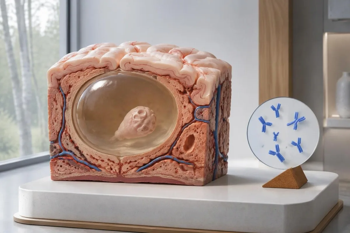
Hướng dẫn của IDSA/ASTMH khuyến nghị xét nghiệm miễn dịch điện di chuyển liên kết với enzym, hay EITB, làm xét nghiệm huyết thanh xác nhận và cảnh báo rõ rằng độ nhạy kém khi chỉ có một tổn thương nhu mô hoặc chỉ có vôi hóa (White và cộng sự, 2018). Không nên áp dụng độ nhạy cao được báo cáo ở bệnh nhân có nhiều nang, bệnh ở não thất hoặc bệnh dưới nhện cho người chỉ có 1 bất thường nhỏ trong não.
Hãy hình dung, chỉ nhằm mục đích minh họa, rằng một xét nghiệm có độ nhạy 55% ở 100 người mắc bệnh chỉ có một nang đã được xác định độc lập. Khoảng 45 người sẽ có kết quả âm tính dù mắc bệnh; ví dụ này minh họa việc bỏ sót phát hiện, chứ không cho biết xác suất bệnh ở bất kỳ người cụ thể nào có kết quả âm tính.
Lặp lại cùng một xét nghiệm sau 2 tuần có thể không khắc phục được đáp ứng yếu do gánh nặng bệnh thấp. Điều này khác với các bệnh nhiễm trùng mà vấn đề chính là lấy mẫu quá sớm, như được giải thích trong hướng dẫn về âm tính giả liên quan đến thời điểm xét nghiệm của chúng tôi; hãy hỏi liệu chẩn đoán hình ảnh hoặc một xét nghiệm được đánh giá rõ hơn có làm thay đổi quyết định tiếp theo hay không.
Vì sao bệnh ấu trùng sán dây lợn ở thần kinh trung ương đã vôi hóa vẫn có thể gây triệu chứng?
Bệnh sán dây thần kinh có vôi hóa có thể gây co giật dù kháng thể âm tính, because the remaining mineralized focus can still be associated with local irritation and episodic surrounding edema. Typical calcifications may measure approximately 1–4 mm, but their small size does not establish whether they are clinically harmless.
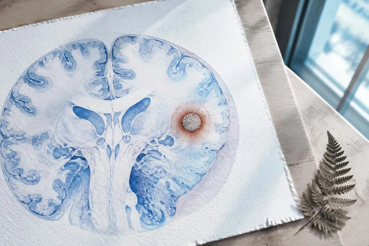
A calcification is generally a remnant rather than a living cyst that antiparasitic medication can eliminate. In the IDSA/ASTMH guideline, calcified-only parenchymal disease is managed symptomatically rather than with antiparasitic drugs; a person with 1 calcified focus and seizures may therefore need antiseizure treatment without needing cyst-killing medication (White et al., 2018).
Surrounding edema can appear intermittently, sometimes long after the original cyst has degenerated. MRI may show this tissue response even when the calcification is more obvious on CT, which is why the 2 scans can tell different but compatible stories rather than one proving that the other is wrong.
Neither a negative antibody result nor a persistent positive result measures seizure risk by itself. The broader lesson resembles other seronegative neurological conditions, discussed in our negative neurological antibody results guide: absence of detectable antibodies is not equivalent to absence of a clinically relevant neurological process.
Thực tế đã thực hiện xét nghiệm kháng thể nào đối với bệnh ấu trùng sán dây lợn?
The useful first question is whether the report describes EITB, ELISA, or another antibody method, because these are not interchangeable tests. A negative index below 1 laboratory's cutoff cannot inherit the performance figures of a different reference-laboratory assay, even when both reports use the name cysticercosis IgG.
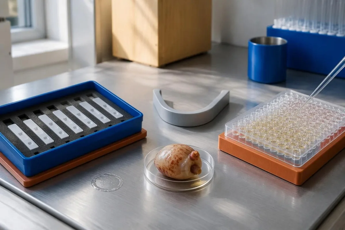
EITB using purified parasite glycoproteins is the preferred serological method in the IDSA/ASTMH guideline, while crude-antigen ELISA methods have recognized sensitivity and specificity limitations (White et al., 2018). Before comparing 2 results, check the method, specimen type, reference interval, and whether the laboratory used a validated confirmatory procedure.
Some laboratories report a qualitative result; others provide an optical-density ratio or numerical index. A value such as 0.8 in a hypothetical assay does not represent 0.8 cysts, an 80% disease probability, or a universal severity score—our kết quả định tính và định lượng guide explains that distinction.
Kantesti AI's role is to help readers preserve the laboratory's original wording rather than reinterpret 1 method through another method's thresholds. Our hướng dẫn xét nghiệm trong phòng thí nghiệm provides broader report-reading context; for this result, keep the actual assay name available when discussing referral or repeat testing.
Tiền sử phơi nhiễm ảnh hưởng thế nào đến mức độ nghi ngờ chẩn đoán?
Neurocysticercosis follows ingestion of Taenia solium eggs, usually through fecal contamination from a person carrying the intestinal tapeworm. Eating undercooked pork can cause intestinal taeniasis, but brain cysts arise through a different transmission step; symptoms may emerge years after exposure rather than within 1 recent trip.

A vegetarian can develop cysticercosis because eggs can contaminate food or hands independently of meat consumption. Useful history includes past residence, household contacts, sanitation conditions, and possible exposure to a tapeworm carrier; asking only about meals eaten during the last 2 weeks can miss the relevant background.
A negative stool examination does not exclude cysts in the brain. Stool testing addresses intestinal tapeworm carriage, whereas CT and MRI assess nervous-system findings; these are 2 different sites and clinical questions, although public-health teams may recommend evaluating contacts when a carrier could be involved.
Normal eosinophils also cannot exclude neurocysticercosis, and raised eosinophils do not identify the brain as the affected site. Our parasite antibody interpretation guide discusses a separate parasite with similar localization limits; the practical point is to avoid using 1 routine cell count as a substitute for targeted neurological evaluation.
Khi nào cơn co giật mới xuất hiện cần được đánh giá cấp cứu?
A first known seizure needs urgent medical assessment regardless of antibody results, and emergency services should be contacted for a first seizure or an ongoing seizure with concerning features. A convulsive seizure lasting 5 minutes, repeated seizures without recovery, breathing difficulty, pregnancy, or significant injury requires emergency care.

Do not wait for the 5-minute mark to seek help after a first known seizure. Keep the person away from hazards, cushion the head if safely possible, do not restrain them or put anything in their mouth, and place them on their side once convulsions stop if they are breathing normally.
Persistent confusion, a severe new headache, fever with neck stiffness, or new weakness increases concern for causes beyond cysticercosis. Sudden one-sided weakness or speech difficulty also demands emergency assessment, as our hướng dẫn về các dấu hiệu cảnh báo đột quỵ explains; 1 negative infection test cannot rule out stroke, meningitis, or a structural brain problem.
After a suspected seizure, stop driving and avoid swimming alone, heights, or operating machinery until a clinician provides advice consistent with local rules. If a witness can safely record the duration and describe movements or recovery, those observations may be more useful than ordering a second antibody test before the first neurological assessment.
CT bổ sung điều gì cho chẩn đoán bệnh ấu trùng sán dây lợn ở thần kinh trung ương?
Noncontrast CT is particularly useful for detecting calcified cyst remnants, including small approximately 1–4 mm foci that MRI may show less clearly. CT also helps assess urgent structural problems such as hydrocephalus, but 1 isolated calcification is not automatically proof of neurocysticercosis or the cause of a seizure.
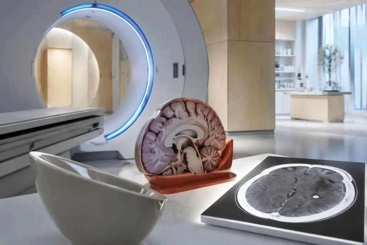
Calcium appears dense on CT, allowing radiologists to identify mineralized remnants and describe their distribution. The IDSA/ASTMH guideline recommends both MRI and noncontrast CT when classifying newly diagnosed neurocysticercosis, because the 2 modalities have complementary strengths rather than being interchangeable alternatives (White et al., 2018).
An incidental calcification needs context: previous granulomatous disease, other causes of mineralization, and scan appearance can influence interpretation. If 1 calcification sits near a suspected seizure focus, concordance with symptoms and EEG may strengthen the explanation, but anatomical proximity alone cannot establish causation.
A laboratory marker used in selected head-injury pathways does not test for brain cysts or replace imaging after an unexplained seizure. Our head-injury CT decisions guide illustrates that narrower setting; ask the clinician whether the present question requires noncontrast CT, contrast imaging, MRI, or a combination of 2 modalities.
MRI phát hiện điều gì mà CT có thể bỏ sót?
MRI is better suited to evaluating viable cysts, surrounding edema, and many ventricular or subarachnoid findings. Specialized 3-dimensional sequences such as CISS or FIESTA can improve visualization of cyst membranes within cerebrospinal fluid spaces, where a cyst may otherwise resemble the surrounding fluid.
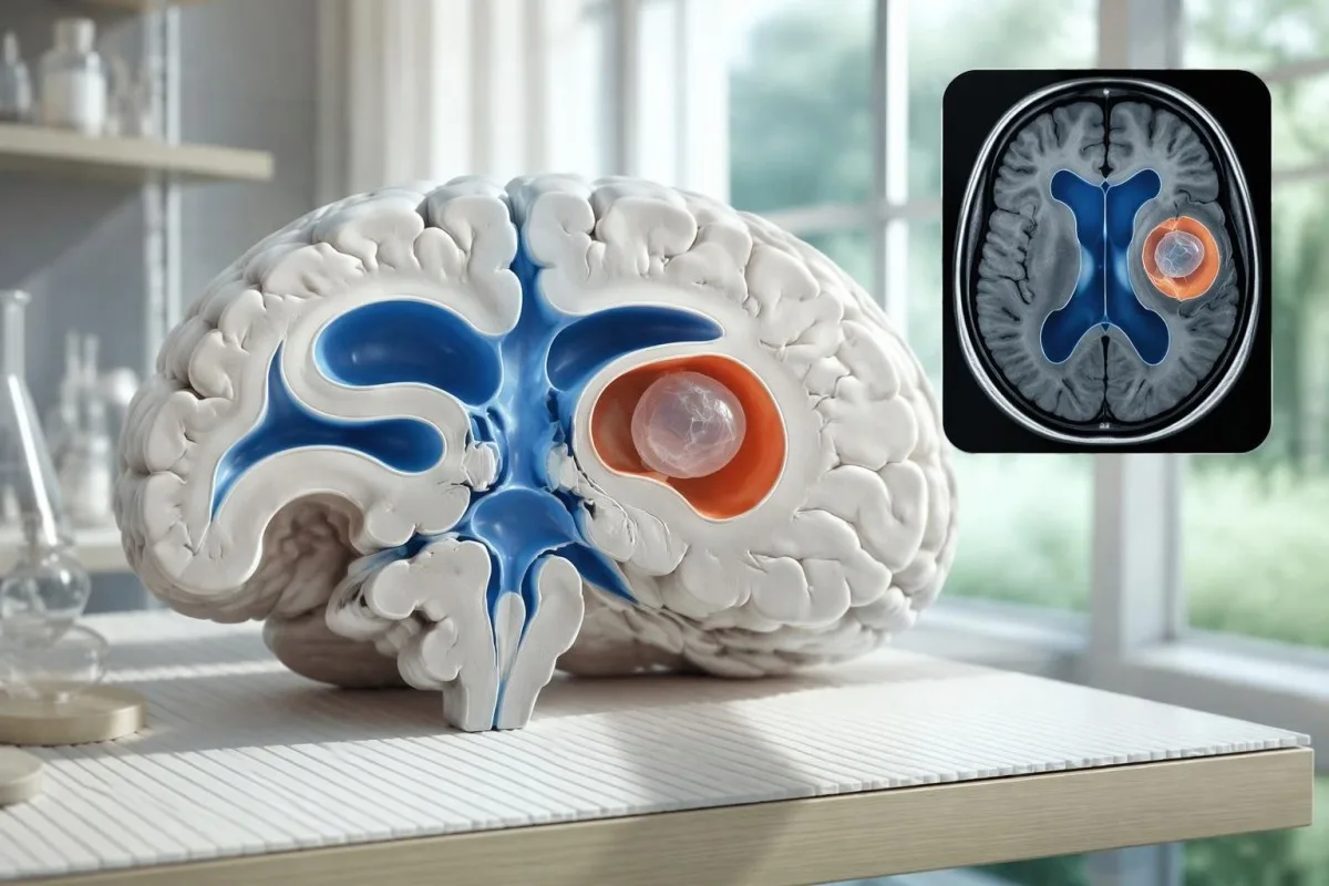
A cyst inside the ventricles can obstruct cerebrospinal fluid flow and cause hydrocephalus even without numerous parenchymal cysts. Headache with vomiting, declining alertness, or new visual symptoms therefore changes urgency; 1 reassuring antibody result should not override symptoms that suggest raised pressure or impaired fluid circulation.
MRI may show a scolex, the small internal structure of the parasite, inside a compatible cyst. This finding can be highly specific when confidently identified, whereas ring enhancement alone has a broader differential; our brain imaging warning signs guide explains why structural abnormalities require interpretation rather than diagnosis from 1 visual feature.
A routine MRI protocol may not answer every cysticercosis question. If ventricular or subarachnoid disease is suspected, discuss whether thin-section 3-dimensional imaging is needed; radiologists choose sequences and contrast according to the clinical question, and a normal scan using 1 protocol does not prove every possible disease compartment has been adequately assessed.
Chẩn đoán bệnh ấu trùng sán dây lợn ở thần kinh trung ương như thế nào khi các xét nghiệm không thống nhất?
Neurocysticercosis diagnosis combines imaging, clinical findings, exposure information, and supporting tests, rather than requiring a positive antibody result in every case. The revised diagnostic criteria published in 2017 give characteristic neuroimaging findings a central role and distinguish strong evidence from less-specific supportive features (Del Brutto et al., 2017).
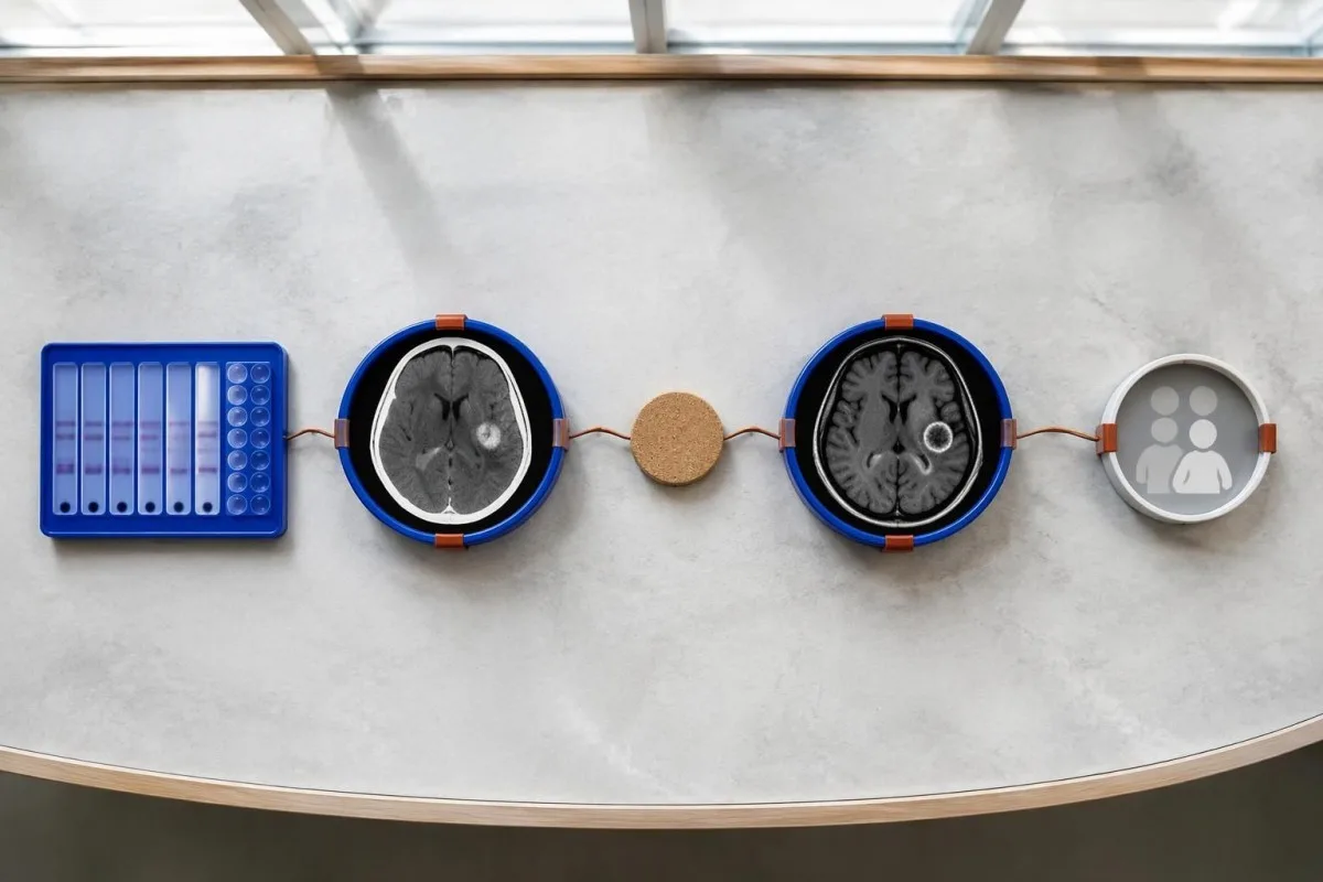
Evidence should be weighted, not tallied. A confidently visualized scolex inside a compatible cyst is different from 1 nonspecific ring-enhancing area, while a positive antibody result is different from direct evidence that a finding is in the brain; our kết quả kháng thể dương tính explains why detection alone cannot establish location.
For a clearly hypothetical probability example, start with a 30% pretest probability and assume a single-cyst assay has 55% sensitivity and 99% specificity. In 1,000 comparable people, about 135 affected and 693 unaffected people would test negative, leaving approximately a 16% probability of disease among negative results—still substantial, although these assumed figures are not a personal risk calculator.
The most useful disagreement to investigate is whether each test is answering the same question. A negative antibody test, a CT calcification, and MRI edema can all be compatible with 1 calcified disease process; unexplained discrepancies should prompt review of the assay and images rather than automatic dismissal of either result.
Những bệnh lý nào khác có thể giống một nang đơn độc trên hình ảnh não?
A single ring-enhancing brain finding is not specific to neurocysticercosis. Other possibilities include tuberculoma, an abscess, a neoplasm, or an inflammatory process; clinicians use appearance, location, immune status, symptoms, and sometimes follow-up imaging or tissue examination to distinguish these possibilities rather than relying on 1 antibody panel.
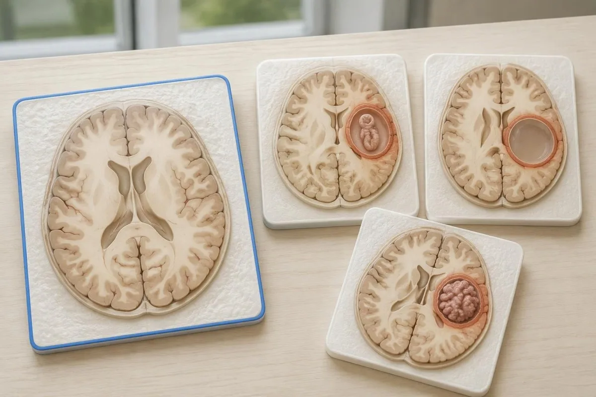
Edema, restricted diffusion, internal structures, and the number of findings all contribute to the differential, but no single nonspecific feature settles every case. Fever and immune suppression may change the suspected cause; a 2 cm ring-enhancing area in a hypothetical immunocompromised person warrants a different investigation from a tiny calcification in an otherwise well person.
Toxoplasmosis is one consideration in selected immunocompromised patients, and a positive IgG result can indicate past exposure rather than prove an active brain process. Our Toxoplasma IgG interpretation guide explains that laboratory distinction; imaging and the patient's immune status determine whether the result is clinically relevant to 1 or more cerebral findings.
New seizures accompanied by rapidly evolving memory problems, psychiatric changes, or abnormal movements can also suggest encephalitis rather than a solitary parasite cyst. Our encephalitis diagnostic pathway describes how MRI, EEG, and cerebrospinal fluid testing answer different questions; a second negative cysticercosis antibody result does not investigate those alternatives.
Xét nghiệm kháng nguyên, PCR hoặc dịch não tủy có thể giải quyết điều chưa chắc chắn không?
Antigen tests, PCR, and cerebrospinal fluid analysis can add information in selected cases, particularly when extraparenchymal disease is suspected. None is a universal rescue test after 1 negative antibody result: availability, assay validation, disease compartment, and safety of lumbar puncture determine whether additional testing is appropriate.
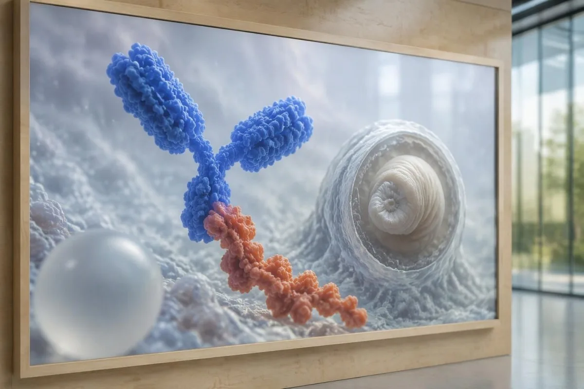
Antibody testing detects the host response, while antigen testing aims to detect parasite-derived material and may better reflect viable disease burden. Antigen detection is often less useful when only 1 small parenchymal cyst or calcifications are present; molecular testing is used in some specialist settings, but performance cannot be generalized across every laboratory or specimen.
Lumbar puncture is not routinely required for an uncomplicated solitary parenchymal finding and can be unsafe when obstructive hydrocephalus or significant mass effect is present. Cerebrospinal fluid tests for other disorders also require disease-specific interpretation, as our CSF band interpretation guide explains; 1 abnormal CSF marker does not identify every possible cause.
Kantesti là một nền tảng giải thích kết quả xét nghiệm máu AI, not a service that orders lumbar punctures, identifies cysts on scans, or confirms neurological infections. Our công nghệ diễn giải AI của chúng tôi explains the laboratory-report workflow; when 2 tests disagree, our role is to clarify their stated meanings and limitations, not select an invasive diagnostic procedure.
Vì sao việc điều trị phụ thuộc vào giai đoạn và vị trí của nang?
Treatment depends on whether cysts are viable, degenerating, calcified, or located outside the brain tissue, not simply on antibody positivity. Selected viable parenchymal cases receive antiparasitic treatment, whereas calcified-only disease generally receives symptom-directed care; ventricular obstruction may require a procedural approach rather than 1 standard medication regimen.
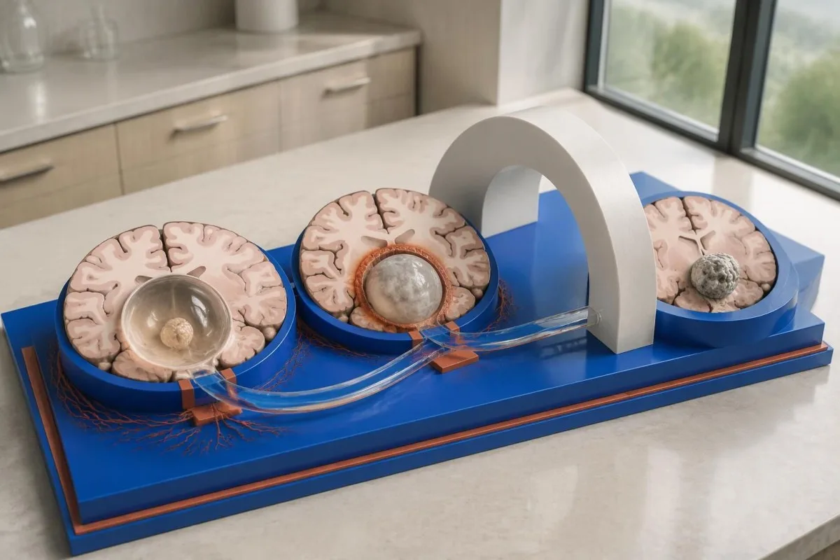
For selected patients with 1–2 viable parenchymal cysts, the IDSA/ASTMH guideline describes albendazole treatment for 10–14 days with appropriate accompanying care. This is a specialist-directed recommendation, not a home-treatment instruction: antiparasitic drugs can provoke an inflammatory response, and untreated hydrocephalus or diffuse cerebral edema changes the order of management (White et al., 2018).
WHO's 2021 guidance focuses on defined presentations of parenchymal neurocysticercosis, while the IDSA/ASTMH guideline also addresses ventricular, subarachnoid, and other forms in detail. Differences in imaging access and disease compartment explain why 1 treatment recommendation cannot be transferred to every patient with an antibody result; the guidelines do not turn negative serology into a rule-out test.
Corticosteroids may accompany antiparasitic therapy, but prolonged use requires additional safety planning, including assessment for relevant latent infections. Our Strongyloides and steroid safety guide explains a separate risk that matters before immune-suppressing treatment; an eye examination is also recommended before antiparasitic therapy to evaluate possible intraocular cysts.
Việc theo dõi nên kiểm tra những gì ngoài kết quả kháng thể?
Follow-up should track seizures, neurological symptoms, imaging changes, and medication safety, rather than use antibody disappearance as proof of recovery. For viable parenchymal cysts, the IDSA/ASTMH guideline suggests repeat MRI at least every 6 months until the cystic component resolves, with earlier reassessment when symptoms change.
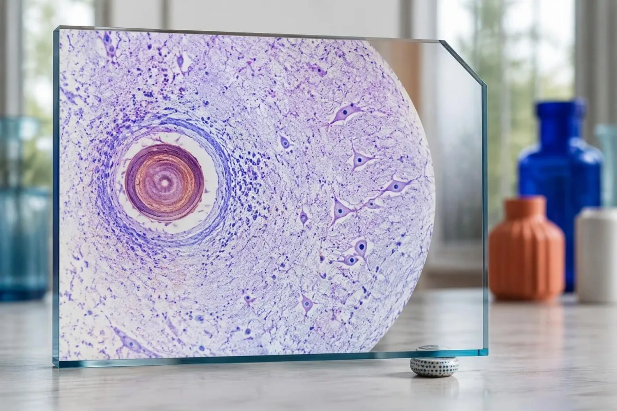
Antibodies may remain detectable after the original disease process has changed, and low-burden disease may never produce a positive result. If 1 new seizure occurs during follow-up, clinicians consider adherence, medication interactions, recurrent edema, and other causes; a rising or falling antibody index alone cannot distinguish among those explanations.
Kantesti AI can help explain reported laboratory results during medication follow-up, but a drug concentration is not a stand-alone measure of seizure control. Our diễn giải nồng độ levetiracetam guide explains why sample timing and kidney function matter; 2 measurements collected at different times after dosing may not be directly comparable.
Monitoring depends on the chosen drug and treatment duration: prolonged albendazole therapy can require liver-test and blood-count surveillance, while some antiseizure medicines can affect sodium. Our 电解质安全指南 discusses those separate laboratory concerns; do not stop an antiseizure medicine or change its dose because 1 cysticercosis antibody result is negative.
Kantesti có thể giải thích điều gì, và nghiên cứu không thể chứng minh điều gì?
AI report interpretation cannot confirm or exclude neurocysticercosis from laboratory results alone. Kantesti là một Công cụ phân tích xét nghiệm máu dựa trên AI whose role here is to organize reported laboratory findings; a negative result still needs clinical context, and 1 new seizure takes priority over reviewing an automated explanation.

Prepare a focused review packet with the original antibody report, assay method, CT or MRI report, symptom timeline, and medication list. These 5 information categories reduce the risk of discussing a result without its context; ask specifically whether the imaging suggests viable, degenerating, calcified, ventricular, or subarachnoid disease and which unanswered question the next test would address.
The 2 Kantesti publications listed below are a repository-deposited global analysis report and an educational RDW guide, not clinical evidence validating neurocysticercosis diagnosis. Our RDW và CBC đi kèm can support interpretation of a separately ordered blood count, but RDW, MCV, and MCHC do not establish whether a brain cyst is present.
Evidence for the neurological recommendations in this article comes from the cited diagnostic criteria and clinical guidelines, not from general AI benchmarking or laboratory-report volume. Our technical validation scope should be read within its stated boundaries; neither 1 successful report explanation nor a large aggregate dataset substitutes for condition-specific diagnostic validation.
Câu hỏi thường gặp
Bạn có thể bị bệnh ấu trùng sán dây lợn ở hệ thần kinh trung ương dù xét nghiệm máu cho kết quả âm tính không?
Đúng vậy, bệnh ấu trùng sán dây lợn ở hệ thần kinh vẫn có thể xuất hiện dù xét nghiệm kháng thể trong máu cho kết quả âm tính, đặc biệt khi chỉ có 1 nang não hoặc bệnh chỉ có tổn thương vôi hóa. Các ước tính được công bố từ các phòng xét nghiệm tham chiếu về EITB thường cho thấy độ nhạy khoảng 50–60% đối với trường hợp chỉ có một nang, thấp hơn đáng kể so với khi có nhiều nang còn sống. Vì vậy, CT, MRI, triệu chứng và tiền sử phơi nhiễm vẫn đóng vai trò then chốt trong chẩn đoán. Cơn co giật mới khởi phát cần được đánh giá khẩn cấp, thay vì yên tâm chỉ vì kết quả âm tính.
Xét nghiệm bệnh ấu trùng sán dây lợn thần kinh nào đáng tin cậy nhất?
Không có xét nghiệm đơn lẻ nào về bệnh ấu trùng sán dây lợn thần kinh có thể trả lời mọi câu hỏi chẩn đoán; chẩn đoán hình ảnh não và xét nghiệm huyết thanh được lựa chọn phù hợp có vai trò bổ trợ cho nhau. EITB là phương pháp kháng thể xác nhận được ưu tiên trong hướng dẫn IDSA/ASTMH, nhưng có thể bỏ sót bệnh chỉ liên quan đến 1 nang. CT không tiêm thuốc cản quang đặc biệt hữu ích để phát hiện vôi hóa, trong khi MRI đánh giá tốt hơn nhiều nang còn sống, phù não và các bất thường khoang dịch. Bác sĩ kết hợp những phát hiện này thay vì xem một kết quả âm tính là kết luận cuối cùng.
Chụp CT hay MRI tốt hơn để chẩn đoán bệnh ấu trùng sán dây lợn thần kinh?
CT và MRI có những ưu thế khác nhau trong chẩn đoán bệnh ấu trùng sán dây lợn thần kinh. CT không tiêm thuốc cản quang thường phát hiện các nốt vôi hóa nhỏ, đường kính khoảng 1–4 mm, rõ hơn, trong khi MRI đánh giá các nang còn sống, phù xung quanh và bệnh lý ở não thất hoặc khoang dưới nhện. Các chuỗi xung MRI 3 chiều chuyên biệt có thể giúp xác định màng nang trong các khoang chứa dịch não tủy. Hướng dẫn của IDSA/ASTMH khuyến nghị sử dụng cả hai phương thức khi phân loại bệnh mới được chẩn đoán.
U nang vôi hóa vẫn có thể gây co giật không?
Di tích nang sán đã vôi hóa vẫn có thể liên quan đến các cơn co giật dù không còn là nang sống. Một số ổ vôi hóa, thường chỉ rộng 1–4 mm, xuất hiện phù nề quanh ổ từng đợt và có thể thấy trên MRI. Kết quả kháng thể âm tính không loại trừ cơ chế này, còn kháng thể dương tính dai dẳng không phản ánh nguy cơ co giật. Bệnh chỉ có tổn thương vôi hóa thường được điều trị theo triệu chứng thay vì dùng thuốc chống ký sinh trùng.
Tôi có nên làm lại xét nghiệm kháng thể bệnh sán dây lợn nếu kết quả âm tính không?
Việc lặp lại xét nghiệm kháng thể bệnh nang sán âm tính không phải lúc nào cũng là bước tiếp theo tốt nhất. Khi chỉ có 1 nang nhỏ hoặc tổn thương vôi hóa, đáp ứng kháng thể yếu có thể vẫn âm tính ngay cả khi lặp lại cùng xét nghiệm sau 2 tuần. Thay vào đó, bác sĩ có thể xem xét lại phương pháp xét nghiệm ban đầu, yêu cầu một xét nghiệm xác nhận được đặc trưng hóa tốt hơn, hoặc ưu tiên chụp CT và MRI. Không nên chờ lặp lại xét nghiệm nếu xuất hiện cơn co giật mới hoặc các triệu chứng thần kinh trở nặng.
Khi nào tôi nên gọi cấp cứu nếu bị co giật lần đầu?
Cơn co giật đầu tiên được ghi nhận cần được đánh giá y tế khẩn cấp và liên hệ với dịch vụ cấp cứu tại địa phương, bất kể kết quả xét nghiệm kháng thể bệnh ấu trùng sán dây lợn ở não âm tính. Cơn co giật co cứng-co giật kéo dài 5 phút, các cơn co giật lặp lại mà người bệnh không tỉnh lại, khó thở, mang thai hoặc chấn thương nghiêm trọng đều là tình trạng cấp cứu. Không ghì giữ người bệnh hoặc cho bất cứ thứ gì vào miệng họ; khi cơn co giật ngừng, hãy đặt họ nằm nghiêng nếu họ thở bình thường. Không lái xe cho đến khi bác sĩ đánh giá sự việc và giải thích các hạn chế áp dụng.
Tìm hiểu cách đọc kết quả xét nghiệm máu của bạn
Tải lên báo cáo xét nghiệm của bạn và AI của Kantesti sẽ giải thích từng dấu ấn sinh học bằng ngôn ngữ dễ hiểu chỉ trong vài giây. Được hơn 2 triệu người trên toàn thế giới sử dụng.
Tài liệu tham khảo
- Klein, T., Mitchell, S., & Weber, H. (2026). Trình phân tích xét nghiệm máu AI: 2,5M xét nghiệm được phân tích | Báo cáo sức khỏe toàn cầu 2026. Nghiên cứu y khoa của Kantesti AI. DOI
- Klein, T., Mitchell, S., & Weber, H. (2026). Xét nghiệm máu RDW: Hướng dẫn đầy đủ về RDW-CV, MCV và MCHC. Nghiên cứu y khoa của Kantesti AI. DOI
- White AC Jr et al. (2018). Diagnosis and Treatment of Neurocysticercosis: 2017 Clinical Practice Guidelines by the Infectious Diseases Society of America (IDSA) and the American Society of Tropical Medicine and Hygiene (ASTMH). Clinical Infectious Diseases. DOI
- Del Brutto OH et al. (2017). Revised diagnostic criteria for neurocysticercosis. Journal of the Neurological Sciences. DOI
- World Health Organization (2021). WHO guidelines on management of Taenia solium neurocysticercosis. Tổ chức Y tế Thế giới.
- 2 triệu+Các xét nghiệm được phân tích
- 127+Các quốc gia
- 100+Ngôn ngữ
Về đội ngũ y tế


Tín hiệu tin cậy E-E-A-T
Kinh nghiệm
Đánh giá lâm sàng do bác sĩ phụ trách đối với quy trình diễn giải kết quả xét nghiệm.
Chuyên môn
Tập trung vào y học xét nghiệm: cách các chỉ dấu sinh học (biomarker) hoạt động trong bối cảnh lâm sàng.
Tính uy quyền
Được Tiến sĩ Thomas Klein đánh giá về mặt y khoa, với sự đóng góp của Tiến sĩ Sarah Mitchell và Giáo sư, Tiến sĩ Hans Weber.
Độ tin cậy
Diễn giải dựa trên bằng chứng, kèm các lộ trình theo dõi rõ ràng để giảm mức độ báo động.





Tuyên bố miễn trừ trách nhiệm y tế
Bài viết này chỉ nhằm mục đích giáo dục và không cấu thành lời khuyên y tế. Luôn tham khảo ý kiến của nhà cung cấp dịch vụ chăm sóc sức khỏe đủ năng lực để đưa ra quyết định chẩn đoán và điều trị.
Đã xuất bản · Liên hệ với ban biên tập
Xuất bản bởi Kantesti Ltd, đăng ký tại Anh và xứ Wales, số công ty. 17090423, London, Vương quốc Anh.
