
In negative test foar neurocysticerkose slút ien harsenscyste of ferkalke sykte net út. CT, MRI en urgente beoardieling fan nije krampen binne wichtiger as gerêststelling op basis fan allinnich antykoadetests.

Wichtichste punten
- Negative antykodearring slute neurocysticerkose net út, benammen as de ôfbylding mar 1 troffen gebiet of ferkalkingen sjen lit.
- EITB fan in referinsjelaboratoarium kin in gefoelichheid fan hast 98% berikke by mear as 1 libbene cyste, mar de prestaasje is flink leger by ien cyste.
- Gefoelichheid by ien cyste is neffens publisearre EITB-skattingen faak likernôch 50–60%; dizze sifers jilde net foar elke kommersjele antykoadetest.
- CT sûnder kontrast is benammen nuttich foar lytse ferkalkingen, dy't faak likernôch 1–4 mm grut binne.
- MRI beoardielet libbene cysten, omlizzend oedeem en sykte yn de ventrikels of subarachnoïdale romte; spesjalisearre 3-diminsjonale sekwinsjes kinne de opspoaring ferbetterje.
- Earste krampen fereaskje in driuwende medyske beoardieling, ûnôfhinklik fan it antistofferesultaat. Belje de helptsjinsten by in earste bekende oanfal of oare needtekenen.
- Fiif minuten is in needdrompel foar in oanhâldende krampachtige oanfal, net in reden om te wachtsjen mei help sykjen.
- Allinnich ferkalkte sykte hat yn ’t algemien gjin antiparasitêre medikaasje nedich, mar behanneling fan de oanfal en fierdere ôfbylding kinne noch wol nedich wêze.
Kin in negative test foar neurocysticerkose de sykte útslute?
A in negative test op neurocysticercose kin de sykte net betrouber útslute, benammen by 1 harsenscyste of ferkalke resten. Antistoffetesten mjitte de ymmúnreaksje, ynstee fan it brein direkt te ûndersykjen; CT en MRI bepale de lokaasje en it stadium fan fertochte befiningen, wylst in earste oanfal driuwende beoardieling fereasket.

De útslach is betingstlik, net absolút. In antistoftest foar cysticercose dy't goed wurket by minsken mei ferskate libbensfetbere cysten, kin min wurkje by ien mei mar 1 lyts troffen gebiet, dus it wurd negative betsjut dat der mei dy test gjin reaksje oantoand is—net dat der nearne yn it senuwstelsel cysten binne.
Beskôgje as hypotetysk foarbyld in 34-jierrige mei in earste oanfal, in negatyf antistofferapport en in iensume ringfoarmich kontrastopladende fynst op MRI. It rapport kin net fêststelle oft dy fynst cysticercose, in oare ynfeksje of in net-ynfektyf proses fertsjintwurdiget; de betsjutting hinget ôf fan de scan, de bleatstellingsskiednis en de neurologyske beoardieling mei-inoar.
Kantesti is in AI bloedtestanalysator dy't helpe kin om rapportearre laboratoariumfynsten te ferklearjen, mar harsensykte net útslute kin op grûn fan 1 negatyf antistofresultaat. Us organisaasje en missy ferklearje ús fokus op laboratoariumynformaasje; lêzers moatte in automatyske útlis fan in rapport net beskôgje as tastimming om de soarch by in oanfal út te stellen.
Wêrom kin antykoadetesting in inkele harsenscyste misse?
A in iensume cyste kin in antistofreaksje feroarsaakje dy't ûnder de deteksjedrompel fan de test leit. Publisearre rûzings fan EITB by referinsjelaboratoaria pleatse de gefoelichheid faak ticht by 50–60% foar in iensume cyste, yn ferliking mei likernôch 98% as der mear as 1 libbensfetbere cyste oanwêzich is; it ferskil wjerspegelet de syktelêst en de prestaasjes fan de test.
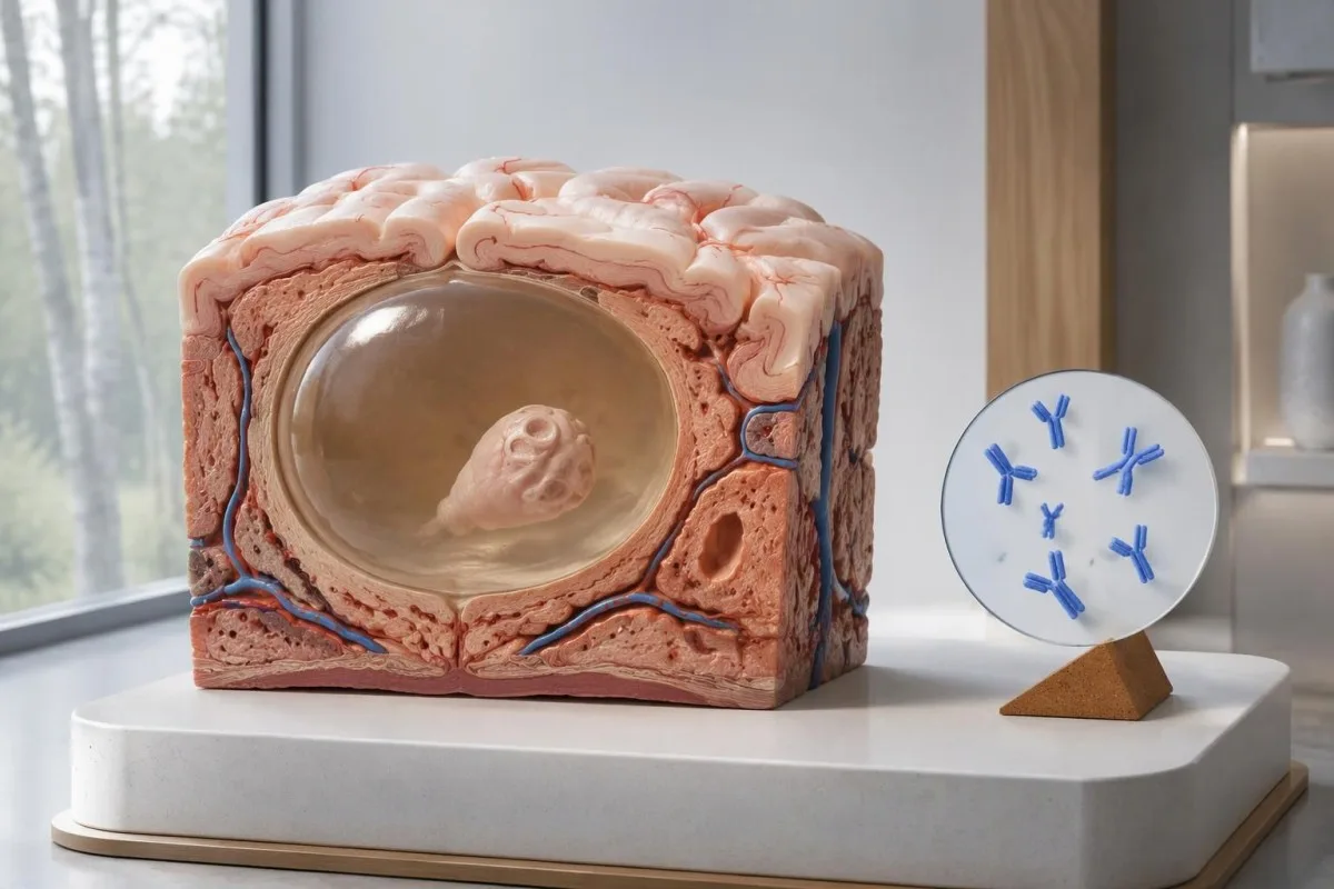
De rjochtline fan IDSA/ASTMH advisearret enzyme-linked immunoelectrotransfer blot, of EITB, as befêstigjende serology en warskôget eksplisyt dat de gefoelichheid min is by mar ien fynst yn it harsensweefsel of allinnich ferkalkingen (White et al., 2018). De hege gefoelichheid dy't rapportearre is by pasjinten mei meardere cysten, sykte yn de harsensfentrikels of subarachnoïdale sykte, mei net tapast wurde op ien mei 1 lytse harsensôfwiking.
Stel jo, inkeld as yllustraasje, foar dat in test 55% gefoelichheid hat by 100 minsken by wa't sykte troch ien cyste al ûnôfhinklik fêststeld is. Likernôch 45 soene negatyf teste, hoewol't se de oandwaning hawwe; dit útwurke foarbyld ferklearret miste deteksje, net de kâns dat in bepaald negatyf resultaat op sykte wiist.
Deselde test nei 2 wiken werhelje kin in swakke reaksje troch in lege syktelêst miskien net ferbetterje. Dat ferskilt fan ynfeksjes wêrby't in betide ôfname it wichtichste probleem is, lykas útlein yn ús hantlieding foar falsk-negative útslaggen yn ferbân mei timing; freegje oft ôfbylding of in better karakterisearre test de folgjende beslissing feroarje soe.
Wêrom kin ferkalke neurocysticerkose symptomatysk bliuwe?
Ferkealke neurocysticercose kin oanfallen feroarsaakje nettsjinsteande negative antistoffen, because the remaining mineralized focus can still be associated with local irritation and episodic surrounding edema. Typical calcifications may measure approximately 1–4 mm, but their small size does not establish whether they are clinically harmless.
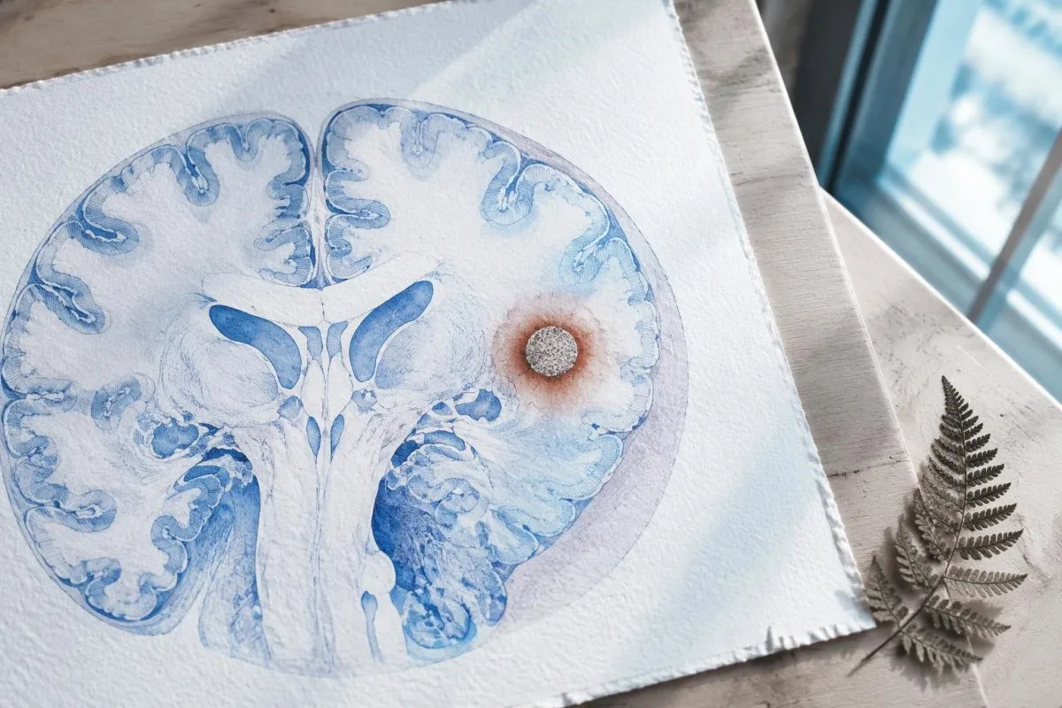
A calcification is generally a remnant rather than a living cyst that antiparasitic medication can eliminate. In the IDSA/ASTMH guideline, calcified-only parenchymal disease is managed symptomatically rather than with antiparasitic drugs; a person with 1 calcified focus and seizures may therefore need antiseizure treatment without needing cyst-killing medication (White et al., 2018).
Surrounding edema can appear intermittently, sometimes long after the original cyst has degenerated. MRI may show this tissue response even when the calcification is more obvious on CT, which is why the 2 scans can tell different but compatible stories rather than one proving that the other is wrong.
Neither a negative antibody result nor a persistent positive result measures seizure risk by itself. The broader lesson resembles other seronegative neurological conditions, discussed in our negative neurological antibody results guide: absence of detectable antibodies is not equivalent to absence of a clinically relevant neurological process.
Hokker antykoadetest foar cysticerkose is eins útfierd?
The useful first question is whether the report describes EITB, ELISA, or another antibody method, because these are not interchangeable tests. A negative index below 1 laboratory's cutoff cannot inherit the performance figures of a different reference-laboratory assay, even when both reports use the name cysticercosis IgG.
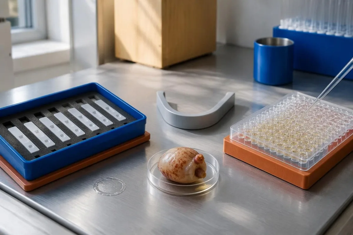
EITB using purified parasite glycoproteins is the preferred serological method in the IDSA/ASTMH guideline, while crude-antigen ELISA methods have recognized sensitivity and specificity limitations (White et al., 2018). Before comparing 2 results, check the method, specimen type, reference interval, and whether the laboratory used a validated confirmatory procedure.
Some laboratories report a qualitative result; others provide an optical-density ratio or numerical index. A value such as 0.8 in a hypothetical assay does not represent 0.8 cysts, an 80% disease probability, or a universal severity score—our kwalitative en kwantitative resultaten guide explains that distinction.
Kantesti AI's role is to help readers preserve the laboratory's original wording rather than reinterpret 1 method through another method's thresholds. Our laboratoariummarkeergids provides broader report-reading context; for this result, keep the actual assay name available when discussing referral or repeat testing.
Hoe feroaret de bleatstellingsskiednis de diagnostyske fertinking?
Neurocysticercosis follows ingestion of Taenia solium eggs, usually through fecal contamination from a person carrying the intestinal tapeworm. Eating undercooked pork can cause intestinal taeniasis, but brain cysts arise through a different transmission step; symptoms may emerge years after exposure rather than within 1 recent trip.

A vegetarian can develop cysticercosis because eggs can contaminate food or hands independently of meat consumption. Useful history includes past residence, household contacts, sanitation conditions, and possible exposure to a tapeworm carrier; asking only about meals eaten during the last 2 weeks can miss the relevant background.
A negative stool examination does not exclude cysts in the brain. Stool testing addresses intestinal tapeworm carriage, whereas CT and MRI assess nervous-system findings; these are 2 different sites and clinical questions, although public-health teams may recommend evaluating contacts when a carrier could be involved.
Normal eosinophils also cannot exclude neurocysticercosis, and raised eosinophils do not identify the brain as the affected site. Our parasite antibody interpretation guide discusses a separate parasite with similar localization limits; the practical point is to avoid using 1 routine cell count as a substitute for targeted neurological evaluation.
Wannear moatte nije krampen mei spoed beoardiele wurde?
A first known seizure needs urgent medical assessment regardless of antibody results, and emergency services should be contacted for a first seizure or an ongoing seizure with concerning features. A convulsive seizure lasting 5 minutes, repeated seizures without recovery, breathing difficulty, pregnancy, or significant injury requires emergency care.

Do not wait for the 5-minute mark to seek help after a first known seizure. Keep the person away from hazards, cushion the head if safely possible, do not restrain them or put anything in their mouth, and place them on their side once convulsions stop if they are breathing normally.
Persistent confusion, a severe new headache, fever with neck stiffness, or new weakness increases concern for causes beyond cysticercosis. Sudden one-sided weakness or speech difficulty also demands emergency assessment, as our gids foar warskôgingstekens fan in beroerte explains; 1 negative infection test cannot rule out stroke, meningitis, or a structural brain problem.
After a suspected seizure, stop driving and avoid swimming alone, heights, or operating machinery until a clinician provides advice consistent with local rules. If a witness can safely record the duration and describe movements or recovery, those observations may be more useful than ordering a second antibody test before the first neurological assessment.
Wat draacht CT by oan de diagnoaze fan neurocysticerkose?
Noncontrast CT is particularly useful for detecting calcified cyst remnants, including small approximately 1–4 mm foci that MRI may show less clearly. CT also helps assess urgent structural problems such as hydrocephalus, but 1 isolated calcification is not automatically proof of neurocysticercosis or the cause of a seizure.
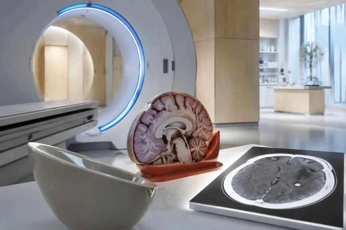
Calcium appears dense on CT, allowing radiologists to identify mineralized remnants and describe their distribution. The IDSA/ASTMH guideline recommends both MRI and noncontrast CT when classifying newly diagnosed neurocysticercosis, because the 2 modalities have complementary strengths rather than being interchangeable alternatives (White et al., 2018).
An incidental calcification needs context: previous granulomatous disease, other causes of mineralization, and scan appearance can influence interpretation. If 1 calcification sits near a suspected seizure focus, concordance with symptoms and EEG may strengthen the explanation, but anatomical proximity alone cannot establish causation.
A laboratory marker used in selected head-injury pathways does not test for brain cysts or replace imaging after an unexplained seizure. Our head-injury CT decisions guide illustrates that narrower setting; ask the clinician whether the present question requires noncontrast CT, contrast imaging, MRI, or a combination of 2 modalities.
Wat lit MRI sjen dat CT miskien mist?
MRI is better suited to evaluating viable cysts, surrounding edema, and many ventricular or subarachnoid findings. Specialized 3-dimensional sequences such as CISS or FIESTA can improve visualization of cyst membranes within cerebrospinal fluid spaces, where a cyst may otherwise resemble the surrounding fluid.
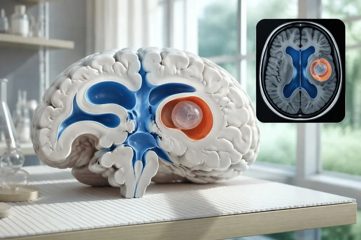
A cyst inside the ventricles can obstruct cerebrospinal fluid flow and cause hydrocephalus even without numerous parenchymal cysts. Headache with vomiting, declining alertness, or new visual symptoms therefore changes urgency; 1 reassuring antibody result should not override symptoms that suggest raised pressure or impaired fluid circulation.
MRI may show a scolex, the small internal structure of the parasite, inside a compatible cyst. This finding can be highly specific when confidently identified, whereas ring enhancement alone has a broader differential; our brain imaging warning signs guide explains why structural abnormalities require interpretation rather than diagnosis from 1 visual feature.
A routine MRI protocol may not answer every cysticercosis question. If ventricular or subarachnoid disease is suspected, discuss whether thin-section 3-dimensional imaging is needed; radiologists choose sequences and contrast according to the clinical question, and a normal scan using 1 protocol does not prove every possible disease compartment has been adequately assessed.
Hoe wurdt neurocysticerkose diagnostisearre as testresultaten inoar tsjinsprekke?
Neurocysticercosis diagnosis combines imaging, clinical findings, exposure information, and supporting tests, rather than requiring a positive antibody result in every case. The revised diagnostic criteria published in 2017 give characteristic neuroimaging findings a central role and distinguish strong evidence from less-specific supportive features (Del Brutto et al., 2017).
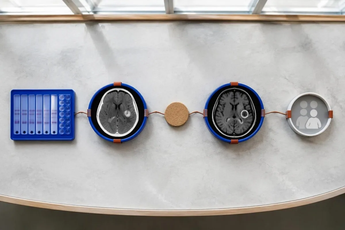
Evidence should be weighted, not tallied. A confidently visualized scolex inside a compatible cyst is different from 1 nonspecific ring-enhancing area, while a positive antibody result is different from direct evidence that a finding is in the brain; our posityf antyklodyresultaatgids explains why detection alone cannot establish location.
For a clearly hypothetical probability example, start with a 30% pretest probability and assume a single-cyst assay has 55% sensitivity and 99% specificity. In 1,000 comparable people, about 135 affected and 693 unaffected people would test negative, leaving approximately a 16% probability of disease among negative results—still substantial, although these assumed figures are not a personal risk calculator.
The most useful disagreement to investigate is whether each test is answering the same question. A negative antibody test, a CT calcification, and MRI edema can all be compatible with 1 calcified disease process; unexplained discrepancies should prompt review of the assay and images rather than automatic dismissal of either result.
Wat oars kin op harsenôfbylding op in inkele cyste lykje?
A single ring-enhancing brain finding is not specific to neurocysticercosis. Other possibilities include tuberculoma, an abscess, a neoplasm, or an inflammatory process; clinicians use appearance, location, immune status, symptoms, and sometimes follow-up imaging or tissue examination to distinguish these possibilities rather than relying on 1 antibody panel.
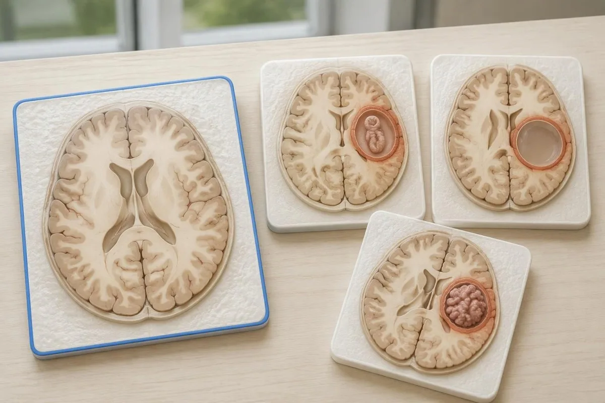
Edema, restricted diffusion, internal structures, and the number of findings all contribute to the differential, but no single nonspecific feature settles every case. Fever and immune suppression may change the suspected cause; a 2 cm ring-enhancing area in a hypothetical immunocompromised person warrants a different investigation from a tiny calcification in an otherwise well person.
Toxoplasmosis is one consideration in selected immunocompromised patients, and a positive IgG result can indicate past exposure rather than prove an active brain process. Our Toxoplasma IgG interpretation guide explains that laboratory distinction; imaging and the patient's immune status determine whether the result is clinically relevant to 1 or more cerebral findings.
New seizures accompanied by rapidly evolving memory problems, psychiatric changes, or abnormal movements can also suggest encephalitis rather than a solitary parasite cyst. Our encephalitis diagnostic pathway describes how MRI, EEG, and cerebrospinal fluid testing answer different questions; a second negative cysticercosis antibody result does not investigate those alternatives.
Kinne antigeentesting, PCR of CSF de ûnwissichheid oplosse?
Antigen tests, PCR, and cerebrospinal fluid analysis can add information in selected cases, particularly when extraparenchymal disease is suspected. None is a universal rescue test after 1 negative antibody result: availability, assay validation, disease compartment, and safety of lumbar puncture determine whether additional testing is appropriate.
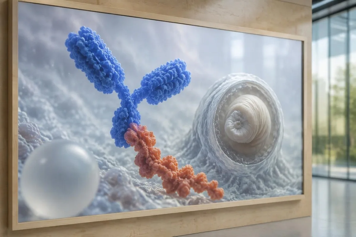
Antibody testing detects the host response, while antigen testing aims to detect parasite-derived material and may better reflect viable disease burden. Antigen detection is often less useful when only 1 small parenchymal cyst or calcifications are present; molecular testing is used in some specialist settings, but performance cannot be generalized across every laboratory or specimen.
Lumbar puncture is not routinely required for an uncomplicated solitary parenchymal finding and can be unsafe when obstructive hydrocephalus or significant mass effect is present. Cerebrospinal fluid tests for other disorders also require disease-specific interpretation, as our CSF band interpretation guide explains; 1 abnormal CSF marker does not identify every possible cause.
Kantesti is in AI bloedtest-útslachplatfoarm, not a service that orders lumbar punctures, identifies cysts on scans, or confirms neurological infections. Our AI-ynterpretaasjetechnology explains the laboratory-report workflow; when 2 tests disagree, our role is to clarify their stated meanings and limitations, not select an invasive diagnostic procedure.
Wêrom hinget de behanneling ôf fan it cystestadium en de lokaasje?
Treatment depends on whether cysts are viable, degenerating, calcified, or located outside the brain tissue, not simply on antibody positivity. Selected viable parenchymal cases receive antiparasitic treatment, whereas calcified-only disease generally receives symptom-directed care; ventricular obstruction may require a procedural approach rather than 1 standard medication regimen.
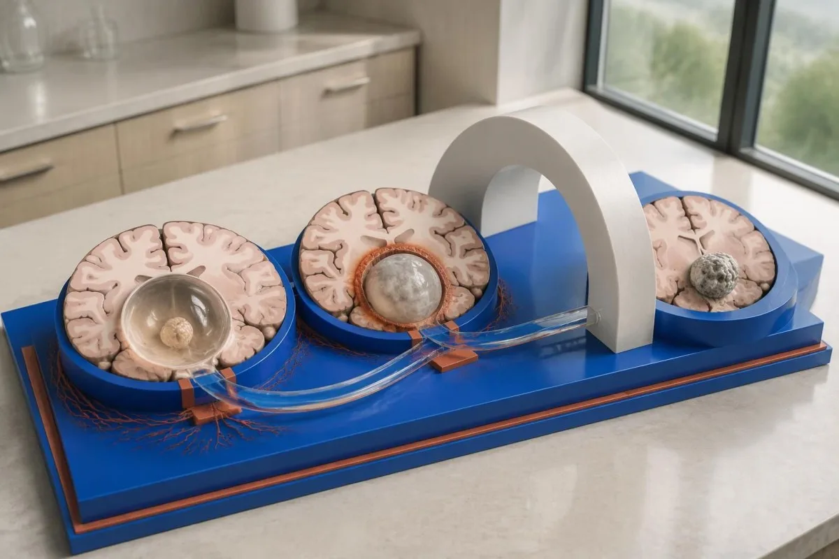
For selected patients with 1–2 viable parenchymal cysts, the IDSA/ASTMH guideline describes albendazole treatment for 10–14 days with appropriate accompanying care. This is a specialist-directed recommendation, not a home-treatment instruction: antiparasitic drugs can provoke an inflammatory response, and untreated hydrocephalus or diffuse cerebral edema changes the order of management (White et al., 2018).
WHO's 2021 guidance focuses on defined presentations of parenchymal neurocysticercosis, while the IDSA/ASTMH guideline also addresses ventricular, subarachnoid, and other forms in detail. Differences in imaging access and disease compartment explain why 1 treatment recommendation cannot be transferred to every patient with an antibody result; the guidelines do not turn negative serology into a rule-out test.
Corticosteroids may accompany antiparasitic therapy, but prolonged use requires additional safety planning, including assessment for relevant latent infections. Our Strongyloides and steroid safety guide explains a separate risk that matters before immune-suppressing treatment; an eye examination is also recommended before antiparasitic therapy to evaluate possible intraocular cysts.
Wat moat de follow-up neist antykoaderesultaten kontrolearje?
Follow-up should track seizures, neurological symptoms, imaging changes, and medication safety, rather than use antibody disappearance as proof of recovery. For viable parenchymal cysts, the IDSA/ASTMH guideline suggests repeat MRI at least every 6 months until the cystic component resolves, with earlier reassessment when symptoms change.
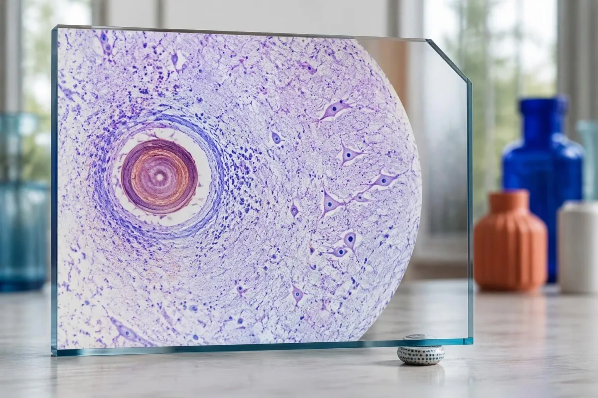
Antibodies may remain detectable after the original disease process has changed, and low-burden disease may never produce a positive result. If 1 new seizure occurs during follow-up, clinicians consider adherence, medication interactions, recurrent edema, and other causes; a rising or falling antibody index alone cannot distinguish among those explanations.
Kantesti AI can help explain reported laboratory results during medication follow-up, but a drug concentration is not a stand-alone measure of seizure control. Our de ynterpretaasje fan levetiracetamnivo's guide explains why sample timing and kidney function matter; 2 measurements collected at different times after dosing may not be directly comparable.
Monitoring depends on the chosen drug and treatment duration: prolonged albendazole therapy can require liver-test and blood-count surveillance, while some antiseizure medicines can affect sodium. Our hantlieding foar elektrolytfeiligens discusses those separate laboratory concerns; do not stop an antiseizure medicine or change its dose because 1 cysticercosis antibody result is negative.
Wat kin Kantesti útlizze, en wat kin ûndersyk net bewize?
AI report interpretation cannot confirm or exclude neurocysticercosis from laboratory results alone. Kantesti is in AI-oandreaune ark foar analyse fan bloedtests whose role here is to organize reported laboratory findings; a negative result still needs clinical context, and 1 new seizure takes priority over reviewing an automated explanation.

Prepare a focused review packet with the original antibody report, assay method, CT or MRI report, symptom timeline, and medication list. These 5 information categories reduce the risk of discussing a result without its context; ask specifically whether the imaging suggests viable, degenerating, calcified, ventricular, or subarachnoid disease and which unanswered question the next test would address.
The 2 Kantesti publications listed below are a repository-deposited global analysis report and an educational RDW guide, not clinical evidence validating neurocysticercosis diagnosis. Our RDW en CBC gids can support interpretation of a separately ordered blood count, but RDW, MCV, and MCHC do not establish whether a brain cyst is present.
Evidence for the neurological recommendations in this article comes from the cited diagnostic criteria and clinical guidelines, not from general AI benchmarking or laboratory-report volume. Our technical validation scope should be read within its stated boundaries; neither 1 successful report explanation nor a large aggregate dataset substitutes for condition-specific diagnostic validation.
Faak stelde fragen
Kinne jo neurocysticerkose hawwe mei in negative bloedtest?
Ja, neurocysticerkose kin oanwêzich wêze nettsjinsteande in negative bloedtest op antistoffen, benammen by 1 harsenscyste of allinnich ferkalkte sykte. Publisearre skatten fan EITB-testen yn referinsjelaboratoaria litte faak in gefoelichheid fan likernôch 50–60% sjen foar ien cyste, oanmerklik leger as de prestaasjes by meardere libbene cysten. CT, MRI, symptomen en de bleatstellingsskiednis bliuwe dêrom sintraal foar de diagnoaze. In nije oanfal freget om in driuwende beoardieling, ynstee fan gerêststelling op grûn fan it negative resultaat.
Wat is de meast betroubere test foar neurocysticerkose?
Gjin inkele test foar neurocysticerkose jout antwurd op alle diagnostyske fragen; harsensôfbylding en passend keazen serology folje inoar oan. EITB is neffens de rjochtline fan IDSA/ASTMH de foarkarsmetoade foar befêstiging mei antistoffen, mar kin sykte misse wêrby't mar 1 cyste belutsen is. CT sûnder kontrast is benammen nuttich foar kalkôfsettings, wylst MRI in protte libbensfetbere cysten, oedeem en ôfwikingen yn floeistofromten better beoardielet. Klinisy kombinearje dizze befiningen ynstee fan ien negative útslach as definityf te beskôgjen.
Is CT of MRI better foar neurocysticercose?
CT en MRI hawwe ferskillende sterke punten by de diagnoaze fan neurocystiserkoaze. CT sûnder kontrast ûntdekt faak lytse kalkôfsettings fan likernôch 1–4 mm dúdliker, wylst MRI libbensfetbere cysten, omlizzend oedeem en sykte yn de harsenskeamers of subarachnoïdale romte beoardielet. Spesjalisearre 3-diminsjonale MRI-sekwinsjes kinne helpe om cystmembranen yn romten mei harsens-rêgemurchfloeistof te identifisearjen. De IDSA/ASTMH-rjochtline advisearret beide ûndersyksmetoaden by it klassifisearjen fan nij diagnostisearre sykte.
Kin in ferkalkte cyste noch altyd oanfallen feroarsaakje?
In ferkalkke oerbliuwsel fan cysticerkose kin ferbûn bliuwe mei krampen, ek al is it net langer in libbensfetbere cyste. Guon ferkalkke foci, faak mar 1–4 mm yn trochsneed, ûntwikkelje tuskenskoften oedeem deromhinne dat sichtber wêze kin op in MRI. Negative antistoffen slute dit meganisme net út, en oanhâldend positive antistoffen jouwe it risiko op krampen net oan. By allinnich ferkalkke sykte wurdt yn it algemien keazen foar behanneling rjochte op de symptomen ynstee fan antiparasitêre medikaasje.
Moat ik in negative antystoftest foar cysticerkose werhelje?
In negative antistofftest foar cysticerkose opnij útfiere is net automatysk de bêste folgjende stap. By 1 lytse cyste of ferkalkte sykte kin in swakke antistofreaksje ek nei in werhelling fan deselde test nei 2 wiken negatyf bliuwe. In dokter kin ynstee de oarspronklike metoade beoardielje, freegje om in better karakterisearre befêstigjende test, of CT en MRI prioriteit jaan. Nije oanfallen of slimmer wurdende neurologyske symptomen moatte net wachtsje op werhelle tests.
Wannear moat ik by in earste epileptyske oanfal needhelp ynskeakelje?
In earste bekende epileptyske oanfal fereasket driuwende medyske beoardieling en kontakt mei de lokale helptsjinsten, ek as in antistoftest foar neurocysticerkose negatyf is. In krampende epileptyske oanfal dy't 5 minuten duorret, werhelle oanfallen sûnder dat de persoan wer by bewustwêzen komt, sykhelproblemen, swierwêzen of in slim letsel is in needgefal. Hâld de persoan net fêst en stopje neat yn har mûle; lis har op ’e side as de krampen ophâlden binne en se normaal sykhellet. Ryd net oant in medyske professional it foarfal beoardiele hat en de jildende beheiningen útlein hat.
Lit jo bloedtest resultaten útlizze
Laad jo laboratoariumrapport op en de AI fan Kantesti leit elke biomarker binnen sekonden út yn gewoane taal. Brûkt troch mear as 2 miljoen minsken wrâldwiid.
Referinsjes
- Klein, T., Mitchell, S., & Weber, H. (2026). AI Bloedtest Analyzer: 2.5M Tests Analiseerd | Global Health Report 2026. Kantesti AI Medysk Undersyk. DOI
- Klein, T., Mitchell, S., & Weber, H. (2026). RDW-bloedtest: folsleine gids foar RDW-CV, MCV en MCHC. Kantesti AI Medysk Undersyk. DOI
- White AC Jr et al. (2018). Diagnosis and Treatment of Neurocysticercosis: 2017 Clinical Practice Guidelines by the Infectious Diseases Society of America (IDSA) and the American Society of Tropical Medicine and Hygiene (ASTMH). Clinical Infectious Diseases. DOI
- Del Brutto OH et al. (2017). Revised diagnostic criteria for neurocysticercosis. Journal of the Neurological Sciences. DOI
- World Health Organization (2021). WHO guidelines on management of Taenia solium neurocysticercosis. Wrald sûnens organisaasje.
- 2M+Analysearre tests
- 127+Lannen
- 100+Talen
Oer it medyske team


E-E-A-T fertrouwensignalen
Ûnderfining
Dokter-oandreaune klinyske resinsje fan lab-ynterpretaasje-wurkprosessen.
Ekspertize
Fokus fan laboratoariummedisyne op hoe’t biomarkers har gedrage yn in klinyske kontekst.
Autoriteit
Medysk beoardiele troch Dr. Thomas Klein, mei bydragen fan Dr. Sarah Mitchell en prof. dr. Hans Weber.
Betrouberens
Bewiis-basearre ynterpretaasje mei dúdlike ferfolchpaadkes om alarm te ferminderjen.





Medyske disclaimer
Dit artikel is allinnich foar ûnderwiisdoelen en foarmet gjin medysk advys. Rieplachtsje altyd in kwalifisearre soarchferliener foar besluten oer diagnoaze en behanneling.
Publisearre · Nim kontakt op mei it redaksjeteam
Publisearre troch Kantesti Ltd, registrearre yn Ingelân en Wales, bedriuwsnûmer. 17090423, Londen, Feriene Keninkryk.
