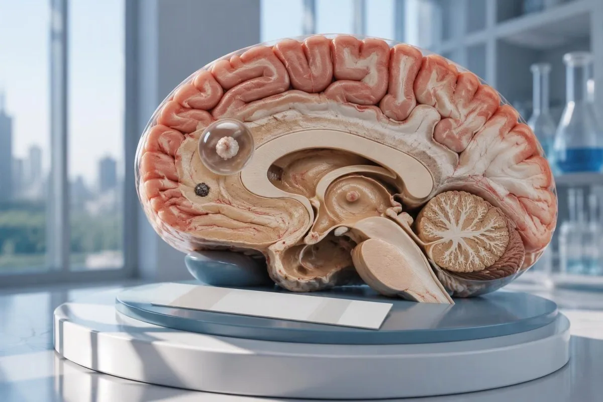
د عصبي سیستیسرکوز منفي ازموینه د مغز د یوه منفرد کیسټ یا کلسیفای شوې ناروغۍ شتون نه ردوي. CT، MRI او د نوو تشنجونو بیړنۍ ارزونه تر یوازې د انټيباډي ازموینې له ډاډ ورکولو ډېر اهمیت لري.

مهم ټکي
- منفي ضد اجسام عصبي سیستیسرکوز نه ردوي، په ځانګړې توګه کله چې انځور اخیستنه یوازې 1 اغېزمنه سیمه یا کلسیفیکیشنونه وښيي.
- د مرجع لابراتوار EITB د 1 څخه د ډېرو ژوندي کیسټونو په شتون کې یې حساسیت 98% ته رسېدای شي، خو د یوه منفرد کیسټ په شتون کې یې فعالیت د پام وړ ټیټ وي.
- د یوه منفرد کیسټ حساسیت په خپرو شویو EITB اټکلونو کې ډېری وخت شاوخوا 50–60% وي؛ دا شمېرې پر هرې سوداګریزې انټيباډي ازموینې نه تطبیقېږي.
- بېکنتراسته CT د کوچنیو کلسیفیکیشنونو لپاره په ځانګړې توګه ګټور دی، چې قطر یې ډېری وخت شاوخوا 1–4 mm وي.
- MRI ژوندي کیسټونه، شاوخوا پړسوب او بطني یا تحتعنکبوتیه ناروغي ارزوي؛ ځانګړې 3-بعدي لړۍ ښايي موندنه ښه کړي.
- لومړني تشنجونه د انټيباډۍ د پایلې په پام کې نیولو پرته، عاجلې طبي ارزونې ته اړتیا لري. د لومړي ځل پېژندل شوي تشنج یا د بیړني حالت د نورو نښو پر مهال بیړنیو خدمتونو ته زنګ ووهئ.
- پنځه دقیقې د روان تشنجي تشنج لپاره د بیړني حالت حد دی، نه دا چې د مرستې غوښتلو تر مخه د انتظار دلیل وي.
- یوازې کلسیفای شوې ناروغي عموماً د پرازیت ضد درملو ته اړتیا نه لري، خو د تشنج درملنه او نورې انځوریزې معاینې بیا هم اړینې کېدای شي.
ایا د عصبي سیستیسرکوز منفي ازموینه دا ناروغي ردولی شي؟
A د عصبي سیسټیسېرکوسس منفي ازموینه په ډاډ سره ناروغي نه شي ردولای، په ځانګړي ډول د 1 دماغي کیسټ یا کلسیفای شوو پاتې شونو په شتون کې. د انټيباډۍ ازموینه د معافیت غبرګون اندازه کوي، نه دا چې دماغ په مستقیم ډول وڅېړي؛ CT او MRI د مشکوکو موندنو ځای او پړاو څرګندوي، په داسې حال کې چې لومړی تشنج عاجلې ارزونې ته اړتیا لري.
پایله مشروطه ده، مطلقه نه ده. د سیسټیسېرکوسس د انټيباډۍ هغه ازموینه چې په څو ژوندي کیسټونو لرونکو کسانو کې ښه کار کوي، ښايي یوازې د 1 کوچنۍ اغېزمنې سیمې لرونکي کس کې کمزوری فعالیت ولري؛ نو د منفي مانا دا ده چې په دې ازموینه کې د موندلو وړ غبرګون نشته—نه دا چې د عصبي سیستم په هېڅ ځای کې کیسټ نشته.
د یوه فرضي مثال په توګه، د 34 کلن کس په اړه فکر وکړئ چې د لومړي ځل تشنج، د انټيباډۍ منفي راپور، او په MRI کې یوه یوازینۍ کړۍډوله روښانه شوې موندنه لري. راپور نه شي پرېکړه کولای چې دا موندنه سیسټیسېرکوسس دی، بله عفونت دی، که غیرعفوني بهیر؛ مانا یې په ګډه د سکن، د مخامخېدو د سابقې او عصبي ارزونې پورې اړه لري.
کانټیسټي یو دی د AI د وینې معاینې شنونکی چې د لابراتواري راپور شویو موندنو په تشریح کې مرسته کولای شي، خو د انټيباډۍ د 1 منفي پایلې له مخې د دماغ ناروغي نه شي ردولای. زموږ سازمان او موخه زموږ د لابراتواري معلوماتو د تمرکز په اړه وضاحت ورکوي؛ لوستونکي باید د اتومات راپور تشریح داسې ونه ګڼي چې د تشنج د درملنې ځنډولو اجازه ورکوي.
ولې د انټيباډي ازموینه د مغز یو منفرد کیسټ نه شي موندلی؟
A یوه کیسټ د ازموینې د موندلو له حد څخه د ټیټ انټيباډۍ غبرګون رامنځته کولای شي. د خپرو شویو اټکلونو له مخې، د مرجع لابراتوار EITB حساسیت د یوې کیسټ لپاره ډېری وخت نږدې 50–60% وي، په پرتله له 98% سره نږدې هغه مهال چې له 1 څخه ډېر ژوندي کیسټونه موجود وي؛ دا توپیر د ناروغۍ د کچې او د ازموینې د فعالیت څرګندونه کوي.
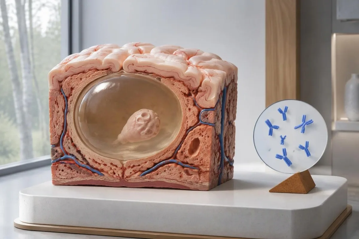
د IDSA/ASTMH لارښود سپارښتنه کوي چې د انزایم په وسیله تړلی ایمونو الیکټروټرانسفر بلاټ، یا EITB، د تاییدوونکې سیرولوژۍ په توګه وکارول شي، او په څرګنده خبرداری ورکوي چې د یوې یوازینۍ پارانشیمي موندنې یا یوازې کلسیفیکېشنونو په شتون کې حساسیت کمزوری دی (White et al., 2018). د څو کیسټونو، بطیني ناروغۍ یا تحتعنکبوتیه ناروغۍ لرونکو ناروغانو لپاره راپور شوی لوړ حساسیت باید د داسې کس په اړه ونه کارول شي چې په دماغ کې یې 1 کوچنۍ غیرعادي موندنه وي.
یوازې د وضاحت لپاره فرض کړئ چې یوه ازموینه په 100 هغو کسانو کې 55% حساسیت لري چې د یوې کیسټ ناروغي یې له مخکې په خپلواکه توګه ثابته شوې ده. نږدې 45 کسان به د ناروغۍ له لرلو سره سره منفي نتیجه واخلي؛ دا کاري بېلګه د نه موندلو بهیر تشریح کوي، نه دا چې د کومې ځانګړې منفي پایلې له مخې د ناروغۍ احتمال څومره دی.
د 2 اونیو وروسته د هماغې ازموینې بیا کول ښايي د ناروغۍ د کمې کچې له امله رامنځته شوی کمزوری غبرګون سم نه کړي. دا له هغو عفونتونو سره توپیر لري چې پکې د نمونې اخیستلو وختيوالی اصلي ستونزه وي، لکه څنګه چې زموږ په د وخت اړوند د کاذبې منفي پایلې لارښود; کې تشریح شوي؛ وپوښتئ چې ایا انځوریزه معاینه یا ښه ځانګړنه لرونکې ازموینه به بله پرېکړه بدله کړي.
ولې کلسیفای شوی عصبي سیستیسرکوز لا هم نښې لرلی شي؟
کلسیفای شوې عصبي سیسټیسېرکوسس د منفي انټيباډیو سره سره د تشنج لامل کېدای شي, because the remaining mineralized focus can still be associated with local irritation and episodic surrounding edema. Typical calcifications may measure approximately 1–4 mm, but their small size does not establish whether they are clinically harmless.
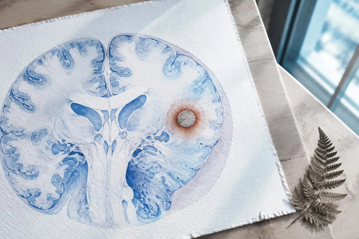
A calcification is generally a remnant rather than a living cyst that antiparasitic medication can eliminate. In the IDSA/ASTMH guideline, calcified-only parenchymal disease is managed symptomatically rather than with antiparasitic drugs; a person with 1 calcified focus and seizures may therefore need antiseizure treatment without needing cyst-killing medication (White et al., 2018).
Surrounding edema can appear intermittently, sometimes long after the original cyst has degenerated. MRI may show this tissue response even when the calcification is more obvious on CT, which is why the 2 scans can tell different but compatible stories rather than one proving that the other is wrong.
Neither a negative antibody result nor a persistent positive result measures seizure risk by itself. The broader lesson resembles other seronegative neurological conditions, discussed in our negative neurological antibody results guide: absence of detectable antibodies is not equivalent to absence of a clinically relevant neurological process.
په حقیقت کې د سیستیسرکوز د انټيباډي کومه ازموینه ترسره شوې وه؟
The useful first question is whether the report describes EITB, ELISA, or another antibody method, because these are not interchangeable tests. A negative index below 1 laboratory's cutoff cannot inherit the performance figures of a different reference-laboratory assay, even when both reports use the name cysticercosis IgG.
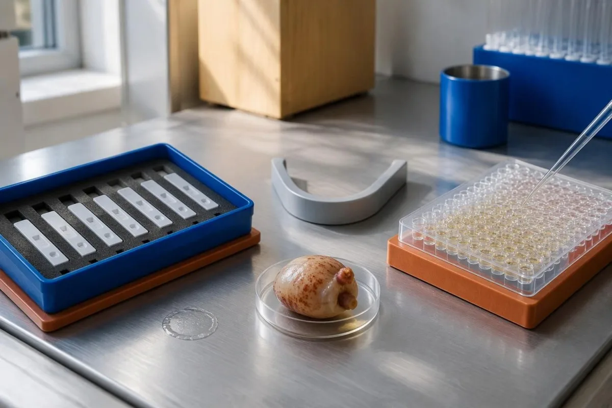
EITB using purified parasite glycoproteins is the preferred serological method in the IDSA/ASTMH guideline, while crude-antigen ELISA methods have recognized sensitivity and specificity limitations (White et al., 2018). Before comparing 2 results, check the method, specimen type, reference interval, and whether the laboratory used a validated confirmatory procedure.
Some laboratories report a qualitative result; others provide an optical-density ratio or numerical index. A value such as 0.8 in a hypothetical assay does not represent 0.8 cysts, an 80% disease probability, or a universal severity score—our کیفیتي او کمي پایلې guide explains that distinction.
Kantesti AI's role is to help readers preserve the laboratory's original wording rather than reinterpret 1 method through another method's thresholds. Our د لابراتوار مارکر لارښود provides broader report-reading context; for this result, keep the actual assay name available when discussing referral or repeat testing.
د تماس سابقه د تشخیص شک څنګه بدلوي؟
Neurocysticercosis follows ingestion of Taenia solium eggs, usually through fecal contamination from a person carrying the intestinal tapeworm. Eating undercooked pork can cause intestinal taeniasis, but brain cysts arise through a different transmission step; symptoms may emerge years after exposure rather than within 1 recent trip.

A vegetarian can develop cysticercosis because eggs can contaminate food or hands independently of meat consumption. Useful history includes past residence, household contacts, sanitation conditions, and possible exposure to a tapeworm carrier; asking only about meals eaten during the last 2 weeks can miss the relevant background.
A negative stool examination does not exclude cysts in the brain. Stool testing addresses intestinal tapeworm carriage, whereas CT and MRI assess nervous-system findings; these are 2 different sites and clinical questions, although public-health teams may recommend evaluating contacts when a carrier could be involved.
Normal eosinophils also cannot exclude neurocysticercosis, and raised eosinophils do not identify the brain as the affected site. Our parasite antibody interpretation guide discusses a separate parasite with similar localization limits; the practical point is to avoid using 1 routine cell count as a substitute for targeted neurological evaluation.
نوي تشنجونه کله بیړنۍ ارزونې ته اړتیا لري؟
A first known seizure needs urgent medical assessment regardless of antibody results, and emergency services should be contacted for a first seizure or an ongoing seizure with concerning features. A convulsive seizure lasting 5 minutes, repeated seizures without recovery, breathing difficulty, pregnancy, or significant injury requires emergency care.

Do not wait for the 5-minute mark to seek help after a first known seizure. Keep the person away from hazards, cushion the head if safely possible, do not restrain them or put anything in their mouth, and place them on their side once convulsions stop if they are breathing normally.
Persistent confusion, a severe new headache, fever with neck stiffness, or new weakness increases concern for causes beyond cysticercosis. Sudden one-sided weakness or speech difficulty also demands emergency assessment, as our د سکتې د خبرداري نښو لارښود explains; 1 negative infection test cannot rule out stroke, meningitis, or a structural brain problem.
After a suspected seizure, stop driving and avoid swimming alone, heights, or operating machinery until a clinician provides advice consistent with local rules. If a witness can safely record the duration and describe movements or recovery, those observations may be more useful than ordering a second antibody test before the first neurological assessment.
CT د عصبي سیستیسرکوز په تشخیص کې څه ورزیاتوي؟
Noncontrast CT is particularly useful for detecting calcified cyst remnants, including small approximately 1–4 mm foci that MRI may show less clearly. CT also helps assess urgent structural problems such as hydrocephalus, but 1 isolated calcification is not automatically proof of neurocysticercosis or the cause of a seizure.
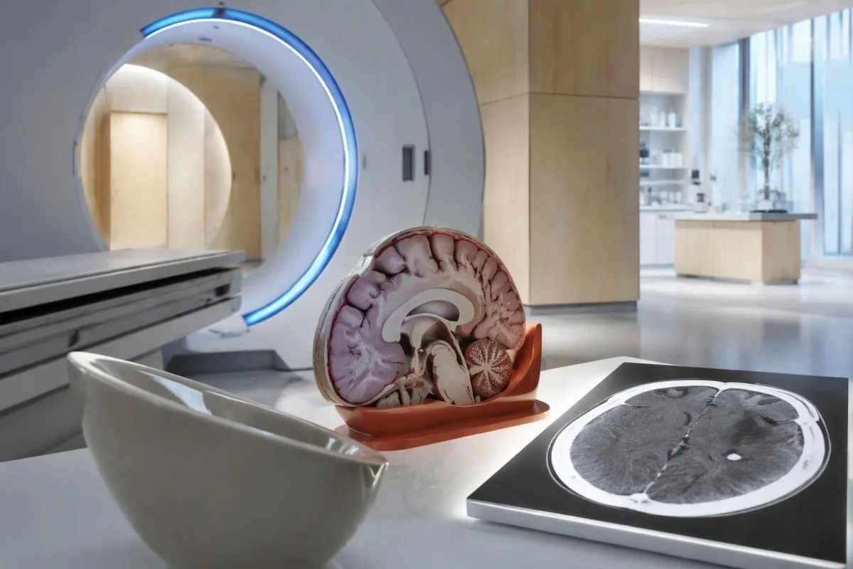
Calcium appears dense on CT, allowing radiologists to identify mineralized remnants and describe their distribution. The IDSA/ASTMH guideline recommends both MRI and noncontrast CT when classifying newly diagnosed neurocysticercosis, because the 2 modalities have complementary strengths rather than being interchangeable alternatives (White et al., 2018).
An incidental calcification needs context: previous granulomatous disease, other causes of mineralization, and scan appearance can influence interpretation. If 1 calcification sits near a suspected seizure focus, concordance with symptoms and EEG may strengthen the explanation, but anatomical proximity alone cannot establish causation.
A laboratory marker used in selected head-injury pathways does not test for brain cysts or replace imaging after an unexplained seizure. Our head-injury CT decisions guide illustrates that narrower setting; ask the clinician whether the present question requires noncontrast CT, contrast imaging, MRI, or a combination of 2 modalities.
MRI څه څرګندوي چې ښايي CT یې ونه مومي؟
MRI is better suited to evaluating viable cysts, surrounding edema, and many ventricular or subarachnoid findings. Specialized 3-dimensional sequences such as CISS or FIESTA can improve visualization of cyst membranes within cerebrospinal fluid spaces, where a cyst may otherwise resemble the surrounding fluid.
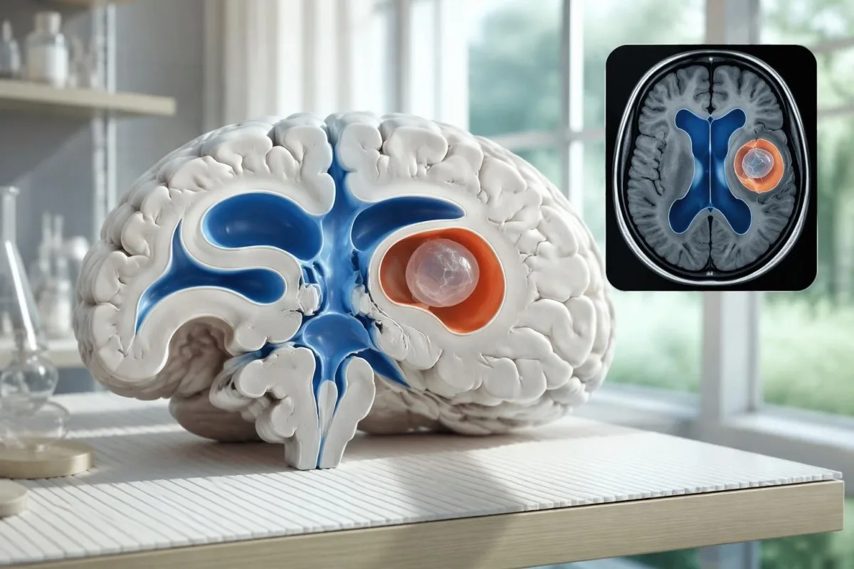
A cyst inside the ventricles can obstruct cerebrospinal fluid flow and cause hydrocephalus even without numerous parenchymal cysts. Headache with vomiting, declining alertness, or new visual symptoms therefore changes urgency; 1 reassuring antibody result should not override symptoms that suggest raised pressure or impaired fluid circulation.
MRI may show a scolex, the small internal structure of the parasite, inside a compatible cyst. This finding can be highly specific when confidently identified, whereas ring enhancement alone has a broader differential; our brain imaging warning signs guide explains why structural abnormalities require interpretation rather than diagnosis from 1 visual feature.
A routine MRI protocol may not answer every cysticercosis question. If ventricular or subarachnoid disease is suspected, discuss whether thin-section 3-dimensional imaging is needed; radiologists choose sequences and contrast according to the clinical question, and a normal scan using 1 protocol does not prove every possible disease compartment has been adequately assessed.
کله چې د ازموینو پایلې سره توپیر ولري، عصبي سیستیسرکوز څنګه تشخیصېږي؟
Neurocysticercosis diagnosis combines imaging, clinical findings, exposure information, and supporting tests, rather than requiring a positive antibody result in every case. The revised diagnostic criteria published in 2017 give characteristic neuroimaging findings a central role and distinguish strong evidence from less-specific supportive features (Del Brutto et al., 2017).
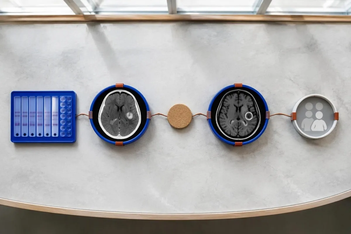
Evidence should be weighted, not tallied. A confidently visualized scolex inside a compatible cyst is different from 1 nonspecific ring-enhancing area, while a positive antibody result is different from direct evidence that a finding is in the brain; our مثبت انټي باډي پایله لارښود explains why detection alone cannot establish location.
For a clearly hypothetical probability example, start with a 30% pretest probability and assume a single-cyst assay has 55% sensitivity and 99% specificity. In 1,000 comparable people, about 135 affected and 693 unaffected people would test negative, leaving approximately a 16% probability of disease among negative results—still substantial, although these assumed figures are not a personal risk calculator.
The most useful disagreement to investigate is whether each test is answering the same question. A negative antibody test, a CT calcification, and MRI edema can all be compatible with 1 calcified disease process; unexplained discrepancies should prompt review of the assay and images rather than automatic dismissal of either result.
د مغز په انځور اخیستنه کې نور څه د یوه منفرد کیسټ په څېر ښکاري؟
A single ring-enhancing brain finding is not specific to neurocysticercosis. Other possibilities include tuberculoma, an abscess, a neoplasm, or an inflammatory process; clinicians use appearance, location, immune status, symptoms, and sometimes follow-up imaging or tissue examination to distinguish these possibilities rather than relying on 1 antibody panel.
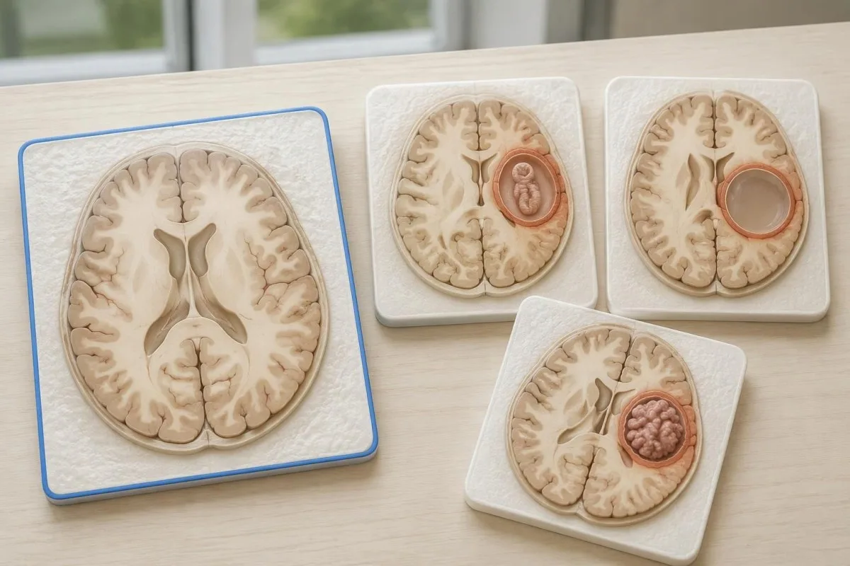
Edema, restricted diffusion, internal structures, and the number of findings all contribute to the differential, but no single nonspecific feature settles every case. Fever and immune suppression may change the suspected cause; a 2 cm ring-enhancing area in a hypothetical immunocompromised person warrants a different investigation from a tiny calcification in an otherwise well person.
Toxoplasmosis is one consideration in selected immunocompromised patients, and a positive IgG result can indicate past exposure rather than prove an active brain process. Our Toxoplasma IgG interpretation guide explains that laboratory distinction; imaging and the patient's immune status determine whether the result is clinically relevant to 1 or more cerebral findings.
New seizures accompanied by rapidly evolving memory problems, psychiatric changes, or abnormal movements can also suggest encephalitis rather than a solitary parasite cyst. Our encephalitis diagnostic pathway describes how MRI, EEG, and cerebrospinal fluid testing answer different questions; a second negative cysticercosis antibody result does not investigate those alternatives.
ایا د انټيجن ازموینه، PCR یا CSF نامعلوم حالت روښانه کولی شي؟
Antigen tests, PCR, and cerebrospinal fluid analysis can add information in selected cases, particularly when extraparenchymal disease is suspected. None is a universal rescue test after 1 negative antibody result: availability, assay validation, disease compartment, and safety of lumbar puncture determine whether additional testing is appropriate.
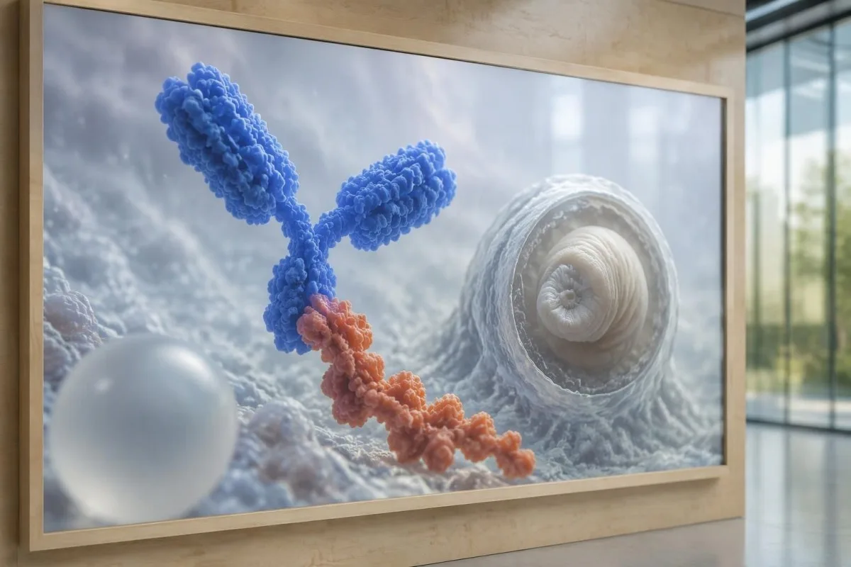
Antibody testing detects the host response, while antigen testing aims to detect parasite-derived material and may better reflect viable disease burden. Antigen detection is often less useful when only 1 small parenchymal cyst or calcifications are present; molecular testing is used in some specialist settings, but performance cannot be generalized across every laboratory or specimen.
Lumbar puncture is not routinely required for an uncomplicated solitary parenchymal finding and can be unsafe when obstructive hydrocephalus or significant mass effect is present. Cerebrospinal fluid tests for other disorders also require disease-specific interpretation, as our CSF band interpretation guide explains; 1 abnormal CSF marker does not identify every possible cause.
کانټیسټي یو دی AI د وینې ازموینې تشریح پلیټفارم, not a service that orders lumbar punctures, identifies cysts on scans, or confirms neurological infections. Our تكنولوجيا تفسير الذكاء الاصطناعي (AI) الخاصة بنا explains the laboratory-report workflow; when 2 tests disagree, our role is to clarify their stated meanings and limitations, not select an invasive diagnostic procedure.
ولې درملنه د کیسټ پر پړاو او ځای پورې تړلې ده؟
Treatment depends on whether cysts are viable, degenerating, calcified, or located outside the brain tissue, not simply on antibody positivity. Selected viable parenchymal cases receive antiparasitic treatment, whereas calcified-only disease generally receives symptom-directed care; ventricular obstruction may require a procedural approach rather than 1 standard medication regimen.
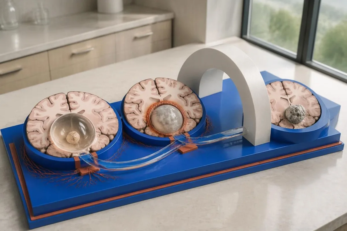
For selected patients with 1–2 viable parenchymal cysts, the IDSA/ASTMH guideline describes albendazole treatment for 10–14 days with appropriate accompanying care. This is a specialist-directed recommendation, not a home-treatment instruction: antiparasitic drugs can provoke an inflammatory response, and untreated hydrocephalus or diffuse cerebral edema changes the order of management (White et al., 2018).
WHO's 2021 guidance focuses on defined presentations of parenchymal neurocysticercosis, while the IDSA/ASTMH guideline also addresses ventricular, subarachnoid, and other forms in detail. Differences in imaging access and disease compartment explain why 1 treatment recommendation cannot be transferred to every patient with an antibody result; the guidelines do not turn negative serology into a rule-out test.
Corticosteroids may accompany antiparasitic therapy, but prolonged use requires additional safety planning, including assessment for relevant latent infections. Our Strongyloides and steroid safety guide explains a separate risk that matters before immune-suppressing treatment; an eye examination is also recommended before antiparasitic therapy to evaluate possible intraocular cysts.
تعقیبي څارنه باید د انټيباډي پایلو سربېره څه وڅاري؟
Follow-up should track seizures, neurological symptoms, imaging changes, and medication safety, rather than use antibody disappearance as proof of recovery. For viable parenchymal cysts, the IDSA/ASTMH guideline suggests repeat MRI at least every 6 months until the cystic component resolves, with earlier reassessment when symptoms change.
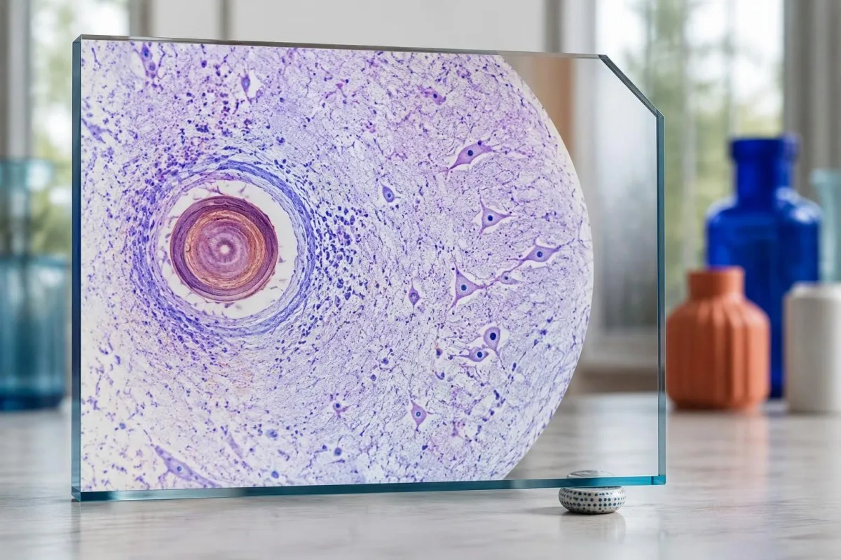
Antibodies may remain detectable after the original disease process has changed, and low-burden disease may never produce a positive result. If 1 new seizure occurs during follow-up, clinicians consider adherence, medication interactions, recurrent edema, and other causes; a rising or falling antibody index alone cannot distinguish among those explanations.
Kantesti AI can help explain reported laboratory results during medication follow-up, but a drug concentration is not a stand-alone measure of seizure control. Our د لیوېټیراسیتام د کچې تفسیر guide explains why sample timing and kidney function matter; 2 measurements collected at different times after dosing may not be directly comparable.
Monitoring depends on the chosen drug and treatment duration: prolonged albendazole therapy can require liver-test and blood-count surveillance, while some antiseizure medicines can affect sodium. Our د الکترولایتونو د خوندیتوب لارښود discusses those separate laboratory concerns; do not stop an antiseizure medicine or change its dose because 1 cysticercosis antibody result is negative.
Kantesti څه تشریح کولی شي، او څېړنې څه نه شي ثابتولی؟
AI report interpretation cannot confirm or exclude neurocysticercosis from laboratory results alone. Kantesti یو د AI پر بنسټ د وینې ازموینې تحلیل وسیله whose role here is to organize reported laboratory findings; a negative result still needs clinical context, and 1 new seizure takes priority over reviewing an automated explanation.
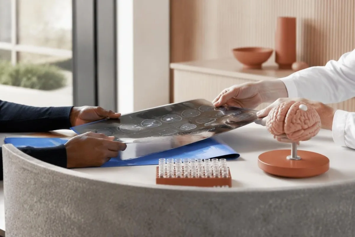
Prepare a focused review packet with the original antibody report, assay method, CT or MRI report, symptom timeline, and medication list. These 5 information categories reduce the risk of discussing a result without its context; ask specifically whether the imaging suggests viable, degenerating, calcified, ventricular, or subarachnoid disease and which unanswered question the next test would address.
The 2 Kantesti publications listed below are a repository-deposited global analysis report and an educational RDW guide, not clinical evidence validating neurocysticercosis diagnosis. Our RDW او CBC لارښود can support interpretation of a separately ordered blood count, but RDW, MCV, and MCHC do not establish whether a brain cyst is present.
Evidence for the neurological recommendations in this article comes from the cited diagnostic criteria and clinical guidelines, not from general AI benchmarking or laboratory-report volume. Our technical validation scope should be read within its stated boundaries; neither 1 successful report explanation nor a large aggregate dataset substitutes for condition-specific diagnostic validation.
پرله پسې پوښتل شوي پوښتنې
ایا د وینې د منفي ازموینې سره هم نیورو سېسټیسرکوز لرل کېدای شي؟
هو، د وینې د انټيباډي د ازموینې د منفي پایلې سربېره هم عصبي سیسټیسرکوسس موجود کېدای شي، په ځانګړي ډول که په مغز کې یو سیسټ وي یا ناروغي یوازې د کلسیفایډ سیسټونو بڼه ولري. د خپرو شویو اټکلونو له مخې، د مرجع لابراتوارونو EITB ازموینه د یوه سیسټ لپاره ډېری وخت نږدې ۵۰–60% حساسیت لري، چې د څو ژوندیو سیسټونو په شتون کې د دې ازموینې له فعالیت څخه ډېر ټیټ دی. له همدې امله، CT، MRI، نښې نښانې او له ناروغۍ سره د مخ کېدو سابقه د تشخیص لپاره مهم پاتې کېږي. نوې تشنج بیړنۍ ارزونې ته اړتیا لري، نه دا چې د منفي پایلې له امله ډاډ ورکړل شي.
د نیوروسیسټیسرکوسس تر ټولو باوري ازموینه کومه ده؟
د نیوروسیسټیکرکوسس هېڅ یوه ازموینه د تشخیص ټولو پوښتنو ته ځواب نه شي ویلای؛ د مغز انځور اخیستنه او په مناسبه توګه غوره شوې سیرولوژي یو د بل بشپړوونکې دي. EITB د IDSA/ASTMH په لارښود کې د تاییدوونکې انټيباډۍ غوره طریقه ده، خو د یوازې ۱ سیسټ لرونکې ناروغۍ په موندلو کې پاتې راتلای شي. له کانټراسټ پرته CT په ځانګړې توګه د کلسیفیکیشنونو لپاره ګټوره ده، خو MRI د ډېرو ژوندیو سیسټونو، اذیما او د مایع د فضا د غیرنورمالیو په ارزونه کې غوره ده. ډاکټران دا موندنې سره یوځای ارزوي، نه دا چې یوه منفي پایله قطعي وګڼي.
د عصبي سیسټیسرکوسس لپاره CT غوره دی که MRI؟
د CT او MRI د نیوروسیستیسرکوز په تشخیص کې بېلابېلې وړتیاوې لري. له کانټراسټ پرته CT اکثره د شاوخوا ۱–۴ mm پلنو کوچنیو کلسیفیکیشنونو په روښانه توګه موندلو کې ښه وي، په داسې حال کې چې MRI ژوندي سیسټونه، د هغوی شاوخوا اذیما، او د بطیني یا عنکبوتیهالوتېزې ناروغۍ ارزونه کوي. د MRI ځانګړي درېبعدي تسلسلونه ښايي د مغزينخاعي مایع په تشو کې د سیسټ د غشاګانو په پېژندلو کې مرسته وکړي. د IDSA/ASTMH لارښود د نوې تشخیص شوې ناروغۍ د ډلبندۍ پر مهال د دواړو معایناتو سپارښتنه کوي.
ایا کلسیفایډ شوې کیسټ لا هم د تشنج لامل کېدای شي؟
د کلسیفایډ سیسټیسرکوز پاتې شونی له تشنجونو سره تړاو لرلای شي، که څه هم نور یو ژوندی سیسټ نه وي. ځینې کلسیفایډ شوي مرکزونه، چې ډېری وخت یوازې ۱–۴ mm قطر لري، د دوی شاوخوا وخت ناوخت پړسوب رامنځته کوي چې ښايي په MRI کې ولیدل شي. د انټيباډي منفي پایلې دا میکانیزم نه ردوي، او د انټيباډي دوامدارې مثبتې پایلې د تشنج د خطر اندازه نه ښيي. یوازې کلسیفایډ شوې ناروغي عموماً د نښو پر بنسټ درملنه غواړي، نه د پرازیت ضد درمل.
ایا باید د سیسټیسیرکوسس د انټيباډي منفي ازموینه تکرار کړم؟
د سیسټیسرکوسس د انټيباډي د منفي ازموینې بیا تکرارول په خپله د بل ګام لپاره تر ټولو غوره انتخاب نه دی. که یو کوچنی سیسټ یا کلسیفیه شوې ناروغي وي، د انټيباډي کمزوری غبرګون ښايي منفي پاتې شي، ان که هماغه ازموینه له ۲ اوونیو وروسته بیا هم تکرار شي. پر ځای یې، ډاکټر ښايي د اصلي ازموینې طریقه وارزوي، د لا ښه مشخصې شوې تاییدي ازموینې غوښتنه وکړي، یا CT او MRI ته لومړیتوب ورکړي. نوې تشنجونه یا د عصبي نښو خرابېدل باید د بیا ازموینې لپاره انتظار ونه کړي.
د لومړي ځل تشنج پر مهال باید کله بیړنۍ مرسته وغواړم؟
لومړنۍ پېژندل شوې تشنجي حمله بېړنۍ طبي ارزونه او له سیمهییزو بیړنیو خدماتو سره اړیکه نیول غواړي، که د نیورو سیستیکرکوز لپاره د انټيباډي ازموینه منفي هم وي. هغه تشنجي حمله چې ۵ دقیقې دوام وکړي، پرلهپسې حملې چې ناروغ ترمنځ بېرته هوښیار نه شي، د ساه اخیستلو ستونزه، امیندوارۍ یا جدي ټپ بیړنی حالت دی. ناروغ مه نیسئ او هېڅ شی یې خولې ته مه ورکوئ؛ کله چې تشنجونه ودرېږي، که عادي ساه اخلي، په اړخ یې څملوئ. تر هغه موټر مه چلوئ چې ډاکټر پېښه ارزولې نه وي او د تطبیق وړ محدودیتونه یې درته نه وي تشریح کړي.
د وینې ازموینې پایلې څنګه ولولئ
خپل لابراتواري راپور پورته کړئ، او د Kantesti AI به په څو ثانیو کې هر بایومارکر په ساده ژبه تشریح کړي. په ټوله نړۍ کې له 2 میلیونو څخه زیاتو کسانو ترې کار اخیستی.
ماخذونه
- کلاین، ټي.، میچل، ایس.، او وېبر، ایچ. (۲۰۲۶). د AI وینې د ازموینې شنونکی: 2.5M ازموینې تحلیل شوې | د نړیوال روغتیا راپور 2026. د Kantesti AI طبي څېړنه. د دفاع وزارت
- کلاین، ټي.، میچل، ایس.، او وېبر، ایچ. (۲۰۲۶). د RDW د وینې ازموینه: د RDW-CV، MCV او MCHC بشپړ لارښود. د Kantesti AI طبي څېړنه. د دفاع وزارت
- White AC Jr et al. (2018). Diagnosis and Treatment of Neurocysticercosis: 2017 Clinical Practice Guidelines by the Infectious Diseases Society of America (IDSA) and the American Society of Tropical Medicine and Hygiene (ASTMH). Clinical Infectious Diseases. د دفاع وزارت
- Del Brutto OH et al. (2017). Revised diagnostic criteria for neurocysticercosis. Journal of the Neurological Sciences. د دفاع وزارت
- World Health Organization (2021). WHO guidelines on management of Taenia solium neurocysticercosis. د روغتیا نړیوال سازمان.
- ۲ میلیونه+تحلیل شوې ازموینې
- 127+هېوادونه
- 100+ژبې
د طبي ټیم په اړه


د E-E-A-T باور نښې
تجربه
د ډاکټر تر مشرۍ لاندې کلینیکي بیاکتنه د لابراتواري تفسیر د کاري بهیرونو لپاره.
تخصص
د لابراتواري طب تمرکز پر دې چې بایومارکرونه په کلینیکي شرایطو کې څنګه چلند کوي.
واک ورکول
د ډاکټر توماس کلاین له خوا طبي بیاکتنه شوې، د ډاکټر سارا میچل او پروفیسور ډاکټر هانس وېبر په مرسته.
اعتبار
د شواهدو پر بنسټ تفسیر د روښانه تعقیبي لارو چارو سره، تر څو اندیښنه کمه شي.





طبي سلبِ مسئولیت
دا مقاله یوازې د زده کړې لپاره ده او طبي مشوره نه جوړوي. د تشخیص او درملنې د پرېکړو لپاره تل د وړ روغتیايي خدمت وړاندې کوونکي سره سلا وکړئ.
خپور شوی · له تحریري ټیم سره اړیکه ونیسئ
خپروونکی یې Kantesti Ltd دی، چې په انګلستان او وېلز کې ثبت شوی، د شرکت شمېره. 17090423, ، لندن، بریتانیا.
