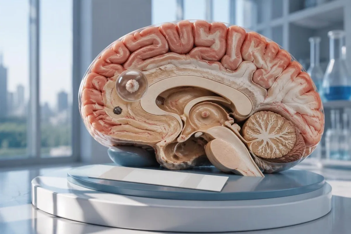
Ang negatibong test alang sa neurocysticercosis wala magwagtang sa posibilidad nga adunay usa ka cyst sa utok o kalkipikadong sakit. Mas hinungdanon ang CT, MRI, ug dinaliang pagsusi sa bag-ong mga seizure kaysa sa pagpasalig nga gibase lamang sa pagsulay sa antibody.

Pangunang mga punto
- Negatibo nga mga antibody dili makapawala sa posibilidad sa neurocysticercosis, ilabi na kon 1 ra ka apektadong bahin o mga kalkipikasyon ang makita sa imaging.
- EITB sa laboratoryong reperensiya mahimong moabot sa 98% ang pagkasensitibo kon adunay labaw sa 1 ka buhing cyst, apan dako kaayong moubos ang kahusayan niini kon usa ra ka cyst.
- Pagkasensitibo sa usa ka cyst kasagarang mga 50–60% sumala sa mga gipatik nga banabana sa EITB; dili magamit kining mga numeroha sa tanang komersiyal nga pagsulay sa antibody.
- CT nga walay contrast labi nang mapuslanon sa gagmayng kalkipikasyon, nga kasagarang mga 1–4 mm ang gilapdon.
- MRI mosusi sa buhing mga cyst, paghubag sa palibot niini, ug sakit sa mga ventricle o subarachnoid nga bahin; ang espesyal nga mga sunodsunod nga hulagway nga 3-dimensional mahimong makapaayo sa pagkatiktik.
- Unang mga seizure nagkinahanglan og dinaliang medikal nga pagtimbang-timbang bisan unsa pa ang resulta sa antibody test. Tawga ang serbisyong pang-emerhensiya kon kini ang unang nailhang seizure o adunay laing timailhan sa emerhensiya.
- Lima ka minutos mao ang sukdanan sa emerhensiya alang sa nagpadayon nga kombulsiyong seizure, dili rason nga maghulat una mangayo og tabang.
- Sakit nga calcified lamang kasagaran dili magkinahanglan og antiparasitic nga tambal, apan mahimo gihapong gikinahanglan ang pagtambal sa seizure ug dugang nga imaging.
Makapawala ba niini ang negatibong test alang sa neurocysticercosis?
A ang negatibong test sa neurocysticercosis dili kasaligan nga makasalikway sa sakit, ilabi na kon adunay 1 ka cyst sa utok o mga salin nga calcified. Gisukod sa antibody testing ang tubag sa immune system imbes nga direktang susihon ang utok; gitino sa CT ug MRI ang nahimutangan ug yugto sa mga kadudahang nakita, samtang ang unang seizure nagkinahanglan og dinaliang pagtimbang-timbang.
Ang resulta adunay mga kondisyon, dili hingpit. Ang cysticercosis antibody test nga maayo og performance sa mga tawo nga adunay daghang buhing cyst mahimong dili maayo og performance sa usa ka tawo nga adunay 1 lamang ka gamayng apektadong bahin, busa ang pulong negatibo nagpasabot nga walay matiktikang tubag sa maong assay—dili nga walay cyst bisan asa sa sistema sa nerbiyos.
Pananglitan lamang, hunahunaa ang usa ka 34-anyos nga unang na-seizure, negatibo ang resulta sa antibody, ug adunay nag-inusarang nakita sa MRI nga adunay singsing nga nagpausbaw sa signal human sa contrast. Dili matino sa report kon ang nakita nagpasabot ba og cysticercosis, laing impeksiyon, o proseso nga dili tungod sa impeksiyon; nagdepende ang kahulogan niini sa scan, kasaysayan sa pagkahayag, ug pagtimbang-timbang sa nerbiyos kon tan-awon nga dungan.
Si Kantesti usa ka AI blood test analyzer nga makatabang sa pagpasabot sa mga resulta sa laboratoryo nga giulat, apan dili niini masalikway ang sakit sa utok tungod lamang sa 1 ka negatibong resulta sa antibody. Ang among organisasyon ug misyon nagpasabot sa among pokus sa impormasyon sa laboratoryo; dili angay isipon sa mga magbabasa nga ang awtomatikong pagpasabot sa report usa ka pasalig nga luwas nga malangan ang pag-atiman sa seizure.
Nganong dili matiktikan sa pagsulay sa antibody ang usa lang ka cyst sa utok?
A Ang usa ka cyst mahimong mopatunghag tubag sa antibody nga ubos sa limitasyon sa pagtiktik sa assay. Kasagarang nagbutang ang mga gipatik nga banabana sa EITB sa mga reference laboratory sa sensitivity duol sa 50–60% alang sa usa ka cyst, kon itandi sa mga 98% kon adunay sobra sa 1 ka buhing cyst; ang kalainan nagpakita sa gidaghanon sa sakit ug performance sa assay.
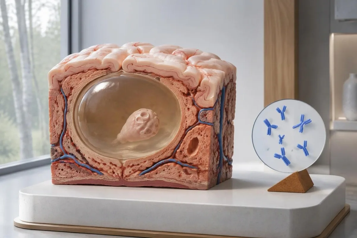
Girekomenda sa giya sa IDSA/ASTMH ang enzyme-linked immunoelectrotransfer blot, o EITB, isip serolohiya nga mokumpirma, ug klarong nagpasidaan nga ubos ang sensitivity kon usa lamang ka parenchymal nga makita o mga calcification lamang ang anaa (White et al., 2018). Dili angay ipahinungod sa usa ka tawo nga adunay 1 ka gamayng abnormalidad sa utok ang taas nga sensitivity nga giulat alang sa mga pasyenteng adunay daghang cyst, sakit sa mga ventricle, o sakit sa subarachnoid.
Hunahunaa, alang lamang sa paghulagway, nga ang usa ka assay adunay 55% nga sensitivity sa 100 ka tawo kansang sakit tungod sa usa ka cyst napamatud-an na pinaagi sa laing paagi. Mokabat sa 45 kanila ang makakuha og negatibong resulta bisan pa og aduna silay sakit; kini nga pananglitan nagpasabot sa dili pagkamatiktik, dili sa kalagmitan nga ang bisan kinsang negatibong resulta nagpasabot og sakit.
Ang pag-usab sa samang assay human sa 2 ka semana mahimong dili makasulbad sa huyang nga tubag tungod sa gamay nga gidaghanon sa sakit. Lahi kini sa mga impeksiyon diin ang sayong pagkuha sa sampol mao ang pangunang problema, sumala sa pagpasabot sa among giya bahin sa sayop nga negatibong resulta tungod sa oras sa pagsulay; pangutan-a kon ang imaging o mas nasabtan pag-ayo nga assay makausab ba sa sunod nga desisyon.
Nganong mahimong magpabiling adunay mga sintomas ang kalkipikadong neurocysticercosis?
Ang neurocysticercosis nga adunay calcification mahimong hinungdan sa seizure bisan pa og negatibo ang antibodies, because the remaining mineralized focus can still be associated with local irritation and episodic surrounding edema. Typical calcifications may measure approximately 1–4 mm, but their small size does not establish whether they are clinically harmless.
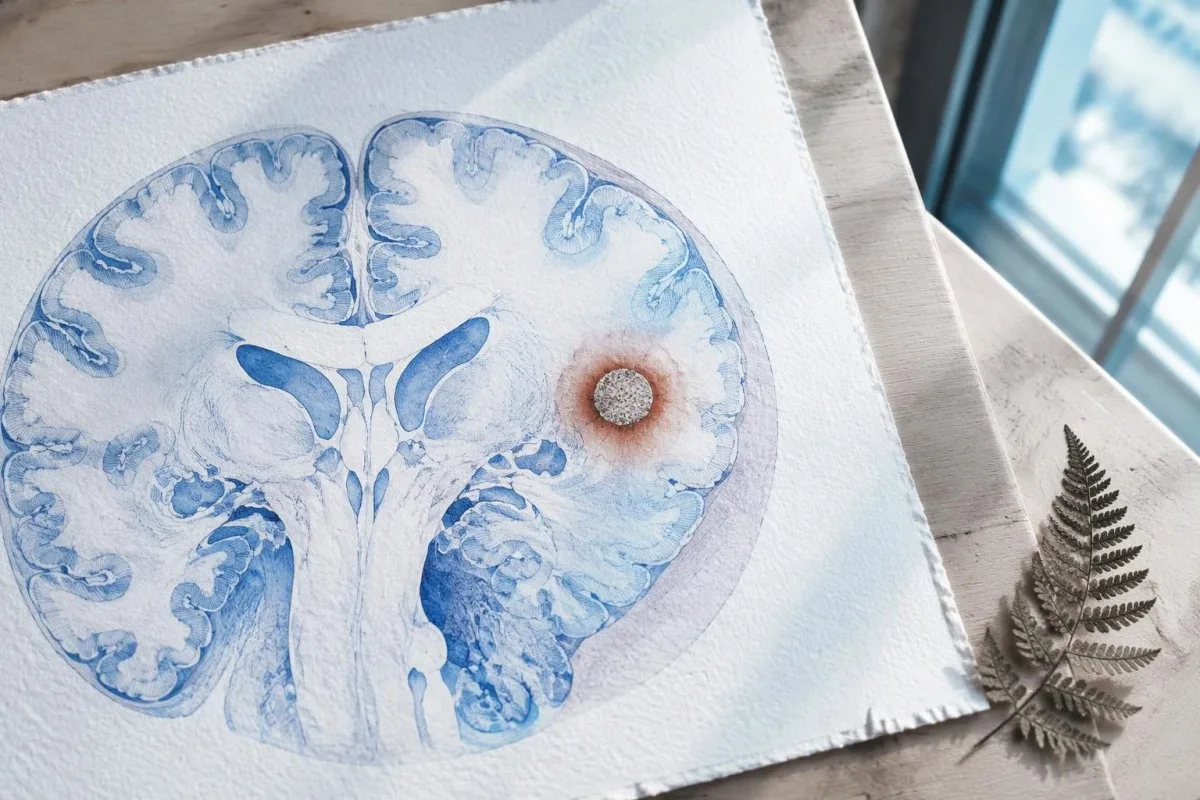
A calcification is generally a remnant rather than a living cyst that antiparasitic medication can eliminate. In the IDSA/ASTMH guideline, calcified-only parenchymal disease is managed symptomatically rather than with antiparasitic drugs; a person with 1 calcified focus and seizures may therefore need antiseizure treatment without needing cyst-killing medication (White et al., 2018).
Surrounding edema can appear intermittently, sometimes long after the original cyst has degenerated. MRI may show this tissue response even when the calcification is more obvious on CT, which is why the 2 scans can tell different but compatible stories rather than one proving that the other is wrong.
Neither a negative antibody result nor a persistent positive result measures seizure risk by itself. The broader lesson resembles other seronegative neurological conditions, discussed in our negative neurological antibody results guide: absence of detectable antibodies is not equivalent to absence of a clinically relevant neurological process.
Unsang pagsulay sa antibody sa cysticercosis ang aktuwal nga gihimo?
The useful first question is whether the report describes EITB, ELISA, or another antibody method, because these are not interchangeable tests. A negative index below 1 laboratory's cutoff cannot inherit the performance figures of a different reference-laboratory assay, even when both reports use the name cysticercosis IgG.
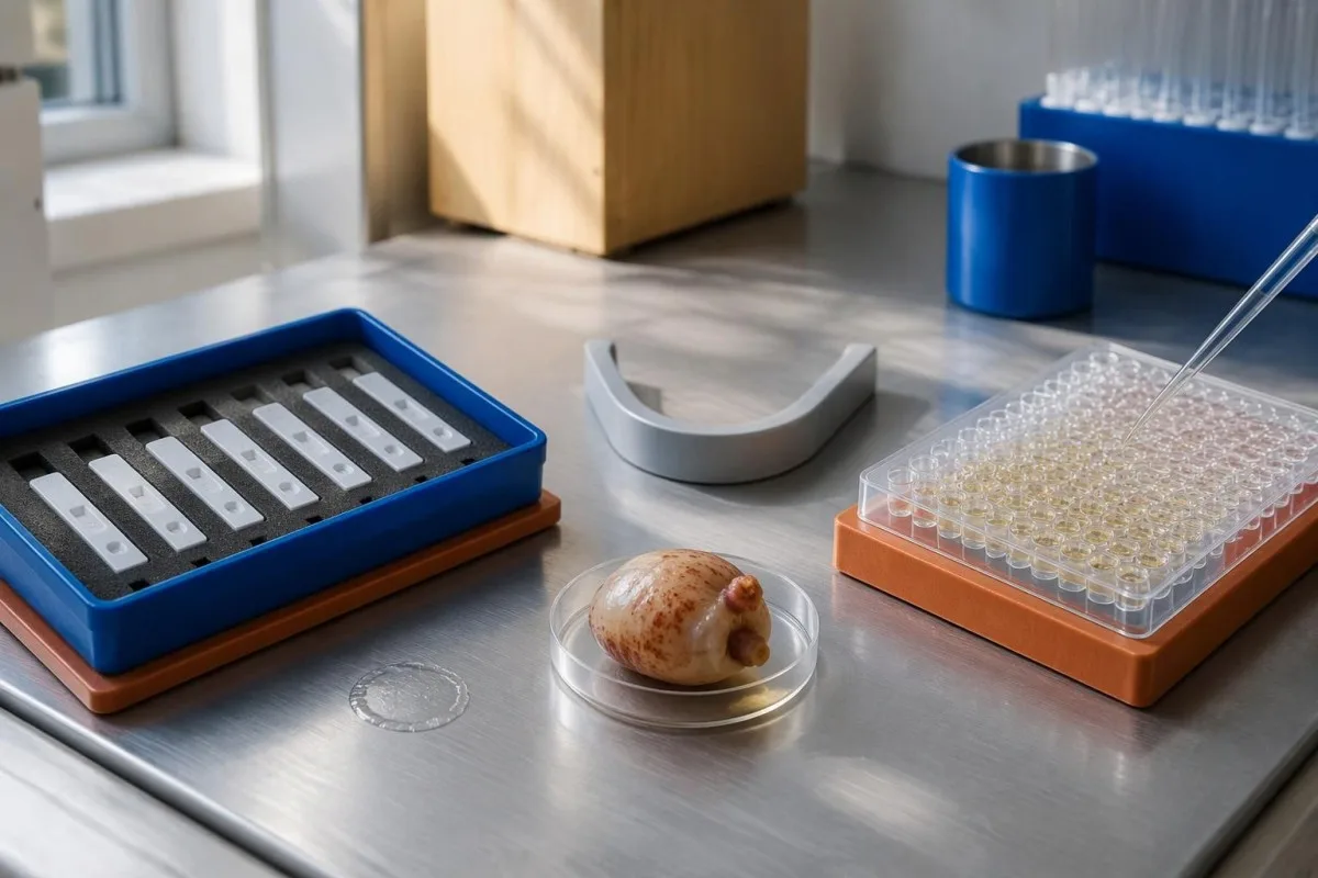
EITB using purified parasite glycoproteins is the preferred serological method in the IDSA/ASTMH guideline, while crude-antigen ELISA methods have recognized sensitivity and specificity limitations (White et al., 2018). Before comparing 2 results, check the method, specimen type, reference interval, and whether the laboratory used a validated confirmatory procedure.
Some laboratories report a qualitative result; others provide an optical-density ratio or numerical index. A value such as 0.8 in a hypothetical assay does not represent 0.8 cysts, an 80% disease probability, or a universal severity score—our qualitative ug quantitative results guide explains that distinction.
Kantesti AI's role is to help readers preserve the laboratory's original wording rather than reinterpret 1 method through another method's thresholds. Our giya sa laboratory marker provides broader report-reading context; for this result, keep the actual assay name available when discussing referral or repeat testing.
Sa unsang paagi mausab sa kasaysayan sa pagkaladlad ang pagduda sa diagnosis?
Neurocysticercosis follows ingestion of Taenia solium eggs, usually through fecal contamination from a person carrying the intestinal tapeworm. Eating undercooked pork can cause intestinal taeniasis, but brain cysts arise through a different transmission step; symptoms may emerge years after exposure rather than within 1 recent trip.

A vegetarian can develop cysticercosis because eggs can contaminate food or hands independently of meat consumption. Useful history includes past residence, household contacts, sanitation conditions, and possible exposure to a tapeworm carrier; asking only about meals eaten during the last 2 weeks can miss the relevant background.
A negative stool examination does not exclude cysts in the brain. Stool testing addresses intestinal tapeworm carriage, whereas CT and MRI assess nervous-system findings; these are 2 different sites and clinical questions, although public-health teams may recommend evaluating contacts when a carrier could be involved.
Normal eosinophils also cannot exclude neurocysticercosis, and raised eosinophils do not identify the brain as the affected site. Our parasite antibody interpretation guide discusses a separate parasite with similar localization limits; the practical point is to avoid using 1 routine cell count as a substitute for targeted neurological evaluation.
Kanus-a kinahanglan dad-on sa emerhensiyang pagsusi ang mga bag-ong seizure?
A first known seizure needs urgent medical assessment regardless of antibody results, and emergency services should be contacted for a first seizure or an ongoing seizure with concerning features. A convulsive seizure lasting 5 minutes, repeated seizures without recovery, breathing difficulty, pregnancy, or significant injury requires emergency care.

Do not wait for the 5-minute mark to seek help after a first known seizure. Keep the person away from hazards, cushion the head if safely possible, do not restrain them or put anything in their mouth, and place them on their side once convulsions stop if they are breathing normally.
Persistent confusion, a severe new headache, fever with neck stiffness, or new weakness increases concern for causes beyond cysticercosis. Sudden one-sided weakness or speech difficulty also demands emergency assessment, as our giya sa mga timailhan sa pasidaan sa stroke explains; 1 negative infection test cannot rule out stroke, meningitis, or a structural brain problem.
After a suspected seizure, stop driving and avoid swimming alone, heights, or operating machinery until a clinician provides advice consistent with local rules. If a witness can safely record the duration and describe movements or recovery, those observations may be more useful than ordering a second antibody test before the first neurological assessment.
Unsay dugang ikatampo sa CT sa pagdayagnos sa neurocysticercosis?
Noncontrast CT is particularly useful for detecting calcified cyst remnants, including small approximately 1–4 mm foci that MRI may show less clearly. CT also helps assess urgent structural problems such as hydrocephalus, but 1 isolated calcification is not automatically proof of neurocysticercosis or the cause of a seizure.
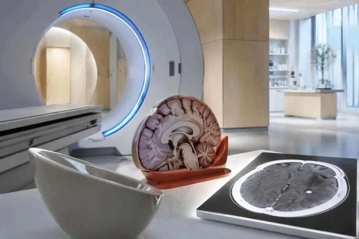
Calcium appears dense on CT, allowing radiologists to identify mineralized remnants and describe their distribution. The IDSA/ASTMH guideline recommends both MRI and noncontrast CT when classifying newly diagnosed neurocysticercosis, because the 2 modalities have complementary strengths rather than being interchangeable alternatives (White et al., 2018).
An incidental calcification needs context: previous granulomatous disease, other causes of mineralization, and scan appearance can influence interpretation. If 1 calcification sits near a suspected seizure focus, concordance with symptoms and EEG may strengthen the explanation, but anatomical proximity alone cannot establish causation.
A laboratory marker used in selected head-injury pathways does not test for brain cysts or replace imaging after an unexplained seizure. Our head-injury CT decisions guide illustrates that narrower setting; ask the clinician whether the present question requires noncontrast CT, contrast imaging, MRI, or a combination of 2 modalities.
Unsay makita sa MRI nga basin dili makita sa CT?
MRI is better suited to evaluating viable cysts, surrounding edema, and many ventricular or subarachnoid findings. Specialized 3-dimensional sequences such as CISS or FIESTA can improve visualization of cyst membranes within cerebrospinal fluid spaces, where a cyst may otherwise resemble the surrounding fluid.
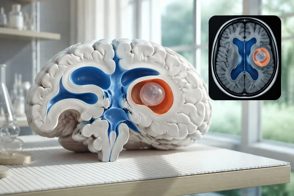
A cyst inside the ventricles can obstruct cerebrospinal fluid flow and cause hydrocephalus even without numerous parenchymal cysts. Headache with vomiting, declining alertness, or new visual symptoms therefore changes urgency; 1 reassuring antibody result should not override symptoms that suggest raised pressure or impaired fluid circulation.
MRI may show a scolex, the small internal structure of the parasite, inside a compatible cyst. This finding can be highly specific when confidently identified, whereas ring enhancement alone has a broader differential; our brain imaging warning signs guide explains why structural abnormalities require interpretation rather than diagnosis from 1 visual feature.
A routine MRI protocol may not answer every cysticercosis question. If ventricular or subarachnoid disease is suspected, discuss whether thin-section 3-dimensional imaging is needed; radiologists choose sequences and contrast according to the clinical question, and a normal scan using 1 protocol does not prove every possible disease compartment has been adequately assessed.
Sa unsang paagi madayagnos ang neurocysticercosis kon dili magkauyon ang mga resulta sa pagsulay?
Neurocysticercosis diagnosis combines imaging, clinical findings, exposure information, and supporting tests, rather than requiring a positive antibody result in every case. The revised diagnostic criteria published in 2017 give characteristic neuroimaging findings a central role and distinguish strong evidence from less-specific supportive features (Del Brutto et al., 2017).
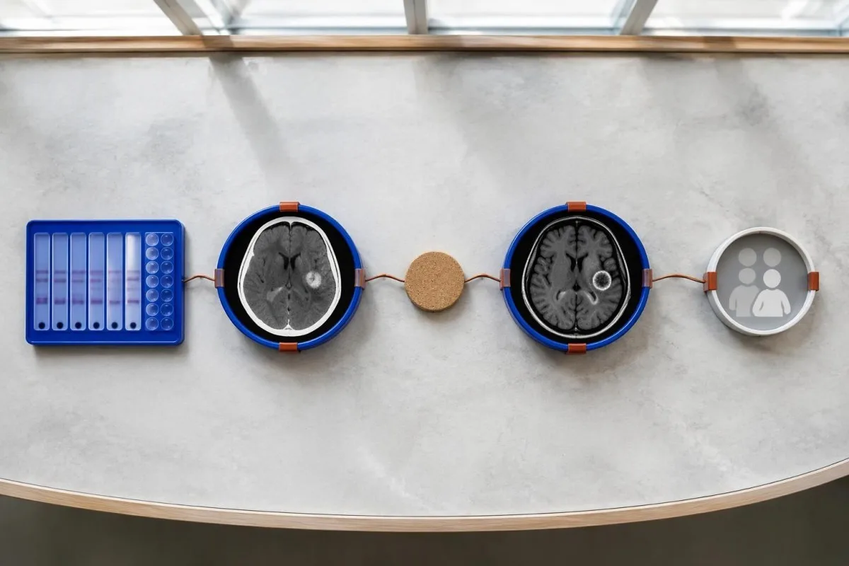
Evidence should be weighted, not tallied. A confidently visualized scolex inside a compatible cyst is different from 1 nonspecific ring-enhancing area, while a positive antibody result is different from direct evidence that a finding is in the brain; our giya sa positibo nga resulta sa antibody explains why detection alone cannot establish location.
For a clearly hypothetical probability example, start with a 30% pretest probability and assume a single-cyst assay has 55% sensitivity and 99% specificity. In 1,000 comparable people, about 135 affected and 693 unaffected people would test negative, leaving approximately a 16% probability of disease among negative results—still substantial, although these assumed figures are not a personal risk calculator.
The most useful disagreement to investigate is whether each test is answering the same question. A negative antibody test, a CT calcification, and MRI edema can all be compatible with 1 calcified disease process; unexplained discrepancies should prompt review of the assay and images rather than automatic dismissal of either result.
Unsa pa ang mahimong mahisama sa usa ka cyst sa mga hulagway sa utok?
A single ring-enhancing brain finding is not specific to neurocysticercosis. Other possibilities include tuberculoma, an abscess, a neoplasm, or an inflammatory process; clinicians use appearance, location, immune status, symptoms, and sometimes follow-up imaging or tissue examination to distinguish these possibilities rather than relying on 1 antibody panel.
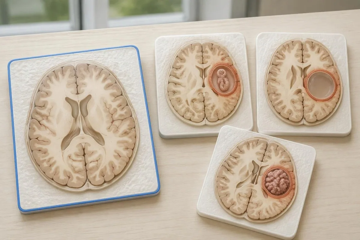
Edema, restricted diffusion, internal structures, and the number of findings all contribute to the differential, but no single nonspecific feature settles every case. Fever and immune suppression may change the suspected cause; a 2 cm ring-enhancing area in a hypothetical immunocompromised person warrants a different investigation from a tiny calcification in an otherwise well person.
Toxoplasmosis is one consideration in selected immunocompromised patients, and a positive IgG result can indicate past exposure rather than prove an active brain process. Our Toxoplasma IgG interpretation guide explains that laboratory distinction; imaging and the patient's immune status determine whether the result is clinically relevant to 1 or more cerebral findings.
New seizures accompanied by rapidly evolving memory problems, psychiatric changes, or abnormal movements can also suggest encephalitis rather than a solitary parasite cyst. Our encephalitis diagnostic pathway describes how MRI, EEG, and cerebrospinal fluid testing answer different questions; a second negative cysticercosis antibody result does not investigate those alternatives.
Masulbad ba sa pagsulay sa antigen, PCR, o CSF ang kawalay-kasigurohan?
Antigen tests, PCR, and cerebrospinal fluid analysis can add information in selected cases, particularly when extraparenchymal disease is suspected. None is a universal rescue test after 1 negative antibody result: availability, assay validation, disease compartment, and safety of lumbar puncture determine whether additional testing is appropriate.
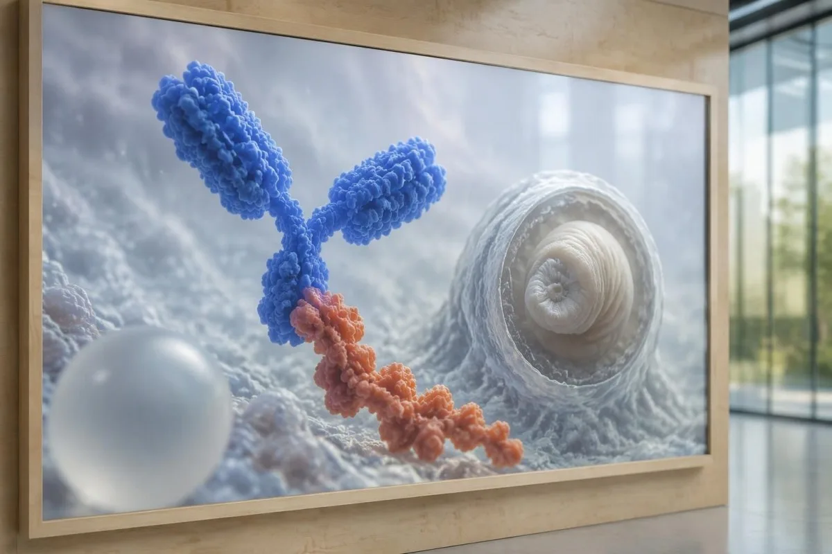
Antibody testing detects the host response, while antigen testing aims to detect parasite-derived material and may better reflect viable disease burden. Antigen detection is often less useful when only 1 small parenchymal cyst or calcifications are present; molecular testing is used in some specialist settings, but performance cannot be generalized across every laboratory or specimen.
Lumbar puncture is not routinely required for an uncomplicated solitary parenchymal finding and can be unsafe when obstructive hydrocephalus or significant mass effect is present. Cerebrospinal fluid tests for other disorders also require disease-specific interpretation, as our CSF band interpretation guide explains; 1 abnormal CSF marker does not identify every possible cause.
Si Kantesti usa ka AI pagsabot sa resulta sa blood test platform, not a service that orders lumbar punctures, identifies cysts on scans, or confirms neurological infections. Our teknolohiya sa interpretasyon sa AI explains the laboratory-report workflow; when 2 tests disagree, our role is to clarify their stated meanings and limitations, not select an invasive diagnostic procedure.
Nganong nagdepende ang pagtambal sa yugto ug lokasyon sa cyst?
Treatment depends on whether cysts are viable, degenerating, calcified, or located outside the brain tissue, not simply on antibody positivity. Selected viable parenchymal cases receive antiparasitic treatment, whereas calcified-only disease generally receives symptom-directed care; ventricular obstruction may require a procedural approach rather than 1 standard medication regimen.
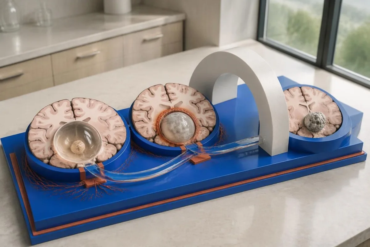
For selected patients with 1–2 viable parenchymal cysts, the IDSA/ASTMH guideline describes albendazole treatment for 10–14 days with appropriate accompanying care. This is a specialist-directed recommendation, not a home-treatment instruction: antiparasitic drugs can provoke an inflammatory response, and untreated hydrocephalus or diffuse cerebral edema changes the order of management (White et al., 2018).
WHO's 2021 guidance focuses on defined presentations of parenchymal neurocysticercosis, while the IDSA/ASTMH guideline also addresses ventricular, subarachnoid, and other forms in detail. Differences in imaging access and disease compartment explain why 1 treatment recommendation cannot be transferred to every patient with an antibody result; the guidelines do not turn negative serology into a rule-out test.
Corticosteroids may accompany antiparasitic therapy, but prolonged use requires additional safety planning, including assessment for relevant latent infections. Our Strongyloides and steroid safety guide explains a separate risk that matters before immune-suppressing treatment; an eye examination is also recommended before antiparasitic therapy to evaluate possible intraocular cysts.
Unsa ang angay bantayan sa follow-up gawas sa mga resulta sa antibody?
Follow-up should track seizures, neurological symptoms, imaging changes, and medication safety, rather than use antibody disappearance as proof of recovery. For viable parenchymal cysts, the IDSA/ASTMH guideline suggests repeat MRI at least every 6 months until the cystic component resolves, with earlier reassessment when symptoms change.
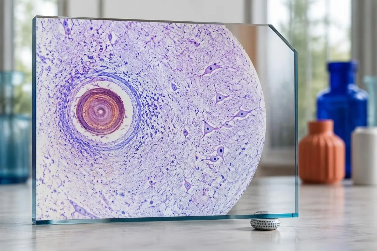
Antibodies may remain detectable after the original disease process has changed, and low-burden disease may never produce a positive result. If 1 new seizure occurs during follow-up, clinicians consider adherence, medication interactions, recurrent edema, and other causes; a rising or falling antibody index alone cannot distinguish among those explanations.
Kantesti AI can help explain reported laboratory results during medication follow-up, but a drug concentration is not a stand-alone measure of seizure control. Our paghubad sa lebel sa levetiracetam guide explains why sample timing and kidney function matter; 2 measurements collected at different times after dosing may not be directly comparable.
Monitoring depends on the chosen drug and treatment duration: prolonged albendazole therapy can require liver-test and blood-count surveillance, while some antiseizure medicines can affect sodium. Our giya sa kaluwasan sa electrolyte discusses those separate laboratory concerns; do not stop an antiseizure medicine or change its dose because 1 cysticercosis antibody result is negative.
Unsa ang mapasabot sa Kantesti, ug unsa ang dili mapamatud-an sa panukiduki?
AI report interpretation cannot confirm or exclude neurocysticercosis from laboratory results alone. Ang Kantesti usa ka AI-powered blood test analysis tool whose role here is to organize reported laboratory findings; a negative result still needs clinical context, and 1 new seizure takes priority over reviewing an automated explanation.

Prepare a focused review packet with the original antibody report, assay method, CT or MRI report, symptom timeline, and medication list. These 5 information categories reduce the risk of discussing a result without its context; ask specifically whether the imaging suggests viable, degenerating, calcified, ventricular, or subarachnoid disease and which unanswered question the next test would address.
The 2 Kantesti publications listed below are a repository-deposited global analysis report and an educational RDW guide, not clinical evidence validating neurocysticercosis diagnosis. Our RDW ug CBC guide can support interpretation of a separately ordered blood count, but RDW, MCV, and MCHC do not establish whether a brain cyst is present.
Evidence for the neurological recommendations in this article comes from the cited diagnostic criteria and clinical guidelines, not from general AI benchmarking or laboratory-report volume. Our technical validation scope should be read within its stated boundaries; neither 1 successful report explanation nor a large aggregate dataset substitutes for condition-specific diagnostic validation.
Mga kanunayng gipangutana
Mahimo ba kang adunay neurocysticercosis bisan negatibo ang resulta sa pagsulay sa dugo?
Oo, mahimong anaa ang neurocysticercosis bisan negatibo ang pagsulay sa mga antibody sa dugo, ilabi na kon adunay 1 ka cyst sa utok o kalkipikado lamang ang sakit. Ang mga banabana gikan sa gipatik nga mga reperensiyang laboratoryo gamit ang EITB kasagarang nagpakita og pagkasensitibo nga mga 50–60% kon usa ra ka cyst, nga mas ubos pag-ayo kaysa sa kaepektibo niini kon daghan ang buhing mga cyst. Busa, nagpabiling mahinungdanon sa pagdayagnos ang CT, MRI, mga sintomas, ug kasaysayan sa pagkaladlad. Ang bag-ong kombulsyon nanginahanglan og dinaliang pagsusi, dili pagpasalig tungod lang sa negatibong resulta.
Unsa ang labing kasaligan nga pagsulay alang sa neurocysticercosis?
Walay usa ka test alang sa neurocysticercosis nga makatubag sa tanang pangutana sa pagdayagnos; nagtinabangay ang imaging sa utok ug ang serology nga gipili sumala sa panginahanglan. Ang EITB mao ang gipalabi nga pamaagi sa pagkumpirma sa mga antibody sa giya sa IDSA/ASTMH, apan mahimong dili niini mamatikdan ang sakit nga usa ra ka cyst ang nalangkit. Labi nang mapuslanon ang CT nga walay contrast sa pag-ila sa mga pagkalime; samtang mas maayo ang MRI sa pagtimbang-timbang sa daghang cyst nga buhi pa, edema, ug mga abnormalidad sa mga luna nga adunay pluwido. Gihiusa sa mga klinisyan ang maong mga kaplag imbis nga isipon nga hingpit nang pamatuod ang usa ka negatibong resulta.
Mas maayo ba ang CT o MRI para sa neurocysticercosis?
Lahi ang mga bentaha sa CT ug MRI sa pagdayagnos sa neurocysticercosis. Kasagaran mas klarong makakita ang CT nga walay contrast sa gagmayng kalipikasyon nga mga 1–4 mm ang gilapdon, samtang gisusi sa MRI ang mga buhing cyst, ang palibot nga edema, ug ang sakit sa mga ventricle o sa subarachnoid nga bahin. Ang mga espesyal nga 3-dimensional nga han-ay sa MRI makatabang sa pag-ila sa mga lamad sa cyst sulod sa mga luna sa cerebrospinal fluid. Girekomenda sa giya sa IDSA/ASTMH ang paggamit sa duha ka pamaagi sa pagklasipikar sa bag-ong nadayagnos nga sakit.
Makapahinabo gihapon ba og kombulsyon ang gikalsipikar nga cyst?
Ang nahibiling calcified nga cysticercosis mahimong magpadayon nga nalangkit sa mga seizure bisan og dili na kini buhi nga cyst. Ang pipila ka calcified nga mga dapit, kasagaran 1–4 mm ra ang gilapdon, makaugmad og panagsang edema sa palibot nga makita sa MRI. Dili makapahikap sa posibilidad niini nga mekanismo ang negatibong resulta sa mga antibody, ug ang padayon nga positibong resulta sa mga antibody dili sukod sa risgo sa seizure. Ang sakit nga calcified lamang kasagaran tambalan sumala sa mga sintomas, imbes nga hatagan og tambal batok sa parasito.
Kinahanglan ba nakong usbon ang negatibong pagsusuri sa antibody sa cysticercosis?
Dili awtomatikong labing maayong sunod nga lakang ang pag-usab sa negatibong resulta sa pagsusuri sa antibody alang sa cysticercosis. Kung adunay 1 ka gamay nga cyst o kalkipikadong sakit, mahimo gihapong negatibo ang huyang nga tubag sa antibody bisan usbon ang samang assay human sa 2 ka semana. Hinuon, mahimong repasohon sa clinician ang orihinal nga pamaagi, mangayo og mas maayong pagkaila nga confirmatory assay, o unahon ang CT ug MRI. Dili angay hulaton ang pagsulay pag-usab kon adunay bag-ong mga seizure o nagkagrabeng mga sintomas sa nerbiyos.
Kanus-a ko mangayo og emerhensiyang tabang kon unang higayon nakong makuyapan tungod sa kombulsyon?
Ang unang nahibalang kombulsyon nagkinahanglan og dinaliang medikal nga pagsusi ug pagkontak sa lokal nga serbisyong pangemerhensya, bisan pa og negatibo ang resulta sa pagsulay sa mga antibody batok sa neurocysticercosis. Pangemerhensya ang kombulsyon nga molungtad og 5 minutos, balik-balik nga mga kombulsyon nga walay pagbalik sa panimuot, kalisod sa pagginhawa, pagmabdos, o dakong kadaot. Ayaw pugngi ang tawo o butangi og bisan unsa ang iyang baba, ug ipahigda siya sa kilid kon mohunong na ang mga kombulsyon ug normal ang iyang pagginhawa. Ayaw pagmaneho hangtod masusi na sa usa ka clinician ang hitabo ug mapasabot ang mga limitasyong angay sundon.
Paipatin-aw ang imong resulta sa blood test
I-upload ang report sa imong laboratoryo ug ipatin-aw sa AI sa Kantesti ang matag biomarker sa yano nga pinulongan sulod sa pipila ka segundo. Gigamit sa kapin sa 2 ka milyon ka tawo sa tibuok kalibotan.
Mga Reperensya
- Klein, T., Mitchell, S., & Weber, H. (2026). AI Blood Test Analyzer: 2.5M Tests Analyzed | Global Health Report 2026. Panukiduki Medikal sa Kantesti AI. DOI
- Klein, T., Mitchell, S., & Weber, H. (2026). Pagsulay sa RDW sa Dugo: Kompletong Giya sa RDW-CV, MCV ug MCHC. Panukiduki Medikal sa Kantesti AI. DOI
- White AC Jr et al. (2018). Diagnosis and Treatment of Neurocysticercosis: 2017 Clinical Practice Guidelines by the Infectious Diseases Society of America (IDSA) and the American Society of Tropical Medicine and Hygiene (ASTMH). Clinical Infectious Diseases. DOI
- Del Brutto OH et al. (2017). Revised diagnostic criteria for neurocysticercosis. Journal of the Neurological Sciences. DOI
- World Health Organization (2021). WHO guidelines on management of Taenia solium neurocysticercosis. Organisasyon sa Kahimsug sa Kalibutan.
- 2M+Mga gisusi nga eksaminasyon
- 127+Mga nasud
- 100+Mga pinulongan
Mahitungod sa medikal nga team


Mga E-E-A-T Trust Signals
Kasinatian
Pagsusi sa klinika nga gipangulohan sa doktor sa mga workflow sa interpretasyon sa lab.
Kahanas
Pokus sa medisina sa laboratoryo kung giunsa paglihok ang mga biomarker sa konteksto sa klinika.
Pagka-awtorisado
Gisusi sa medisina ni Dr. Thomas Klein, uban sa mga kontribusyon ni Dr. Sarah Mitchell ug Prof. Dr. Hans Weber.
Kasaligan
Interpretasyon nga base sa ebidensya, nga adunay klaro nga mga agianan sa sunod nga buhat aron makunhuran ang kabalaka.





Disclaimer sa medisina
Kini nga artikulo para sa katuyoan sa edukasyon ra ug dili kini mosangpot sa medikal nga tambag. Kanunay mokonsulta sa usa ka kwalipikado nga healthcare provider alang sa mga desisyon sa diagnosis ug pagtambal.
Gibilin · Kontaka ang team sa editoryal
Gipatik sa Kantesti Ltd, narehistro sa England ug Wales, numero sa kompanya. 17090423, London, United Kingdom.
