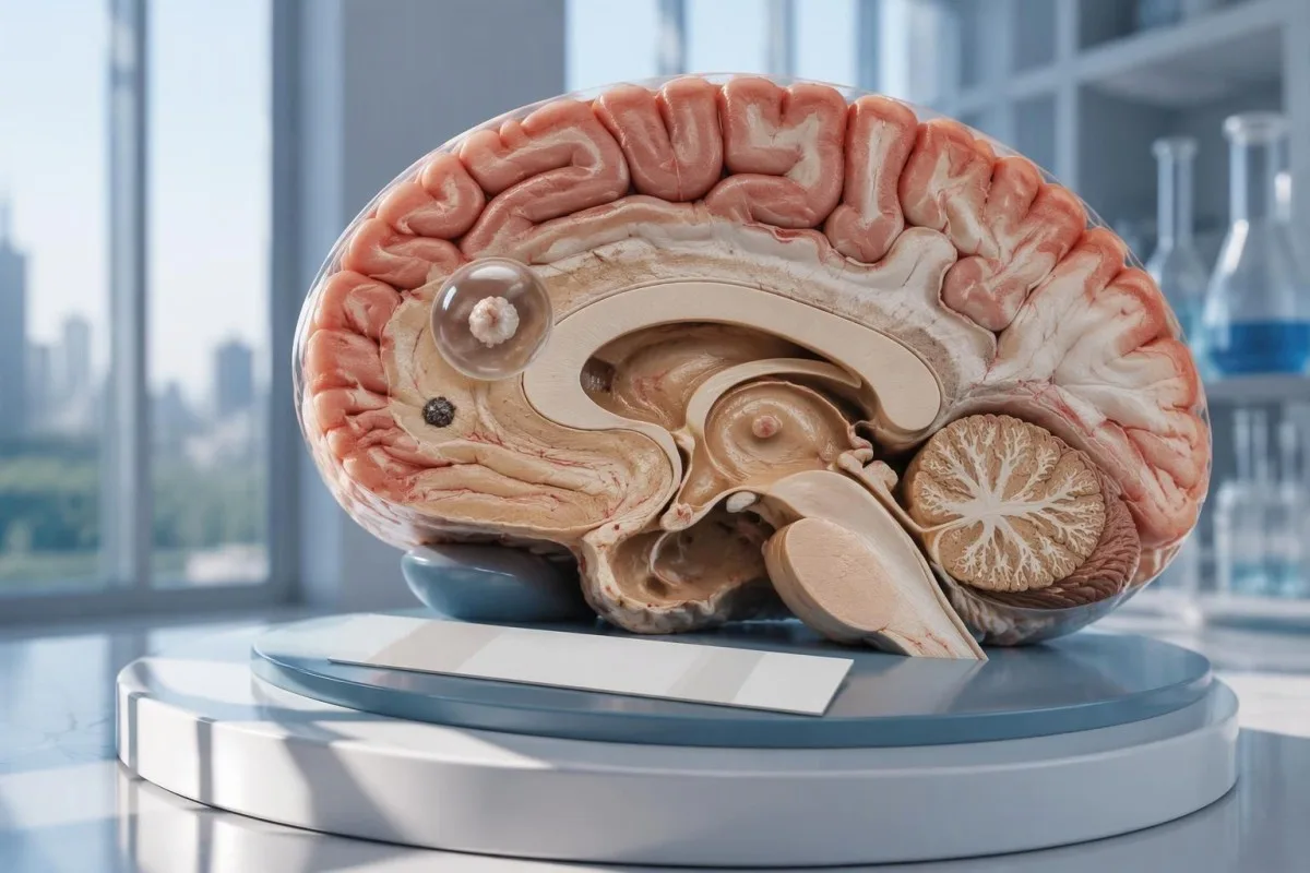
Отрицательный тест на нейроцистицеркоз не исключает наличие одиночной кисты головного мозга или кальцинированного заболевания. КТ, МРТ и срочная оценка при впервые возникших судорогах важнее, чем успокоение на основании одного лишь тестирования на антитела.

Основные выводы
- Отрицательные антитела не исключают нейроцистицеркоз, особенно если на снимках выявлена лишь 1 пораженная область или кальцинаты.
- Референс-лабораторный EITB может иметь чувствительность до 98% при наличии более 1 жизнеспособной кисты, однако при одиночной кисте этот показатель значительно ниже.
- Чувствительность при одиночной кисте в опубликованных оценках для EITB часто составляет приблизительно 50–60%; эти показатели не относятся ко всем коммерческим тестам на антитела.
- Нативная КТ особенно эффективна для выявления небольших кальцинатов, размер которых часто составляет приблизительно 1–4 мм.
- МРТ позволяет оценить жизнеспособные кисты, окружающий отек и поражение желудочков или субарахноидального пространства; специализированные трехмерные последовательности могут повысить выявляемость.
- Первые судорожные приступы требуют срочного медицинского обследования независимо от результата анализа на антитела. Вызовите скорую помощь при первом известном приступе судорог или появлении других экстренных симптомов.
- Пять минут порог экстренного вмешательства при продолжающемся судорожном приступе, а не повод ждать, прежде чем обратиться за помощью.
- Только кальцифицированная форма заболевания обычно не требует противопаразитарной терапии, но лечение судорог и дополнительная визуализация всё же могут быть необходимы.
Может ли отрицательный тест на нейроцистицеркоз исключить заболевание?
A отрицательный тест на нейроцистицеркоз не позволяет надёжно исключить заболевание, особенно при наличии 1 кисты головного мозга или кальцифицированных остатков. Анализ на антитела измеряет иммунный ответ, а не исследует непосредственно головной мозг; КТ и МРТ определяют расположение и стадию подозрительных изменений, а первый приступ судорог требует срочного обследования.

Результат условный, а не абсолютный. Анализ на антитела к цистицеркозу, который хорошо работает у людей с несколькими жизнеспособными кистами, может плохо работать у человека всего с 1 небольшим поражённым участком, поэтому слово отрицательный означает отсутствие обнаруживаемого ответа в этом анализе, а не отсутствие кист где-либо в нервной системе.
Для условного примера рассмотрим 34-летнего человека с первым приступом судорог, отрицательным результатом анализа на антитела и одиночным кольцевидно контрастирующимся очагом на МРТ. Результат анализа не позволяет установить, является ли этот очаг проявлением цистицеркоза, другой инфекции или неинфекционного процесса; его значение определяется совокупностью данных сканирования, сведений о возможном заражении и неврологического обследования.
Кантести — это Анализатор анализа крови на основе искусственного интеллекта который может помочь объяснить полученные лабораторные результаты, но не позволяет исключить заболевание головного мозга на основании 1 отрицательного результата анализа на антитела. Наша организация и миссия объясняют нашу специализацию на лабораторной информации; читателям не следует воспринимать автоматическое объяснение результатов как разрешение отложить помощь при судорогах.
Почему тестирование на антитела может не выявить одиночную кисту головного мозга?
A одиночная киста может вызвать ответ на антитела ниже порога обнаружения анализа. Согласно опубликованным оценкам, чувствительность EITB в референсных лабораториях часто составляет около 50–60% при одиночной кисте по сравнению с приблизительно 98% при наличии более 1 жизнеспособной кисты; различие отражает степень заболевания и эффективность анализа.
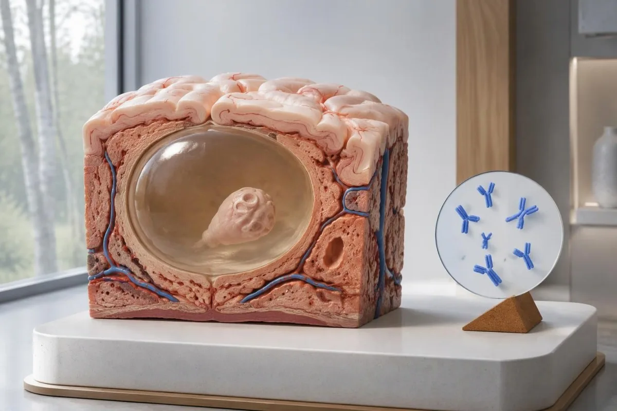
Руководство IDSA/ASTMH рекомендует иммуноблоттинг с фермент-связанным иммунопереносом, или EITB, в качестве подтверждающего серологического исследования и прямо предупреждает о низкой чувствительности при единичном паренхиматозном очаге или только кальцификатах (White et al., 2018). Высокие показатели чувствительности, полученные у пациентов с несколькими кистами, поражением желудочков или субарахноидального пространства, нельзя переносить на случай 1 небольшого патологического изменения в головном мозге.
Представим исключительно для наглядности, что чувствительность анализа составляет 55% у 100 человек, у которых наличие заболевания с одиночной кистой уже было независимо подтверждено. Примерно 45 из них получат отрицательный результат, несмотря на наличие заболевания; этот расчёт иллюстрирует пропуски при выявлении, а не вероятность того, что конкретный отрицательный результат означает наличие заболевания.
Повторение того же анализа через 2 недели может не устранить проблему слабого ответа, обусловленного низкой степенью заболевания. Это отличается от инфекций, при которых основная проблема — слишком ранний забор образца, как объясняется в нашем руководстве по ложноотрицательным результатам, связанным со сроками проведения анализа; уточните, повлияют ли результаты визуализации или более надёжно охарактеризованного анализа на дальнейшее решение.
Почему кальцинированный нейроцистицеркоз может оставаться симптомным?
Кальцифицированный нейроцистицеркоз может вызывать судороги даже при отрицательных результатах анализа на антитела, because the remaining mineralized focus can still be associated with local irritation and episodic surrounding edema. Typical calcifications may measure approximately 1–4 mm, but their small size does not establish whether they are clinically harmless.
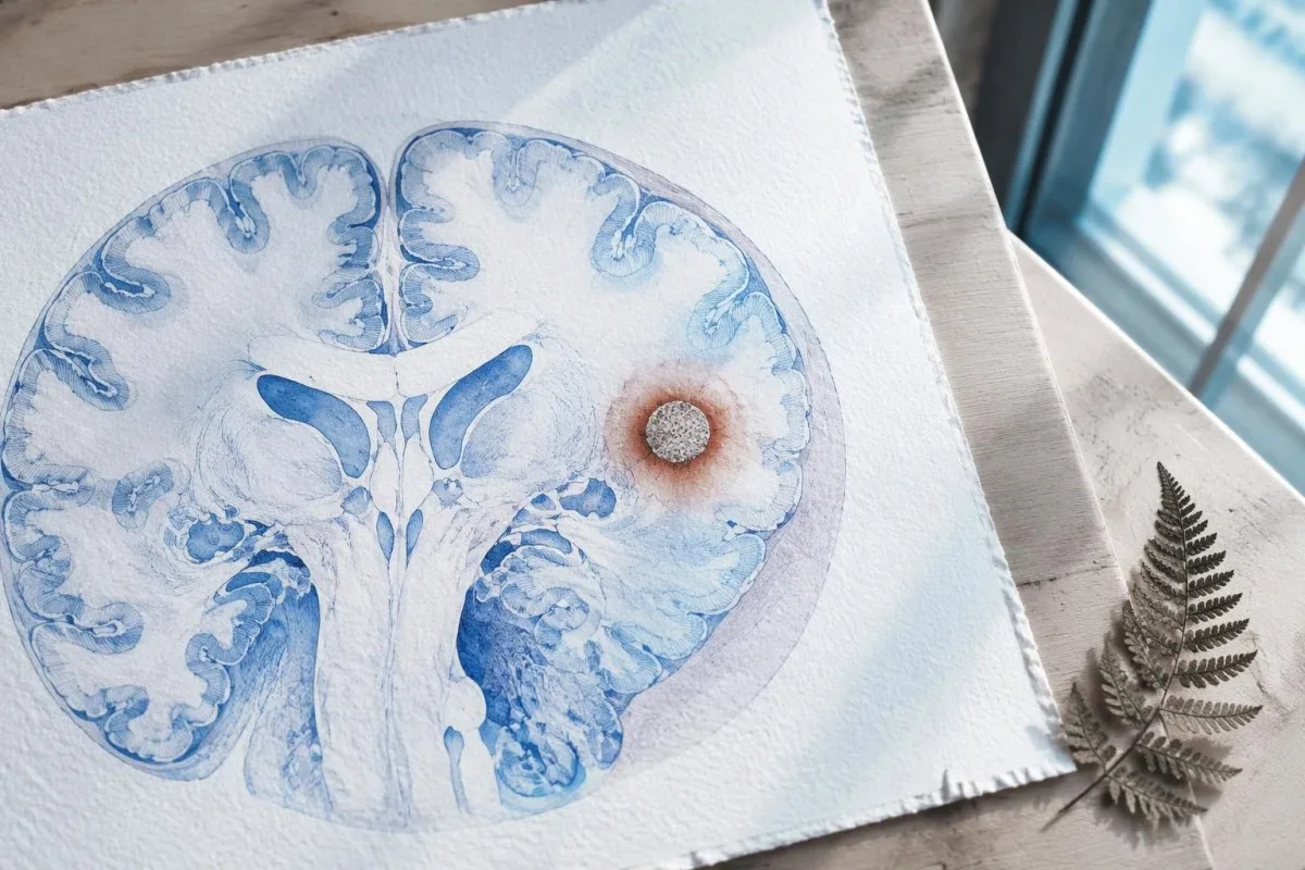
A calcification is generally a remnant rather than a living cyst that antiparasitic medication can eliminate. In the IDSA/ASTMH guideline, calcified-only parenchymal disease is managed symptomatically rather than with antiparasitic drugs; a person with 1 calcified focus and seizures may therefore need antiseizure treatment without needing cyst-killing medication (White et al., 2018).
Surrounding edema can appear intermittently, sometimes long after the original cyst has degenerated. MRI may show this tissue response even when the calcification is more obvious on CT, which is why the 2 scans can tell different but compatible stories rather than one proving that the other is wrong.
Neither a negative antibody result nor a persistent positive result measures seizure risk by itself. The broader lesson resembles other seronegative neurological conditions, discussed in our negative neurological antibody results guide: absence of detectable antibodies is not equivalent to absence of a clinically relevant neurological process.
Какой именно тест на антитела к цистицеркозу был выполнен?
The useful first question is whether the report describes EITB, ELISA, or another antibody method, because these are not interchangeable tests. A negative index below 1 laboratory's cutoff cannot inherit the performance figures of a different reference-laboratory assay, even when both reports use the name cysticercosis IgG.
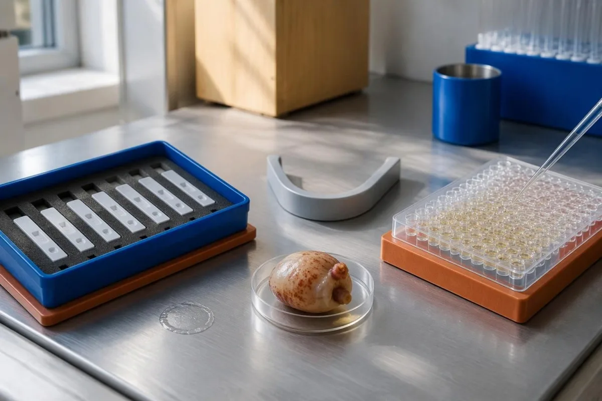
EITB using purified parasite glycoproteins is the preferred serological method in the IDSA/ASTMH guideline, while crude-antigen ELISA methods have recognized sensitivity and specificity limitations (White et al., 2018). Before comparing 2 results, check the method, specimen type, reference interval, and whether the laboratory used a validated confirmatory procedure.
Some laboratories report a qualitative result; others provide an optical-density ratio or numerical index. A value such as 0.8 in a hypothetical assay does not represent 0.8 cysts, an 80% disease probability, or a universal severity score—our качественные и количественные результаты guide explains that distinction.
Kantesti AI's role is to help readers preserve the laboratory's original wording rather than reinterpret 1 method through another method's thresholds. Our руководство по лабораторным маркерам provides broader report-reading context; for this result, keep the actual assay name available when discussing referral or repeat testing.
Как анамнез контактов меняет диагностическую настороженность?
Neurocysticercosis follows ingestion of Taenia solium eggs, usually through fecal contamination from a person carrying the intestinal tapeworm. Eating undercooked pork can cause intestinal taeniasis, but brain cysts arise through a different transmission step; symptoms may emerge years after exposure rather than within 1 recent trip.

A vegetarian can develop cysticercosis because eggs can contaminate food or hands independently of meat consumption. Useful history includes past residence, household contacts, sanitation conditions, and possible exposure to a tapeworm carrier; asking only about meals eaten during the last 2 weeks can miss the relevant background.
A negative stool examination does not exclude cysts in the brain. Stool testing addresses intestinal tapeworm carriage, whereas CT and MRI assess nervous-system findings; these are 2 different sites and clinical questions, although public-health teams may recommend evaluating contacts when a carrier could be involved.
Normal eosinophils also cannot exclude neurocysticercosis, and raised eosinophils do not identify the brain as the affected site. Our parasite antibody interpretation guide discusses a separate parasite with similar localization limits; the practical point is to avoid using 1 routine cell count as a substitute for targeted neurological evaluation.
В каких случаях впервые возникшие судороги требуют экстренной оценки?
A first known seizure needs urgent medical assessment regardless of antibody results, and emergency services should be contacted for a first seizure or an ongoing seizure with concerning features. A convulsive seizure lasting 5 minutes, repeated seizures without recovery, breathing difficulty, pregnancy, or significant injury requires emergency care.

Do not wait for the 5-minute mark to seek help after a first known seizure. Keep the person away from hazards, cushion the head if safely possible, do not restrain them or put anything in their mouth, and place them on their side once convulsions stop if they are breathing normally.
Persistent confusion, a severe new headache, fever with neck stiffness, or new weakness increases concern for causes beyond cysticercosis. Sudden one-sided weakness or speech difficulty also demands emergency assessment, as our руководством по предупреждающим признакам инсульта explains; 1 negative infection test cannot rule out stroke, meningitis, or a structural brain problem.
After a suspected seizure, stop driving and avoid swimming alone, heights, or operating machinery until a clinician provides advice consistent with local rules. If a witness can safely record the duration and describe movements or recovery, those observations may be more useful than ordering a second antibody test before the first neurological assessment.
Что дает КТ для диагностики нейроцистицеркоза?
Noncontrast CT is particularly useful for detecting calcified cyst remnants, including small approximately 1–4 mm foci that MRI may show less clearly. CT also helps assess urgent structural problems such as hydrocephalus, but 1 isolated calcification is not automatically proof of neurocysticercosis or the cause of a seizure.
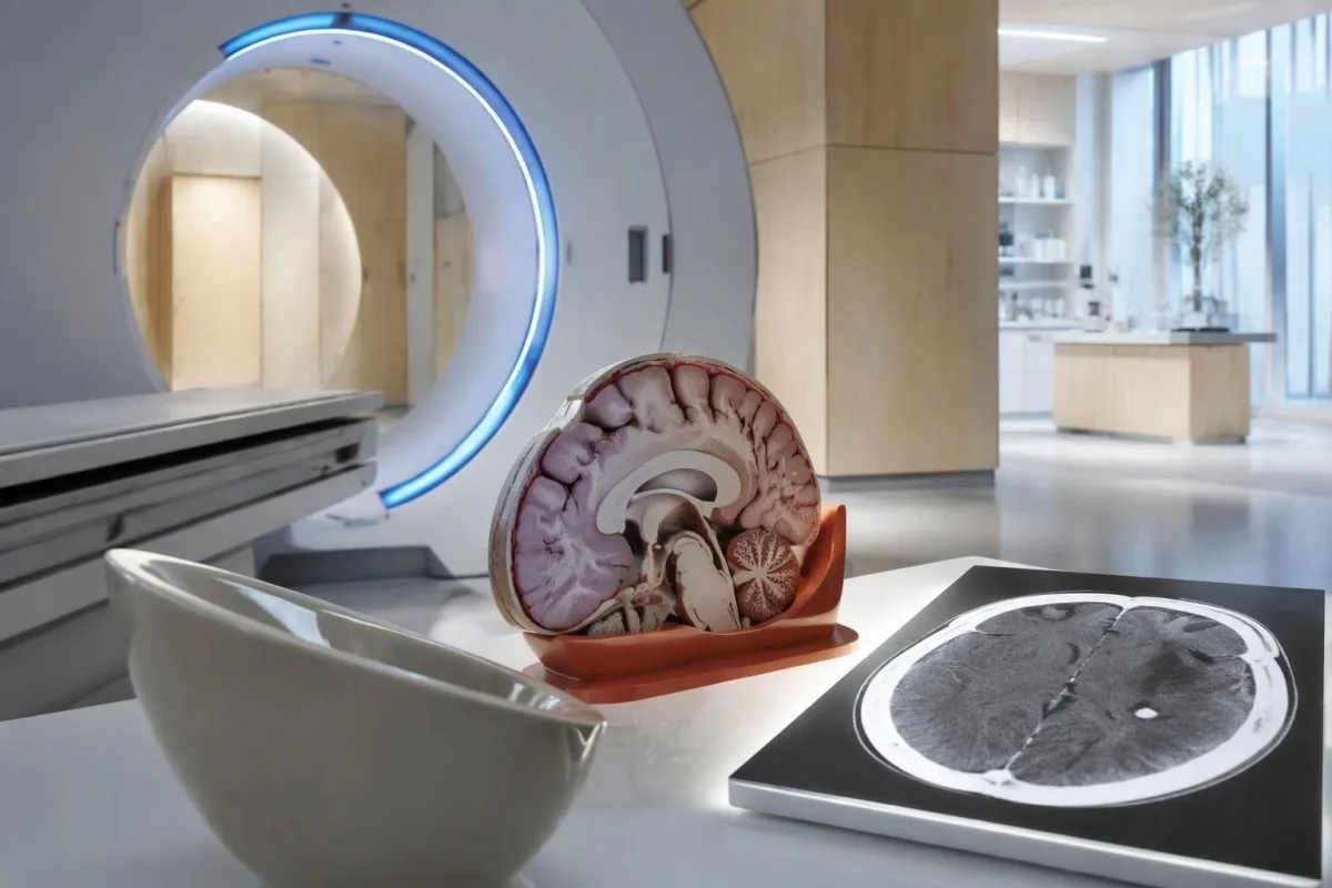
Calcium appears dense on CT, allowing radiologists to identify mineralized remnants and describe their distribution. The IDSA/ASTMH guideline recommends both MRI and noncontrast CT when classifying newly diagnosed neurocysticercosis, because the 2 modalities have complementary strengths rather than being interchangeable alternatives (White et al., 2018).
An incidental calcification needs context: previous granulomatous disease, other causes of mineralization, and scan appearance can influence interpretation. If 1 calcification sits near a suspected seizure focus, concordance with symptoms and EEG may strengthen the explanation, but anatomical proximity alone cannot establish causation.
A laboratory marker used in selected head-injury pathways does not test for brain cysts or replace imaging after an unexplained seizure. Our head-injury CT decisions guide illustrates that narrower setting; ask the clinician whether the present question requires noncontrast CT, contrast imaging, MRI, or a combination of 2 modalities.
Что выявляет МРТ, что может быть не видно на КТ?
MRI is better suited to evaluating viable cysts, surrounding edema, and many ventricular or subarachnoid findings. Specialized 3-dimensional sequences such as CISS or FIESTA can improve visualization of cyst membranes within cerebrospinal fluid spaces, where a cyst may otherwise resemble the surrounding fluid.
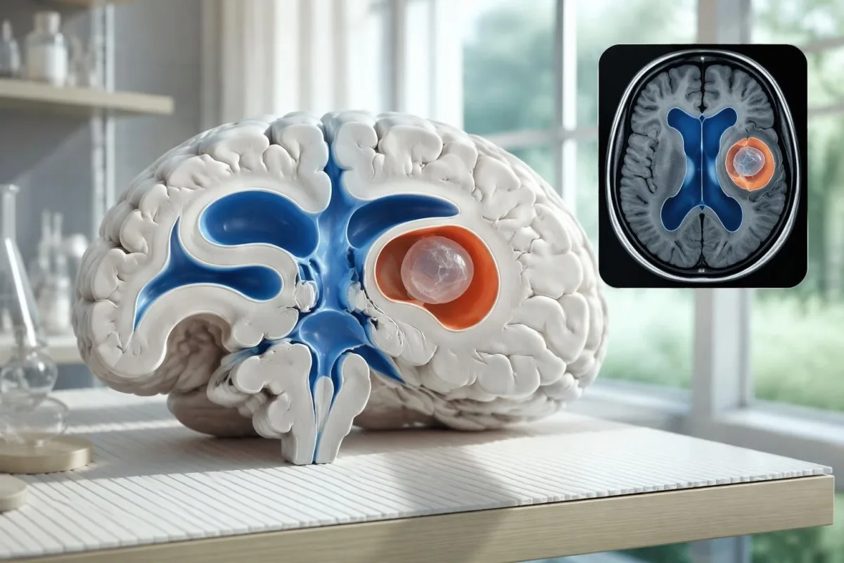
A cyst inside the ventricles can obstruct cerebrospinal fluid flow and cause hydrocephalus even without numerous parenchymal cysts. Headache with vomiting, declining alertness, or new visual symptoms therefore changes urgency; 1 reassuring antibody result should not override symptoms that suggest raised pressure or impaired fluid circulation.
MRI may show a scolex, the small internal structure of the parasite, inside a compatible cyst. This finding can be highly specific when confidently identified, whereas ring enhancement alone has a broader differential; our brain imaging warning signs guide explains why structural abnormalities require interpretation rather than diagnosis from 1 visual feature.
A routine MRI protocol may not answer every cysticercosis question. If ventricular or subarachnoid disease is suspected, discuss whether thin-section 3-dimensional imaging is needed; radiologists choose sequences and contrast according to the clinical question, and a normal scan using 1 protocol does not prove every possible disease compartment has been adequately assessed.
Как диагностируют нейроцистицеркоз, если результаты тестов расходятся?
Neurocysticercosis diagnosis combines imaging, clinical findings, exposure information, and supporting tests, rather than requiring a positive antibody result in every case. The revised diagnostic criteria published in 2017 give characteristic neuroimaging findings a central role and distinguish strong evidence from less-specific supportive features (Del Brutto et al., 2017).
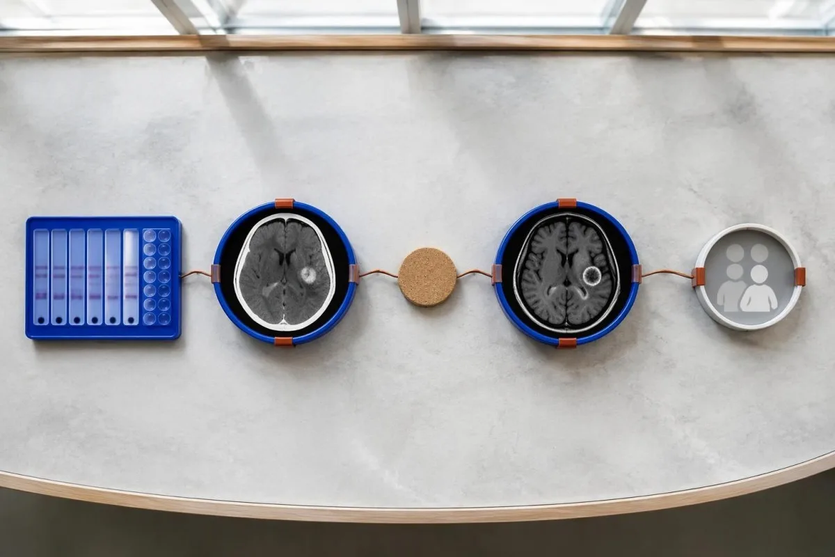
Evidence should be weighted, not tallied. A confidently visualized scolex inside a compatible cyst is different from 1 nonspecific ring-enhancing area, while a positive antibody result is different from direct evidence that a finding is in the brain; our руководство по положительному результату антител explains why detection alone cannot establish location.
For a clearly hypothetical probability example, start with a 30% pretest probability and assume a single-cyst assay has 55% sensitivity and 99% specificity. In 1,000 comparable people, about 135 affected and 693 unaffected people would test negative, leaving approximately a 16% probability of disease among negative results—still substantial, although these assumed figures are not a personal risk calculator.
The most useful disagreement to investigate is whether each test is answering the same question. A negative antibody test, a CT calcification, and MRI edema can all be compatible with 1 calcified disease process; unexplained discrepancies should prompt review of the assay and images rather than automatic dismissal of either result.
Какие еще образования могут напоминать одиночную кисту на снимках головного мозга?
A single ring-enhancing brain finding is not specific to neurocysticercosis. Other possibilities include tuberculoma, an abscess, a neoplasm, or an inflammatory process; clinicians use appearance, location, immune status, symptoms, and sometimes follow-up imaging or tissue examination to distinguish these possibilities rather than relying on 1 antibody panel.
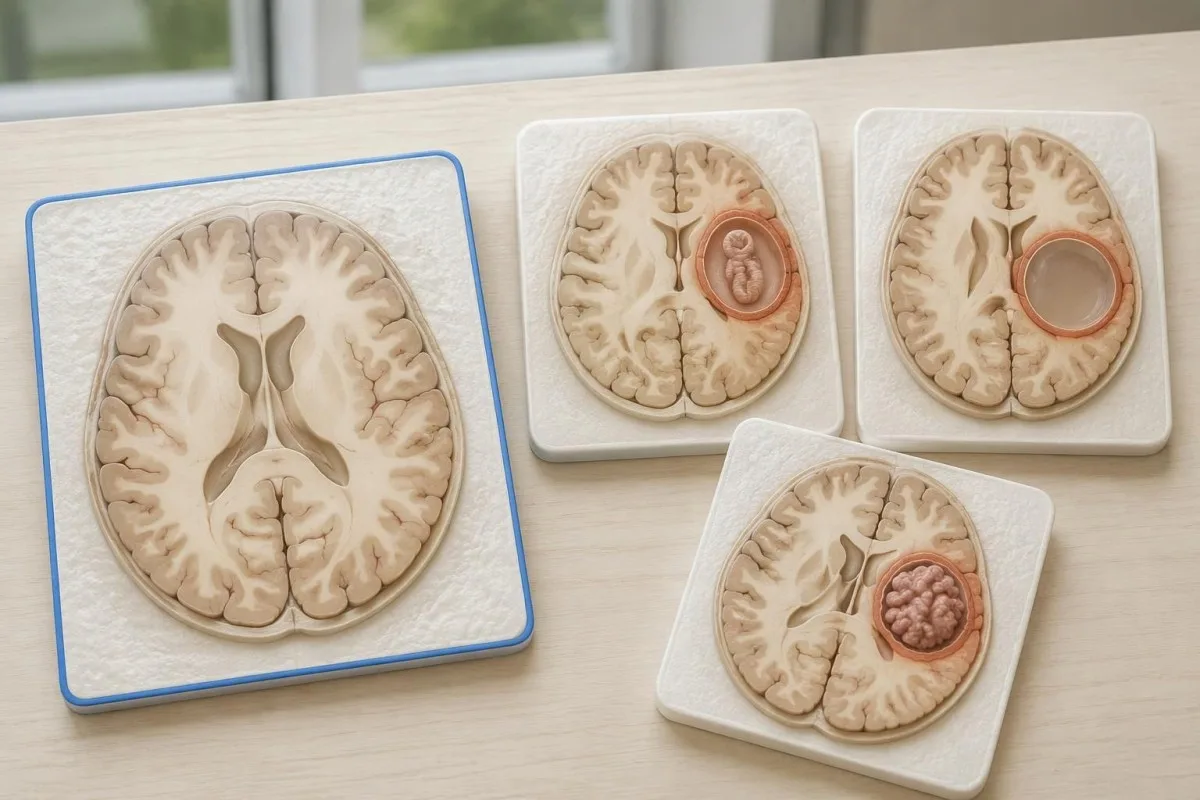
Edema, restricted diffusion, internal structures, and the number of findings all contribute to the differential, but no single nonspecific feature settles every case. Fever and immune suppression may change the suspected cause; a 2 cm ring-enhancing area in a hypothetical immunocompromised person warrants a different investigation from a tiny calcification in an otherwise well person.
Toxoplasmosis is one consideration in selected immunocompromised patients, and a positive IgG result can indicate past exposure rather than prove an active brain process. Our Toxoplasma IgG interpretation guide explains that laboratory distinction; imaging and the patient's immune status determine whether the result is clinically relevant to 1 or more cerebral findings.
New seizures accompanied by rapidly evolving memory problems, psychiatric changes, or abnormal movements can also suggest encephalitis rather than a solitary parasite cyst. Our encephalitis diagnostic pathway describes how MRI, EEG, and cerebrospinal fluid testing answer different questions; a second negative cysticercosis antibody result does not investigate those alternatives.
Могут ли анализ на антиген, ПЦР или исследование ЦСЖ разрешить неопределенность?
Antigen tests, PCR, and cerebrospinal fluid analysis can add information in selected cases, particularly when extraparenchymal disease is suspected. None is a universal rescue test after 1 negative antibody result: availability, assay validation, disease compartment, and safety of lumbar puncture determine whether additional testing is appropriate.
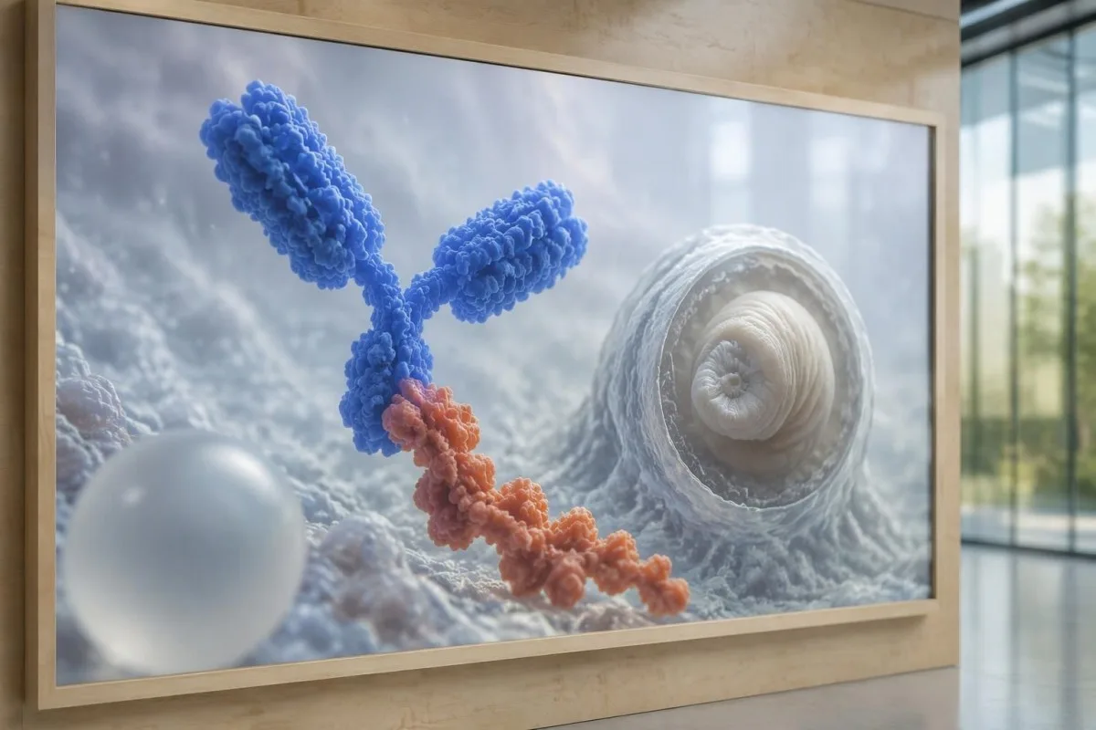
Antibody testing detects the host response, while antigen testing aims to detect parasite-derived material and may better reflect viable disease burden. Antigen detection is often less useful when only 1 small parenchymal cyst or calcifications are present; molecular testing is used in some specialist settings, but performance cannot be generalized across every laboratory or specimen.
Lumbar puncture is not routinely required for an uncomplicated solitary parenchymal finding and can be unsafe when obstructive hydrocephalus or significant mass effect is present. Cerebrospinal fluid tests for other disorders also require disease-specific interpretation, as our CSF band interpretation guide explains; 1 abnormal CSF marker does not identify every possible cause.
Кантести — это Платформа для расшифровки анализа крови AI, not a service that orders lumbar punctures, identifies cysts on scans, or confirms neurological infections. Our технология интерпретации с использованием ИИ explains the laboratory-report workflow; when 2 tests disagree, our role is to clarify their stated meanings and limitations, not select an invasive diagnostic procedure.
Почему лечение зависит от стадии развития и локализации кисты?
Treatment depends on whether cysts are viable, degenerating, calcified, or located outside the brain tissue, not simply on antibody positivity. Selected viable parenchymal cases receive antiparasitic treatment, whereas calcified-only disease generally receives symptom-directed care; ventricular obstruction may require a procedural approach rather than 1 standard medication regimen.
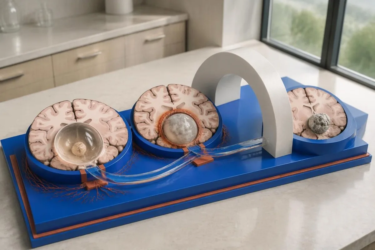
For selected patients with 1–2 viable parenchymal cysts, the IDSA/ASTMH guideline describes albendazole treatment for 10–14 days with appropriate accompanying care. This is a specialist-directed recommendation, not a home-treatment instruction: antiparasitic drugs can provoke an inflammatory response, and untreated hydrocephalus or diffuse cerebral edema changes the order of management (White et al., 2018).
WHO's 2021 guidance focuses on defined presentations of parenchymal neurocysticercosis, while the IDSA/ASTMH guideline also addresses ventricular, subarachnoid, and other forms in detail. Differences in imaging access and disease compartment explain why 1 treatment recommendation cannot be transferred to every patient with an antibody result; the guidelines do not turn negative serology into a rule-out test.
Corticosteroids may accompany antiparasitic therapy, but prolonged use requires additional safety planning, including assessment for relevant latent infections. Our Strongyloides and steroid safety guide explains a separate risk that matters before immune-suppressing treatment; an eye examination is also recommended before antiparasitic therapy to evaluate possible intraocular cysts.
За чем следует наблюдать при последующем контроле помимо результатов на антитела?
Follow-up should track seizures, neurological symptoms, imaging changes, and medication safety, rather than use antibody disappearance as proof of recovery. For viable parenchymal cysts, the IDSA/ASTMH guideline suggests repeat MRI at least every 6 months until the cystic component resolves, with earlier reassessment when symptoms change.

Antibodies may remain detectable after the original disease process has changed, and low-burden disease may never produce a positive result. If 1 new seizure occurs during follow-up, clinicians consider adherence, medication interactions, recurrent edema, and other causes; a rising or falling antibody index alone cannot distinguish among those explanations.
Kantesti AI can help explain reported laboratory results during medication follow-up, but a drug concentration is not a stand-alone measure of seizure control. Our интерпретация уровня леветирацетама guide explains why sample timing and kidney function matter; 2 measurements collected at different times after dosing may not be directly comparable.
Monitoring depends on the chosen drug and treatment duration: prolonged albendazole therapy can require liver-test and blood-count surveillance, while some antiseizure medicines can affect sodium. Our руководство по безопасности электролитов discusses those separate laboratory concerns; do not stop an antiseizure medicine or change its dose because 1 cysticercosis antibody result is negative.
Что может объяснить Kantesti и чего не могут доказать исследования?
AI report interpretation cannot confirm or exclude neurocysticercosis from laboratory results alone. Kantesti — это Инструмент анализа результатов анализа крови с ИИ whose role here is to organize reported laboratory findings; a negative result still needs clinical context, and 1 new seizure takes priority over reviewing an automated explanation.

Prepare a focused review packet with the original antibody report, assay method, CT or MRI report, symptom timeline, and medication list. These 5 information categories reduce the risk of discussing a result without its context; ask specifically whether the imaging suggests viable, degenerating, calcified, ventricular, or subarachnoid disease and which unanswered question the next test would address.
The 2 Kantesti publications listed below are a repository-deposited global analysis report and an educational RDW guide, not clinical evidence validating neurocysticercosis diagnosis. Our руководство по RDW и CBC can support interpretation of a separately ordered blood count, but RDW, MCV, and MCHC do not establish whether a brain cyst is present.
Evidence for the neurological recommendations in this article comes from the cited diagnostic criteria and clinical guidelines, not from general AI benchmarking or laboratory-report volume. Our technical validation scope should be read within its stated boundaries; neither 1 successful report explanation nor a large aggregate dataset substitutes for condition-specific diagnostic validation.
Часто задаваемые вопросы
Может ли быть нейроцистицеркоз при отрицательном анализе крови?
Да, нейроцистицеркоз может присутствовать даже при отрицательном анализе крови на антитела, особенно при наличии одной кисты головного мозга или только кальцифицированных очагов. Согласно опубликованным оценкам, чувствительность EITB, проводимого в референс-лабораториях, при наличии одной кисты часто составляет примерно 50–60%, что значительно ниже, чем при множественных жизнеспособных кистах. Поэтому КТ, МРТ, симптомы и сведения о возможном заражении по-прежнему играют ключевую роль в диагностике. При впервые возникшем приступе необходимо срочно обратиться за медицинской помощью, а не полагаться на отрицательный результат анализа.
Какой тест на нейроцистицеркоз наиболее надежен?
Ни один тест на нейроцистицеркоз не позволяет ответить на все диагностические вопросы; методы визуализации головного мозга и правильно подобранные серологические исследования дополняют друг друга. Согласно рекомендациям IDSA/ASTMH, EITB является предпочтительным методом подтверждения диагноза с помощью выявления антител, однако он может не выявить заболевание, если присутствует только 1 киста. КТ без контрастирования особенно полезна для выявления кальцификатов, тогда как МРТ лучше позволяет оценить множество жизнеспособных кист, отёк и изменения в ликворных пространствах. Врачи сопоставляют эти результаты, а не считают один отрицательный результат окончательным.
Что лучше при нейроцистицеркозе: КТ или МРТ?
КТ и МРТ имеют разные преимущества при диагностике нейроцистицеркоза. КТ без контрастного усиления часто позволяет чётче выявить мелкие кальцинаты размером примерно 1–4 мм, тогда как МРТ оценивает жизнеспособные цисты, окружающий отёк и поражение желудочков или субарахноидального пространства. Специализированные трёхмерные последовательности МРТ могут помочь выявить оболочки цист в пространствах, заполненных спинномозговой жидкостью. Рекомендации IDSA/ASTMH предписывают использовать оба метода при классификации впервые диагностированного заболевания.
Может ли кальцинированная киста по-прежнему вызывать судороги?
Кальцинированный остаток цистицерка может по-прежнему быть связан с судорогами, хотя жизнеспособной кистой он уже не является. Вокруг некоторых кальцинированных очагов, часто всего 1–4 мм в диаметре, периодически развивается отёк, который может быть виден на МРТ. Отрицательный результат анализа на антитела не исключает этот механизм, а сохраняющийся положительный результат не отражает риск судорог. При заболевании, проявляющемся только кальцинированными очагами, обычно назначают симптоматическое лечение, а не противопаразитарные препараты.
Нужно ли повторить отрицательный анализ на антитела к цистицеркозу?
Повторное проведение отрицательного анализа на антитела к цистицеркозу не всегда является оптимальным следующим шагом. При одной небольшой кисте или кальцинированной форме заболевания слабый антительный ответ может оставаться отрицательным, даже если повторить тот же анализ через 2 недели. Вместо этого врач может пересмотреть первоначальный метод исследования, назначить более точный подтверждающий анализ или отдать приоритет КТ и МРТ. При новых приступах или ухудшении неврологических симптомов не следует ждать повторного анализа.
Когда следует обратиться за неотложной помощью при первом приступе?
A first known seizure warrants urgent medical assessment and contacting local emergency services, regardless of a negative neurocysticercosis antibody test. A convulsive seizure lasting 5 minutes, repeated seizures without recovery, breathing difficulty, pregnancy, or significant injury is an emergency. Do not restrain the person or put anything in their mouth, and place them on their side once convulsions stop if they are breathing normally. Do not drive until a clinician has assessed the event and explained the applicable restrictions.
Получите объяснение результатов анализа крови
Загрузите отчёт лаборатории, и ИИ Kantesti за считаные секунды объяснит каждый биомаркер простым языком. Им пользуются более 2 миллионов человек по всему миру.
Ссылки
- Кляйн, Т., Митчелл, С., и Вебер, Х. (2026). Анализатор анализов крови с помощью ИИ: 2,5 млн тестов проанализировано | Global Health Report 2026. Kantesti AI Медицинские исследования. DOI
- Кляйн, Т., Митчелл, С., и Вебер, Х. (2026). Анализ крови на RDW: полное руководство по RDW-CV, MCV и MCHC. Kantesti AI Медицинские исследования. DOI
- White AC Jr et al. (2018). Diagnosis and Treatment of Neurocysticercosis: 2017 Clinical Practice Guidelines by the Infectious Diseases Society of America (IDSA) and the American Society of Tropical Medicine and Hygiene (ASTMH). Clinical Infectious Diseases. DOI
- Del Brutto OH et al. (2017). Revised diagnostic criteria for neurocysticercosis. Journal of the Neurological Sciences. DOI
- World Health Organization (2021). WHO guidelines on management of Taenia solium neurocysticercosis. Всемирная организация здравоохранения.
- 2М+Проанализированные исследования
- 127+Страны
- 100+Языки
О медицинской команде


Сигналы доверия E-E-A-T
Опыт
Клинический обзор рабочих процессов интерпретации лабораторных данных под руководством врача.
Экспертиза
Акцент на лабораторной медицине: как биомаркеры ведут себя в клиническом контексте.
Авторитетность
Медицинскую проверку провёл доктор Томас Кляйн при участии доктора Сары Митчелл и профессора, доктора Ганса Вебера.
Доверие
Интерпретация на основе доказательств с понятными маршрутами дальнейших действий, чтобы снизить тревожность.





Медицинская оговорка
Эта статья предназначена только для образовательных целей и не является медицинской консультацией. Всегда консультируйтесь с квалифицированным специалистом здравоохранения по вопросам диагностики и решений по лечению.
Опубликовано · Связаться с редакцией
Издатель — Kantesti Ltd, зарегистрированная в Англии и Уэльсе компания, рег. №. 17090423, Лондон, Великобритания.
