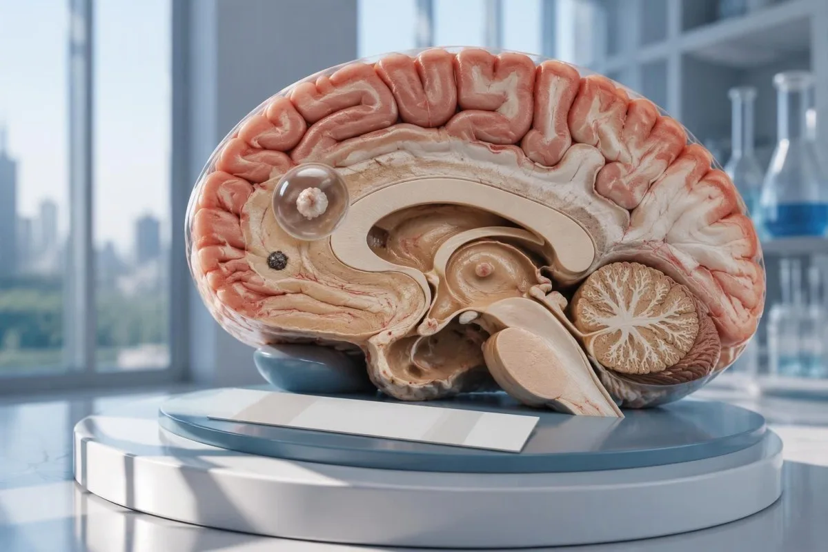
Kipimo hasi cha neurocysticercosis hakiondoi uwezekano wa uvimbe mmoja wa kistiki kwenye ubongo au ugonjwa wenye kalkifikasheni. CT, MRI na tathmini ya haraka ya kifafa kipya ni muhimu zaidi kuliko kutulizwa tu na kipimo cha kingamwili.

Mambo muhimu ya kukumbuka
- Kingamwili hasi haziondoi uwezekano wa neurocysticercosis, hasa picha zinapoonyesha eneo 1 tu lililoathirika au kalkifikasheni.
- EITB ya maabara ya marejeo inaweza kufikia usikivu wa 98% kukiwa na zaidi ya uvimbe 1 wa kistiki unaoweza kuishi, lakini utendaji wake huwa chini zaidi kukiwa na uvimbe mmoja wa kistiki.
- Usikivu wa uvimbe mmoja wa kistiki mara nyingi huwa takriban 50–60% katika makadirio ya EITB yaliyochapishwa; takwimu hizi hazitumiki kwa kila kipimo cha kibiashara cha kingamwili.
- CT isiyo na kontrasti ni muhimu hasa kwa kalkifikasheni ndogo, ambazo mara nyingi huwa na upana wa takriban 1–4 mm.
- MRI hutathmini uvimbe wa kistiki unaoweza kuishi, uvimbe wa tishu zinazouzunguka, na ugonjwa wa ventrikali au nafasi ya subaraknoida; mfululizo maalumu wa picha za pande 3 unaweza kuboresha ugunduzi.
- Kifafa cha kwanza kunahitaji tathmini ya haraka ya kitabibu bila kujali matokeo ya kipimo cha kingamwili. Piga simu kwa huduma za dharura ikiwa ni mshtuko wa kwanza wa kifafa unaojulikana au kuna dalili nyingine za dharura.
- Dakika tano ni kizingiti cha dharura kwa mshtuko wa kifafa wa kukakamaa unaoendelea, si sababu ya kusubiri kabla ya kutafuta msaada.
- Ugonjwa wenye mabaki yaliyokalsifika pekee kwa kawaida hauhitaji dawa za kuua vimelea, lakini matibabu ya kifafa na uchunguzi zaidi wa picha bado yanaweza kuhitajika.
Je, kipimo hasi cha neurocysticercosis kinaweza kuondoa uwezekano wa ugonjwa huo?
A kipimo hasi cha neurocysticercosis hakiwezi kuondoa ugonjwa huo kwa uhakika, hasa kukiwa na kivimbe 1 kwenye ubongo au mabaki yaliyokalsifika. Kipimo cha kingamwili hupima mwitikio wa kinga badala ya kuchunguza ubongo moja kwa moja; CT na MRI hutambua mahali na hatua ya matokeo yanayotiliwa shaka, huku mshtuko wa kwanza wa kifafa ukihitaji tathmini ya haraka.

Matokeo haya ni ya masharti, si ya uhakika kabisa. Kipimo cha kingamwili cha cysticercosis kinachofanya kazi vizuri kwa watu wenye vivimbe kadhaa hai kinaweza kufanya kazi vibaya kwa mtu mwenye eneo 1 tu dogo lililoathirika, kwa hiyo neno hasi linamaanisha hakuna mwitikio unaoweza kugunduliwa kwa kutumia kipimo hicho—si kwamba hakuna vivimbe popote kwenye mfumo wa neva.
Kwa mfano wa kubuni, fikiria mtu wa miaka 34 aliyepata mshtuko wa kwanza wa kifafa, ripoti hasi ya kingamwili, na eneo moja linalojitokeza kama pete kwenye MRI. Ripoti hiyo haiwezi kubainisha iwapo eneo hilo linawakilisha cysticercosis, maambukizi mengine au hali isiyotokana na maambukizi; maana yake hutegemea kwa pamoja uchunguzi wa picha, historia ya kukumbwa na maambukizi na tathmini ya mfumo wa neva.
Kantesti ni Mchambuzi wa mtihani wa damu wa AI ambayo inaweza kusaidia kueleza matokeo ya maabara yaliyoripotiwa, lakini haiwezi kuondoa uwezekano wa ugonjwa wa ubongo kutokana na matokeo 1 hasi ya kingamwili. Yetu shirika na dhamira vinaeleza mwelekeo wetu wa kutoa taarifa za maabara; wasomaji hawapaswi kuchukulia maelezo ya kiotomatiki ya ripoti kuwa ruhusa ya kuchelewesha huduma ya kifafa.
Kwa nini kipimo cha kingamwili kinaweza kukosa uvimbe mmoja wa kistiki kwenye ubongo?
A kivimbe kimoja kinaweza kusababisha mwitikio wa kingamwili ulio chini ya kiwango cha ugunduzi cha kipimo. Makadirio yaliyochapishwa ya EITB katika maabara za marejeo mara nyingi huweka unyeti karibu na 50–60% kwa kivimbe kimoja, ikilinganishwa na takriban 98% kukiwa na zaidi ya kivimbe 1 hai; tofauti hiyo inaonyesha kiwango cha ugonjwa na utendaji wa kipimo.
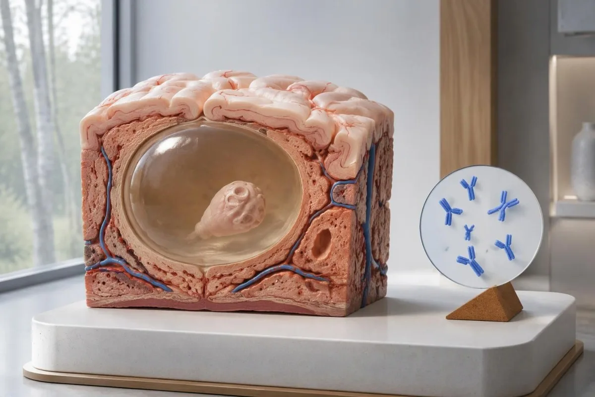
Mwongozo wa IDSA/ASTMH unapendekeza kipimo cha immunoelectrotransfer kinachohusishwa na kimeng'enya, au EITB, kama kipimo cha serolojia cha uthibitisho na unaonya wazi kwamba unyeti ni mdogo kukiwa na eneo moja tu lililoathirika kwenye ubongo au mabaki yaliyokalsifika pekee (White et al., 2018). Unyeti wa juu ulioripotiwa kwa wagonjwa wenye vivimbe vingi, ugonjwa wa ventrikali au ugonjwa wa nafasi ya subaraknoidi haupaswi kuhusishwa na mtu mwenye hitilafu ndogo 1 kwenye ubongo.
Kwa mfano wa kufafanua tu, fikiria kwamba kipimo kina unyeti wa 55% kwa watu 100 ambao ugonjwa wa kivimbe kimoja tayari umethibitishwa kwa njia huru. Takriban watu 45 wangepata matokeo hasi licha ya kuwa na ugonjwa huo; mfano huu unaeleza kutogunduliwa kwa ugonjwa, si uwezekano kwamba matokeo yoyote hasi yanamaanisha ugonjwa.
Kurudia kipimo kilekile baada ya wiki 2 huenda kusisahihishe mwitikio dhaifu unaosababishwa na kiwango kidogo cha ugonjwa. Hali hii ni tofauti na maambukizi ambayo tatizo kuu ni kupima mapema mno, kama ilivyoelezwa katika mwongozo wetu wa matokeo hasi ya uongo yanayohusiana na muda; uliza iwapo uchunguzi wa picha au kipimo kilichothibitishwa vyema zaidi kingebadilisha uamuzi unaofuata.
Kwa nini neurocysticercosis yenye kalkifikasheni inaweza kuendelea kusababisha dalili?
Neurocysticercosis yenye mabaki yaliyokalsifika inaweza kusababisha kifafa licha ya kingamwili hasi, because the remaining mineralized focus can still be associated with local irritation and episodic surrounding edema. Typical calcifications may measure approximately 1–4 mm, but their small size does not establish whether they are clinically harmless.
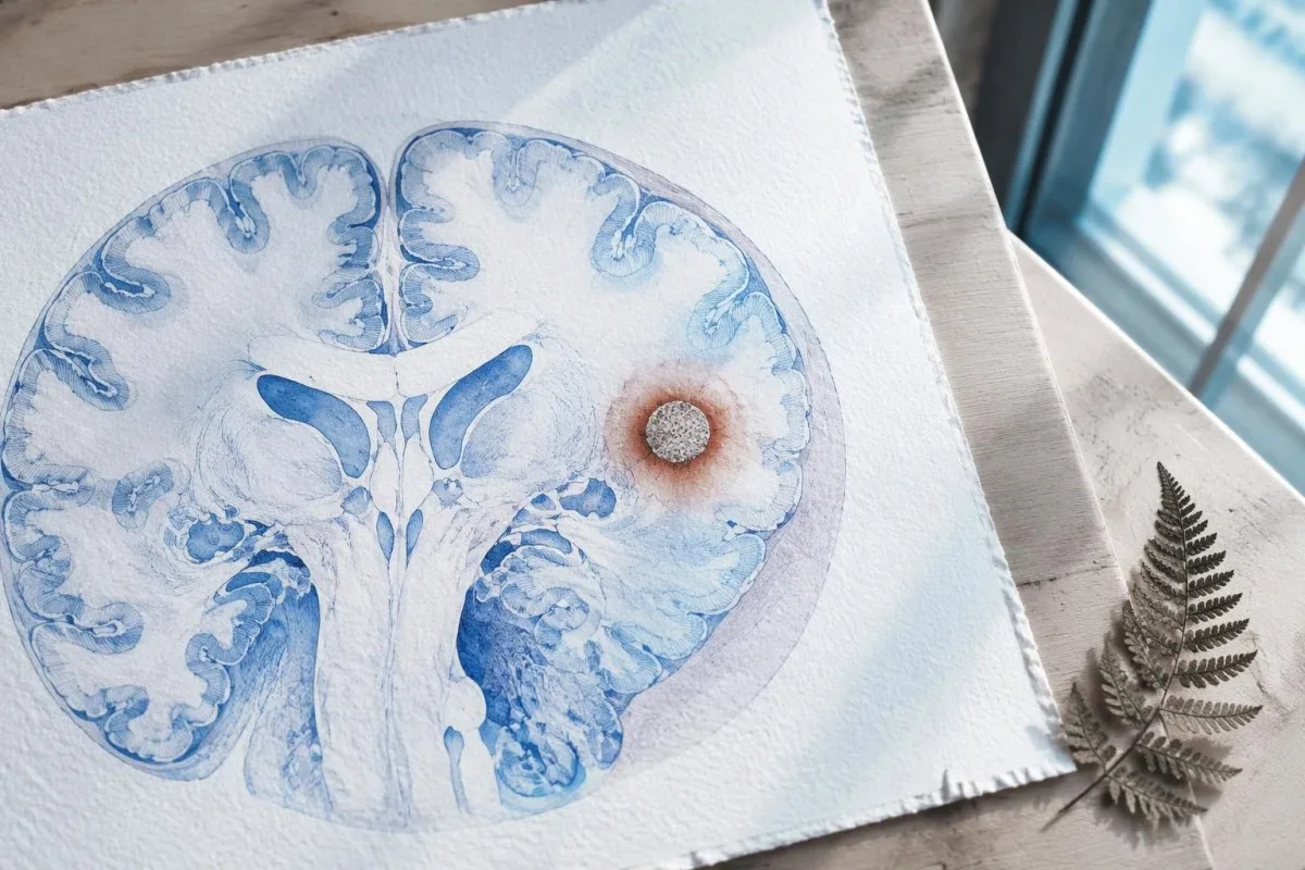
A calcification is generally a remnant rather than a living cyst that antiparasitic medication can eliminate. In the IDSA/ASTMH guideline, calcified-only parenchymal disease is managed symptomatically rather than with antiparasitic drugs; a person with 1 calcified focus and seizures may therefore need antiseizure treatment without needing cyst-killing medication (White et al., 2018).
Surrounding edema can appear intermittently, sometimes long after the original cyst has degenerated. MRI may show this tissue response even when the calcification is more obvious on CT, which is why the 2 scans can tell different but compatible stories rather than one proving that the other is wrong.
Neither a negative antibody result nor a persistent positive result measures seizure risk by itself. The broader lesson resembles other seronegative neurological conditions, discussed in our negative neurological antibody results guide: absence of detectable antibodies is not equivalent to absence of a clinically relevant neurological process.
Ni kipimo gani hasa cha kingamwili za cysticercosis kilichofanywa?
The useful first question is whether the report describes EITB, ELISA, or another antibody method, because these are not interchangeable tests. A negative index below 1 laboratory's cutoff cannot inherit the performance figures of a different reference-laboratory assay, even when both reports use the name cysticercosis IgG.
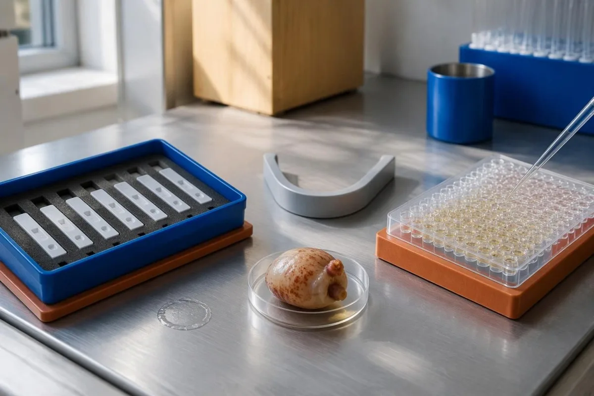
EITB using purified parasite glycoproteins is the preferred serological method in the IDSA/ASTMH guideline, while crude-antigen ELISA methods have recognized sensitivity and specificity limitations (White et al., 2018). Before comparing 2 results, check the method, specimen type, reference interval, and whether the laboratory used a validated confirmatory procedure.
Some laboratories report a qualitative result; others provide an optical-density ratio or numerical index. A value such as 0.8 in a hypothetical assay does not represent 0.8 cysts, an 80% disease probability, or a universal severity score—our matokeo ya ubora na wingi guide explains that distinction.
Kantesti AI's role is to help readers preserve the laboratory's original wording rather than reinterpret 1 method through another method's thresholds. Our mwongozo wa alama za maabara provides broader report-reading context; for this result, keep the actual assay name available when discussing referral or repeat testing.
Historia ya kukabiliwa na maambukizi hubadilishaje tuhuma za utambuzi?
Neurocysticercosis follows ingestion of Taenia solium eggs, usually through fecal contamination from a person carrying the intestinal tapeworm. Eating undercooked pork can cause intestinal taeniasis, but brain cysts arise through a different transmission step; symptoms may emerge years after exposure rather than within 1 recent trip.

A vegetarian can develop cysticercosis because eggs can contaminate food or hands independently of meat consumption. Useful history includes past residence, household contacts, sanitation conditions, and possible exposure to a tapeworm carrier; asking only about meals eaten during the last 2 weeks can miss the relevant background.
A negative stool examination does not exclude cysts in the brain. Stool testing addresses intestinal tapeworm carriage, whereas CT and MRI assess nervous-system findings; these are 2 different sites and clinical questions, although public-health teams may recommend evaluating contacts when a carrier could be involved.
Normal eosinophils also cannot exclude neurocysticercosis, and raised eosinophils do not identify the brain as the affected site. Our parasite antibody interpretation guide discusses a separate parasite with similar localization limits; the practical point is to avoid using 1 routine cell count as a substitute for targeted neurological evaluation.
Ni wakati gani kifafa kipya huhitaji tathmini ya dharura?
A first known seizure needs urgent medical assessment regardless of antibody results, and emergency services should be contacted for a first seizure or an ongoing seizure with concerning features. A convulsive seizure lasting 5 minutes, repeated seizures without recovery, breathing difficulty, pregnancy, or significant injury requires emergency care.

Do not wait for the 5-minute mark to seek help after a first known seizure. Keep the person away from hazards, cushion the head if safely possible, do not restrain them or put anything in their mouth, and place them on their side once convulsions stop if they are breathing normally.
Persistent confusion, a severe new headache, fever with neck stiffness, or new weakness increases concern for causes beyond cysticercosis. Sudden one-sided weakness or speech difficulty also demands emergency assessment, as our ishara za tahadhari za kiharusi explains; 1 negative infection test cannot rule out stroke, meningitis, or a structural brain problem.
After a suspected seizure, stop driving and avoid swimming alone, heights, or operating machinery until a clinician provides advice consistent with local rules. If a witness can safely record the duration and describe movements or recovery, those observations may be more useful than ordering a second antibody test before the first neurological assessment.
CT huongeza nini katika utambuzi wa neurocysticercosis?
Noncontrast CT is particularly useful for detecting calcified cyst remnants, including small approximately 1–4 mm foci that MRI may show less clearly. CT also helps assess urgent structural problems such as hydrocephalus, but 1 isolated calcification is not automatically proof of neurocysticercosis or the cause of a seizure.
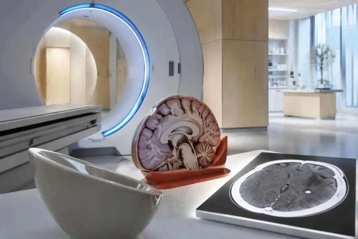
Calcium appears dense on CT, allowing radiologists to identify mineralized remnants and describe their distribution. The IDSA/ASTMH guideline recommends both MRI and noncontrast CT when classifying newly diagnosed neurocysticercosis, because the 2 modalities have complementary strengths rather than being interchangeable alternatives (White et al., 2018).
An incidental calcification needs context: previous granulomatous disease, other causes of mineralization, and scan appearance can influence interpretation. If 1 calcification sits near a suspected seizure focus, concordance with symptoms and EEG may strengthen the explanation, but anatomical proximity alone cannot establish causation.
A laboratory marker used in selected head-injury pathways does not test for brain cysts or replace imaging after an unexplained seizure. Our head-injury CT decisions guide illustrates that narrower setting; ask the clinician whether the present question requires noncontrast CT, contrast imaging, MRI, or a combination of 2 modalities.
MRI huonyesha nini ambacho CT inaweza kukosa?
MRI is better suited to evaluating viable cysts, surrounding edema, and many ventricular or subarachnoid findings. Specialized 3-dimensional sequences such as CISS or FIESTA can improve visualization of cyst membranes within cerebrospinal fluid spaces, where a cyst may otherwise resemble the surrounding fluid.
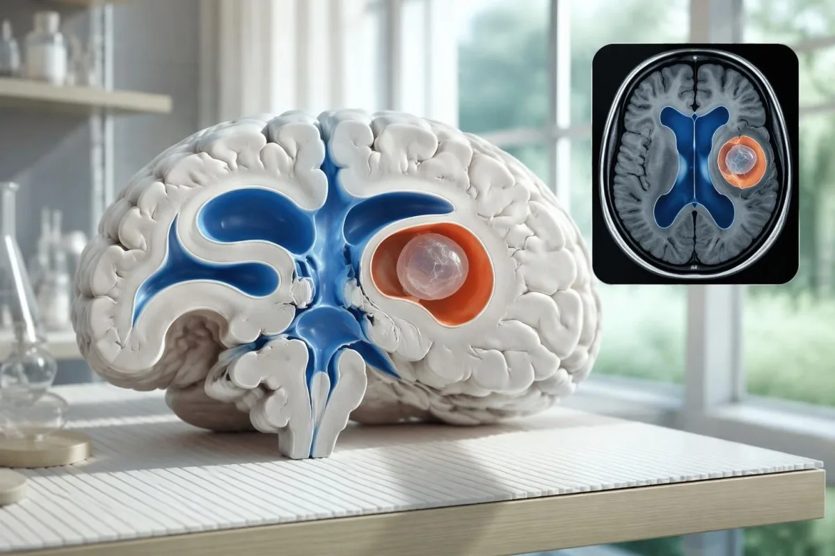
A cyst inside the ventricles can obstruct cerebrospinal fluid flow and cause hydrocephalus even without numerous parenchymal cysts. Headache with vomiting, declining alertness, or new visual symptoms therefore changes urgency; 1 reassuring antibody result should not override symptoms that suggest raised pressure or impaired fluid circulation.
MRI may show a scolex, the small internal structure of the parasite, inside a compatible cyst. This finding can be highly specific when confidently identified, whereas ring enhancement alone has a broader differential; our brain imaging warning signs guide explains why structural abnormalities require interpretation rather than diagnosis from 1 visual feature.
A routine MRI protocol may not answer every cysticercosis question. If ventricular or subarachnoid disease is suspected, discuss whether thin-section 3-dimensional imaging is needed; radiologists choose sequences and contrast according to the clinical question, and a normal scan using 1 protocol does not prove every possible disease compartment has been adequately assessed.
Neurocysticercosis hutambuliwaje vipimo vinapotofautiana?
Neurocysticercosis diagnosis combines imaging, clinical findings, exposure information, and supporting tests, rather than requiring a positive antibody result in every case. The revised diagnostic criteria published in 2017 give characteristic neuroimaging findings a central role and distinguish strong evidence from less-specific supportive features (Del Brutto et al., 2017).
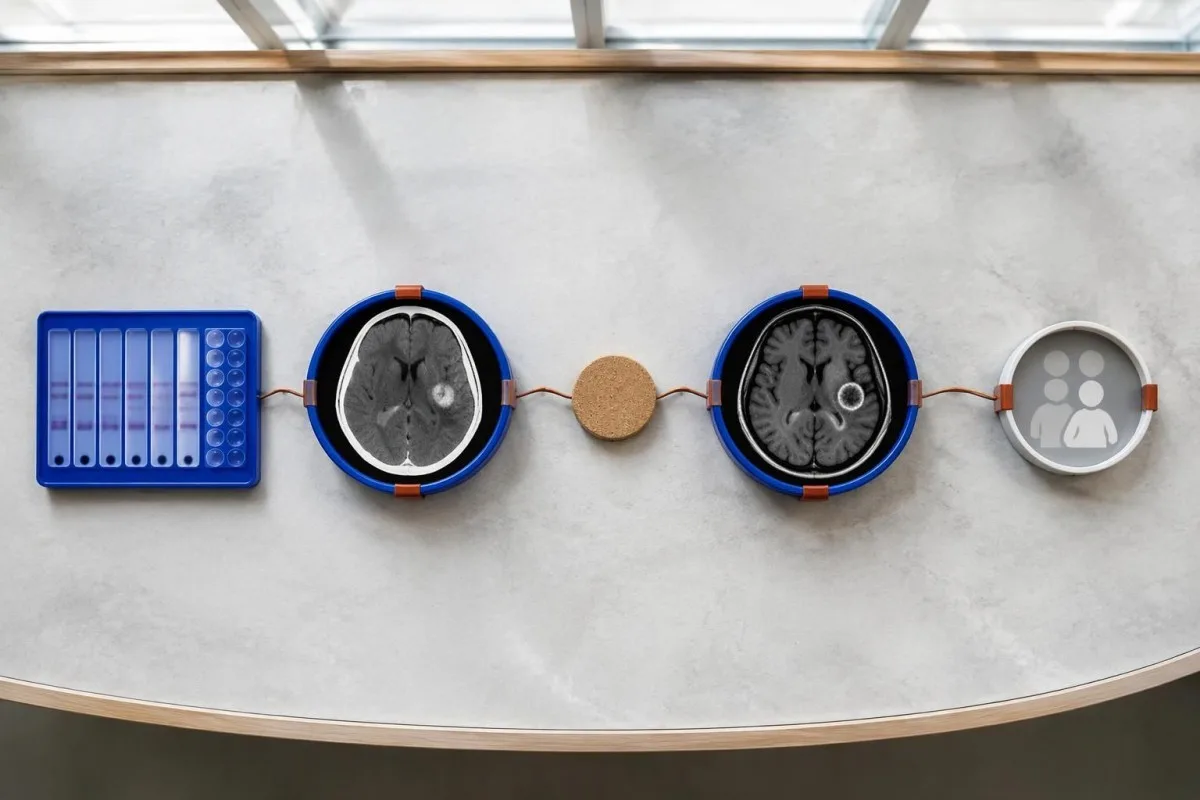
Evidence should be weighted, not tallied. A confidently visualized scolex inside a compatible cyst is different from 1 nonspecific ring-enhancing area, while a positive antibody result is different from direct evidence that a finding is in the brain; our matokeo chanya ya kingamwili explains why detection alone cannot establish location.
For a clearly hypothetical probability example, start with a 30% pretest probability and assume a single-cyst assay has 55% sensitivity and 99% specificity. In 1,000 comparable people, about 135 affected and 693 unaffected people would test negative, leaving approximately a 16% probability of disease among negative results—still substantial, although these assumed figures are not a personal risk calculator.
The most useful disagreement to investigate is whether each test is answering the same question. A negative antibody test, a CT calcification, and MRI edema can all be compatible with 1 calcified disease process; unexplained discrepancies should prompt review of the assay and images rather than automatic dismissal of either result.
Ni nini kingine kinachoweza kufanana na uvimbe mmoja wa kistiki kwenye picha za ubongo?
A single ring-enhancing brain finding is not specific to neurocysticercosis. Other possibilities include tuberculoma, an abscess, a neoplasm, or an inflammatory process; clinicians use appearance, location, immune status, symptoms, and sometimes follow-up imaging or tissue examination to distinguish these possibilities rather than relying on 1 antibody panel.
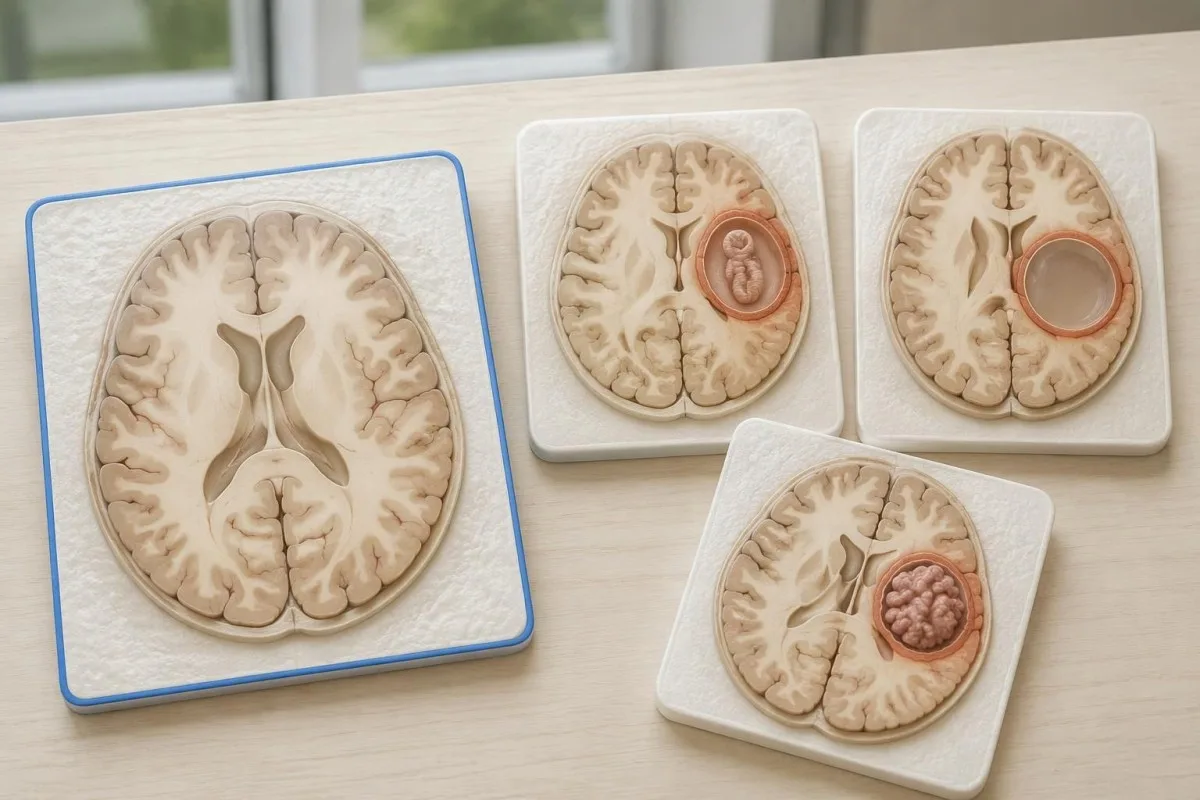
Edema, restricted diffusion, internal structures, and the number of findings all contribute to the differential, but no single nonspecific feature settles every case. Fever and immune suppression may change the suspected cause; a 2 cm ring-enhancing area in a hypothetical immunocompromised person warrants a different investigation from a tiny calcification in an otherwise well person.
Toxoplasmosis is one consideration in selected immunocompromised patients, and a positive IgG result can indicate past exposure rather than prove an active brain process. Our Toxoplasma IgG interpretation guide explains that laboratory distinction; imaging and the patient's immune status determine whether the result is clinically relevant to 1 or more cerebral findings.
New seizures accompanied by rapidly evolving memory problems, psychiatric changes, or abnormal movements can also suggest encephalitis rather than a solitary parasite cyst. Our encephalitis diagnostic pathway describes how MRI, EEG, and cerebrospinal fluid testing answer different questions; a second negative cysticercosis antibody result does not investigate those alternatives.
Je, kipimo cha antijeni, PCR au CSF kinaweza kuondoa kutokuwa na uhakika?
Antigen tests, PCR, and cerebrospinal fluid analysis can add information in selected cases, particularly when extraparenchymal disease is suspected. None is a universal rescue test after 1 negative antibody result: availability, assay validation, disease compartment, and safety of lumbar puncture determine whether additional testing is appropriate.
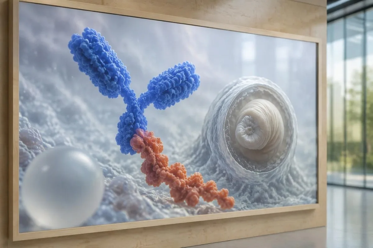
Antibody testing detects the host response, while antigen testing aims to detect parasite-derived material and may better reflect viable disease burden. Antigen detection is often less useful when only 1 small parenchymal cyst or calcifications are present; molecular testing is used in some specialist settings, but performance cannot be generalized across every laboratory or specimen.
Lumbar puncture is not routinely required for an uncomplicated solitary parenchymal finding and can be unsafe when obstructive hydrocephalus or significant mass effect is present. Cerebrospinal fluid tests for other disorders also require disease-specific interpretation, as our CSF band interpretation guide explains; 1 abnormal CSF marker does not identify every possible cause.
Kantesti ni jukwaa la tafsiri ya vipimo vya damu la AI, not a service that orders lumbar punctures, identifies cysts on scans, or confirms neurological infections. Our teknolojia ya kutafsiri AI explains the laboratory-report workflow; when 2 tests disagree, our role is to clarify their stated meanings and limitations, not select an invasive diagnostic procedure.
Kwa nini matibabu hutegemea hatua na eneo la uvimbe wa kistiki?
Treatment depends on whether cysts are viable, degenerating, calcified, or located outside the brain tissue, not simply on antibody positivity. Selected viable parenchymal cases receive antiparasitic treatment, whereas calcified-only disease generally receives symptom-directed care; ventricular obstruction may require a procedural approach rather than 1 standard medication regimen.
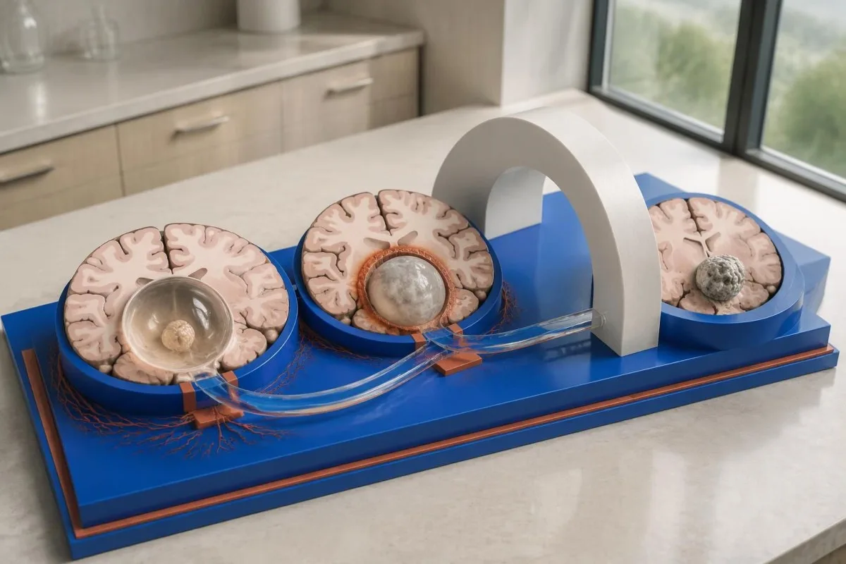
For selected patients with 1–2 viable parenchymal cysts, the IDSA/ASTMH guideline describes albendazole treatment for 10–14 days with appropriate accompanying care. This is a specialist-directed recommendation, not a home-treatment instruction: antiparasitic drugs can provoke an inflammatory response, and untreated hydrocephalus or diffuse cerebral edema changes the order of management (White et al., 2018).
WHO's 2021 guidance focuses on defined presentations of parenchymal neurocysticercosis, while the IDSA/ASTMH guideline also addresses ventricular, subarachnoid, and other forms in detail. Differences in imaging access and disease compartment explain why 1 treatment recommendation cannot be transferred to every patient with an antibody result; the guidelines do not turn negative serology into a rule-out test.
Corticosteroids may accompany antiparasitic therapy, but prolonged use requires additional safety planning, including assessment for relevant latent infections. Our Strongyloides and steroid safety guide explains a separate risk that matters before immune-suppressing treatment; an eye examination is also recommended before antiparasitic therapy to evaluate possible intraocular cysts.
Ufuatiliaji unapaswa kufuatilia nini zaidi ya matokeo ya kingamwili?
Follow-up should track seizures, neurological symptoms, imaging changes, and medication safety, rather than use antibody disappearance as proof of recovery. For viable parenchymal cysts, the IDSA/ASTMH guideline suggests repeat MRI at least every 6 months until the cystic component resolves, with earlier reassessment when symptoms change.
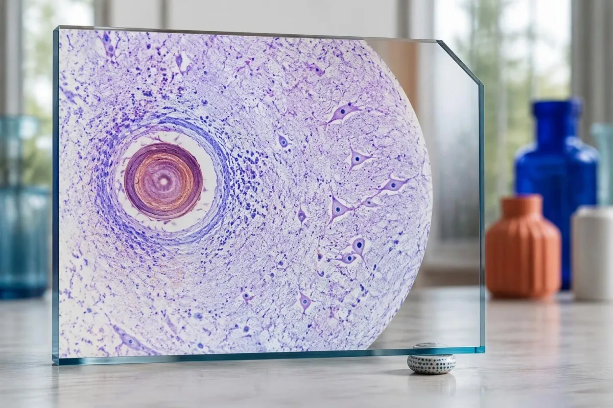
Antibodies may remain detectable after the original disease process has changed, and low-burden disease may never produce a positive result. If 1 new seizure occurs during follow-up, clinicians consider adherence, medication interactions, recurrent edema, and other causes; a rising or falling antibody index alone cannot distinguish among those explanations.
Kantesti AI can help explain reported laboratory results during medication follow-up, but a drug concentration is not a stand-alone measure of seizure control. Our tafsiri ya kiwango cha levetiracetam guide explains why sample timing and kidney function matter; 2 measurements collected at different times after dosing may not be directly comparable.
Monitoring depends on the chosen drug and treatment duration: prolonged albendazole therapy can require liver-test and blood-count surveillance, while some antiseizure medicines can affect sodium. Our mwongozo wa usalama wa elektrolaiti discusses those separate laboratory concerns; do not stop an antiseizure medicine or change its dose because 1 cysticercosis antibody result is negative.
Kantesti inaweza kueleza nini, na utafiti hauwezi kuthibitisha nini?
AI report interpretation cannot confirm or exclude neurocysticercosis from laboratory results alone. Kantesti ni Zana ya uchambuzi wa vipimo vya damu inayotumia AI whose role here is to organize reported laboratory findings; a negative result still needs clinical context, and 1 new seizure takes priority over reviewing an automated explanation.

Prepare a focused review packet with the original antibody report, assay method, CT or MRI report, symptom timeline, and medication list. These 5 information categories reduce the risk of discussing a result without its context; ask specifically whether the imaging suggests viable, degenerating, calcified, ventricular, or subarachnoid disease and which unanswered question the next test would address.
The 2 Kantesti publications listed below are a repository-deposited global analysis report and an educational RDW guide, not clinical evidence validating neurocysticercosis diagnosis. Our Mwongozo wa RDW na CBC can support interpretation of a separately ordered blood count, but RDW, MCV, and MCHC do not establish whether a brain cyst is present.
Evidence for the neurological recommendations in this article comes from the cited diagnostic criteria and clinical guidelines, not from general AI benchmarking or laboratory-report volume. Our technical validation scope should be read within its stated boundaries; neither 1 successful report explanation nor a large aggregate dataset substitutes for condition-specific diagnostic validation.
Maswali ya mara kwa mara
Je, unaweza kuwa na neurocysticercosis licha ya kipimo cha damu kuwa hasi?
Ndiyo, neurocysticercosis inaweza kuwepo licha ya kipimo hasi cha kingamwili kwenye damu, hasa ikiwa kuna uvimbe mmoja kwenye ubongo au ugonjwa wenye uvimbe uliokalsifika pekee. Makadirio yaliyochapishwa kutoka maabara za marejeo yanaonyesha mara nyingi kuwa EITB ina unyeti wa takriban 50–60% kwa uvimbe mmoja, ambao ni wa chini sana kuliko utendaji wake kwa uvimbe mwingi ulio hai. Kwa hiyo, CT, MRI, dalili na historia ya kukabiliwa na maambukizi hubaki kuwa mambo muhimu katika utambuzi. Kifafa kipya kinahitaji tathmini ya haraka badala ya kutulizwa na matokeo hasi.
Ni kipimo gani cha kuaminika zaidi cha neurocysticercosis?
Hakuna kipimo kimoja cha neurocysticercosis kinachojibu maswali yote ya uchunguzi; picha za ubongo na vipimo vya serolojia vilivyochaguliwa ipasavyo hukamilishana. EITB ndiyo njia inayopendelewa ya kuthibitisha kingamwili katika mwongozo wa IDSA/ASTMH, lakini inaweza kukosa kugundua ugonjwa unaohusisha uvimbe mmoja tu. CT bila kutumia dutu ya kutofautisha ni muhimu hasa kwa kutambua maeneo yaliyokakamaa, ilhali MRI hutathmini vyema zaidi uvimbe mwingi ulio hai, uvimbe wa tishu na hitilafu za nafasi za majimaji. Madaktari huchanganya matokeo haya badala ya kuchukulia matokeo hasi ya kipimo kimoja kuwa ya mwisho.
Je, CT au MRI ni bora zaidi kwa neurocysticercosis?
CT na MRI zina uwezo tofauti katika utambuzi wa neurocysticercosis. CT isiyotumia kontrasti mara nyingi hugundua kwa uwazi zaidi kalshifikesheni ndogo zenye ukubwa wa takriban mm 1–4, huku MRI ikitathmini cysti hai, uvimbe wa tishu zinazozizunguka, na ugonjwa kwenye ventrikali au nafasi za subaraknoida. Mfuatano maalum wa MRI wa pande tatu unaweza kusaidia kutambua utando wa cysti katika nafasi zenye maji ya cerebrospinal. Mwongozo wa IDSA/ASTMH unapendekeza matumizi ya mbinu zote mbili wakati wa kuainisha ugonjwa uliogunduliwa hivi karibuni.
Je, uvimbe wenye ukokotoaji bado unaweza kusababisha kifafa?
Mabaki ya cysticercosis yaliyokwishakokotoa yanaweza kuendelea kuhusishwa na kifafa cha mshtuko ingawa si uvimbe hai tena. Baadhi ya maeneo yaliyokokotoa, ambayo mara nyingi huwa na upana wa mm 1–4 tu, hupata uvimbe wa muda mfupi kwenye tishu zinazoyazunguka ambao unaweza kuonekana kwenye MRI. Matokeo hasi ya vipimo vya kingamwili hayazuii utaratibu huu, na matokeo chanya ya kingamwili yanayoendelea hayapimi hatari ya kifafa cha mshtuko. Ugonjwa unaohusisha maeneo yaliyokokotoa pekee kwa kawaida hutibiwa kwa kulenga dalili badala ya kutumia dawa za kuua vimelea.
Je, ninapaswa kurudia kipimo cha kingamwili za cysticercosis kilichoonyesha matokeo hasi?
Kurudia kipimo hasi cha kingamwili dhidi ya cysticercosis si lazima kuwe hatua bora inayofuata. Ikiwa kuna uvimbe mdogo 1 au ugonjwa wenye ukokotoaji, mwitikio hafifu wa kingamwili unaweza kubaki hasi hata kipimo kilekile kikifanywa tena baada ya wiki 2. Badala yake, daktari anaweza kuchunguza mbinu iliyotumika awali, kuomba kipimo cha uthibitisho kilichobainishwa vyema zaidi, au kuweka kipaumbele kwa CT na MRI. Kifafa kipya au dalili za mfumo wa neva zinazozidi kuwa mbaya hazipaswi kusubiri vipimo virudiwe.
Ninapaswa kutafuta msaada wa dharura lini kwa mara ya kwanza ninapopata kifafa?
Mshtuko wa kifafa wa kwanza unaojulikana unahitaji tathmini ya haraka ya kitabibu na kuwasiliana na huduma za dharura za eneo lako, hata kama kipimo cha kingamwili za neurocysticercosis ni hasi. Mshtuko wa kifafa wa kukakamaa unaodumu kwa dakika 5, mishtuko inayojirudia bila mtu kupata fahamu tena, shida ya kupumua, ujauzito au jeraha kubwa ni dharura. Usimzuie mtu huyo wala kuweka kitu chochote mdomoni mwake, na mlaze ubavuni baada ya kukakamaa kukoma ikiwa anapumua kawaida. Usiendeshe gari hadi daktari atathmini tukio hilo na kueleza vizuizi vinavyotumika.
Pata maelezo ya jinsi ya kusoma matokeo ya vipimo vya damu
Pakia ripoti yako ya maabara na AI ya Kantesti itaeleza kila kiashiria cha kibayolojia kwa lugha rahisi ndani ya sekunde chache. Inatumiwa na zaidi ya watu milioni 2 duniani kote.
Marejeleo
- Klein, T., Mitchell, S., & Weber, H. (2026). Kichanganuzi cha Uchambuzi wa Damu kwa AI: Vipimo 2.5M Vilivyochambuliwa | Ripoti ya Afya ya Kimataifa 2026. Utafiti wa Kimatibabu wa AI ya Kantesti. DOI
- Klein, T., Mitchell, S., & Weber, H. (2026). Kipimo cha RDW katika Damu: Mwongozo Kamili wa RDW-CV, MCV na MCHC. Utafiti wa Kimatibabu wa AI ya Kantesti. DOI
- White AC Jr et al. (2018). Diagnosis and Treatment of Neurocysticercosis: 2017 Clinical Practice Guidelines by the Infectious Diseases Society of America (IDSA) and the American Society of Tropical Medicine and Hygiene (ASTMH). Magonjwa ya Kuambukiza ya Kliniki. DOI
- Del Brutto OH et al. (2017). Revised diagnostic criteria for neurocysticercosis. Journal of the Neurological Sciences. DOI
- World Health Organization (2021). WHO guidelines on management of Taenia solium neurocysticercosis. Shirika la Afya Ulimwenguni.
- 2M+Vipimo vilivyochanganuliwa
- 127+Nchi
- 100+Lugha
Kuhusu timu ya matibabu


E-E-A-T Trust Signals
Uzoefu
Mapitio ya kimatibabu inayoongozwa na daktari ya mifumo ya tafsiri ya maabara.
Utaalamu
Kuzingatia dawa za maabara kuhusu jinsi viashiria (biomarkers) vinavyobadilika katika muktadha wa kliniki.
Mamlaka
Imekaguliwa kimatibabu na Dkt. Thomas Klein, kwa michango kutoka kwa Dkt. Sarah Mitchell na Prof. Dkt. Hans Weber.
Uaminifu
Tafsiri inayotegemea ushahidi yenye njia zilizo wazi za ufuatiliaji ili kupunguza tahadhari za hofu.





Kanusho la kimatibabu
Makala haya ni kwa madhumuni ya elimu tu na si ushauri wa kimatibabu. Wasiliana na mtoa huduma aliyehitimu kila wakati kwa maamuzi ya utambuzi na matibabu.
Imechapishwa · Wasiliana na timu ya uhariri
Imechapishwa na Kantesti Ltd, imesajiliwa nchini Uingereza na Wales, nambari ya kampuni. 17090423, London, Uingereza.
