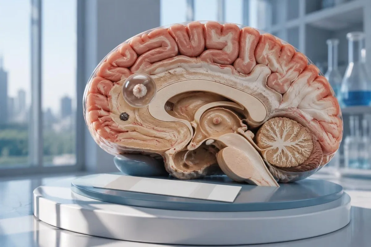
Chan eil deuchainn àicheil airson neurocysticercosis a’ cur às do chiste aonaranach san eanchainn no do ghalar le calcachadh. Tha CT, MRI agus measadh èiginneach air glacaidhean ùra nas cudromaiche na fois inntinn bho dheuchainn antibodies a-mhàin.

Prìomh phuingean
- Dìth-chumadh àicheil na cuir às do neurocysticercosis, gu h-àraidh nuair nach eil ìomhaighean a’ sealltainn ach 1 raon air a bheil buaidh no calcachaidhean.
- EITB obair-lann iomraidh faodaidh e tighinn faisg air mothalachd 98% nuair a tha barrachd air 1 chiste bheò ann, ach tha an coileanadh gu math nas ìsle nuair nach eil ann ach aon chiste.
- Mothalachd airson ciste shingilte tha i gu tric mu 50–60% ann an tuairmsean EITB a chaidh fhoillseachadh; chan eil na figearan seo iomchaidh airson gach deuchainn antibodies mhalairteach.
- CT gun choimeasair tha e gu h-àraidh feumail airson calcachaidhean beaga, a tha gu tric mu 1–4 mm de leud.
- MRI a’ measadh chisteachan beò, sèid mun cuairt orra, agus galar sna ventricles no san àite fo-arachnoid; dh’fhaodadh sreathan sònraichte trì-thaobhach lorg a dhèanamh nas fheàrr.
- Ciad ghlacaidhean feumadh measadh meidigeach èiginneach ge b’ e toradh an deuchainn antibody a bh’ ann. Cuir fios gu na seirbheisean èiginn ma ’s e seo a’ chiad ghlacadh aithnichte no ma tha comharran èiginn eile ann.
- Còig mionaidean ’s e seo an stairsneach èiginn airson grèim-clisgeach leantainneach, chan e adhbhar airson feitheamh mus iarrar cuideachadh.
- Galar le calcachadh a-mhàin mar as trice chan eil feum air cungaidh an aghaidh pharasitean air a shon, ach dh’fhaodadh gum bi feum fhathast air làimhseachadh nan glacaidhean agus tuilleadh ìomhaigheachd.
An urrainn do dheuchainn àicheil airson neurocysticercosis an galar a chur às?
A chan urrainn do dheuchainn àicheil airson neurocysticercosis an galar a chur às gu h-earbsach, gu h-àraidh nuair a tha 1 ciste san eanchainn no fuigheall calcichte ann. Bidh deuchainn antibody a’ tomhas freagairt an t-siostaim dìona seach a bhith a’ sgrùdadh na h-eanchainn gu dìreach; bidh CT agus MRI a’ dearbhadh far a bheil na toraidhean amharasach agus dè an ìre aig a bheil iad, agus tha feum air measadh èiginneach às dèidh a’ chiad ghlacaidh.

Tha an toradh cumhach, chan e iomlan. Dh’fhaodadh gun dèan deuchainn antibody airson cysticercosis a tha èifeachdach ann an daoine aig a bheil grunn chistichean beò gu dona ann an neach aig nach eil ach 1 raon beag air a bheil buaidh, agus mar sin tha am facal àicheil a’ ciallachadh nach eil freagairt a ghabhas lorg leis an deuchainn sin ann—chan eil e a’ ciallachadh nach eil cistichean an àite sam bith san t-siostam nearbhach.
Mar eisimpleir beachd-bharail, smaoinich air neach 34-bliadhna aig a bheil a’ chiad ghlacadh, aithisg antibody àicheil, agus lorg aonaranach a tha a’ neartachadh ann an cumadh fàinne air MRI. Chan urrainn don aithisg dearbhadh a bheil an lorg sin a’ comharrachadh cysticercosis, galar eile, no pròiseas neo-ghalarach; tha a bhrìgh an urra ris an sganaidh, eachdraidh nochdaidh agus measadh eanchainneach còmhla.
Tha Kantesti na Anailisiche deuchainn fala AI a chuidicheas le bhith a’ mìneachadh toraidhean obair-lann a chaidh aithris, ach chan urrainn dha galar na h-eanchainn a chur às stèidhichte air 1 toradh antibody àicheil. Tha ar buidheann agus misean a’ mìneachadh ar fòcas air fiosrachadh obair-lann; cha bu chòir do luchd-leughaidh mìneachadh fèin-obrachail air aithisg a mheas mar chead dàil a chur air cùram airson glacaidhean.
Carson a dh’fhaodadh deuchainn antibodies ciste aonaranach san eanchainn a chall?
A faodaidh aon chiste freagairt antibody a chruthachadh a tha nas ìsle na stairsneach lorg na deuchainn. Tha tuairmsean foillsichte air EITB ann an obair-lannan iomraidh gu tric a’ cur na mothalachd faisg air 50–60% airson aon chiste, an coimeas ri mu 98% nuair a tha barrachd na 1 chiste bheò an làthair; tha an diofar a’ nochdadh ìre an eallaich galair agus èifeachdas na deuchainn.
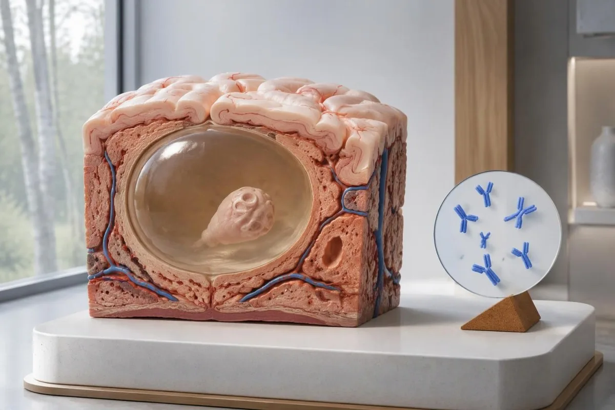
Tha stiùireadh IDSA/ASTMH a’ moladh blot immunoelectrotransfer ceangailte ri enzyme, no EITB, mar dheuchainn shèireòlach dhearbhach, agus tha e a’ toirt rabhadh gu soilleir gu bheil mothalachd ìosal nuair nach eil ann ach aon lorg sa pharenchìma no calcachaidhean a-mhàin (White et al., 2018). Cha bu chòir an àrd-mhothalachd a chaidh aithris airson euslaintich aig a bheil grunn chistichean, galar anns na ventricles, no galar fon arachnoid a chur an sàs ann an neach aig a bheil 1 neo-riaghailteachd bheag san eanchainn.
Smaoinich, airson dealbhachaidh a-mhàin, gu bheil mothalachd 55% aig deuchainn ann an 100 neach aig a bheil galar aon-chiste a chaidh a dhearbhadh mar-thà gu neo-eisimeileach. Bhiodh mu 45 a’ faighinn toradh àicheil a dh’aindeoin ’s gu bheil an galar orra; tha an eisimpleir obraichte seo a’ mìneachadh mar a dh’fhaodar an galar a sheachnadh, chan e an coltachd gu bheil galar air cùl toradh àicheil neach sònraichte sam bith.
Is dòcha nach ceartaich ath-dhèanamh na h-aon deuchainn an dèidh 2 sheachdain freagairt lag a dh’adhbhraich eallach galair ìosal. Tha sin eadar-dhealaichte bho ghalaran far a bheil samplachadh tràth na phrìomh dhuilgheadas, mar a tha air a mhìneachadh nar iùl air toraidhean meallta àicheil co-cheangailte ri àm an t-samplachaidh; faighnich am biodh ìomhaigheachd no deuchainn a chaidh a chomharrachadh nas fheàrr ag atharrachadh an ath cheum.
Carson a dh’fhaodas neurocysticercosis le calcachadh a bhith na adhbhar chomharran fhathast?
Faodaidh neurocysticercosis le calcachadh glacaidhean adhbhrachadh a dh’aindeoin antibodies àicheil, because the remaining mineralized focus can still be associated with local irritation and episodic surrounding edema. Typical calcifications may measure approximately 1–4 mm, but their small size does not establish whether they are clinically harmless.
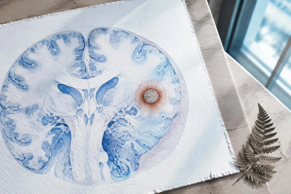
A calcification is generally a remnant rather than a living cyst that antiparasitic medication can eliminate. In the IDSA/ASTMH guideline, calcified-only parenchymal disease is managed symptomatically rather than with antiparasitic drugs; a person with 1 calcified focus and seizures may therefore need antiseizure treatment without needing cyst-killing medication (White et al., 2018).
Surrounding edema can appear intermittently, sometimes long after the original cyst has degenerated. MRI may show this tissue response even when the calcification is more obvious on CT, which is why the 2 scans can tell different but compatible stories rather than one proving that the other is wrong.
Neither a negative antibody result nor a persistent positive result measures seizure risk by itself. The broader lesson resembles other seronegative neurological conditions, discussed in our negative neurological antibody results guide: absence of detectable antibodies is not equivalent to absence of a clinically relevant neurological process.
Dè an deuchainn antibodies airson cysticercosis a chaidh a dhèanamh dha-rìribh?
The useful first question is whether the report describes EITB, ELISA, or another antibody method, because these are not interchangeable tests. A negative index below 1 laboratory's cutoff cannot inherit the performance figures of a different reference-laboratory assay, even when both reports use the name cysticercosis IgG.
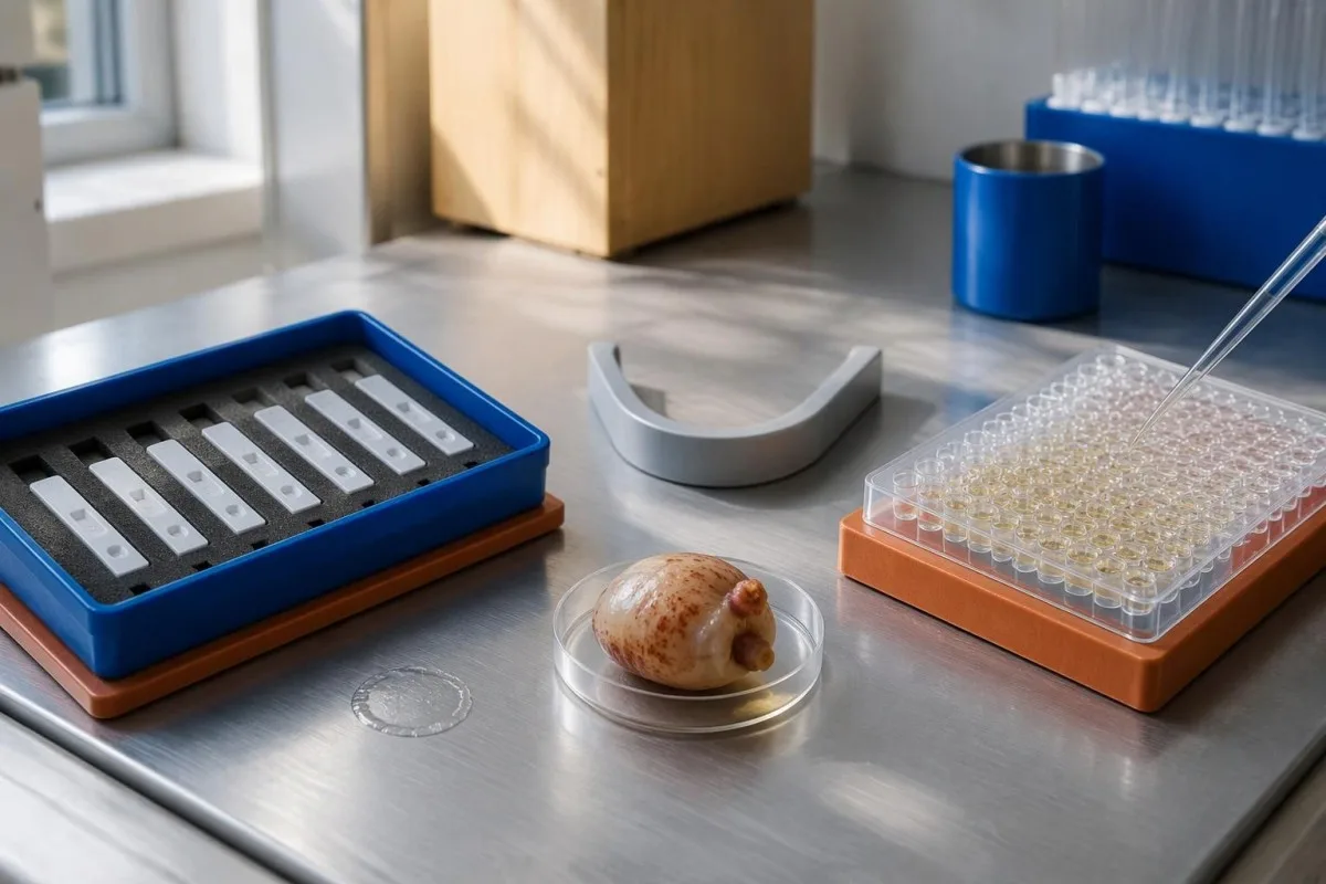
EITB using purified parasite glycoproteins is the preferred serological method in the IDSA/ASTMH guideline, while crude-antigen ELISA methods have recognized sensitivity and specificity limitations (White et al., 2018). Before comparing 2 results, check the method, specimen type, reference interval, and whether the laboratory used a validated confirmatory procedure.
Some laboratories report a qualitative result; others provide an optical-density ratio or numerical index. A value such as 0.8 in a hypothetical assay does not represent 0.8 cysts, an 80% disease probability, or a universal severity score—our toraidhean càileachdail agus cainneachdail guide explains that distinction.
Kantesti AI's role is to help readers preserve the laboratory's original wording rather than reinterpret 1 method through another method's thresholds. Our stiùireadh comharran obair-lann provides broader report-reading context; for this result, keep the actual assay name available when discussing referral or repeat testing.
Ciamar a dh’atharraicheas eachdraidh nochdaidh amharas breithneachail?
Neurocysticercosis follows ingestion of Taenia solium eggs, usually through fecal contamination from a person carrying the intestinal tapeworm. Eating undercooked pork can cause intestinal taeniasis, but brain cysts arise through a different transmission step; symptoms may emerge years after exposure rather than within 1 recent trip.

A vegetarian can develop cysticercosis because eggs can contaminate food or hands independently of meat consumption. Useful history includes past residence, household contacts, sanitation conditions, and possible exposure to a tapeworm carrier; asking only about meals eaten during the last 2 weeks can miss the relevant background.
A negative stool examination does not exclude cysts in the brain. Stool testing addresses intestinal tapeworm carriage, whereas CT and MRI assess nervous-system findings; these are 2 different sites and clinical questions, although public-health teams may recommend evaluating contacts when a carrier could be involved.
Normal eosinophils also cannot exclude neurocysticercosis, and raised eosinophils do not identify the brain as the affected site. Our parasite antibody interpretation guide discusses a separate parasite with similar localization limits; the practical point is to avoid using 1 routine cell count as a substitute for targeted neurological evaluation.
Cuin a dh’fheumas glacaidhean ùra measadh èiginneach?
A first known seizure needs urgent medical assessment regardless of antibody results, and emergency services should be contacted for a first seizure or an ongoing seizure with concerning features. A convulsive seizure lasting 5 minutes, repeated seizures without recovery, breathing difficulty, pregnancy, or significant injury requires emergency care.

Do not wait for the 5-minute mark to seek help after a first known seizure. Keep the person away from hazards, cushion the head if safely possible, do not restrain them or put anything in their mouth, and place them on their side once convulsions stop if they are breathing normally.
Persistent confusion, a severe new headache, fever with neck stiffness, or new weakness increases concern for causes beyond cysticercosis. Sudden one-sided weakness or speech difficulty also demands emergency assessment, as our stiùireadh mu shoidhnichean rabhaidh stròic explains; 1 negative infection test cannot rule out stroke, meningitis, or a structural brain problem.
After a suspected seizure, stop driving and avoid swimming alone, heights, or operating machinery until a clinician provides advice consistent with local rules. If a witness can safely record the duration and describe movements or recovery, those observations may be more useful than ordering a second antibody test before the first neurological assessment.
Dè tha CT a’ cur ri breithneachadh neurocysticercosis?
Noncontrast CT is particularly useful for detecting calcified cyst remnants, including small approximately 1–4 mm foci that MRI may show less clearly. CT also helps assess urgent structural problems such as hydrocephalus, but 1 isolated calcification is not automatically proof of neurocysticercosis or the cause of a seizure.
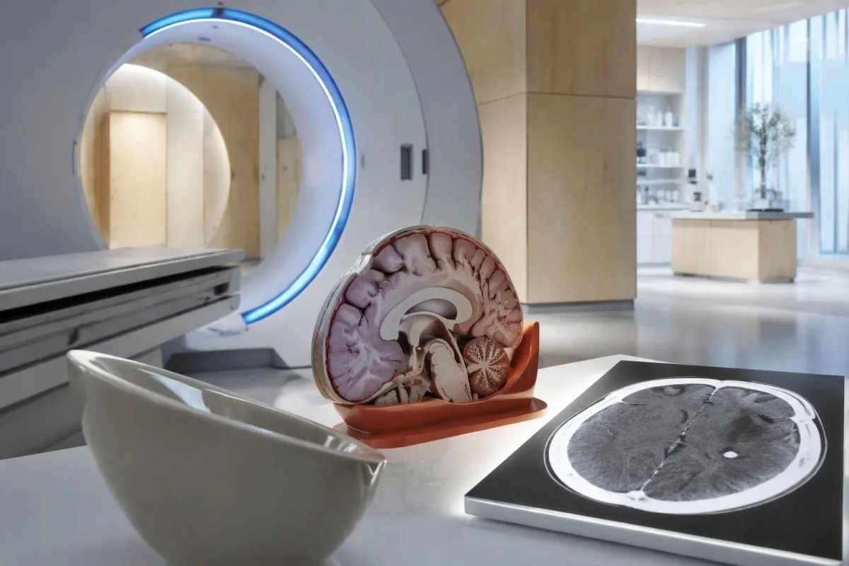
Calcium appears dense on CT, allowing radiologists to identify mineralized remnants and describe their distribution. The IDSA/ASTMH guideline recommends both MRI and noncontrast CT when classifying newly diagnosed neurocysticercosis, because the 2 modalities have complementary strengths rather than being interchangeable alternatives (White et al., 2018).
An incidental calcification needs context: previous granulomatous disease, other causes of mineralization, and scan appearance can influence interpretation. If 1 calcification sits near a suspected seizure focus, concordance with symptoms and EEG may strengthen the explanation, but anatomical proximity alone cannot establish causation.
A laboratory marker used in selected head-injury pathways does not test for brain cysts or replace imaging after an unexplained seizure. Our head-injury CT decisions guide illustrates that narrower setting; ask the clinician whether the present question requires noncontrast CT, contrast imaging, MRI, or a combination of 2 modalities.
Dè tha MRI a’ sealltainn a dh’fhaodadh CT a chall?
MRI is better suited to evaluating viable cysts, surrounding edema, and many ventricular or subarachnoid findings. Specialized 3-dimensional sequences such as CISS or FIESTA can improve visualization of cyst membranes within cerebrospinal fluid spaces, where a cyst may otherwise resemble the surrounding fluid.
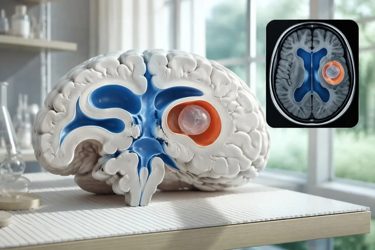
A cyst inside the ventricles can obstruct cerebrospinal fluid flow and cause hydrocephalus even without numerous parenchymal cysts. Headache with vomiting, declining alertness, or new visual symptoms therefore changes urgency; 1 reassuring antibody result should not override symptoms that suggest raised pressure or impaired fluid circulation.
MRI may show a scolex, the small internal structure of the parasite, inside a compatible cyst. This finding can be highly specific when confidently identified, whereas ring enhancement alone has a broader differential; our brain imaging warning signs guide explains why structural abnormalities require interpretation rather than diagnosis from 1 visual feature.
A routine MRI protocol may not answer every cysticercosis question. If ventricular or subarachnoid disease is suspected, discuss whether thin-section 3-dimensional imaging is needed; radiologists choose sequences and contrast according to the clinical question, and a normal scan using 1 protocol does not prove every possible disease compartment has been adequately assessed.
Ciamar a thèid neurocysticercosis a bhreithneachadh nuair a tha na deuchainnean ag eas-aontachadh?
Neurocysticercosis diagnosis combines imaging, clinical findings, exposure information, and supporting tests, rather than requiring a positive antibody result in every case. The revised diagnostic criteria published in 2017 give characteristic neuroimaging findings a central role and distinguish strong evidence from less-specific supportive features (Del Brutto et al., 2017).
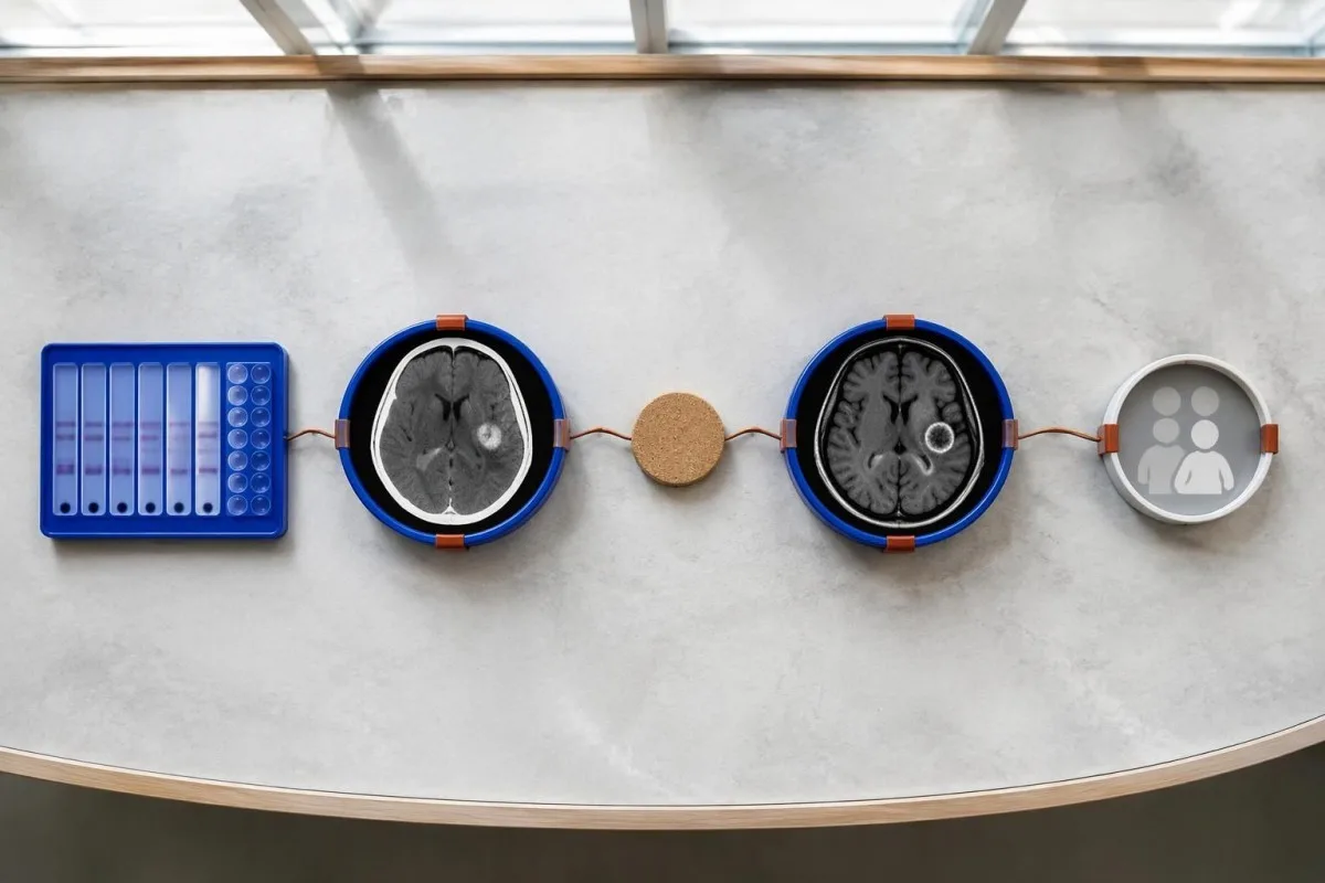
Evidence should be weighted, not tallied. A confidently visualized scolex inside a compatible cyst is different from 1 nonspecific ring-enhancing area, while a positive antibody result is different from direct evidence that a finding is in the brain; our toradh antibody adhartach explains why detection alone cannot establish location.
For a clearly hypothetical probability example, start with a 30% pretest probability and assume a single-cyst assay has 55% sensitivity and 99% specificity. In 1,000 comparable people, about 135 affected and 693 unaffected people would test negative, leaving approximately a 16% probability of disease among negative results—still substantial, although these assumed figures are not a personal risk calculator.
The most useful disagreement to investigate is whether each test is answering the same question. A negative antibody test, a CT calcification, and MRI edema can all be compatible with 1 calcified disease process; unexplained discrepancies should prompt review of the assay and images rather than automatic dismissal of either result.
Dè eile a dh’fhaodadh a bhith coltach ri ciste aonaranach ann an ìomhaighean na h-eanchainn?
A single ring-enhancing brain finding is not specific to neurocysticercosis. Other possibilities include tuberculoma, an abscess, a neoplasm, or an inflammatory process; clinicians use appearance, location, immune status, symptoms, and sometimes follow-up imaging or tissue examination to distinguish these possibilities rather than relying on 1 antibody panel.
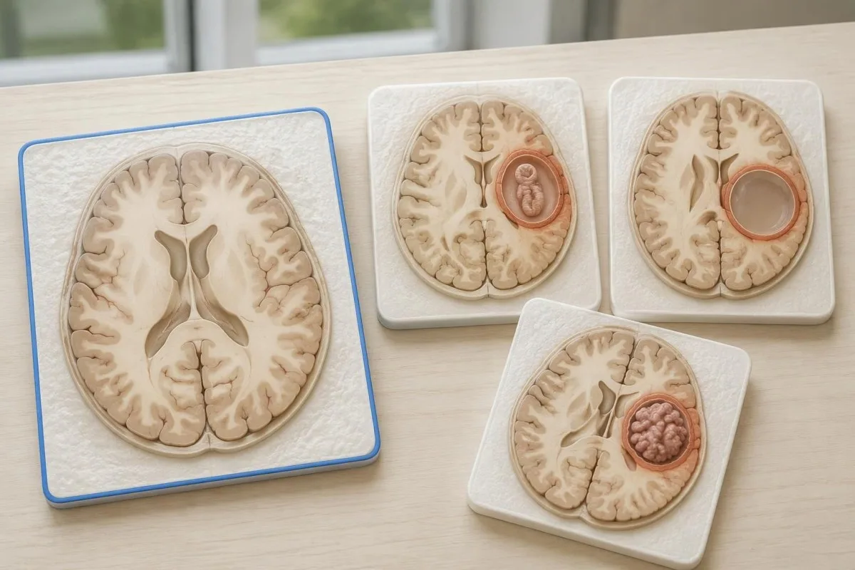
Edema, restricted diffusion, internal structures, and the number of findings all contribute to the differential, but no single nonspecific feature settles every case. Fever and immune suppression may change the suspected cause; a 2 cm ring-enhancing area in a hypothetical immunocompromised person warrants a different investigation from a tiny calcification in an otherwise well person.
Toxoplasmosis is one consideration in selected immunocompromised patients, and a positive IgG result can indicate past exposure rather than prove an active brain process. Our Toxoplasma IgG interpretation guide explains that laboratory distinction; imaging and the patient's immune status determine whether the result is clinically relevant to 1 or more cerebral findings.
New seizures accompanied by rapidly evolving memory problems, psychiatric changes, or abnormal movements can also suggest encephalitis rather than a solitary parasite cyst. Our encephalitis diagnostic pathway describes how MRI, EEG, and cerebrospinal fluid testing answer different questions; a second negative cysticercosis antibody result does not investigate those alternatives.
An urrainn do dheuchainn antigen, PCR no CSF mì-chinnt fhuasgladh?
Antigen tests, PCR, and cerebrospinal fluid analysis can add information in selected cases, particularly when extraparenchymal disease is suspected. None is a universal rescue test after 1 negative antibody result: availability, assay validation, disease compartment, and safety of lumbar puncture determine whether additional testing is appropriate.
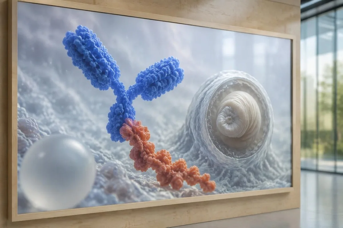
Antibody testing detects the host response, while antigen testing aims to detect parasite-derived material and may better reflect viable disease burden. Antigen detection is often less useful when only 1 small parenchymal cyst or calcifications are present; molecular testing is used in some specialist settings, but performance cannot be generalized across every laboratory or specimen.
Lumbar puncture is not routinely required for an uncomplicated solitary parenchymal finding and can be unsafe when obstructive hydrocephalus or significant mass effect is present. Cerebrospinal fluid tests for other disorders also require disease-specific interpretation, as our CSF band interpretation guide explains; 1 abnormal CSF marker does not identify every possible cause.
Tha Kantesti na àrd-ùrlar mìneachaidh deuchainn fala AI, not a service that orders lumbar punctures, identifies cysts on scans, or confirms neurological infections. Our teicneòlas mìneachaidh AI explains the laboratory-report workflow; when 2 tests disagree, our role is to clarify their stated meanings and limitations, not select an invasive diagnostic procedure.
Carson a tha làimhseachadh an urra ri ìre agus àite na ciste?
Treatment depends on whether cysts are viable, degenerating, calcified, or located outside the brain tissue, not simply on antibody positivity. Selected viable parenchymal cases receive antiparasitic treatment, whereas calcified-only disease generally receives symptom-directed care; ventricular obstruction may require a procedural approach rather than 1 standard medication regimen.
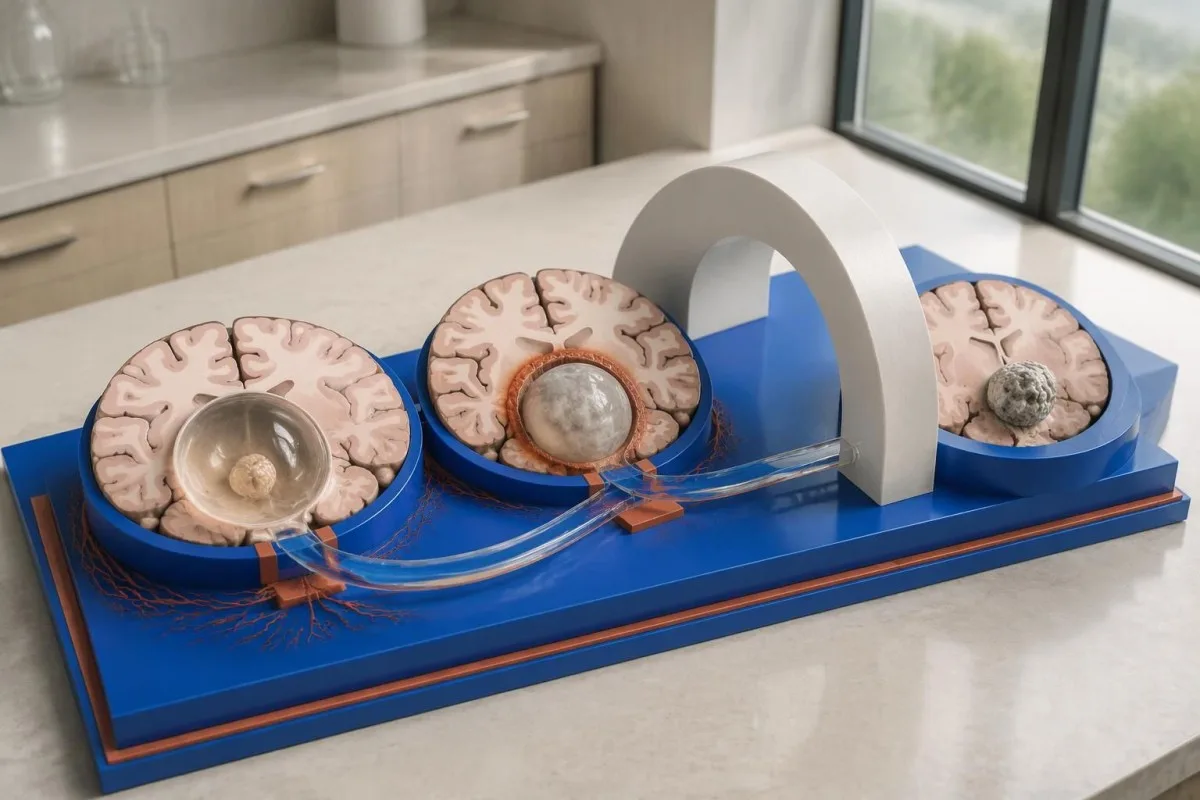
For selected patients with 1–2 viable parenchymal cysts, the IDSA/ASTMH guideline describes albendazole treatment for 10–14 days with appropriate accompanying care. This is a specialist-directed recommendation, not a home-treatment instruction: antiparasitic drugs can provoke an inflammatory response, and untreated hydrocephalus or diffuse cerebral edema changes the order of management (White et al., 2018).
WHO's 2021 guidance focuses on defined presentations of parenchymal neurocysticercosis, while the IDSA/ASTMH guideline also addresses ventricular, subarachnoid, and other forms in detail. Differences in imaging access and disease compartment explain why 1 treatment recommendation cannot be transferred to every patient with an antibody result; the guidelines do not turn negative serology into a rule-out test.
Corticosteroids may accompany antiparasitic therapy, but prolonged use requires additional safety planning, including assessment for relevant latent infections. Our Strongyloides and steroid safety guide explains a separate risk that matters before immune-suppressing treatment; an eye examination is also recommended before antiparasitic therapy to evaluate possible intraocular cysts.
Dè bu chòir sùil a chumail air rè leanmhainn a bharrachd air toraidhean antibodies?
Follow-up should track seizures, neurological symptoms, imaging changes, and medication safety, rather than use antibody disappearance as proof of recovery. For viable parenchymal cysts, the IDSA/ASTMH guideline suggests repeat MRI at least every 6 months until the cystic component resolves, with earlier reassessment when symptoms change.
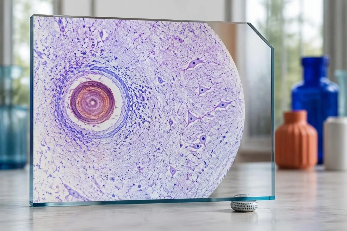
Antibodies may remain detectable after the original disease process has changed, and low-burden disease may never produce a positive result. If 1 new seizure occurs during follow-up, clinicians consider adherence, medication interactions, recurrent edema, and other causes; a rising or falling antibody index alone cannot distinguish among those explanations.
Kantesti AI can help explain reported laboratory results during medication follow-up, but a drug concentration is not a stand-alone measure of seizure control. Our mìneachadh ìre levetiracetam guide explains why sample timing and kidney function matter; 2 measurements collected at different times after dosing may not be directly comparable.
Monitoring depends on the chosen drug and treatment duration: prolonged albendazole therapy can require liver-test and blood-count surveillance, while some antiseizure medicines can affect sodium. Our stiùireadh sàbhailteachd electrolytes discusses those separate laboratory concerns; do not stop an antiseizure medicine or change its dose because 1 cysticercosis antibody result is negative.
Dè as urrainn do Kantesti a mhìneachadh, agus dè nach urrainn do rannsachadh dearbhadh?
AI report interpretation cannot confirm or exclude neurocysticercosis from laboratory results alone. Tha Kantesti na Inneal mion-sgrùdaidh deuchainn fala le cumhachd AI whose role here is to organize reported laboratory findings; a negative result still needs clinical context, and 1 new seizure takes priority over reviewing an automated explanation.

Prepare a focused review packet with the original antibody report, assay method, CT or MRI report, symptom timeline, and medication list. These 5 information categories reduce the risk of discussing a result without its context; ask specifically whether the imaging suggests viable, degenerating, calcified, ventricular, or subarachnoid disease and which unanswered question the next test would address.
The 2 Kantesti publications listed below are a repository-deposited global analysis report and an educational RDW guide, not clinical evidence validating neurocysticercosis diagnosis. Our RDW agus stiùireadh CBC can support interpretation of a separately ordered blood count, but RDW, MCV, and MCHC do not establish whether a brain cyst is present.
Evidence for the neurological recommendations in this article comes from the cited diagnostic criteria and clinical guidelines, not from general AI benchmarking or laboratory-report volume. Our technical validation scope should be read within its stated boundaries; neither 1 successful report explanation nor a large aggregate dataset substitutes for condition-specific diagnostic validation.
Ceistean cumanta
An urrainn dhut neurocysticercosis a bhith ort ged a tha deuchainn fala àicheil agad?
Faodaidh neurocysticercosis a bhith an làthair a dh’aindeoin deuchainn àicheil airson antibodies san fhuil, gu h-àraidh ma tha 1 chist san eanchainn no ma tha an galar air a àireamhachadh a-mhàin. Mar as trice, tha tuairmsean foillsichte bho obair-lann iomraidh airson EITB a’ sealltainn cugallachd de mu 50–60% airson aon chist, mòran nas ìsle na an coileanadh aige nuair a tha grunn chisteachan obrachail ann. Mar sin, tha CT, MRI, na comharraidhean agus eachdraidh nochdaidh fhathast aig cridhe a’ bhreithneachaidh. Feumar measadh èiginneach a dhèanamh air grèim ùr, seach a bhith an urra ri fois-inntinn bho thoradh àicheil.
Dè an deuchainn as earbsaiche airson neurocysticercosis?
Chan eil aon deuchainn airson neurocysticercosis a’ freagairt gach ceist dhearbhaidh; tha ìomhaighean eanchainn agus deuchainnean seròlais air an taghadh gu h-iomchaidh a’ cur ri chèile. Is e EITB an dòigh dearbhaidh antibody as fheàrr leotha ann an stiùireadh IDSA/ASTMH, ach faodaidh e fàiligeadh ann a bhith a’ lorg galair anns nach eil ach 1 chiste. Tha CT gun chaochladh gu h-àraidh feumail airson àireamhachadh, agus tha MRI nas fheàrr airson measadh a dhèanamh air mòran chistean obrachail, sèid agus neo-riaghailteachdan ann an àiteachan làn lionn. Bidh luchd-clionaigeach a’ cothlamadh nan toraidhean sin an àite a bhith a’ gabhail ri aon toradh àicheil mar dhearbhadh deireannach.
A bheil CT no MRI nas fheàrr airson neurocysticercosis?
Tha neartan eadar-dhealaichte aig CT agus MRI ann a bhith a’ breithneachadh neurocysticercosis. Gu tric, lorgaidh CT gun àidseant coimeasachaidh calcachaidhean beaga, mu 1–4 mm tarsainn, nas soilleire, ach nì MRI measadh air cystan beò, sèid san stuth mun cuairt orra, agus galair sna ventricles no san àite fo-arachnoideach. Dh’fhaodadh sreathan sònraichte de MRI trì-thaobhach cuideachadh le bhith ag aithneachadh memblanan cyst ann an àiteachan lionn cerebrospinal. Tha stiùireadh IDSA/ASTMH a’ moladh an dà dhòigh nuair a thathar a’ seòrsachadh galair a chaidh a dhearbhadh às ùr.
An urrainn do chist a tha air calcachadh fhathast glacaidhean adhbharachadh?
Faodaidh fuigheall calcified de cysticercosis fuireach co-cheangailte ri glacaidhean ged nach eil e na chist a ghabhas mairsinn tuilleadh. Bidh edema mun cuairt a’ leasachadh bho àm gu àm ann an cuid de fhòcasan calcified, nach eil ach 1–4 mm tarsainn gu tric, agus dh’fhaodadh sin a bhith ri fhaicinn air MRI. Chan eil antibodies àicheil a’ cur às don uidheamachd seo, agus chan eil antibodies a tha fhathast deimhinneach a’ tomhas cunnart glacaidhean. Mar as trice, thèid galar calcified a-mhàin a làimhseachadh a rèir nan comharraidhean seach le cungaidh an-aghaidh dìosganaich.
Am bu chòir dhomh deuchainn antibody àicheil airson cysticercosis ath-aithris?
Chan eil e gu fèin-obrachail nas fheàrr an ath cheum a bhith ag ath-dhèanamh deuchainn àicheil airson antibodies an aghaidh cysticercosis. Ma tha 1 chist bheag no galar calcichte ann, faodaidh freagairt lag nan antibodies fuireach àicheil fiù ’s ma thèid an aon deuchainn ath-dhèanamh an dèidh 2 sheachdain. Dh’fhaodadh clionaigiche ath-sgrùdadh a dhèanamh air a’ mhodh thùsail, deuchainn dhearbhaidh a tha nas fheàrr a chomharrachadh iarraidh, no prìomhachas a thoirt do CT agus MRI. Cha bu chòir dàil a chur air measadh glacaidhean ùra no comharraidhean eanchainn a tha a’ fàs nas miosa gus an tèid an deuchainn ath-dhèanamh.
Cuin a bu chòir dhomh cuideachadh èiginn iarraidh airson a’ chiad ghlacaidh?
Feumaidh a’ chiad ghlacadh aithnichte measadh meidigeach èiginneach agus fios a chur gu seirbheisean èiginn ionadail, ge bith dè an toradh àicheil air deuchainn antibody airson neurocysticercosis. Tha glacaidh chonbhasach a mhaireas 5 mionaidean, glacaidhean a bhios a’ tachairt a-rithist gun faighinn seachad orra, duilgheadas analach, torrachas no leòn mòr nan èiginn. Na cuir bacadh air an neach agus na cuir càil na bheul; cuir air a thaobh e nuair a stadas na glacaidhean, ma tha e a’ tarraing anail gu h-àbhaisteach. Na dràibh gus am bi clionaigear air measadh a dhèanamh air na thachair agus air na cuingeachaidhean iomchaidh a mhìneachadh.
Faigh mìneachadh air na toraidhean deuchainn fala agad
Luchdaich suas aithisg an obair-lann agad agus mìnichidh AI Kantesti gach bith-chomharradh ann an cànan soilleir taobh a-staigh diogan. Air a chleachdadh le còrr is 2 mhillean neach air feadh an t-saoghail.
Tùsan
- Klein, T., Mitchell, S., & Weber, H. (2026). Neach-anailis Deuchainn Fala AI: 2.5M Deuchainnean air an Mion-sgrùdadh | Aithisg Slàinte Cruinne 2026. Rannsachadh Meidigeach AI Kantesti. DOI
- Klein, T., Mitchell, S., & Weber, H. (2026). Deuchainn fala RDW: stiùireadh iomlan air RDW-CV, MCV agus MCHC. Rannsachadh Meidigeach AI Kantesti. DOI
- White AC Jr et al. (2018). Diagnosis and Treatment of Neurocysticercosis: 2017 Clinical Practice Guidelines by the Infectious Diseases Society of America (IDSA) and the American Society of Tropical Medicine and Hygiene (ASTMH). Galaran Clionaigeach Gabhaltach. DOI
- Del Brutto OH et al. (2017). Revised diagnostic criteria for neurocysticercosis. Journal of the Neurological Sciences. DOI
- World Health Organization (2021). WHO guidelines on management of Taenia solium neurocysticercosis. Buidheann Slàinte na Cruinne.
- 2M+Deuchainnean air an sgrùdadh
- 127+Dùthchannan
- 100+Cànanan
Mu dheidhinn an sgioba mheidigeach


Comharran earbsa E-E-A-T
Eòlas
Lèirmheas clionaigeach air a stiùireadh le lighiche air sruthan-obrach mìneachaidh obair-lann.
Eòlas
Fòcas air leigheas obair-lann air mar a bhios bith-chomharraidhean (biomarkers) a’ giùlan ann an co-theacsa clionaigeach.
Ùghdarrasachd
Ath-sgrùdadh meidigeach leis an Dr Thomas Klein, le tabhartasan bhon Dr Sarah Mitchell agus an t-Oll. Dr Hans Weber.
Earbsachd
Mìneachadh stèidhichte air fianais le slighean leanmhainn soilleir gus dragh a lughdachadh.





Àicheadh meidigeach
Tha an artaigil seo dìreach airson adhbharan foghlaim agus chan eil e a’ dèanamh comhairle mheidigeach. Cuir fios an-còmhnaidh gu solaraiche cùram slàinte teisteanasach airson co-dhùnaidhean breithneachaidh is leigheis.
Foillsichte · Cuir fios chun sgioba dheasachaidh
Foillsichte le Kantesti Ltd, clàraichte ann an Sasainn agus sa Chuimrigh, àireamh companaidh. 17090423, Lunnainn, an Rìoghachd Aonaichte.
