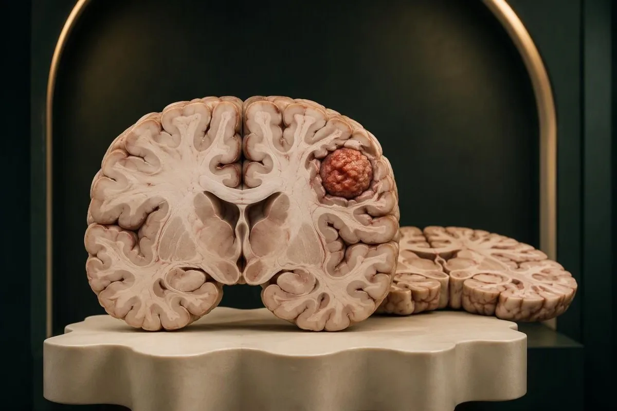
Progressive neurological changes matter more than a headache checklist. The safest next step depends on what changed, how quickly it changed, and whether brain function is affected.


- Brain tumor symptoms can include progressive weakness, vision changes, language difficulties, altered behavior, or a first seizure; no single symptom establishes the diagnosis.
- Headaches alone rarely indicate a brain tumor, particularly when the pattern is longstanding and the neurological examination is normal.
- Comharraidhean èiginn include sudden one-sided weakness, new speech difficulty, a thunderclap headache, or reduced consciousness—even if symptoms improve within minutes.
- First seizure in adults needs urgent medical assessment; call emergency services for a first observed seizure, a seizure lasting 5 minutes, or repeated seizures without recovery.
- Progressive neurological loss warrants prompt assessment; NICE recommends considering urgent brain MRI within 2 weeks for adults with progressive, subacute loss of central neurological function.
- Obair-fala àbhaisteach cannot rule a brain tumor in or out. Normal CBC, glucose, electrolytes, and inflammatory markers do not replace neurological examination or imaging.
- Brain MRI generally provides more detail than CT for suspected tumors, while CT is often the faster first examination during an emergency.
- Safety while waiting means avoiding driving after a suspected seizure and seeking reassessment immediately if weakness, confusion, vision loss, or vomiting develops.
Which brain tumor symptoms warrant assessment?
Brain tumor symptoms that warrant assessment are new, persistent, or progressive changes in neurological function, including weakness, speech difficulty, vision loss, altered behavior, and a first seizure. Headaches alone rarely indicate a tumor; sudden neurological symptoms need emergency care, and normal routine bloodwork cannot exclude a tumor.
A symptom’s trajectory is often more useful than its intensity. Difficulty using 1 hand that worsens over several weeks deserves assessment even without pain, whereas a severe but familiar migraine with full recovery may follow an established, non-tumor pattern.
Brain tumors can affect function through local pressure, surrounding swelling, or obstruction of cerebrospinal fluid. Those are 3 different mechanisms, so symptoms do not reliably reveal either the growth’s size or whether it is malignant; a nonmalignant growth can still require treatment.
I’m Thomas Klein, Kantesti’s Chief Medical Officer, and my practical advice is to describe the lost ability rather than simply say you feel unwell. Compare today with 2–4 weeks ago: typing, reading, walking, remembering appointments, and finding words provide much clearer clinical information.
Tha Kantesti na Anailisiche deuchainn fala AI that helps explain laboratory findings, not a brain tumor detection service. Our UK organization separates laboratory interpretation from neurological diagnosis: an explanation delivered in about 60 seconds cannot replace an examination or scan.
Early signs of brain tumor: patterns, not a checklist
Early signs of brain tumor are not a fixed sequence, and some tumors cause no symptoms initially. A change affecting 2 linked abilities—such as word-finding and reading—can be more informative than an isolated symptom, although many other neurological conditions cause similar patterns.
Repeated errors in a previously familiar task deserve a concrete description. A hypothetical 54-year-old who begins missing words on one side of a page and misjudging doorways needs a different assessment from someone occasionally forgetting names after poor sleep.
Symptoms may evolve over days, weeks, or months; there is no safe minimum duration to wait when function is deteriorating. A 3-day change with worsening balance can be more urgent than a mild symptom present unchanged for 3 years.
Progression is not always smooth. A person may compensate for a subtle visual deficit until a task exposes it, and relatives may notice changes 1–2 weeks before the person does; that difference does not automatically mean rapid growth.
Memory supplements are not a diagnostic trial for unexplained cognitive change. Our discussion of memory supplement evidence explains their limitations, but a new functional decline over 2 weeks should be assessed rather than treated with a shopping list.
Why headaches alone rarely indicate a brain tumor
Brain tumor headache symptoms overlap with migraine, tension-type headache, sleep problems, and other common conditions. A longstanding headache pattern without neurological changes is less concerning than a new headache accompanied by weakness, visual deterioration, repeated vomiting, or a first seizure.
Morning headache is not a tumor signature. Sleep apnea, teeth grinding, medication effects, and caffeine withdrawal can also cause it; similarly, pain worsened by coughing or bending warrants assessment but does not identify 1 particular cause.
A headache diary is useful when it records function as well as pain. Over 7–14 days, note onset, duration, medication use, vomiting, and any trouble speaking or seeing—but do not keep collecting entries while a new neurological deficit worsens.
Fatigue and headache can accompany iron deficiency, sometimes before anemia appears. Our guide to de ferritin ìosal explains that distinction; finding low iron does not explain away a progressive problem involving 1 arm or one visual field.
Headache affecting 2 household members, especially around a heater or fuel-burning appliance, raises a different urgent concern: carbon monoxide exposure. Leave the environment and seek emergency advice; our carbon monoxide testing guide explains why ordinary oxygen readings can be misleading.
Emergency neurological warning signs: act now
Sudden weakness, new speech difficulty, a thunderclap headache, reduced consciousness, or rapidly worsening neurological symptoms require emergency assessment. A headache reaching maximum intensity within about 1 minute is a thunderclap headache; do not assume these symptoms are a tumor or wait for bloodwork.
A sudden facial droop or loss of strength in 1 arm may represent stroke, including when it resolves. Emergency treatment decisions depend on timing, so record when the person was last known to be well and call the local emergency number.
Severe headache with fever and neck stiffness, or headache with increasing drowsiness and repeated vomiting, also needs urgent care. These combinations can reflect several dangerous conditions; even 1 episode of collapse changes the assessment substantially.
Weakness is not always caused by a problem inside the brain. Rapidly ascending weakness involving both legs, particularly with breathing or swallowing difficulty, requires emergency assessment; our comharran rabhaidh laigse èiginneach explain an important peripheral nerve alternative.
Do not drive yourself if you have new neurological impairment. In the UK, call 999 or 112 for an emergency; elsewhere use the local emergency number, and tell the dispatcher about sudden onset, seizures, breathing changes, and any reduced responsiveness.
First seizure in adults: assessment and immediate safety
A first seizure in adults requires urgent assessment, but a seizure does not automatically mean a brain tumor. Call emergency services for a first observed seizure, any seizure lasting 5 minutes, or repeated seizures without recovery; structural brain changes are only one possible cause.
Some seizures involve 1 side of the body, a brief speech arrest, or impaired awareness rather than whole-body shaking. A witness’s description of the first movement, responsiveness, duration, and recovery can help distinguish a seizure from fainting or another event.
During a convulsive seizure, protect the person from nearby hazards, cushion the head, and time the event. Do not restrain them or put anything in their mouth; turn them onto their side when movements stop, and give nothing by mouth until fully alert.
NICE recommends specialist assessment within 2 sheachdain after a first suspected seizure and, when an EEG is requested, ideally performing it within 72 uair a thìde of the seizure (NICE, 2022, NG217). A normal EEG does not exclude epilepsy; a 12-lead ECG helps identify cardiac conditions that can resemble seizures.
Laboratory testing can identify seizure triggers such as low sodium emergencies, but it cannot exclude a structural cause. If treatment is prescribed, our levetiracetam monitoring guide explains why 1 drug level is not a measure of seizure control or tumor status.
Vision changes that need eye and neurological assessment
Persistent double vision, loss of part of the visual field, or progressive visual deterioration warrants assessment. Sudden visual loss needs emergency care; a change affecting 1 eye suggests a different anatomical pathway from loss of the same side of vision in both eyes.
Visual field loss can be surprisingly easy to miss because central vision remains clear. Someone may read small print yet repeatedly overlook objects on 1 side; describing these collisions is more useful than reporting only that their vision feels strange.
Raised intracranial pressure can produce papilledema, meaning swelling of the optic discs. It requires urgent assessment because several conditions can cause it; the absence of papilledema on 1 examination does not rule out a brain tumor.
An eye examination and a neurological examination answer different questions. Our guide to visual field testing explains how clinicians map peripheral vision, while a scan examines structures that an eye pressure measurement cannot assess.
Double vision that disappears when either eye is covered is usually binocular and concerns eye alignment. Double vision persisting in 1 uncovered eye is more often an optical eye problem, but new symptoms with headache, weakness, or eyelid drooping should not be self-triaged.
Progressive weakness, balance problems, and sensory changes
Progressive loss of strength, coordination, or sensation needs a clinical examination, especially when it affects one side or interferes with walking. Problems developing over 2 weeks should not be dismissed as aging or anxiety; sudden onset belongs in emergency care.
Weakness means reduced power, not simply feeling exhausted. Being unable to lift 1 foot, repeatedly dropping a cup, or losing the ability to fasten buttons provides a clearer neurological clue than general tiredness after work.
Balance problems may originate in the inner ear, peripheral nerves, cerebellum, or medication effects. A clinician tests several systems rather than ordering 1 universal dizziness test; unsteadiness accompanied by new slurred speech or severe headache requires urgent reassessment.
Symmetrical tingling in both feet suggests a different pattern from numbness in 1 arm and the same side of the face. Nutritional deficiencies can cause neurological problems; our copper deficiency guide explains one treatable alternative without making it a substitute for examination.
Falls change the urgency because they introduce injury risk as well as diagnostic uncertainty. After 1 unexplained fall with new imbalance, avoid ladders and unsupported stairs, ask someone to accompany you, and arrange assessment rather than attempting repeated home balance challenges.
Personality, memory, and language changes: what matters
New changes in judgment, language, memory, or behavior warrant assessment when they are persistent, progressive, or affect daily function. A 2-week decline in familiar tasks carries different significance from occasional forgetfulness; sudden confusion is an emergency, not a scheduled memory appointment.
Language difficulty is not the same as forgetting a name. Substituting unrelated words, struggling to understand ordinary instructions, or repeatedly losing the meaning of a sentence suggests impaired language processing; note 2–3 actual examples for the appointment.
Personality changes are nonspecific and can reflect depression, poor sleep, medications, substance use, or neurological disease. Our guide to mood-related blood tests discusses treatable contributors, but normal results do not explain away 1 newly lost everyday ability.
A family member’s observations can help establish the timeline without turning the visit into an argument. Ask them to describe what changed over the last 4 weeks—missed bills, unusual disinhibition, getting lost—not simply whether the person seems different.
Sudden confusion developing over hours can arise from metabolic illness, medication toxicity, seizures, stroke, or other acute conditions. Even if there were milder memory concerns for 6 months, the abrupt change needs fresh assessment rather than being attributed to the older problem.
What a neurological examination can—and cannot—show
A neurological examination tests brain and nerve function to identify patterns and guide imaging. It commonly assesses at least 6 domains: mental function, cranial nerves, strength, sensation, reflexes, and coordination or gait; a normal examination lowers concern in some settings but cannot exclude every tumor.
The clinician looks for agreement between findings rather than 1 isolated abnormality. Weakness, altered reflexes, and a compatible sensory pattern together may localize a problem more confidently than a symptom description alone; pain or fatigue can affect performance.
A normal examination is a snapshot, not a guarantee about the next 3 months. Small growths, intermittently symptomatic conditions, and problems in less easily tested brain functions may not produce clear findings at an initial visit.
NICE advises considering urgent direct-access brain MRI within 2 sheachdain for adults with progressive, subacute loss of central neurological function, or CT if MRI is contraindicated (NICE, 2015, NG12). That recommendation concerns the clinical pattern—not whether a CBC is abnormal.
At Kantesti, we distinguish technical laboratory interpretation from evidence supporting a neurological diagnosis. Our inbhean dearbhaidh clionaigeach provide context for methodology; even excellent performance on 1 laboratory task cannot establish accuracy for detecting a condition outside that task.
MRI versus CT: how brain imaging guides diagnosis
MRI is usually the preferred detailed examination for a suspected brain tumor, while CT is often used first in emergencies because it is faster and more accessible. These 2 tests are complementary; the choice depends on symptoms, urgency, safety, and the question the clinician needs answered.
NICE describes standard structural MRI for suspected glioma as including T2-weighted, FLAIR, diffusion-weighted, and T1 imaging before and after contrast when appropriate (NICE, 2018, NG99). These 4 sequence groups reveal different tissue properties; contrast enhancement alone does not prove malignancy.
A normal emergency CT does not answer every question that a dedicated MRI can address. If progressive symptoms continue after 1 CT examination, the next step is clinical reassessment—not assuming the scan was useless or that every possible structural cause has been excluded.
MRI abnormalities also occur in conditions unrelated to tumors. Our explanation of MOG antibody interpretation illustrates why imaging and clinical findings must agree; 1 bright area on a scan is not a diagnosis by itself.
Cerebrospinal fluid testing answers selected questions but is not a routine substitute for brain imaging. The oligoclonal band guide explains one use; clinicians assess lumbar puncture safety first because some intracranial pressure patterns make the procedure unsafe.
Can routine bloodwork detect or exclude a brain tumor?
Routine bloodwork cannot rule a brain tumor in or out. A normal complete blood count, metabolic panel, CRP, or ESR does not exclude an intracranial growth; neither an abnormal result nor a panel containing 20 normal measurements establishes what is happening inside the brain.
Blood tests are still useful for identifying alternative or additional causes. Glucose below 54 mg/dL, or 3.0 mmol/L, is clinically significant hypoglycemia and can impair brain function; resolving low glucose does not automatically account for a separate, longstanding focal neurological deficit.
Sodium is commonly around 135–145 mmol/L in adult laboratories, and levels below 125 mmol/L represent profound hyponatremia. Symptoms and speed of decline determine urgency; our stiùireadh eadar-mhìneachaidh electrolyte explains why a number cannot be interpreted without the clinical situation.
Vitamin B12 deficiency can cause neurological symptoms without obvious anemia. Our discussion of toraidhean B12 crìche covers follow-up testing, but 1 low-normal B12 result must not become a convenient explanation for worsening one-sided weakness.
Tha Kantesti na àrd-ùrlar mìneachaidh deuchainn fala AI that can help explain reported laboratory values and trends. As of October 22, 2026, routine blood panels and consumer blood-based cancer tests are not established replacements for neurological examination and appropriate imaging when brain tumor symptoms are suspected.
Pituitary symptoms and what happens after an abnormal scan
Some pituitary growths produce measurable hormone abnormalities, but most brain tumors do not have a routine diagnostic blood marker. After an abnormal scan, specialists integrate imaging, symptoms, and sometimes tissue and molecular testing; 1 elevated hormone result does not establish the nature of a growth.
Headache with loss of peripheral vision and hormone-related symptoms can prompt pituitary-focused assessment. Our guide to comharraidhean prolactin àrd explains relevant patterns, but medications, pregnancy, stress, and other factors can also raise prolactin; 1 measurement may need confirmation.
Pituitary dysfunction can make a familiar thyroid screening result misleading. A low free T4 with a normal TSH needs contextual assessment, as explained in discordant thyroid test results; 2 apparently reassuring numbers do not necessarily mean the pituitary-thyroid system is functioning normally.
An MRI impression may suggest meningioma, glioma, metastasis, or another condition without definitively classifying it. For gliomas, markers such as IDH status and 1p/19q codeletion contribute to classification; these are specialized tissue-based investigations, not a standard primary-care blood panel.
Not every incidental growth requires immediate tissue sampling or treatment. A small, asymptomatic suspected meningioma may be monitored, while a symptomatic growth affecting vision or causing swelling can require faster action; decisions weigh at least 3 factors—location, behavior, and the person’s overall health.
When a scheduled evaluation is reasonable—and when it is not
A scheduled appointment is reasonable for stable, non-emergency symptoms without new neurological loss. Progressive weakness, language difficulty, or visual deterioration should prompt contact today for expedited assessment; do not book a routine appointment 6 weeks away and simply wait for function to worsen.
A familiar headache pattern, unchanged and without red flags, can usually be discussed in a scheduled clinical visit. A newly changing pattern over 2–4 weeks deserves a sooner appointment, and the clinician may lower the threshold for imaging with a cancer history or abnormal examination.
Bring a short timeline rather than an enormous symptom printout. Record the first date, the change over the last 14 days, specific lost abilities, medication changes, and relevant cancer history; a witness should attend after a suspected seizure if possible.
A suspected seizure changes safety advice immediately. Do not drive until you have received medical and jurisdiction-specific licensing advice; avoid swimming alone, baths alone, heights, and hazardous machinery, even if there has been only 1 event and you currently feel well.
Tha Kantesti na Inneal mion-sgrùdaidh deuchainn fala le cumhachd AI, not an emergency triage service or an interpreter of brain MRI images. Our stiùireadh teicneòlais AI explains the laboratory workflow; reviewing results in about 60 seconds must never delay assessment of a neurological red flag.
Research publications, evidence boundaries, and next steps
Brain tumor assessment should be guided by neurological findings, appropriate imaging, and condition-specific clinical evidence. The 2 laboratory publications listed below address iron and coagulation testing; they are background resources, not evidence that a blood test or AI tool can detect or exclude a brain tumor.
Ar foillseachadh mu sgrùdaidhean iarainn concerns laboratory evaluation of iron status, while our coagulation testing publication addresses clotting measurements. These 2 resources may support broader medical assessment, but neither validates brain tumor screening or provides a neurological rule-out threshold.
A DOI identifies a deposited document; it does not, by itself, establish peer review or clinical effectiveness. Kantesti’s 2 Zenodo resources are therefore separated from the NICE guidelines in the references, and the ResearchGate and Academia.edu discovery links are title searches rather than verified publication copies.
For medical accountability, our Bòrd Comhairleachaidh Meidigeach page describes the physicians associated with Kantesti’s clinical work. As Thomas Klein, I would keep 3 questions central: what ability changed, how fast it changed, and whether the current presentation requires emergency assessment.
The next action should fit the symptom, not the reassuring appearance of a report. Call emergency services for sudden neurological loss or a first observed seizure; contact a clinician promptly for progressive changes, and bring any existing results without waiting for 1 more blood test.
Ceistean Bitheanta
Dè na comharran tràtha a th’ air tumhair eanchainn?
Faodaidh comharran tràtha tumhair eanchainn a bhith a’ gabhail a-steach laigse a tha a’ fàs nas miosa, call raon lèirsinn, duilgheadas cànain, atharrachaidhean ann an giùlan, no a’ chiad ghlacadh, ach chan eil aon chiad chomharra ann a tha cumanta do na h-uile. Bu chòir measadh luath a dhèanamh air call ùr ann an gnìomhachd a tha a’ fàs thar 2–4 seachdainean, fiù ’s às aonais ceann goirt. Feumar cùram èiginn seach coinneamh a chaidh a chur air dòigh ma thig laigse obann, duilgheadas cainnte, troimh-chèile no call lèirsinn ort. Tha iomadh suidheachadh nach eil co-cheangailte ri tumhair ag adhbharachadh comharraidhean coltach ris, agus mar sin tha am breithneachadh an urra ri sgrùdadh agus ìomhaighean meidigeach iomchaidh.
Cò ris a tha ceann goirt bho thumhair eanchainn coltach?
0 Tha ceann goirt bho thumhair eanchainn gun fhaireachdainn sònraichte earbsach, agus dh’fhaodadh e a bhith coltach ri migraine no ceann goirt seòrsa teannachaidh. Tha barrachd adhbhar dragha ann nuair a thig ceann goirt ùr no a tha ag atharrachadh an cois comharraidhean eanchainn a tha a’ fàs nas miosa, cur a-mach a-rithist no a’ chiad ghlacadh. Chan eil pian sa mhadainn no pian nuair a thathar a’ casadaich ag aithneachadh tumhair leis fhèin. Feumar measadh èiginneach a dhèanamh air ceann goirt a ruigeas an dian as àirde aige taobh a-staigh mu 1 mhionaid, oir dh’fhaodadh adhbhar cunnartach eile a bhith aige.
An urrainn do dheuchainnean fala àbhaisteach a bhith a’ cur às do thumhair eanchainn?
Chan urrainn do dheuchainnean fala àbhaisteach àbhaisteach a bhith a’ cur às do chomas tumhair eanchainn. Bidh CBC, pannal meatabolaigeach, CRP agus ESR a’ measadh phròiseasan eadar-dhealaichte agus chan eil iad a’ sealltainn structar na h-eanchainn, fiù ’s nuair a tha 20 tomhas no barrachd a chaidh aithris àbhaisteach. Faodaidh obair-fala nithean a tha a’ cur ris an t-suidheachadh aithneachadh, leithid glùcois ìosal, neo-riaghailteachdan ann an electrolytes no dìth bhiotamain. Tha comharraidhean eanchainneach leantainneach no a tha a’ fàs nas miosa fhathast feumach air measadh clionaigeach agus, nuair a tha sin iomchaidh, ìomhaigheachd den eanchainn.
A bheil a’ chiad ghlacadh ann an inbheach a’ ciallachadh gu bheil tumhair eanchainn air?
Chan eil a’ chiad ghlacadh ann an inbheach gu riatanach a’ ciallachadh gu bheil tumhair eanchainn ann, ach feumar measadh èiginneach a dhèanamh. Am measg nan adhbharan a dh’fhaodadh a bhith ann tha tinneas tuiteamach, buairidhean mheatabolaig, buaidh chungaidhean-leigheis no stuthan, stròc, agus ana-cainnt structarail san eanchainn. Cuir fios gu na seirbheisean èiginn ma chì thu cuideigin a’ faighinn a’ chiad ghlacaidh aca, ma mhaireas grèim 5 mionaidean, no ma bhios grunn ghlacaidhean ann gun a bhith a’ faighinn seachad orra eatarra. Tha NICE a’ moladh measadh le speisealaiche taobh a-staigh 2 sheachdain às dèidh a’ chiad ghlacaidh a thathar an amharas; seachain dràibheadh gus am faigh thu comhairle mheidigeach agus comhairle mu chead dràibhidh ionadail.
A bheil MRI no CT nas fheàrr airson tumhair eanchainn a dhearbhadh?
0 Mar as trice bheir MRI fiosrachadh nas mionaidiche seachad nuair a tha amharas ann gu bheil tumhair eanchainn ann, agus gu tric ’s e CT a’ chiad deuchainn as luaithe ann an èiginn. Bidh MRI structarail àbhaisteach a’ cleachdadh grunn shreathan, nam measg T2, FLAIR, ìomhaigheachd sgaoilidh, agus ìomhaigheachd T1 ro agus às dèidh coimeas nuair a tha sin iomchaidh. Chan eil toradh àbhaisteach bho 1 CT èiginn gu riatanach a’ fuasgladh chomharran eanchainn leantainneach is adhartach. Faodaidh ìomhaigheachd breithneachadh a mholadh, ach feumaidh cuid de dh’fhàsan taghte deuchainnean maothraidh agus moileciuil airson an seòrsachadh.
How quickly should progressive neurological symptoms be assessed?
Progressive weakness, language difficulty, balance impairment, or visual deterioration should prompt contact with a clinician today for an expedited assessment. NICE recommends considering urgent brain MRI within 2 weeks for adults with progressive, subacute loss of central neurological function, or CT when MRI is contraindicated. That 2-week imaging recommendation is not permission to delay emergency care. Sudden symptoms, reduced consciousness, a first observed seizure, or rapid deterioration require emergency assessment.
Faigh Mion-sgrùdadh Deuchainn Fala le Cumhachd AI an-diugh
Thig còmhla ri còrr is 2 mhillean neach air feadh an t-saoghail a tha a’ earbsa Kantesti airson mion-sgrùdadh sa bhad, ceart air deuchainnean obair-lann. Luchdaich suas na toraidhean deuchainn fala agad agus faigh mìneachadh coileanta air biomarcair 15,000+ ann an diogan.
📚 Foillseachaidhean Rannsachaidh le Iomraidhean
Klein, T., Mitchell, S., & Weber, H. (2026). Stiùireadh Sgrùdaidhean Iarainn: TIBC, Sàthadh Iarainn & Comas Ceangail. Rannsachadh Leigheis AI Kantesti.
Klein, T., Mitchell, S., & Weber, H. (2026). Raon Àbhaisteach aPTT: D-Dimer, Pròtain C Stiùireadh air Clotadh Fuil. Rannsachadh Leigheis AI Kantesti.
📖 Iomraidhean Meidigeach Taobh a-muigh
Institiud Nàiseanta airson Slàinte is Cùram Sàr-mhathais (2015). Aimsir aillse: aithne agus iomradh. Clàr-làraich NICE NG12.
Institiud Nàiseanta airson Slàinte is Sàr-mhathais ann an Cùram (2022). Epilepsies in children, young people and adults. NICE guideline NG217.
Institiud Nàiseanta airson Slàinte is Cùram Sàr-mhath (NICE) (2018). Brain tumours (primary) and brain metastases in over 16s. NICE guideline NG99.
⚕️ Àicheadh Meidigeach
Tha an artaigil seo dìreach airson adhbharan foghlaim agus chan eil e a’ dèanamh comhairle mheidigeach. Cuir fios an-còmhnaidh gu solaraiche cùram slàinte teisteanasach airson co-dhùnaidhean breithneachaidh is leigheis.
Comharran earbsa E-E-A-T
Eòlas
Lèirmheas clionaigeach air a stiùireadh le lighiche air sruthan-obrach mìneachaidh obair-lann.
Eòlas
Fòcas air leigheas obair-lann air mar a bhios bith-chomharraidhean (biomarkers) a’ giùlan ann an co-theacsa clionaigeach.
Ùghdarrasachd
Air a sgrìobhadh le Dr. Thomas Klein le ath-sgrùdadh le Dr. Sarah Mitchell agus Prof. Dr. Hans Weber.
Earbsachd
Mìneachadh stèidhichte air fianais le slighean leanmhainn soilleir gus dragh a lughdachadh.