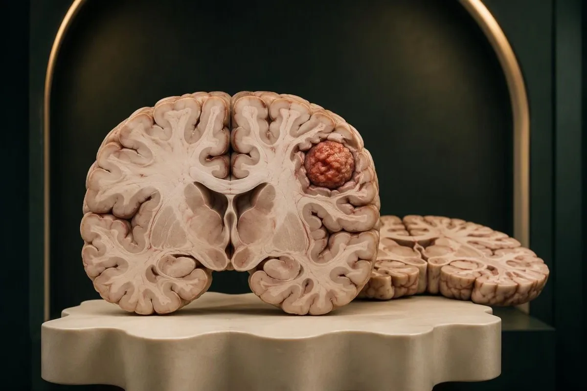
Progressive neurological changes matter more than a headache checklist. The safest next step depends on what changed, how quickly it changed, and whether brain function is affected.


- Brain tumor symptoms can include progressive weakness, vision changes, language difficulties, altered behavior, or a first seizure; no single symptom establishes the diagnosis.
- Headaches alone rarely indicate a brain tumor, particularly when the pattern is longstanding and the neurological examination is normal.
- Экстраныя сімптомы include sudden one-sided weakness, new speech difficulty, a thunderclap headache, or reduced consciousness—even if symptoms improve within minutes.
- First seizure in adults needs urgent medical assessment; call emergency services for a first observed seizure, a seizure lasting 5 minutes, or repeated seizures without recovery.
- Progressive neurological loss warrants prompt assessment; NICE recommends considering urgent brain MRI within 2 weeks for adults with progressive, subacute loss of central neurological function.
- Звычайны аналіз крыві cannot rule a brain tumor in or out. Normal CBC, glucose, electrolytes, and inflammatory markers do not replace neurological examination or imaging.
- Brain MRI generally provides more detail than CT for suspected tumors, while CT is often the faster first examination during an emergency.
- Safety while waiting means avoiding driving after a suspected seizure and seeking reassessment immediately if weakness, confusion, vision loss, or vomiting develops.
Which brain tumor symptoms warrant assessment?
Brain tumor symptoms that warrant assessment are new, persistent, or progressive changes in neurological function, including weakness, speech difficulty, vision loss, altered behavior, and a first seizure. Headaches alone rarely indicate a tumor; sudden neurological symptoms need emergency care, and normal routine bloodwork cannot exclude a tumor.
A symptom’s trajectory is often more useful than its intensity. Difficulty using 1 hand that worsens over several weeks deserves assessment even without pain, whereas a severe but familiar migraine with full recovery may follow an established, non-tumor pattern.
Brain tumors can affect function through local pressure, surrounding swelling, or obstruction of cerebrospinal fluid. Those are 3 different mechanisms, so symptoms do not reliably reveal either the growth’s size or whether it is malignant; a nonmalignant growth can still require treatment.
I’m Thomas Klein, Kantesti’s Chief Medical Officer, and my practical advice is to describe the lost ability rather than simply say you feel unwell. Compare today with 2–4 weeks ago: typing, reading, walking, remembering appointments, and finding words provide much clearer clinical information.
Кантэсці — гэта Аналізатар крыві са штучным інтэлектам that helps explain laboratory findings, not a brain tumor detection service. Our UK organization separates laboratory interpretation from neurological diagnosis: an explanation delivered in about 60 seconds cannot replace an examination or scan.
Early signs of brain tumor: patterns, not a checklist
Early signs of brain tumor are not a fixed sequence, and some tumors cause no symptoms initially. A change affecting 2 linked abilities—such as word-finding and reading—can be more informative than an isolated symptom, although many other neurological conditions cause similar patterns.
Repeated errors in a previously familiar task deserve a concrete description. A hypothetical 54-year-old who begins missing words on one side of a page and misjudging doorways needs a different assessment from someone occasionally forgetting names after poor sleep.
Symptoms may evolve over days, weeks, or months; there is no safe minimum duration to wait when function is deteriorating. A 3-day change with worsening balance can be more urgent than a mild symptom present unchanged for 3 years.
Progression is not always smooth. A person may compensate for a subtle visual deficit until a task exposes it, and relatives may notice changes 1–2 weeks before the person does; that difference does not automatically mean rapid growth.
Memory supplements are not a diagnostic trial for unexplained cognitive change. Our discussion of memory supplement evidence explains their limitations, but a new functional decline over 2 weeks should be assessed rather than treated with a shopping list.
Why headaches alone rarely indicate a brain tumor
Brain tumor headache symptoms overlap with migraine, tension-type headache, sleep problems, and other common conditions. A longstanding headache pattern without neurological changes is less concerning than a new headache accompanied by weakness, visual deterioration, repeated vomiting, or a first seizure.
Morning headache is not a tumor signature. Sleep apnea, teeth grinding, medication effects, and caffeine withdrawal can also cause it; similarly, pain worsened by coughing or bending warrants assessment but does not identify 1 particular cause.
A headache diary is useful when it records function as well as pain. Over 7–14 days, note onset, duration, medication use, vomiting, and any trouble speaking or seeing—but do not keep collecting entries while a new neurological deficit worsens.
Fatigue and headache can accompany iron deficiency, sometimes before anemia appears. Our guide to сімптомы нізкага ферытыну explains that distinction; finding low iron does not explain away a progressive problem involving 1 arm or one visual field.
Headache affecting 2 household members, especially around a heater or fuel-burning appliance, raises a different urgent concern: carbon monoxide exposure. Leave the environment and seek emergency advice; our carbon monoxide testing guide explains why ordinary oxygen readings can be misleading.
Emergency neurological warning signs: act now
Sudden weakness, new speech difficulty, a thunderclap headache, reduced consciousness, or rapidly worsening neurological symptoms require emergency assessment. A headache reaching maximum intensity within about 1 minute is a thunderclap headache; do not assume these symptoms are a tumor or wait for bloodwork.
A sudden facial droop or loss of strength in 1 arm may represent stroke, including when it resolves. Emergency treatment decisions depend on timing, so record when the person was last known to be well and call the local emergency number.
Severe headache with fever and neck stiffness, or headache with increasing drowsiness and repeated vomiting, also needs urgent care. These combinations can reflect several dangerous conditions; even 1 episode of collapse changes the assessment substantially.
Weakness is not always caused by a problem inside the brain. Rapidly ascending weakness involving both legs, particularly with breathing or swallowing difficulty, requires emergency assessment; our трывожных прыкметах небяспечнай слабасці explain an important peripheral nerve alternative.
Do not drive yourself if you have new neurological impairment. In the UK, call 999 or 112 for an emergency; elsewhere use the local emergency number, and tell the dispatcher about sudden onset, seizures, breathing changes, and any reduced responsiveness.
First seizure in adults: assessment and immediate safety
A first seizure in adults requires urgent assessment, but a seizure does not automatically mean a brain tumor. Call emergency services for a first observed seizure, any seizure lasting 5 minutes, or repeated seizures without recovery; structural brain changes are only one possible cause.
Some seizures involve 1 side of the body, a brief speech arrest, or impaired awareness rather than whole-body shaking. A witness’s description of the first movement, responsiveness, duration, and recovery can help distinguish a seizure from fainting or another event.
During a convulsive seizure, protect the person from nearby hazards, cushion the head, and time the event. Do not restrain them or put anything in their mouth; turn them onto their side when movements stop, and give nothing by mouth until fully alert.
NICE recommends specialist assessment within 2 тыдні after a first suspected seizure and, when an EEG is requested, ideally performing it within 72 гадзіны of the seizure (NICE, 2022, NG217). A normal EEG does not exclude epilepsy; a 12-lead ECG helps identify cardiac conditions that can resemble seizures.
Laboratory testing can identify seizure triggers such as low sodium emergencies, but it cannot exclude a structural cause. If treatment is prescribed, our levetiracetam monitoring guide explains why 1 drug level is not a measure of seizure control or tumor status.
Vision changes that need eye and neurological assessment
Persistent double vision, loss of part of the visual field, or progressive visual deterioration warrants assessment. Sudden visual loss needs emergency care; a change affecting 1 eye suggests a different anatomical pathway from loss of the same side of vision in both eyes.
Visual field loss can be surprisingly easy to miss because central vision remains clear. Someone may read small print yet repeatedly overlook objects on 1 side; describing these collisions is more useful than reporting only that their vision feels strange.
Raised intracranial pressure can produce papilledema, meaning swelling of the optic discs. It requires urgent assessment because several conditions can cause it; the absence of papilledema on 1 examination does not rule out a brain tumor.
An eye examination and a neurological examination answer different questions. Our guide to visual field testing explains how clinicians map peripheral vision, while a scan examines structures that an eye pressure measurement cannot assess.
Double vision that disappears when either eye is covered is usually binocular and concerns eye alignment. Double vision persisting in 1 uncovered eye is more often an optical eye problem, but new symptoms with headache, weakness, or eyelid drooping should not be self-triaged.
Progressive weakness, balance problems, and sensory changes
Progressive loss of strength, coordination, or sensation needs a clinical examination, especially when it affects one side or interferes with walking. Problems developing over 2 weeks should not be dismissed as aging or anxiety; sudden onset belongs in emergency care.
Weakness means reduced power, not simply feeling exhausted. Being unable to lift 1 foot, repeatedly dropping a cup, or losing the ability to fasten buttons provides a clearer neurological clue than general tiredness after work.
Balance problems may originate in the inner ear, peripheral nerves, cerebellum, or medication effects. A clinician tests several systems rather than ordering 1 universal dizziness test; unsteadiness accompanied by new slurred speech or severe headache requires urgent reassessment.
Symmetrical tingling in both feet suggests a different pattern from numbness in 1 arm and the same side of the face. Nutritional deficiencies can cause neurological problems; our copper deficiency guide explains one treatable alternative without making it a substitute for examination.
Falls change the urgency because they introduce injury risk as well as diagnostic uncertainty. After 1 unexplained fall with new imbalance, avoid ladders and unsupported stairs, ask someone to accompany you, and arrange assessment rather than attempting repeated home balance challenges.
Personality, memory, and language changes: what matters
New changes in judgment, language, memory, or behavior warrant assessment when they are persistent, progressive, or affect daily function. A 2-week decline in familiar tasks carries different significance from occasional forgetfulness; sudden confusion is an emergency, not a scheduled memory appointment.
Language difficulty is not the same as forgetting a name. Substituting unrelated words, struggling to understand ordinary instructions, or repeatedly losing the meaning of a sentence suggests impaired language processing; note 2–3 actual examples for the appointment.
Personality changes are nonspecific and can reflect depression, poor sleep, medications, substance use, or neurological disease. Our guide to mood-related blood tests discusses treatable contributors, but normal results do not explain away 1 newly lost everyday ability.
A family member’s observations can help establish the timeline without turning the visit into an argument. Ask them to describe what changed over the last 4 weeks—missed bills, unusual disinhibition, getting lost—not simply whether the person seems different.
Sudden confusion developing over hours can arise from metabolic illness, medication toxicity, seizures, stroke, or other acute conditions. Even if there were milder memory concerns for 6 months, the abrupt change needs fresh assessment rather than being attributed to the older problem.
What a neurological examination can—and cannot—show
A neurological examination tests brain and nerve function to identify patterns and guide imaging. It commonly assesses at least 6 domains: mental function, cranial nerves, strength, sensation, reflexes, and coordination or gait; a normal examination lowers concern in some settings but cannot exclude every tumor.
The clinician looks for agreement between findings rather than 1 isolated abnormality. Weakness, altered reflexes, and a compatible sensory pattern together may localize a problem more confidently than a symptom description alone; pain or fatigue can affect performance.
A normal examination is a snapshot, not a guarantee about the next 3 months. Small growths, intermittently symptomatic conditions, and problems in less easily tested brain functions may not produce clear findings at an initial visit.
NICE advises considering urgent direct-access brain MRI within 2 тыдні for adults with progressive, subacute loss of central neurological function, or CT if MRI is contraindicated (NICE, 2015, NG12). That recommendation concerns the clinical pattern—not whether a CBC is abnormal.
At Kantesti, we distinguish technical laboratory interpretation from evidence supporting a neurological diagnosis. Our стандартамі клінічнай валідацыі provide context for methodology; even excellent performance on 1 laboratory task cannot establish accuracy for detecting a condition outside that task.
MRI versus CT: how brain imaging guides diagnosis
MRI is usually the preferred detailed examination for a suspected brain tumor, while CT is often used first in emergencies because it is faster and more accessible. These 2 tests are complementary; the choice depends on symptoms, urgency, safety, and the question the clinician needs answered.
NICE describes standard structural MRI for suspected glioma as including T2-weighted, FLAIR, diffusion-weighted, and T1 imaging before and after contrast when appropriate (NICE, 2018, NG99). These 4 sequence groups reveal different tissue properties; contrast enhancement alone does not prove malignancy.
A normal emergency CT does not answer every question that a dedicated MRI can address. If progressive symptoms continue after 1 CT examination, the next step is clinical reassessment—not assuming the scan was useless or that every possible structural cause has been excluded.
MRI abnormalities also occur in conditions unrelated to tumors. Our explanation of MOG antibody interpretation illustrates why imaging and clinical findings must agree; 1 bright area on a scan is not a diagnosis by itself.
Cerebrospinal fluid testing answers selected questions but is not a routine substitute for brain imaging. The oligoclonal band guide explains one use; clinicians assess lumbar puncture safety first because some intracranial pressure patterns make the procedure unsafe.
Can routine bloodwork detect or exclude a brain tumor?
Routine bloodwork cannot rule a brain tumor in or out. A normal complete blood count, metabolic panel, CRP, or ESR does not exclude an intracranial growth; neither an abnormal result nor a panel containing 20 normal measurements establishes what is happening inside the brain.
Blood tests are still useful for identifying alternative or additional causes. Glucose below 54 mg/dL, or 3.0 mmol/L, is clinically significant hypoglycemia and can impair brain function; resolving low glucose does not automatically account for a separate, longstanding focal neurological deficit.
Sodium is commonly around 135–145 ммоль/л in adult laboratories, and levels below 125 ммоль/л represent profound hyponatremia. Symptoms and speed of decline determine urgency; our кіраўніцтва па інтэрпрэтацыі электралітаў explains why a number cannot be interpreted without the clinical situation.
Vitamin B12 deficiency can cause neurological symptoms without obvious anemia. Our discussion of памежныя вынікі B12 covers follow-up testing, but 1 low-normal B12 result must not become a convenient explanation for worsening one-sided weakness.
Кантэсці — гэта Платформа AI для расшыфроўкі аналізу крыві that can help explain reported laboratory values and trends. As of October 22, 2026, routine blood panels and consumer blood-based cancer tests are not established replacements for neurological examination and appropriate imaging when brain tumor symptoms are suspected.
Pituitary symptoms and what happens after an abnormal scan
Some pituitary growths produce measurable hormone abnormalities, but most brain tumors do not have a routine diagnostic blood marker. After an abnormal scan, specialists integrate imaging, symptoms, and sometimes tissue and molecular testing; 1 elevated hormone result does not establish the nature of a growth.
Headache with loss of peripheral vision and hormone-related symptoms can prompt pituitary-focused assessment. Our guide to сімптомы высокага пролактіну explains relevant patterns, but medications, pregnancy, stress, and other factors can also raise prolactin; 1 measurement may need confirmation.
Pituitary dysfunction can make a familiar thyroid screening result misleading. A low free T4 with a normal TSH needs contextual assessment, as explained in discordant thyroid test results; 2 apparently reassuring numbers do not necessarily mean the pituitary-thyroid system is functioning normally.
An MRI impression may suggest meningioma, glioma, metastasis, or another condition without definitively classifying it. For gliomas, markers such as IDH status and 1p/19q codeletion contribute to classification; these are specialized tissue-based investigations, not a standard primary-care blood panel.
Not every incidental growth requires immediate tissue sampling or treatment. A small, asymptomatic suspected meningioma may be monitored, while a symptomatic growth affecting vision or causing swelling can require faster action; decisions weigh at least 3 factors—location, behavior, and the person’s overall health.
When a scheduled evaluation is reasonable—and when it is not
A scheduled appointment is reasonable for stable, non-emergency symptoms without new neurological loss. Progressive weakness, language difficulty, or visual deterioration should prompt contact today for expedited assessment; do not book a routine appointment 6 weeks away and simply wait for function to worsen.
A familiar headache pattern, unchanged and without red flags, can usually be discussed in a scheduled clinical visit. A newly changing pattern over 2–4 weeks deserves a sooner appointment, and the clinician may lower the threshold for imaging with a cancer history or abnormal examination.
Bring a short timeline rather than an enormous symptom printout. Record the first date, the change over the last 14 days, specific lost abilities, medication changes, and relevant cancer history; a witness should attend after a suspected seizure if possible.
A suspected seizure changes safety advice immediately. Do not drive until you have received medical and jurisdiction-specific licensing advice; avoid swimming alone, baths alone, heights, and hazardous machinery, even if there has been only 1 event and you currently feel well.
Кантэсці — гэта Інструмент аналізу вынікаў аналізу крыві з падтрымкай AI, not an emergency triage service or an interpreter of brain MRI images. Our кіраўніцтва па тэхналогіі AI explains the laboratory workflow; reviewing results in about 60 seconds must never delay assessment of a neurological red flag.
Research publications, evidence boundaries, and next steps
Brain tumor assessment should be guided by neurological findings, appropriate imaging, and condition-specific clinical evidence. The 2 laboratory publications listed below address iron and coagulation testing; they are background resources, not evidence that a blood test or AI tool can detect or exclude a brain tumor.
Наш публікацыя па даследаваннях жалеза concerns laboratory evaluation of iron status, while our coagulation testing publication addresses clotting measurements. These 2 resources may support broader medical assessment, but neither validates brain tumor screening or provides a neurological rule-out threshold.
A DOI identifies a deposited document; it does not, by itself, establish peer review or clinical effectiveness. Kantesti’s 2 Zenodo resources are therefore separated from the NICE guidelines in the references, and the ResearchGate and Academia.edu discovery links are title searches rather than verified publication copies.
For medical accountability, our Медыцынская кансультатыўная рада page describes the physicians associated with Kantesti’s clinical work. As Thomas Klein, I would keep 3 questions central: what ability changed, how fast it changed, and whether the current presentation requires emergency assessment.
The next action should fit the symptom, not the reassuring appearance of a report. Call emergency services for sudden neurological loss or a first observed seizure; contact a clinician promptly for progressive changes, and bring any existing results without waiting for 1 more blood test.
Часта задаваныя пытанні
Якія раннія прыкметы пухліны галаўнога мозгу?
0. Раннія прыкметы пухліны галаўнога мозгу могуць уключаць нарастальную слабасць, выпадзенне палёў зроку, цяжкасці з маўленнем, змены ў паводзінах або першы эпілептычны прыступ, але універсальнага першага сімптому няма. Новая страта функцый, якая развіваецца на працягу 2–4 тыдняў, патрабуе неадкладнай ацэнкі нават без галаўнога болю. Раптоўная слабасць, цяжкасці з маўленнем, спутанасць свядомасці або страта зроку патрабуюць экстраннай медыцынскай дапамогі, а не планавага прыёму. Падобныя сімптомы выклікаюць многія захворванні, не звязаныя з пухлінамі, таму дыягназ залежыць ад абследавання і належнай візуалізацыі.
Як адчуваецца галаўны боль пры пухліне мозгу?
0 Галаўны боль пры пухліне мозгу не мае надзейна адметных асаблівасцяў і можа нагадваць мігрэнь або галаўны боль напружання. Настаражвае з’яўленне новага або змененага галаўнога болю разам з прагрэсавальнымі неўралагічнымі сімптомамі, паўторнымі ванітамі або першым прыступам сутаргаў. Боль раніцай або пры кашлі сам па сабе не сведчыць пра пухліну. Галаўны боль, інтэнсіўнасць якога дасягае максімуму прыкладна за 1 хвіліну, патрабуе неадкладнай ацэнкі, бо можа мець іншую небяспечную прычыну.
Ці могуць звычайныя аналізы крыві выключыць пухліну мозгу?
Звычайныя планавыя аналізы крыві не могуць выключыць пухліну галаўнога мозга. CBC, біяхімічны аналіз крыві, CRP і ESR ацэньваюць розныя працэсы і не паказваюць структуру мозга, нават калі 20 або больш паказчыкаў у выніках знаходзяцца ў межах нормы. Аналізы крыві могуць выявіць фактары, якія спрыяюць праблеме, напрыклад нізкі ўзровень глюкозы, парушэнні электралітнага балансу або дэфіцыт вітамінаў. Устойлівыя або прагрэсавальныя неўралагічныя сімптомы па-ранейшаму патрабуюць клінічнай ацэнкі і, калі ёсць паказанні, візуалізацыі галаўнога мозга.
Ці азначае першы прыступ у дарослага наяўнасць пухліны мозгу?
Першы прыступ у дарослага не абавязкова азначае пухліну мозгу, але патрабуе тэрміновай ацэнкі. Магчымыя прычыны ўключаюць эпілепсію, метабалічныя парушэнні, дзеянне лекаў або псіхаактыўных рэчываў, інсульт і структурныя анамаліі мозгу. Выклічце экстраныя службы пры першым заўважаным прыступе, прыступе працягласцю 5 хвілін або паўторных прыступах без аднаўлення прытомнасці. NICE рэкамендуе прайсці абследаванне ў спецыяліста на працягу 2 тыдняў пасля першага меркаванага прыступу; не сядайце за руль, пакуль не атрымаеце медыцынскія рэкамендацыі і не высветліце мясцовыя патрабаванні да кіравання транспартным сродкам.
Што лепш для дыягностыкі пухліны галаўнога мозгу — МРТ ці КТ?
МРТ звычайна дае больш падрабязную інфармацыю пры падазрэнні на пухліну галаўнога мозгу, тады як КТ часта з'яўляецца хутчэйшым першым даследаваннем у экстранай сітуацыі. Стандартнае структурнае МРТ выкарыстоўвае некалькі паслядоўнасцей, у тым ліку T2, FLAIR, дыфузійную візуалізацыю і T1-візуалізацыю да і пасля ўвядзення кантрасту, калі гэта мэтазгодна. Нармальны вынік 1 экстранай КТ не абавязкова дазваляе растлумачыць устойлівыя прагрэсавальныя неўралагічныя сімптомы. Візуалізацыя можа падказаць дыягназ, але для класіфікацыі асобных утварэнняў патрэбныя даследаванне тканіны і малекулярнае тэставанне.
How quickly should progressive neurological symptoms be assessed?
Progressive weakness, language difficulty, balance impairment, or visual deterioration should prompt contact with a clinician today for an expedited assessment. NICE recommends considering urgent brain MRI within 2 weeks for adults with progressive, subacute loss of central neurological function, or CT when MRI is contraindicated. That 2-week imaging recommendation is not permission to delay emergency care. Sudden symptoms, reduced consciousness, a first observed seizure, or rapid deterioration require emergency assessment.
Атрымаць аналіз крыві з AI сёння
Далучайцеся больш чым да 2 мільёнаў карыстальнікаў па ўсім свеце, якія давяраюць Kantesti для імгненнага, дакладнага аналізу лабараторных тэстаў. Загрузіце вынікі аналізу крыві і атрымайце поўную інтэрпрэтацыю біямаркераў 15,000+ за лічаныя секунды.
📚 Публікацыі даследаванняў са спасылкамі
Кляйн, Т., Мітчэл, С., і Вебер, Г. (2026). Кіраўніцтва па даследаваннях жалеза: TIBC, насычэнне жалезам і звязвальная здольнасць. Kantesti AI Medical Research.
Кляйн, Т., Мітчэл, С., і Вебер, Г. (2026). Нармальны дыяпазон аЧТЧ: D-дымер, бялок С, кіраўніцтва па згусальнасці крыві. Kantesti AI Medical Research.
📖 Знешнія медыцынскія спасылкі
Нацыянальны інстытут аховы здароўя і дасканаласці ў медыцыне (NICE) (2015). Падазрэнне на рак: распазнаванне і накіраванне. Кіраўніцтва NICE NG12.
Нацыянальны інстытут здароўя і дасканаласці ў медыцыне (2022). Epilepsies in children, young people and adults. NICE guideline NG217.
Нацыянальны інстытут аховы здароўя і дасканаласці ў медыцыне (2018). Brain tumours (primary) and brain metastases in over 16s. NICE guideline NG99.
⚕️ Медыцынская адмова ад адказнасці
Гэты артыкул прызначаны толькі для адукацыйных мэт і не з’яўляецца медычнай парадай. Заўсёды кансультуйцеся з кваліфікаваным спецыялістам аховы здароўя для прыняцця рашэнняў па дыягностыцы і лячэнні.
Сігналы даверу E-E-A-T
Вопыт
Клінічны агляд, які вядзе лекар, працэсаў інтэрпрэтацыі лабараторных паказчыкаў.
Экспертыза
Фокус лабараторнай медыцыны на тым, як біямаркеры паводзяць сябе ў клінічным кантэксце.
Аўтарытэтнасць
Напісана доктарам Томасам Кляйнам, з аглядам доктара Сара Мітчэл і праф. доктара Ганса Вебера.
Надзейнасць
Інтэрпрэтацыя на аснове доказаў з выразнымі шляхамі далейшых дзеянняў, каб паменшыць трывогу.