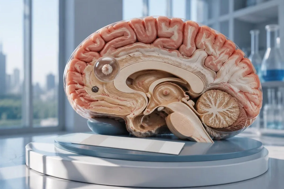
Negatīvs neirocisticerkozes tests neizslēdz atsevišķu smadzeņu cistu vai kalcificētu slimības formu. CT, MRI un steidzama jaunu krampju izvērtēšana ir svarīgāka par mierinājumu, ko sniedz tikai antivielu tests.

Galvenie secinājumi
- Negatīvas antivielas neizslēdz neirocisticerkozi, īpaši tad, ja attēldiagnostikā redzams tikai 1 skarts apvidus vai kalcifikācijas.
- References laboratorijas EITB jutība var tuvoties 98%, ja ir vairāk nekā 1 dzīvotspējīga cista, taču atsevišķas cistas gadījumā tās veiktspēja ir būtiski zemāka.
- Jutība atsevišķas cistas gadījumā publicētajos EITB aprēķinos bieži ir aptuveni 50–60%; šie skaitļi neattiecas uz visiem komerciālajiem antivielu testiem.
- CT bez kontrastvielas ir īpaši noderīga sīku kalcifikāciju noteikšanai, kuru izmērs bieži ir aptuveni 1–4 mm.
- MRI izvērtē dzīvotspējīgas cistas, apkārtējo tūsku un ventrikulāru vai subarahnoidālu slimību; specializētas trīsdimensiju sekvences var uzlabot noteikšanu.
- Pirmreizēji krampji nepieciešama neatliekama ārsta izvērtēšana neatkarīgi no antivielu rezultāta. Pirmās zināmās lēkmes vai citu neatliekamu pazīmju gadījumā izsauciet neatliekamo medicīnisko palīdzību.
- Piecas minūtes ir neatliekamas palīdzības slieksnis nepārejošas konvulsīvas lēkmes gadījumā, nevis iemesls gaidīt, pirms vērsties pēc palīdzības.
- Tikai kalcificēta slimība parasti nav jāārstē ar pretparazītu medikamentiem, taču var būt nepieciešama lēkmju ārstēšana un papildu attēldiagnostika.
Vai negatīvs neirocisticerkozes tests var to izslēgt?
A negatīvs neirocisticerkozes tests nevar droši izslēgt šo slimību, jo īpaši tad, ja smadzenēs ir 1 cista vai kalcificētas paliekas. Antivielu tests nosaka imūnreakciju, nevis tieši pārbauda smadzenes; CT un MRI nosaka aizdomīgo atradņu atrašanās vietu un stadiju, savukārt pirmās lēkmes gadījumā nepieciešama neatliekama izvērtēšana.

Rezultāts ir nosacīts, nevis absolūts. Cisticerkozes antivielu tests, kas labi darbojas cilvēkiem ar vairākām dzīvotspējīgām cistām, var darboties slikti cilvēkiem, kuriem ir tikai 1 neliels skarts apvidus, tāpēc vārds negatīvs nozīmē, ka ar šo testu nav konstatēta reakcija, nevis to, ka nervu sistēmā nav cistu.
Kā hipotētisku piemēru aplūkosim 34 gadus vecu cilvēku, kuram bijusi pirmā lēkme, antivielu analīzes rezultāts ir negatīvs un MRI konstatēts viens gredzenveida kontrastvielu uzkrājošs veidojums. Analīzes rezultāts neļauj noteikt, vai šis veidojums liecina par cisticerkozi, citu infekciju vai neinfekciozu procesu; tā nozīme ir jāvērtē, ņemot vērā skenēšanas rezultātus, saskares ar infekciju anamnēzi un neiroloģisko novērtējumu kopumā.
Kantesti ir AI asins analīžu analizators kas var palīdzēt izskaidrot ziņotos laboratorisko analīžu rezultātus, taču tas nevar izslēgt smadzeņu slimību, pamatojoties uz 1 negatīvu antivielu rezultātu. Mūsu organizācija un misija izskaidro mūsu pievēršanos laboratoriskajai informācijai; lasītājiem nevajadzētu uztvert automatizētu analīžu rezultātu skaidrojumu kā atļauju atlikt palīdzības meklēšanu lēkmju gadījumā.
Kāpēc antivielu tests var neuzrādīt atsevišķu smadzeņu cistu?
A viena cista var izraisīt antivielu atbildreakciju, kas ir zemāka par testa noteikšanas slieksni. Publicētajos atsauces laboratoriju EITB veiktspējas aprēķinos jutība vienas cistas gadījumā bieži ir tuvu 50–60%, salīdzinot ar aptuveni 98%, ja ir vairāk nekā 1 dzīvotspējīga cista; atšķirība atspoguļo slimības apmēru un testa veiktspēju.
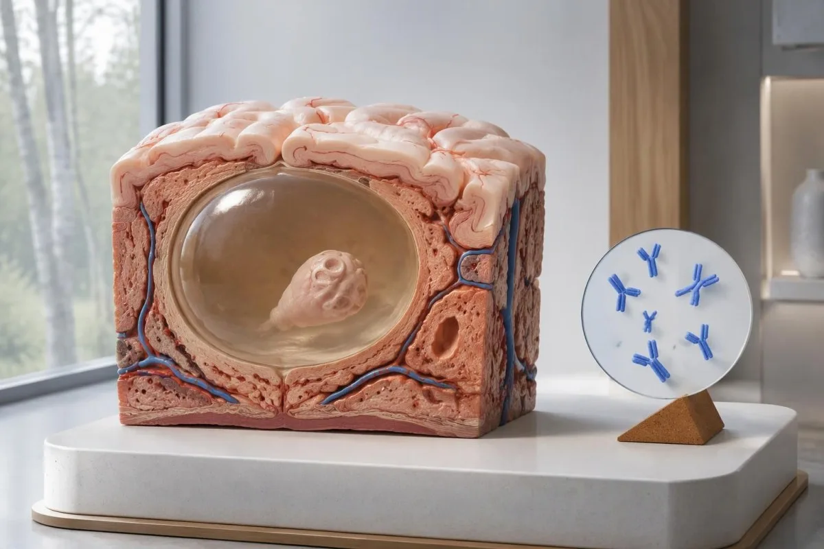
IDSA/ASTMH vadlīnijas iesaka ar enzīmu saistīto imūnelektrotransfera blotu, jeb EITB kā apstiprinošu seroloģisko testu un skaidri brīdina, ka tā jutība ir zema, ja ir tikai viens parenhīmas bojājums vai kalcifikācijas (White et al., 2018). Augstu jutību, kas ziņota pacientiem ar vairākām cistām, ventrikulāru slimību vai subarahnoidālu slimību, nedrīkst attiecināt uz cilvēku ar 1 nelielu smadzeņu anomāliju.
Iztēlojieties tikai ilustratīva piemēra nolūkā, ka testa jutība ir 55% 100 cilvēkiem, kuriem vienas cistas izraisīta slimība jau ir neatkarīgi apstiprināta. Aptuveni 45 cilvēkiem rezultāts būtu negatīvs, lai gan viņiem būtu šī slimība; šis aprēķina piemērs skaidro, kāpēc slimību var neuzrādīt, nevis to, kāda ir varbūtība, ka konkrēts negatīvs rezultāts liecina par slimību.
Tā paša testa atkārtošana pēc 2 nedēļām var nenovērst vāju reakciju, ko izraisa neliels slimības apmērs. Tas atšķiras no infekcijām, kuru gadījumā galvenā problēma ir pārāk agrīna parauga paņemšana, kā paskaidrots mūsu ceļvedī par laika izvēles izraisītiem kļūdaini negatīviem rezultātiem; noskaidrojiet, vai attēldiagnostikas vai labāk raksturota testa rezultāti mainītu turpmāko rīcību.
Kāpēc kalcificēta neirocisticerkoze var izraisīt simptomus?
Kalcificēta neirocisticerkoze var izraisīt lēkmes arī tad, ja antivielu tests ir negatīvs, because the remaining mineralized focus can still be associated with local irritation and episodic surrounding edema. Typical calcifications may measure approximately 1–4 mm, but their small size does not establish whether they are clinically harmless.
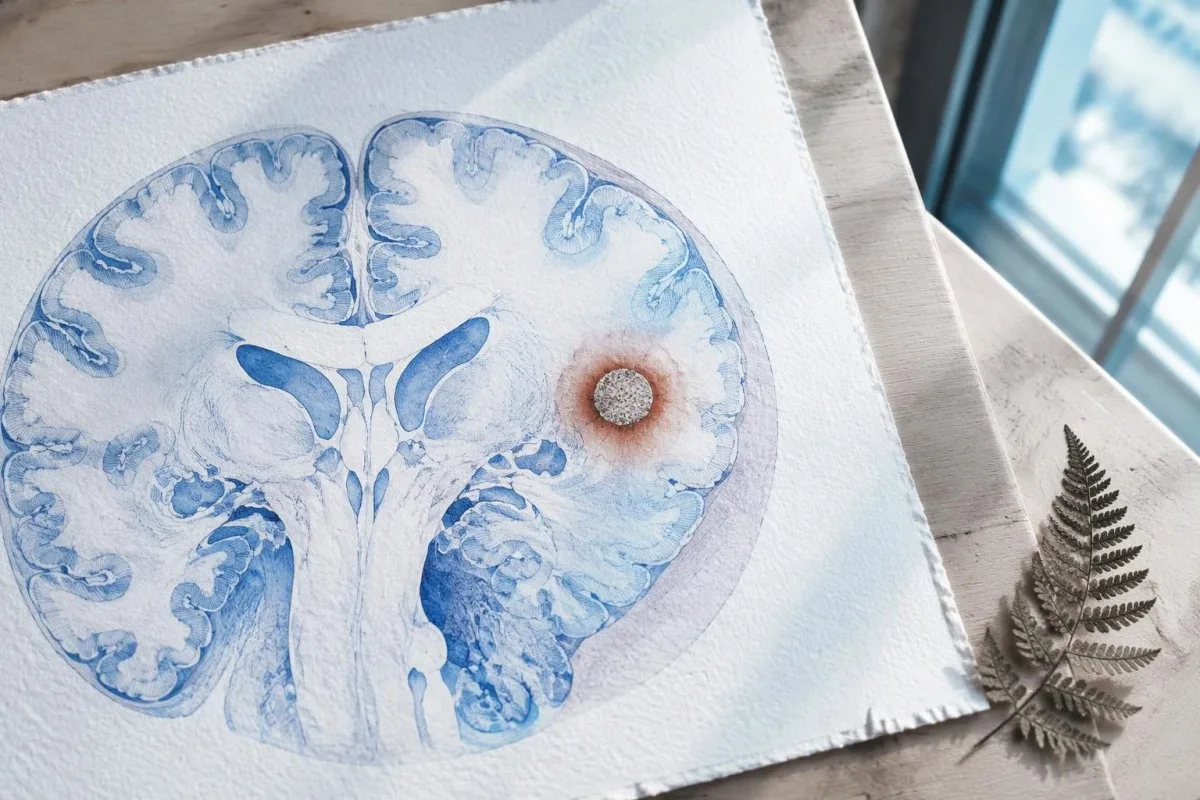
A calcification is generally a remnant rather than a living cyst that antiparasitic medication can eliminate. In the IDSA/ASTMH guideline, calcified-only parenchymal disease is managed symptomatically rather than with antiparasitic drugs; a person with 1 calcified focus and seizures may therefore need antiseizure treatment without needing cyst-killing medication (White et al., 2018).
Surrounding edema can appear intermittently, sometimes long after the original cyst has degenerated. MRI may show this tissue response even when the calcification is more obvious on CT, which is why the 2 scans can tell different but compatible stories rather than one proving that the other is wrong.
Neither a negative antibody result nor a persistent positive result measures seizure risk by itself. The broader lesson resembles other seronegative neurological conditions, discussed in our negative neurological antibody results guide: absence of detectable antibodies is not equivalent to absence of a clinically relevant neurological process.
Kurš cisticerkozes antivielu tests tika veikts?
The useful first question is whether the report describes EITB, ELISA, or another antibody method, because these are not interchangeable tests. A negative index below 1 laboratory's cutoff cannot inherit the performance figures of a different reference-laboratory assay, even when both reports use the name cysticercosis IgG.
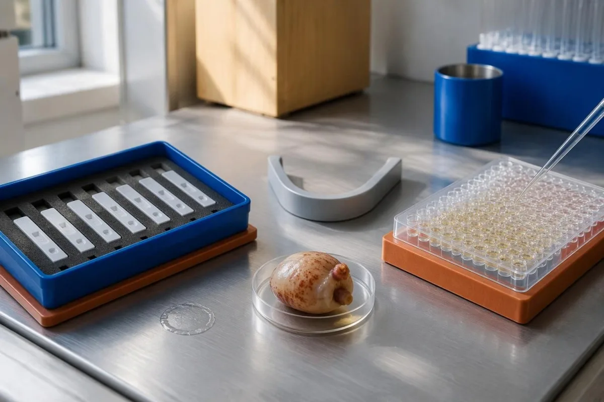
EITB using purified parasite glycoproteins is the preferred serological method in the IDSA/ASTMH guideline, while crude-antigen ELISA methods have recognized sensitivity and specificity limitations (White et al., 2018). Before comparing 2 results, check the method, specimen type, reference interval, and whether the laboratory used a validated confirmatory procedure.
Some laboratories report a qualitative result; others provide an optical-density ratio or numerical index. A value such as 0.8 in a hypothetical assay does not represent 0.8 cysts, an 80% disease probability, or a universal severity score—our kvalitatīvi un kvantitatīvi rezultāti guide explains that distinction.
Kantesti AI's role is to help readers preserve the laboratory's original wording rather than reinterpret 1 method through another method's thresholds. Our laboratorijas marķieru ceļvedis provides broader report-reading context; for this result, keep the actual assay name available when discussing referral or repeat testing.
Kā anamnēzes dati par iespējamu saskari maina diagnostiskās aizdomas?
Neurocysticercosis follows ingestion of Taenia solium eggs, usually through fecal contamination from a person carrying the intestinal tapeworm. Eating undercooked pork can cause intestinal taeniasis, but brain cysts arise through a different transmission step; symptoms may emerge years after exposure rather than within 1 recent trip.

A vegetarian can develop cysticercosis because eggs can contaminate food or hands independently of meat consumption. Useful history includes past residence, household contacts, sanitation conditions, and possible exposure to a tapeworm carrier; asking only about meals eaten during the last 2 weeks can miss the relevant background.
A negative stool examination does not exclude cysts in the brain. Stool testing addresses intestinal tapeworm carriage, whereas CT and MRI assess nervous-system findings; these are 2 different sites and clinical questions, although public-health teams may recommend evaluating contacts when a carrier could be involved.
Normal eosinophils also cannot exclude neurocysticercosis, and raised eosinophils do not identify the brain as the affected site. Our parasite antibody interpretation guide discusses a separate parasite with similar localization limits; the practical point is to avoid using 1 routine cell count as a substitute for targeted neurological evaluation.
Kad jaunu krampju gadījumā nepieciešama neatliekama izvērtēšana?
A first known seizure needs urgent medical assessment regardless of antibody results, and emergency services should be contacted for a first seizure or an ongoing seizure with concerning features. A convulsive seizure lasting 5 minutes, repeated seizures without recovery, breathing difficulty, pregnancy, or significant injury requires emergency care.

Do not wait for the 5-minute mark to seek help after a first known seizure. Keep the person away from hazards, cushion the head if safely possible, do not restrain them or put anything in their mouth, and place them on their side once convulsions stop if they are breathing normally.
Persistent confusion, a severe new headache, fever with neck stiffness, or new weakness increases concern for causes beyond cysticercosis. Sudden one-sided weakness or speech difficulty also demands emergency assessment, as our insulta brīdinājuma pazīmju ceļvedi explains; 1 negative infection test cannot rule out stroke, meningitis, or a structural brain problem.
After a suspected seizure, stop driving and avoid swimming alone, heights, or operating machinery until a clinician provides advice consistent with local rules. If a witness can safely record the duration and describe movements or recovery, those observations may be more useful than ordering a second antibody test before the first neurological assessment.
Ko CT sniedz neirocisticerkozes diagnostikā?
Noncontrast CT is particularly useful for detecting calcified cyst remnants, including small approximately 1–4 mm foci that MRI may show less clearly. CT also helps assess urgent structural problems such as hydrocephalus, but 1 isolated calcification is not automatically proof of neurocysticercosis or the cause of a seizure.
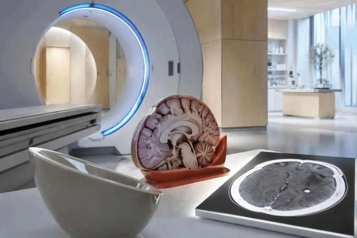
Calcium appears dense on CT, allowing radiologists to identify mineralized remnants and describe their distribution. The IDSA/ASTMH guideline recommends both MRI and noncontrast CT when classifying newly diagnosed neurocysticercosis, because the 2 modalities have complementary strengths rather than being interchangeable alternatives (White et al., 2018).
An incidental calcification needs context: previous granulomatous disease, other causes of mineralization, and scan appearance can influence interpretation. If 1 calcification sits near a suspected seizure focus, concordance with symptoms and EEG may strengthen the explanation, but anatomical proximity alone cannot establish causation.
A laboratory marker used in selected head-injury pathways does not test for brain cysts or replace imaging after an unexplained seizure. Our head-injury CT decisions guide illustrates that narrower setting; ask the clinician whether the present question requires noncontrast CT, contrast imaging, MRI, or a combination of 2 modalities.
Ko atklāj MRI, ko CT var nepamanīt?
MRI is better suited to evaluating viable cysts, surrounding edema, and many ventricular or subarachnoid findings. Specialized 3-dimensional sequences such as CISS or FIESTA can improve visualization of cyst membranes within cerebrospinal fluid spaces, where a cyst may otherwise resemble the surrounding fluid.
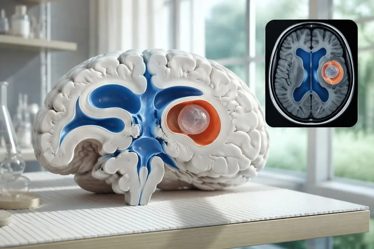
A cyst inside the ventricles can obstruct cerebrospinal fluid flow and cause hydrocephalus even without numerous parenchymal cysts. Headache with vomiting, declining alertness, or new visual symptoms therefore changes urgency; 1 reassuring antibody result should not override symptoms that suggest raised pressure or impaired fluid circulation.
MRI may show a scolex, the small internal structure of the parasite, inside a compatible cyst. This finding can be highly specific when confidently identified, whereas ring enhancement alone has a broader differential; our brain imaging warning signs guide explains why structural abnormalities require interpretation rather than diagnosis from 1 visual feature.
A routine MRI protocol may not answer every cysticercosis question. If ventricular or subarachnoid disease is suspected, discuss whether thin-section 3-dimensional imaging is needed; radiologists choose sequences and contrast according to the clinical question, and a normal scan using 1 protocol does not prove every possible disease compartment has been adequately assessed.
Kā diagnosticē neirocisticerkozi, ja testu rezultāti nesaskan?
Neurocysticercosis diagnosis combines imaging, clinical findings, exposure information, and supporting tests, rather than requiring a positive antibody result in every case. The revised diagnostic criteria published in 2017 give characteristic neuroimaging findings a central role and distinguish strong evidence from less-specific supportive features (Del Brutto et al., 2017).
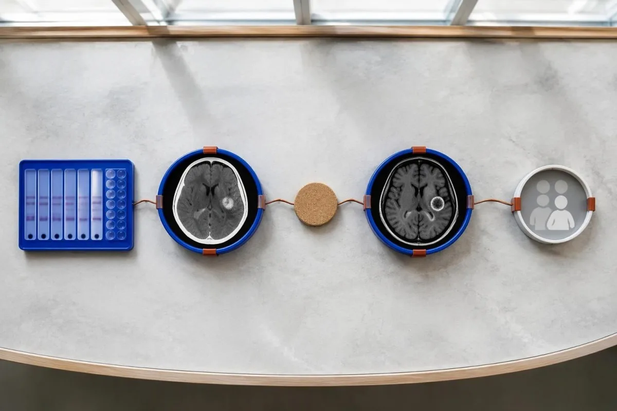
Evidence should be weighted, not tallied. A confidently visualized scolex inside a compatible cyst is different from 1 nonspecific ring-enhancing area, while a positive antibody result is different from direct evidence that a finding is in the brain; our pozitīva antivielu rezultātu rokasgrāmata explains why detection alone cannot establish location.
For a clearly hypothetical probability example, start with a 30% pretest probability and assume a single-cyst assay has 55% sensitivity and 99% specificity. In 1,000 comparable people, about 135 affected and 693 unaffected people would test negative, leaving approximately a 16% probability of disease among negative results—still substantial, although these assumed figures are not a personal risk calculator.
The most useful disagreement to investigate is whether each test is answering the same question. A negative antibody test, a CT calcification, and MRI edema can all be compatible with 1 calcified disease process; unexplained discrepancies should prompt review of the assay and images rather than automatic dismissal of either result.
Kas vēl smadzeņu attēldiagnostikā var līdzināties atsevišķai cistai?
A single ring-enhancing brain finding is not specific to neurocysticercosis. Other possibilities include tuberculoma, an abscess, a neoplasm, or an inflammatory process; clinicians use appearance, location, immune status, symptoms, and sometimes follow-up imaging or tissue examination to distinguish these possibilities rather than relying on 1 antibody panel.
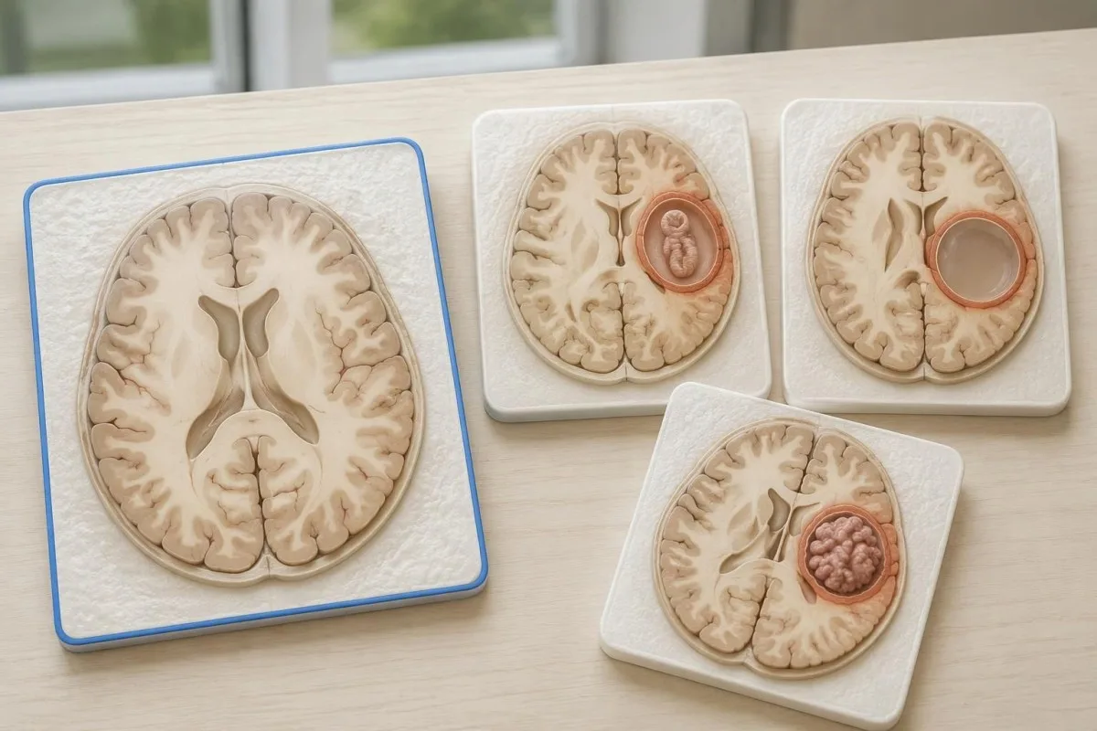
Edema, restricted diffusion, internal structures, and the number of findings all contribute to the differential, but no single nonspecific feature settles every case. Fever and immune suppression may change the suspected cause; a 2 cm ring-enhancing area in a hypothetical immunocompromised person warrants a different investigation from a tiny calcification in an otherwise well person.
Toxoplasmosis is one consideration in selected immunocompromised patients, and a positive IgG result can indicate past exposure rather than prove an active brain process. Our Toxoplasma IgG interpretation guide explains that laboratory distinction; imaging and the patient's immune status determine whether the result is clinically relevant to 1 or more cerebral findings.
New seizures accompanied by rapidly evolving memory problems, psychiatric changes, or abnormal movements can also suggest encephalitis rather than a solitary parasite cyst. Our encephalitis diagnostic pathway describes how MRI, EEG, and cerebrospinal fluid testing answer different questions; a second negative cysticercosis antibody result does not investigate those alternatives.
Vai antigēnu tests, PCR vai CSF var kliedēt neskaidrību?
Antigen tests, PCR, and cerebrospinal fluid analysis can add information in selected cases, particularly when extraparenchymal disease is suspected. None is a universal rescue test after 1 negative antibody result: availability, assay validation, disease compartment, and safety of lumbar puncture determine whether additional testing is appropriate.
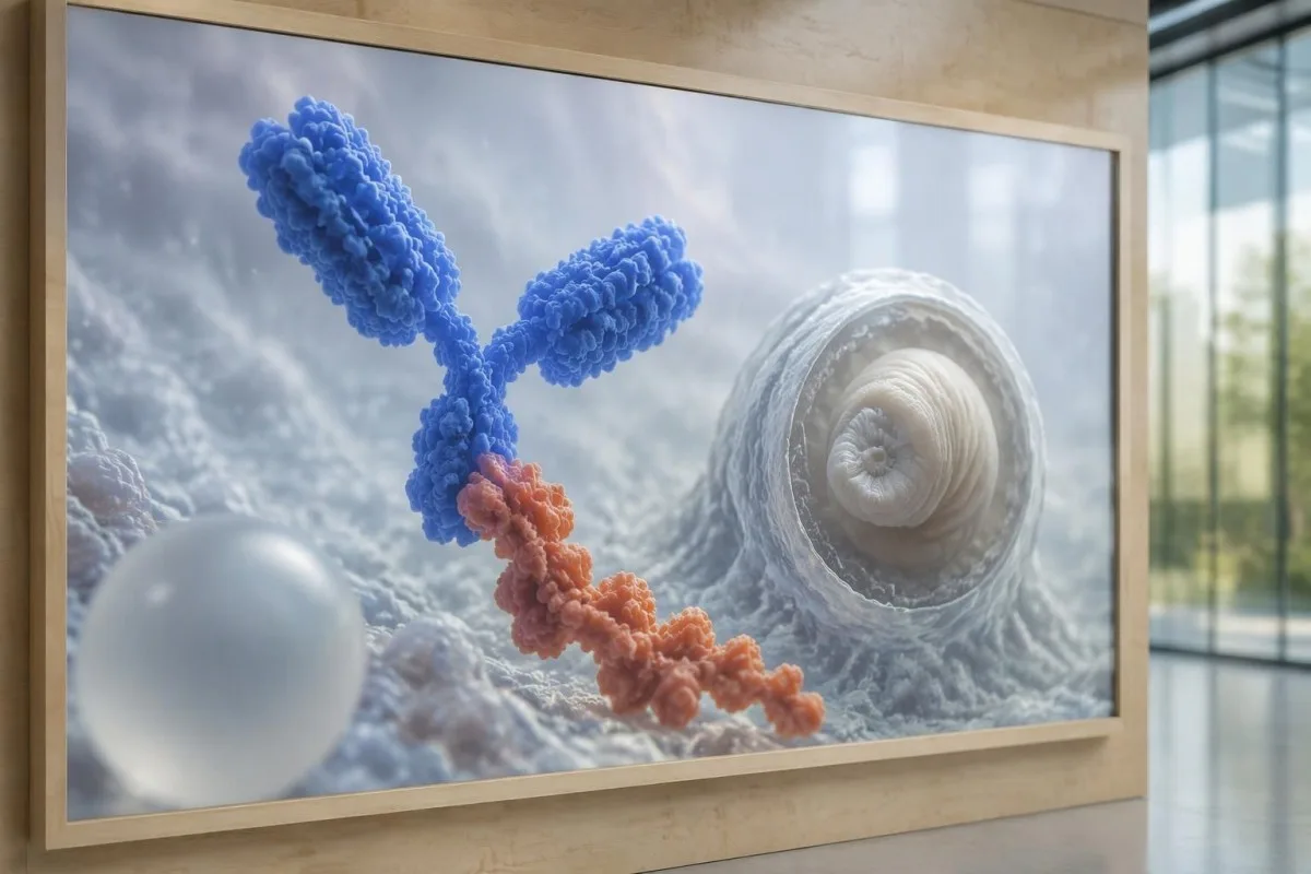
Antibody testing detects the host response, while antigen testing aims to detect parasite-derived material and may better reflect viable disease burden. Antigen detection is often less useful when only 1 small parenchymal cyst or calcifications are present; molecular testing is used in some specialist settings, but performance cannot be generalized across every laboratory or specimen.
Lumbar puncture is not routinely required for an uncomplicated solitary parenchymal finding and can be unsafe when obstructive hydrocephalus or significant mass effect is present. Cerebrospinal fluid tests for other disorders also require disease-specific interpretation, as our CSF band interpretation guide explains; 1 abnormal CSF marker does not identify every possible cause.
Kantesti ir AI asins analīžu rezultātu platforma, not a service that orders lumbar punctures, identifies cysts on scans, or confirms neurological infections. Our AI interpretācijas tehnoloģija explains the laboratory-report workflow; when 2 tests disagree, our role is to clarify their stated meanings and limitations, not select an invasive diagnostic procedure.
Kāpēc ārstēšana ir atkarīga no cistas stadijas un atrašanās vietas?
Treatment depends on whether cysts are viable, degenerating, calcified, or located outside the brain tissue, not simply on antibody positivity. Selected viable parenchymal cases receive antiparasitic treatment, whereas calcified-only disease generally receives symptom-directed care; ventricular obstruction may require a procedural approach rather than 1 standard medication regimen.
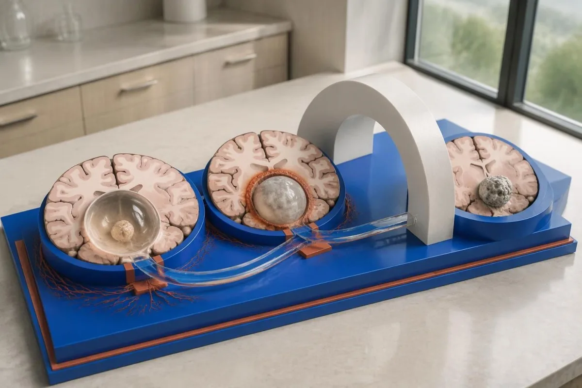
For selected patients with 1–2 viable parenchymal cysts, the IDSA/ASTMH guideline describes albendazole treatment for 10–14 days with appropriate accompanying care. This is a specialist-directed recommendation, not a home-treatment instruction: antiparasitic drugs can provoke an inflammatory response, and untreated hydrocephalus or diffuse cerebral edema changes the order of management (White et al., 2018).
WHO's 2021 guidance focuses on defined presentations of parenchymal neurocysticercosis, while the IDSA/ASTMH guideline also addresses ventricular, subarachnoid, and other forms in detail. Differences in imaging access and disease compartment explain why 1 treatment recommendation cannot be transferred to every patient with an antibody result; the guidelines do not turn negative serology into a rule-out test.
Corticosteroids may accompany antiparasitic therapy, but prolonged use requires additional safety planning, including assessment for relevant latent infections. Our Strongyloides and steroid safety guide explains a separate risk that matters before immune-suppressing treatment; an eye examination is also recommended before antiparasitic therapy to evaluate possible intraocular cysts.
Kas novērošanas laikā jāuzrauga papildus antivielu rezultātiem?
Follow-up should track seizures, neurological symptoms, imaging changes, and medication safety, rather than use antibody disappearance as proof of recovery. For viable parenchymal cysts, the IDSA/ASTMH guideline suggests repeat MRI at least every 6 months until the cystic component resolves, with earlier reassessment when symptoms change.
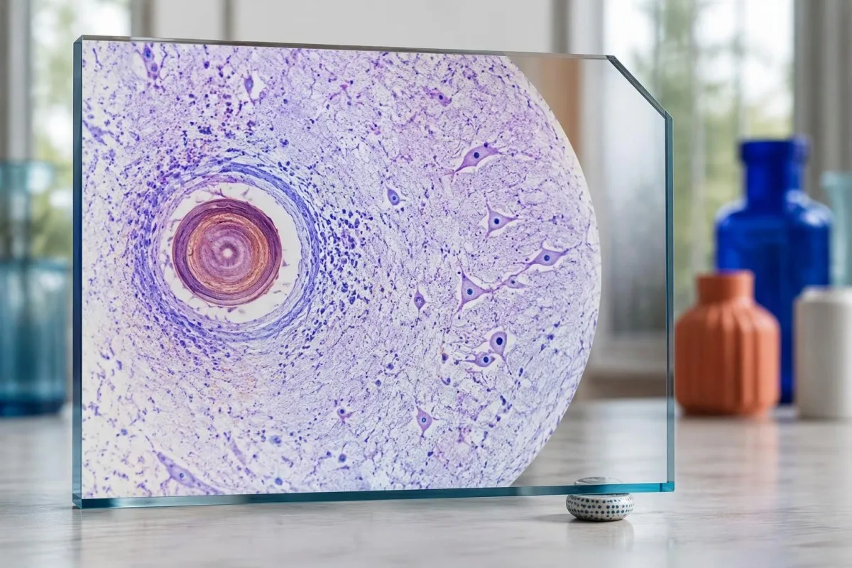
Antibodies may remain detectable after the original disease process has changed, and low-burden disease may never produce a positive result. If 1 new seizure occurs during follow-up, clinicians consider adherence, medication interactions, recurrent edema, and other causes; a rising or falling antibody index alone cannot distinguish among those explanations.
Kantesti AI can help explain reported laboratory results during medication follow-up, but a drug concentration is not a stand-alone measure of seizure control. Our levetiracetāma līmeņa interpretācija guide explains why sample timing and kidney function matter; 2 measurements collected at different times after dosing may not be directly comparable.
Monitoring depends on the chosen drug and treatment duration: prolonged albendazole therapy can require liver-test and blood-count surveillance, while some antiseizure medicines can affect sodium. Our elektrolītu drošuma ceļvedis discusses those separate laboratory concerns; do not stop an antiseizure medicine or change its dose because 1 cysticercosis antibody result is negative.
Ko Kantesti var izskaidrot un ko pētījumi nevar pierādīt?
AI report interpretation cannot confirm or exclude neurocysticercosis from laboratory results alone. Kantesti ir an ar AI balstīts asins analīzes rīks whose role here is to organize reported laboratory findings; a negative result still needs clinical context, and 1 new seizure takes priority over reviewing an automated explanation.

Prepare a focused review packet with the original antibody report, assay method, CT or MRI report, symptom timeline, and medication list. These 5 information categories reduce the risk of discussing a result without its context; ask specifically whether the imaging suggests viable, degenerating, calcified, ventricular, or subarachnoid disease and which unanswered question the next test would address.
The 2 Kantesti publications listed below are a repository-deposited global analysis report and an educational RDW guide, not clinical evidence validating neurocysticercosis diagnosis. Our RDW un CBC ceļvedis can support interpretation of a separately ordered blood count, but RDW, MCV, and MCHC do not establish whether a brain cyst is present.
Evidence for the neurological recommendations in this article comes from the cited diagnostic criteria and clinical guidelines, not from general AI benchmarking or laboratory-report volume. Our technical validation scope should be read within its stated boundaries; neither 1 successful report explanation nor a large aggregate dataset substitutes for condition-specific diagnostic validation.
Bieži uzdotie jautājumi
Vai neurocisticerkozes gadījumā asins analīžu rezultāti var būt negatīvi?
Jā, neirocisticerkoze var būt arī tad, ja asins antivielu tests ir negatīvs, jo īpaši tad, ja ir viena smadzeņu cista vai tikai pārkaļķojumi. Publicētie references laboratoriju EITB testa jutīguma novērtējumi liecina, ka vienas cistas gadījumā tā ir aptuveni 50–60% — ievērojami zemāka nekā vairāku dzīvotspējīgu cistu gadījumā. Tāpēc diagnozes noteikšanā būtiska nozīme joprojām ir CT, MRI, simptomiem un informācijai par iespējamo saskari ar infekcijas avotu. Ja rodas jauna lēkme, nepieciešama steidzama medicīniska izvērtēšana, nevis mierinājums, ko sniedz negatīvais testa rezultāts.
Kāds ir visdrošākais neirocisticerkozes tests?
Neviens atsevišķs neirocisticerkozes tests nesniedz atbildes uz visiem diagnostiskajiem jautājumiem; smadzeņu attēldiagnostika un atbilstoši izvēlēti seroloģiskie izmeklējumi viens otru papildina. IDSA/ASTMH vadlīnijās par vēlamo apstiprinošo antivielu noteikšanas metodi atzīts EITB, taču tas var neuzrādīt slimību, ja ir tikai 1 cista. Datortomogrāfija bez kontrastvielas ir īpaši noderīga kalcifikātu noteikšanai, savukārt magnētiskās rezonanses izmeklējums ļauj labāk novērtēt daudzas dzīvotspējīgas cistas, tūsku un šķidruma telpu anomālijas. Ārsti apvieno šos atradumus, nevis uzskata vienu negatīvu rezultātu par galīgu.
Vai neirocisticerkozes diagnosticēšanai labāka ir datortomogrāfija vai magnētiskā rezonanse?
CT un MRI ir atšķirīgas priekšrocības neirocisticerkozes diagnostikā. CT izmeklējumā bez kontrastvielas bieži vien skaidrāk var noteikt nelielus, aptuveni 1–4 mm lielus kalcifikātus, savukārt MRI ļauj izvērtēt dzīvotspējīgas cistas, apkārtējo tūsku un ventrikulāru vai subarahnoidālu slimību. Specializētas trīsdimensiju MRI sekvences var palīdzēt noteikt cistu membrānas cerebrospinālā šķidruma telpās. IDSA/ASTMH vadlīnijas iesaka izmantot abas metodes, klasificējot no jauna diagnosticētu slimību.
Vai pārkaļķojusies cista joprojām var izraisīt krampjus?
Kalcificēta cisticerkozes palieka var būt saistīta ar krampju lēkmēm, lai gan tā vairs nav dzīvotspējīga cista. Ap dažiem kalcificētiem perēkļiem, kuru diametrs bieži ir tikai 1–4 mm, rodas periodiska tūska, kas var būt redzama MRI. Negatīvs antivielu tests neizslēdz šo mehānismu, un pastāvīgi pozitīvs antivielu tests nenorāda uz krampju lēkmju risku. Ja slimība izpaužas tikai ar kalcificētiem perēkļiem, parasti nozīmē simptomātisku ārstēšanu, nevis pretparazītu medikamentus.
Vai man vajadzētu atkārtot negatīvu cisticerkozes antivielu testu?
Atkārtot negatīvu cisticerkozes antivielu testu ne vienmēr ir labākais nākamais solis. Ja ir 1 mazs cistisks bojājums vai kalcificēta slimība, vāja antivielu atbildes reakcija var saglabāties negatīva pat tad, ja to pašu testu atkārto pēc 2 nedēļām. Tā vietā ārsts var pārskatīt sākotnējo metodi, pieprasīt labāk raksturotu apstiprinošu testu vai noteikt par prioritāti CT un MRI. Ja rodas jaunas lēkmes vai pastiprinās neiroloģiskie simptomi, nedrīkst gaidīt atkārtotu testēšanu.
Kad man meklēt neatliekamo palīdzību pirmās lēkmes gadījumā?
Pirmreizēja zināma epileptiska lēkme prasa steidzamu medicīnisku izvērtēšanu un sazināšanos ar vietējiem neatliekamās palīdzības dienestiem neatkarīgi no negatīva neirocisticerkozes antivielu testa rezultāta. Krampju lēkme, kas ilgst 5 minūtes, atkārtotas lēkmes bez samaņas atgūšanas starp tām, apgrūtināta elpošana, grūtniecība vai nopietna trauma ir neatliekama situācija. Neierobežojiet cilvēka kustības un nelieciet viņam mutē neko, bet, kad krampji ir beigušies, noguldiet viņu uz sāniem, ja viņš elpo normāli. Nevadiet transportlīdzekli, kamēr ārsts nav izvērtējis notikušo un izskaidrojis piemērojamos ierobežojumus.
Uzziniet, kā lasīt asins analīžu rezultātus
Augšupielādējiet laboratorijas pārskatu, un Kantesti mākslīgais intelekts dažu sekunžu laikā vienkāršā valodā izskaidros katru biomarķieri. To izmanto vairāk nekā 2 miljoni cilvēku visā pasaulē.
Atsauces
- Klein, T., Mitchell, S., & Weber, H. (2026). AI asins analīžu analizators: analizēti 2,5M testi | Global Health Report 2026. Kantesti mākslīgā intelekta medicīniskie pētījumi. DOI
- Klein, T., Mitchell, S., & Weber, H. (2026). RDW asins analīze: pilnīgs ceļvedis par RDW-CV, MCV un MCHC. Kantesti mākslīgā intelekta medicīniskie pētījumi. DOI
- White AC Jr et al. (2018). Diagnosis and Treatment of Neurocysticercosis: 2017 Clinical Practice Guidelines by the Infectious Diseases Society of America (IDSA) and the American Society of Tropical Medicine and Hygiene (ASTMH). Klīniskās infekcijas slimības. DOI
- Del Brutto OH et al. (2017). Revised diagnostic criteria for neurocysticercosis. Journal of the Neurological Sciences. DOI
- World Health Organization (2021). WHO guidelines on management of Taenia solium neurocysticercosis. Pasaules Veselības organizācija.
- 2M+Analizētie testi
- 127+Valstis
- 100+Valodas
Par medicīnas komandu


E-E-A-T uzticēšanās signāli
Pieredze
Ārstu vadīta klīniskā laboratorijas interpretācijas darbplūsmu pārskatīšana.
Ekspertīze
Laboratorijas medicīnas fokuss uz to, kā biomarķieri uzvedas klīniskā kontekstā.
Autoritāte
Medicīniski recenzējis Dr. Thomas Klein, piedaloties Dr. Sarah Mitchell un Prof. Dr. Hans Weber.
Uzticamība
Uz pierādījumiem balstīta interpretācija ar skaidriem turpmākās rīcības ceļiem, lai mazinātu trauksmi.





Medicīniskā atruna
Šis raksts ir paredzēts tikai izglītojošiem nolūkiem un nav medicīnisks padoms. Vienmēr konsultējieties ar kvalificētu veselības aprūpes speciālistu, lai pieņemtu lēmumus par diagnostiku un ārstēšanu.
Publicēts · Sazinieties ar redakcijas komandu
Publicējis Kantesti Ltd, reģistrēts Anglijā un Velsā, uzņēmuma reģistrācijas Nr. 17090423, Londona, Apvienotā Karaliste.
