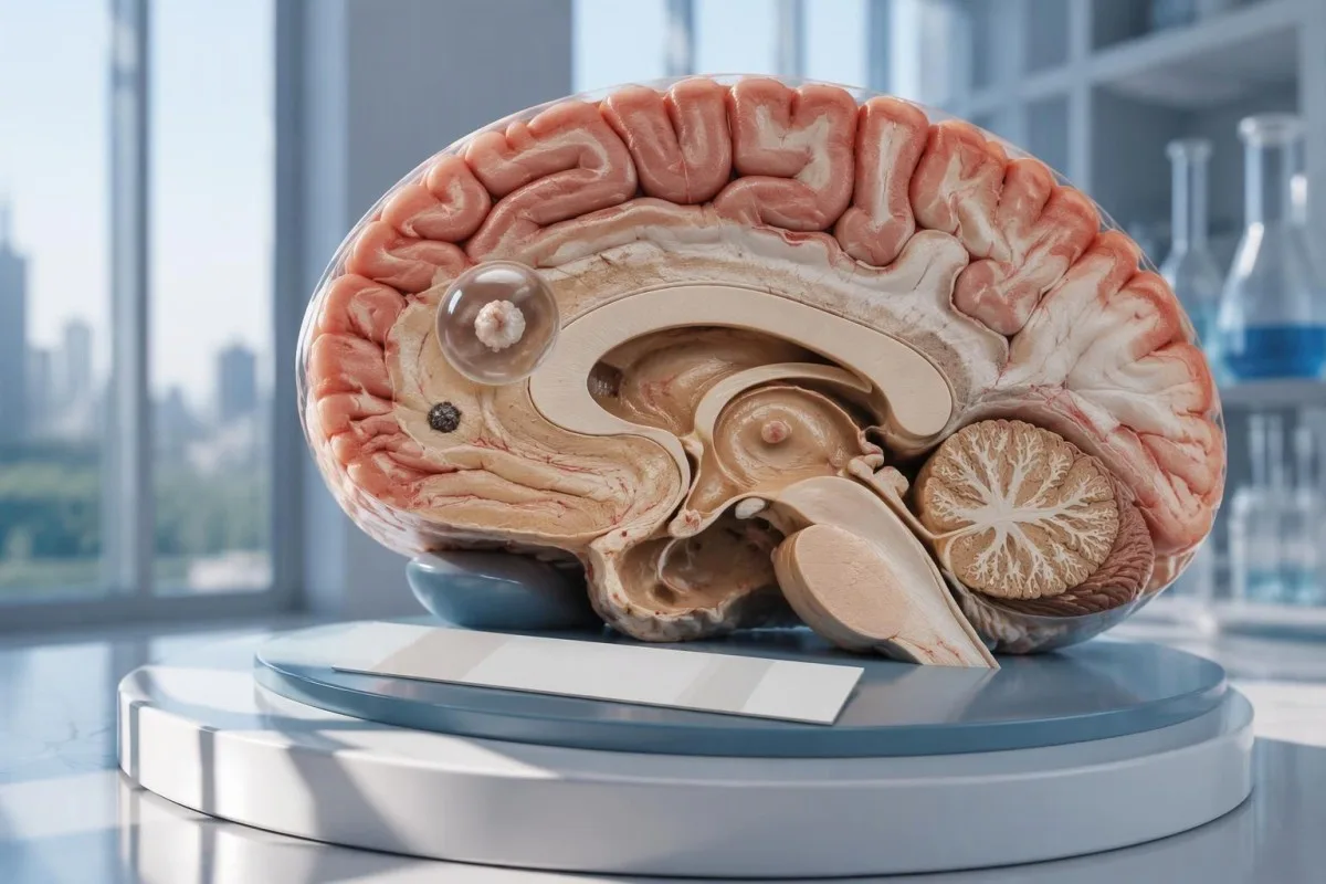
Negativan test na neurocisticerkozu ne isključuje pojedinačnu cistu u mozgu niti kalcifikovanu bolest. CT, MRI i hitna procena novih epileptičnih napada važniji su od umirujućeg rezultata samog testiranja na antitela.

Кључне поруке
- Негативна антитела ne isključuju neurocisticerkozu, naročito kada snimci pokazuju samo 1 zahvaćeno područje ili kalcifikacije.
- EITB u referentnoj laboratoriji može da dostigne osetljivost od 98% kada postoji više od 1 vijabilne ciste, ali je njegova uspešnost znatno niža kod pojedinačne ciste.
- Osetljivost kod pojedinačne ciste prema objavljenim procenama za EITB često iznosi približno 50–60%; ove vrednosti ne važe za svaki komercijalni test na antitela.
- CT bez kontrasta naročito je koristan za otkrivanje sitnih kalcifikacija, koje su često prečnika približno 1–4 mm.
- МРИ procenjuje vijabilne ciste, okolni edem i bolest u komorama ili subarahnoidnom prostoru; specijalizovane trodimenzionalne sekvence mogu da poboljšaju detekciju.
- Prvi epileptični napadi zahtevaju hitnu lekarsku procenu bez obzira na rezultat testiranja na antitela. Pozovite hitnu pomoć ako se prvi put javi poznati napad ili se pojave drugi znaci hitnog stanja.
- Pet minuta predstavlja prag za hitno postupanje kod konvulzivnog napada koji je u toku, a ne razlog da se čeka pre nego što se zatraži pomoć.
- Samo kalcifikovana bolest uglavnom ne zahteva antiparazitske lekove, ali lečenje napada i dodatno snimanje i dalje mogu biti neophodni.
Može li negativan test na neurocisticerkozu da je isključi?
A negativan test na neurocisticerkozu ne može pouzdano da isključi bolest, naročito kada postoji 1 moždana cista ili kalcifikovani ostaci. Testiranje na antitela meri imunski odgovor, a ne neposredno ispituje mozak; CT i MRI utvrđuju lokaciju i stadijum sumnjivih promena, dok prvi napad zahteva hitnu procenu.

Rezultat je uslovan, a ne apsolutan. Test na antitela na cisticerkozu koji daje dobre rezultate kod osoba sa nekoliko živih cista može biti slabo pouzdan kod nekoga ko ima samo 1 malo zahvaćeno područje, pa reč негативно znači da ovim testom nije otkriven odgovor, a ne da nigde u nervnom sistemu nema cista.
Kao hipotetički primer, razmotrimo osobu od 34 godine koja je imala prvi napad, dobila negativan nalaz antitela i kod koje je MRI pokazao jednu promenu sa prstenastim pojačanjem. Nalaz ne može da utvrdi da li je ta promena posledica cisticerkoze, druge infekcije ili nezaraznog procesa; njegovo značenje zavisi od snimka, podataka o izloženosti i neurološke procene zajedno.
Кантести је АИ анализатор крви koji može pomoći u tumačenju prijavljenih laboratorijskih nalaza, ali ne može isključiti bolest mozga na osnovu 1 negativnog rezultata testa na antitela. Naša organizacija i misija objašnjavaju naš fokus na laboratorijske informacije; čitaoci ne bi trebalo da smatraju automatsko objašnjenje nalaza odobrenjem da odlože zbrinjavanje napada.
Zašto testiranje na antitela može da ne otkrije pojedinačnu cistu u mozgu?
A jedna cista može izazvati odgovor antitela ispod praga detekcije testa. Objavljene procene osetljivosti EITB-a u referentnim laboratorijama često ukazuju na vrednosti blizu 50–60% za jednu cistu, u poređenju sa približno 98% kada je prisutno više od 1 žive ciste; razlika odražava obim bolesti i učinak testa.
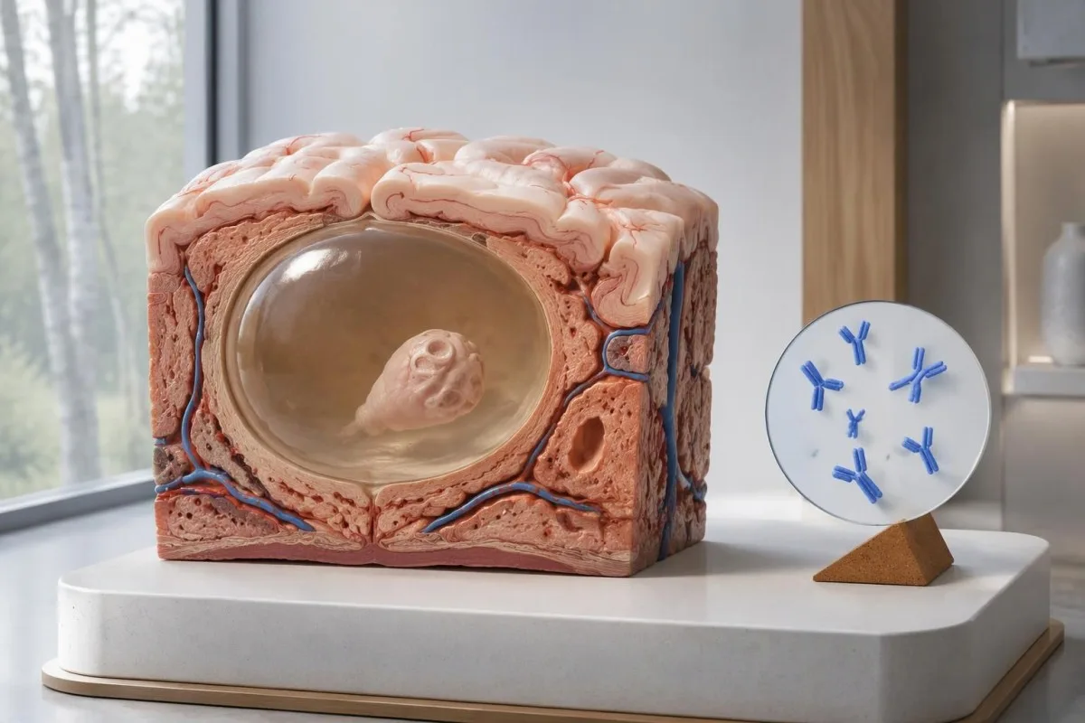
Smernice IDSA/ASTMH preporučuju enzimski povezani imunoelektrotransfer bloting, odnosno EITB, kao potvrdnu serologiju i izričito upozoravaju da je osetljivost slaba kod samo jedne promene u parenhimu ili samo kalcifikacija (White et al., 2018). Visoka osetljivost prijavljena kod pacijenata sa više cista, ventrikularnom bolešću ili subarahnoidalnom bolešću ne treba da se pripisuje osobi sa 1 malom moždanom abnormalnošću.
Zamislimo, isključivo radi ilustracije, da test ima osetljivost od 55% kod 100 osoba kod kojih je bolest sa jednom cistom već nezavisno potvrđena. Približno 45 osoba imalo bi negativan rezultat uprkos tome što imaju bolest; ovaj primer objašnjava propušteno otkrivanje, a ne verovatnoću da bilo koji pojedinačni negativan rezultat ukazuje na bolest.
Ponavljanje istog testa nakon 2 nedelje možda neće ispraviti slab odgovor uzrokovan malim obimom bolesti. To se razlikuje od infekcija kod kojih je glavni problem prerano uzorkovanje, kao što je objašnjeno u našem vodiču za lažno negativne rezultate povezane sa vremenom testiranja; pitajte da li bi snimanje ili pouzdanije okarakterisan test promenili sledeću odluku.
Zašto kalcifikovana neurocisticerkoza može i dalje da izaziva simptome?
Kalcifikovana neurocisticerkoza može izazvati napade uprkos negativnim rezultatima testiranja na antitela, because the remaining mineralized focus can still be associated with local irritation and episodic surrounding edema. Typical calcifications may measure approximately 1–4 mm, but their small size does not establish whether they are clinically harmless.
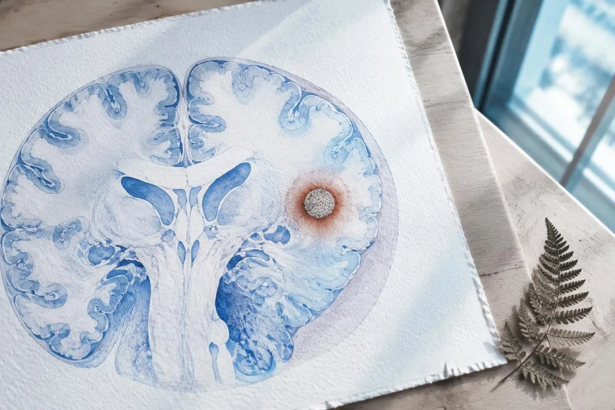
A calcification is generally a remnant rather than a living cyst that antiparasitic medication can eliminate. In the IDSA/ASTMH guideline, calcified-only parenchymal disease is managed symptomatically rather than with antiparasitic drugs; a person with 1 calcified focus and seizures may therefore need antiseizure treatment without needing cyst-killing medication (White et al., 2018).
Surrounding edema can appear intermittently, sometimes long after the original cyst has degenerated. MRI may show this tissue response even when the calcification is more obvious on CT, which is why the 2 scans can tell different but compatible stories rather than one proving that the other is wrong.
Neither a negative antibody result nor a persistent positive result measures seizure risk by itself. The broader lesson resembles other seronegative neurological conditions, discussed in our negative neurological antibody results guide: absence of detectable antibodies is not equivalent to absence of a clinically relevant neurological process.
Koji je test na antitela na cisticerkozu zapravo urađen?
The useful first question is whether the report describes EITB, ELISA, or another antibody method, because these are not interchangeable tests. A negative index below 1 laboratory's cutoff cannot inherit the performance figures of a different reference-laboratory assay, even when both reports use the name cysticercosis IgG.
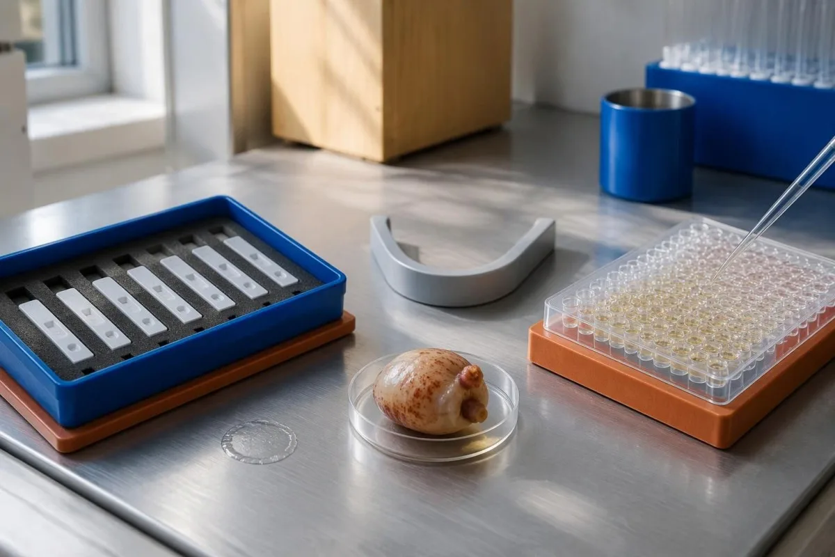
EITB using purified parasite glycoproteins is the preferred serological method in the IDSA/ASTMH guideline, while crude-antigen ELISA methods have recognized sensitivity and specificity limitations (White et al., 2018). Before comparing 2 results, check the method, specimen type, reference interval, and whether the laboratory used a validated confirmatory procedure.
Some laboratories report a qualitative result; others provide an optical-density ratio or numerical index. A value such as 0.8 in a hypothetical assay does not represent 0.8 cysts, an 80% disease probability, or a universal severity score—our kvalitativni i kvantitativni rezultati guide explains that distinction.
Kantesti AI's role is to help readers preserve the laboratory's original wording rather than reinterpret 1 method through another method's thresholds. Our vodič za laboratorijske markere provides broader report-reading context; for this result, keep the actual assay name available when discussing referral or repeat testing.
Kako istorija izloženosti utiče na dijagnostičku sumnju?
Neurocysticercosis follows ingestion of Taenia solium eggs, usually through fecal contamination from a person carrying the intestinal tapeworm. Eating undercooked pork can cause intestinal taeniasis, but brain cysts arise through a different transmission step; symptoms may emerge years after exposure rather than within 1 recent trip.

A vegetarian can develop cysticercosis because eggs can contaminate food or hands independently of meat consumption. Useful history includes past residence, household contacts, sanitation conditions, and possible exposure to a tapeworm carrier; asking only about meals eaten during the last 2 weeks can miss the relevant background.
A negative stool examination does not exclude cysts in the brain. Stool testing addresses intestinal tapeworm carriage, whereas CT and MRI assess nervous-system findings; these are 2 different sites and clinical questions, although public-health teams may recommend evaluating contacts when a carrier could be involved.
Normal eosinophils also cannot exclude neurocysticercosis, and raised eosinophils do not identify the brain as the affected site. Our parasite antibody interpretation guide discusses a separate parasite with similar localization limits; the practical point is to avoid using 1 routine cell count as a substitute for targeted neurological evaluation.
Kada novi epileptični napadi zahtevaju hitnu procenu?
A first known seizure needs urgent medical assessment regardless of antibody results, and emergency services should be contacted for a first seizure or an ongoing seizure with concerning features. A convulsive seizure lasting 5 minutes, repeated seizures without recovery, breathing difficulty, pregnancy, or significant injury requires emergency care.

Do not wait for the 5-minute mark to seek help after a first known seizure. Keep the person away from hazards, cushion the head if safely possible, do not restrain them or put anything in their mouth, and place them on their side once convulsions stop if they are breathing normally.
Persistent confusion, a severe new headache, fever with neck stiffness, or new weakness increases concern for causes beyond cysticercosis. Sudden one-sided weakness or speech difficulty also demands emergency assessment, as our prepoznavanje znakova upozorenja za moždani udar explains; 1 negative infection test cannot rule out stroke, meningitis, or a structural brain problem.
After a suspected seizure, stop driving and avoid swimming alone, heights, or operating machinery until a clinician provides advice consistent with local rules. If a witness can safely record the duration and describe movements or recovery, those observations may be more useful than ordering a second antibody test before the first neurological assessment.
Šta CT doprinosi dijagnozi neurocisticerkoze?
Noncontrast CT is particularly useful for detecting calcified cyst remnants, including small approximately 1–4 mm foci that MRI may show less clearly. CT also helps assess urgent structural problems such as hydrocephalus, but 1 isolated calcification is not automatically proof of neurocysticercosis or the cause of a seizure.
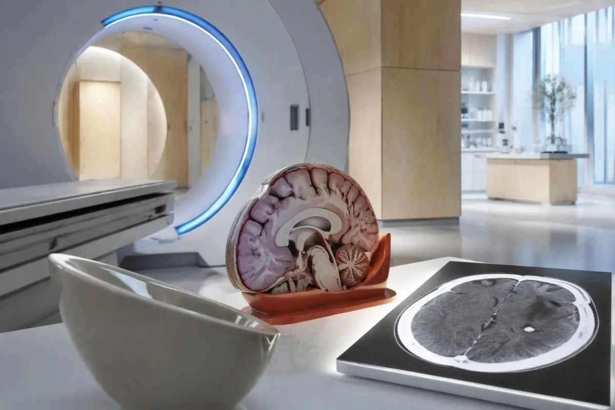
Calcium appears dense on CT, allowing radiologists to identify mineralized remnants and describe their distribution. The IDSA/ASTMH guideline recommends both MRI and noncontrast CT when classifying newly diagnosed neurocysticercosis, because the 2 modalities have complementary strengths rather than being interchangeable alternatives (White et al., 2018).
An incidental calcification needs context: previous granulomatous disease, other causes of mineralization, and scan appearance can influence interpretation. If 1 calcification sits near a suspected seizure focus, concordance with symptoms and EEG may strengthen the explanation, but anatomical proximity alone cannot establish causation.
A laboratory marker used in selected head-injury pathways does not test for brain cysts or replace imaging after an unexplained seizure. Our head-injury CT decisions guide illustrates that narrower setting; ask the clinician whether the present question requires noncontrast CT, contrast imaging, MRI, or a combination of 2 modalities.
Šta MRI otkriva što CT može da propusti?
MRI is better suited to evaluating viable cysts, surrounding edema, and many ventricular or subarachnoid findings. Specialized 3-dimensional sequences such as CISS or FIESTA can improve visualization of cyst membranes within cerebrospinal fluid spaces, where a cyst may otherwise resemble the surrounding fluid.
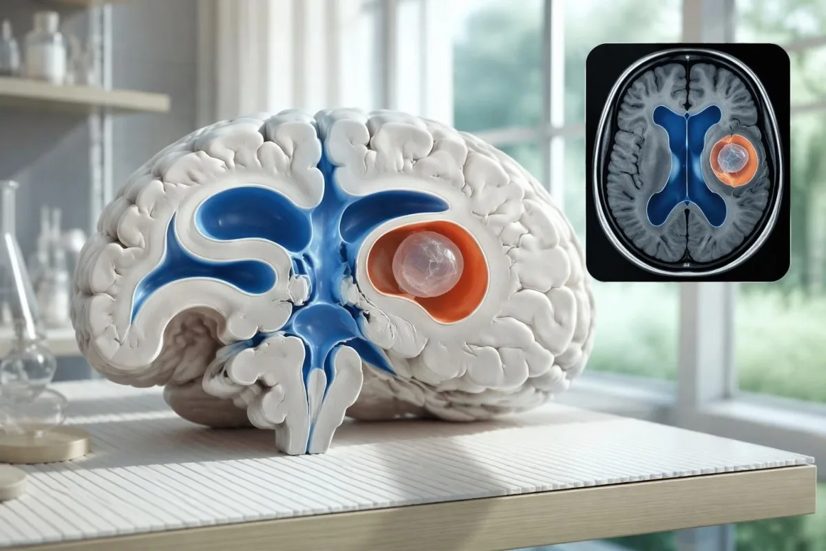
A cyst inside the ventricles can obstruct cerebrospinal fluid flow and cause hydrocephalus even without numerous parenchymal cysts. Headache with vomiting, declining alertness, or new visual symptoms therefore changes urgency; 1 reassuring antibody result should not override symptoms that suggest raised pressure or impaired fluid circulation.
MRI may show a scolex, the small internal structure of the parasite, inside a compatible cyst. This finding can be highly specific when confidently identified, whereas ring enhancement alone has a broader differential; our brain imaging warning signs guide explains why structural abnormalities require interpretation rather than diagnosis from 1 visual feature.
A routine MRI protocol may not answer every cysticercosis question. If ventricular or subarachnoid disease is suspected, discuss whether thin-section 3-dimensional imaging is needed; radiologists choose sequences and contrast according to the clinical question, and a normal scan using 1 protocol does not prove every possible disease compartment has been adequately assessed.
Kako se dijagnostikuje neurocisticerkoza kada se testovi ne slažu?
Neurocysticercosis diagnosis combines imaging, clinical findings, exposure information, and supporting tests, rather than requiring a positive antibody result in every case. The revised diagnostic criteria published in 2017 give characteristic neuroimaging findings a central role and distinguish strong evidence from less-specific supportive features (Del Brutto et al., 2017).
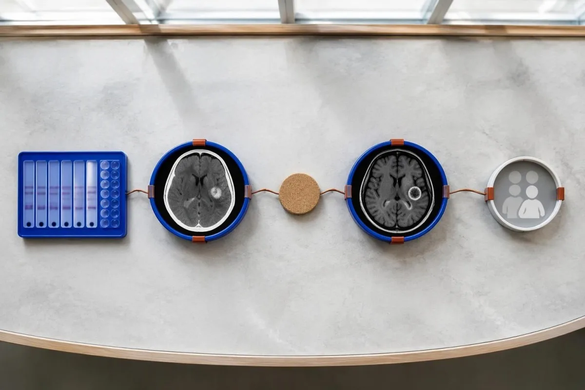
Evidence should be weighted, not tallied. A confidently visualized scolex inside a compatible cyst is different from 1 nonspecific ring-enhancing area, while a positive antibody result is different from direct evidence that a finding is in the brain; our водич за позитивне резултате антитела explains why detection alone cannot establish location.
For a clearly hypothetical probability example, start with a 30% pretest probability and assume a single-cyst assay has 55% sensitivity and 99% specificity. In 1,000 comparable people, about 135 affected and 693 unaffected people would test negative, leaving approximately a 16% probability of disease among negative results—still substantial, although these assumed figures are not a personal risk calculator.
The most useful disagreement to investigate is whether each test is answering the same question. A negative antibody test, a CT calcification, and MRI edema can all be compatible with 1 calcified disease process; unexplained discrepancies should prompt review of the assay and images rather than automatic dismissal of either result.
Šta još na snimcima mozga može da liči na pojedinačnu cistu?
A single ring-enhancing brain finding is not specific to neurocysticercosis. Other possibilities include tuberculoma, an abscess, a neoplasm, or an inflammatory process; clinicians use appearance, location, immune status, symptoms, and sometimes follow-up imaging or tissue examination to distinguish these possibilities rather than relying on 1 antibody panel.
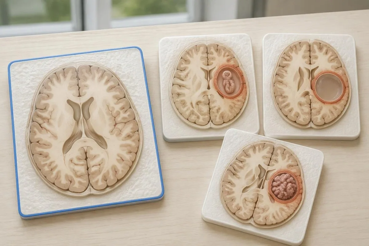
Edema, restricted diffusion, internal structures, and the number of findings all contribute to the differential, but no single nonspecific feature settles every case. Fever and immune suppression may change the suspected cause; a 2 cm ring-enhancing area in a hypothetical immunocompromised person warrants a different investigation from a tiny calcification in an otherwise well person.
Toxoplasmosis is one consideration in selected immunocompromised patients, and a positive IgG result can indicate past exposure rather than prove an active brain process. Our Toxoplasma IgG interpretation guide explains that laboratory distinction; imaging and the patient's immune status determine whether the result is clinically relevant to 1 or more cerebral findings.
New seizures accompanied by rapidly evolving memory problems, psychiatric changes, or abnormal movements can also suggest encephalitis rather than a solitary parasite cyst. Our encephalitis diagnostic pathway describes how MRI, EEG, and cerebrospinal fluid testing answer different questions; a second negative cysticercosis antibody result does not investigate those alternatives.
Mogu li testiranje na antigen, PCR ili likvor da razreše nedoumicu?
Antigen tests, PCR, and cerebrospinal fluid analysis can add information in selected cases, particularly when extraparenchymal disease is suspected. None is a universal rescue test after 1 negative antibody result: availability, assay validation, disease compartment, and safety of lumbar puncture determine whether additional testing is appropriate.
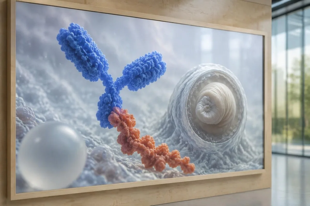
Antibody testing detects the host response, while antigen testing aims to detect parasite-derived material and may better reflect viable disease burden. Antigen detection is often less useful when only 1 small parenchymal cyst or calcifications are present; molecular testing is used in some specialist settings, but performance cannot be generalized across every laboratory or specimen.
Lumbar puncture is not routinely required for an uncomplicated solitary parenchymal finding and can be unsafe when obstructive hydrocephalus or significant mass effect is present. Cerebrospinal fluid tests for other disorders also require disease-specific interpretation, as our CSF band interpretation guide explains; 1 abnormal CSF marker does not identify every possible cause.
Кантести је AI платформа за тумачење крвне слике, not a service that orders lumbar punctures, identifies cysts on scans, or confirms neurological infections. Our технологија за тумачење вештачке интелигенције explains the laboratory-report workflow; when 2 tests disagree, our role is to clarify their stated meanings and limitations, not select an invasive diagnostic procedure.
Zašto lečenje zavisi od stadijuma ciste i njene lokacije?
Treatment depends on whether cysts are viable, degenerating, calcified, or located outside the brain tissue, not simply on antibody positivity. Selected viable parenchymal cases receive antiparasitic treatment, whereas calcified-only disease generally receives symptom-directed care; ventricular obstruction may require a procedural approach rather than 1 standard medication regimen.
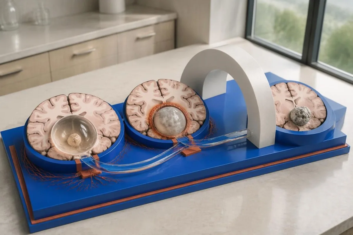
For selected patients with 1–2 viable parenchymal cysts, the IDSA/ASTMH guideline describes albendazole treatment for 10–14 days with appropriate accompanying care. This is a specialist-directed recommendation, not a home-treatment instruction: antiparasitic drugs can provoke an inflammatory response, and untreated hydrocephalus or diffuse cerebral edema changes the order of management (White et al., 2018).
WHO's 2021 guidance focuses on defined presentations of parenchymal neurocysticercosis, while the IDSA/ASTMH guideline also addresses ventricular, subarachnoid, and other forms in detail. Differences in imaging access and disease compartment explain why 1 treatment recommendation cannot be transferred to every patient with an antibody result; the guidelines do not turn negative serology into a rule-out test.
Corticosteroids may accompany antiparasitic therapy, but prolonged use requires additional safety planning, including assessment for relevant latent infections. Our Strongyloides and steroid safety guide explains a separate risk that matters before immune-suppressing treatment; an eye examination is also recommended before antiparasitic therapy to evaluate possible intraocular cysts.
Šta treba pratiti tokom kontrolnih pregleda, pored rezultata na antitela?
Follow-up should track seizures, neurological symptoms, imaging changes, and medication safety, rather than use antibody disappearance as proof of recovery. For viable parenchymal cysts, the IDSA/ASTMH guideline suggests repeat MRI at least every 6 months until the cystic component resolves, with earlier reassessment when symptoms change.
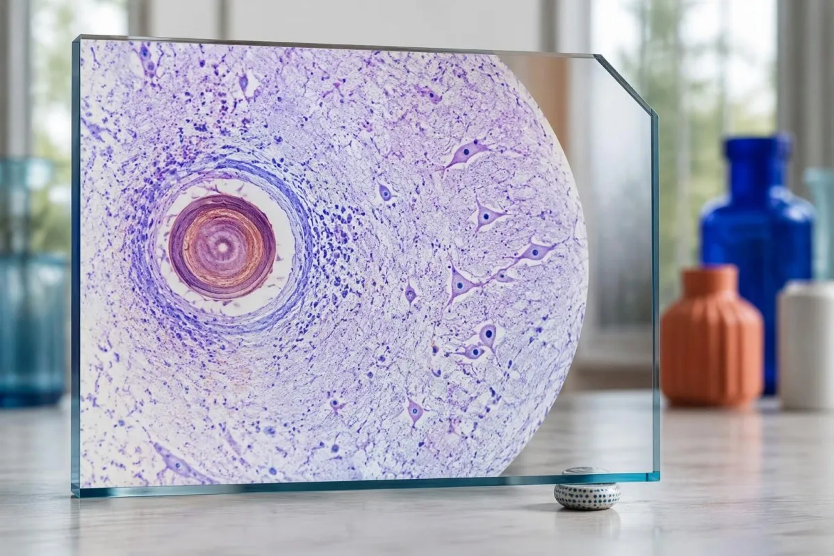
Antibodies may remain detectable after the original disease process has changed, and low-burden disease may never produce a positive result. If 1 new seizure occurs during follow-up, clinicians consider adherence, medication interactions, recurrent edema, and other causes; a rising or falling antibody index alone cannot distinguish among those explanations.
Kantesti AI can help explain reported laboratory results during medication follow-up, but a drug concentration is not a stand-alone measure of seizure control. Our tumačenje nivoa levetiracetama guide explains why sample timing and kidney function matter; 2 measurements collected at different times after dosing may not be directly comparable.
Monitoring depends on the chosen drug and treatment duration: prolonged albendazole therapy can require liver-test and blood-count surveillance, while some antiseizure medicines can affect sodium. Our водич за безбедност електролита discusses those separate laboratory concerns; do not stop an antiseizure medicine or change its dose because 1 cysticercosis antibody result is negative.
Šta Kantesti može da objasni, a šta istraživanja ne mogu da dokažu?
AI report interpretation cannot confirm or exclude neurocysticercosis from laboratory results alone. Kantesti je an Алат за анализу крвних тестова уз помоћ AI whose role here is to organize reported laboratory findings; a negative result still needs clinical context, and 1 new seizure takes priority over reviewing an automated explanation.

Prepare a focused review packet with the original antibody report, assay method, CT or MRI report, symptom timeline, and medication list. These 5 information categories reduce the risk of discussing a result without its context; ask specifically whether the imaging suggests viable, degenerating, calcified, ventricular, or subarachnoid disease and which unanswered question the next test would address.
The 2 Kantesti publications listed below are a repository-deposited global analysis report and an educational RDW guide, not clinical evidence validating neurocysticercosis diagnosis. Our RDW i CBC vodič can support interpretation of a separately ordered blood count, but RDW, MCV, and MCHC do not establish whether a brain cyst is present.
Evidence for the neurological recommendations in this article comes from the cited diagnostic criteria and clinical guidelines, not from general AI benchmarking or laboratory-report volume. Our technical validation scope should be read within its stated boundaries; neither 1 successful report explanation nor a large aggregate dataset substitutes for condition-specific diagnostic validation.
Često postavljana pitanja
Može li se imati neurocisticerkoza uz negativan test krvi?
Da, neurocisticerkoza može biti prisutna i pored negativnog testa na antitela iz krvi, naročito kada postoji 1 moždana cista ili samo kalcifikovana bolest. Objavljene procene referentnih laboratorija za EITB često ukazuju na osetljivost od približno 50–60% kod jedne ciste, što je znatno niže od njegove uspešnosti kod više vijabilnih cista. Zato CT, MRI, simptomi i podaci o izloženosti i dalje imaju ključnu ulogu u dijagnozi. Novi epileptični napad zahteva hitnu procenu, a ne oslanjanje na negativan rezultat kao razlog za umirenje.
Koji je najpouzdaniji test za neurocisticerkozu?
Nijedan pojedinačni test za neurocisticerkozu ne daje odgovor na sva dijagnostička pitanja; snimanje mozga i pravilno odabrana serološka ispitivanja se dopunjuju. EITB je preferirana metoda za potvrdu prisustva antitela prema smernicama IDSA/ASTMH, ali može da ne otkrije bolest koja zahvata samo 1 cistu. CT bez kontrastnog sredstva naročito je koristan za otkrivanje kalcifikacija, dok MRI bolje procenjuje mnoge vijabilne ciste, edem i abnormalnosti prostora ispunjenih tečnošću. Kliničari kombinuju ove nalaze, umesto da jedan negativan rezultat smatraju konačnim.
Da li je CT ili MRI bolji za neurocisticerkozu?
CT i MRI imaju različite prednosti u dijagnostici neurocisticerkoze. CT bez kontrasta često jasnije otkriva male kalcifikacije, prečnika približno 1–4 mm, dok MRI omogućava procenu vijabilnih cista, okolnog edema i bolesti komora ili subarahnoidnog prostora. Specijalizovane trodimenzionalne MRI sekvence mogu pomoći u otkrivanju membrana cista u prostorima ispunjenim cerebrospinalnom tečnošću. Smernice IDSA/ASTMH preporučuju obe metode pri klasifikaciji novodijagnostikovane bolesti.
Može li kalcifikovana cista i dalje izazivati napade?
Kalcifikovani ostatak cisticerka može ostati povezan s epileptičkim napadima iako više nije vitalna cista. Oko nekih kalcifikovanih žarišta, često prečnika svega 1–4 mm, razvija se povremeni okolni edem, koji se može uočiti na MR snimku. Negativna antitela ne isključuju ovaj mehanizam, a trajno pozitivna antitela ne ukazuju na rizik od epileptičkih napada. Kod bolesti s isključivo kalcifikovanim žarištima primenjuje se lečenje usmereno na simptome, a ne antiparazitska terapija.
Da li treba da ponovim negativan test na antitela na cisticerkozu?
Ponavljanje negativnog testa na antitela protiv cisticerkoze nije automatski najbolji sledeći korak. Kod 1 male ciste ili kalcifikovane bolesti, slab odgovor antitela može ostati negativan čak i ako se isti test ponovi nakon 2 nedelje. Lekar može umesto toga da razmotri prvobitnu metodu, zatraži bolje karakterisan potvrdni test ili da prednost CT-u i MR-u. Pojava novih napada ili pogoršanje neuroloških simptoma ne treba da čekaju ponavljanje testiranja.
Kada treba da potražim hitnu pomoć zbog prvog epileptičnog napada?
Prvi poznati epileptički napad zahteva hitnu medicinsku procenu i pozivanje lokalne hitne službe, bez obzira na negativan test na antitela protiv neurocisticerkoze. Konvulzivni napad koji traje 5 minuta, ponovljeni napadi bez oporavka svesti, otežano disanje, trudnoća ili ozbiljna povreda predstavljaju hitan slučaj. Ne sputavajte osobu i ne stavljajte joj ništa u usta; kada konvulzije prestanu, položite je na bok ako normalno diše. Nemojte voziti dok lekar ne proceni događaj i objasni koja ograničenja važe.
Сазнајте како да читате крвну слику
Отпремите свој лабораторијски извештај, а AI алата Kantesti ће за неколико секунди објаснити сваки биомаркер једноставним језиком. Користи га више од 2 милиона људи широм света.
Референце
- Klein, T., Mitchell, S., & Weber, H. (2026). AI анализа крвне слике: 2,5M тестова анализирано | Global Health Report 2026. Kantesti AI Medical Research. ДОИ
- Klein, T., Mitchell, S., & Weber, H. (2026). RDW у крвној слици: комплетан водич за RDW-CV, MCV и MCHC. Kantesti AI Medical Research. ДОИ
- White AC Jr et al. (2018). Diagnosis and Treatment of Neurocysticercosis: 2017 Clinical Practice Guidelines by the Infectious Diseases Society of America (IDSA) and the American Society of Tropical Medicine and Hygiene (ASTMH). Clinical Infectious Diseases. ДОИ
- Del Brutto OH et al. (2017). Revised diagnostic criteria for neurocysticercosis. Journal of the Neurological Sciences. ДОИ
- World Health Organization (2021). WHO guidelines on management of Taenia solium neurocysticercosis. Светска здравствена организација.
- 2 милиона+Analizirani testovi
- 127+Земље
- 100+Језици
O medicinskom timu


Е-Е-А-Т сигнали поверења
Искуство
Клиничка ревизија процеса тумачења лабораторијских налаза коју води лекар.
Експертиза
Фокус на лабораторијску медицину: како се биомаркери понашају у клиничком контексту.
Ауторитативност
Medicinski pregledao dr Thomas Klein, uz doprinos dr Sarah Mitchell i prof. dr Hansa Webera.
Поузданост
Тумачење засновано на доказима, са јасним путевима праћења како би се смањила узнемиреност.





Medicinsko odricanje od odgovornosti
Овај чланак је само у образовне сврхе и не представља медицински савет. За одлуке о дијагнози и лечењу увек се консултујте са квалификованим здравственим радником.
Objavljeno · Kontaktirajte urednički tim
Objavio Kantesti Ltd, registrovan u Engleskoj i Velsu, broj kompanije. 17090423, London, Ujedinjeno Kraljevstvo.
