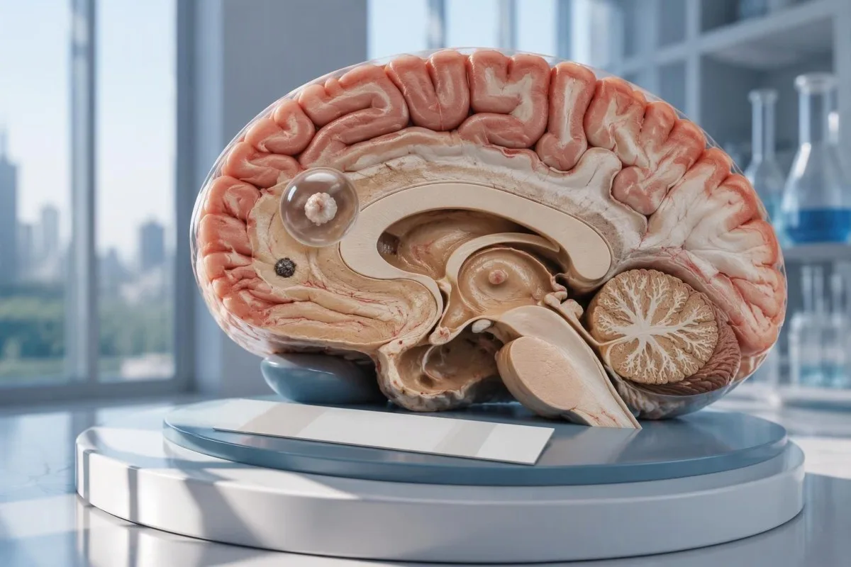
Un tèst negatiu de neurocisticercòsi exclutz pas un quist cerebral unic o una malautiá calcificada. La TC, l’IRM e una avaloracion urgenta de las crisis convulsivas recentas comptan mai que lo rassicurament basat unicament sus la recèrca d’anticòsses.

Punts essencials
- Anticòs negatius excluisson pas la neurocisticercòsi, subretot quand l’imatge mòstra sonque 1 zòna afectada o de calcificacions.
- EITB de laboratòri de referéncia pòt aténher una sensibilitat de 98% amb mai d’1 quist viable, mas las performanças son fòrça mai feblas amb un quist unic.
- Sensibilitat per un quist unic es sovent d’aperaquí 50–60% segon las estimacions publicadas per EITB; aquelas chifras s’aplican pas a totes los tèsts comercials d’anticòsses.
- TC sens contraste es particularament utila per detectar de pichonas calcificacions, que fan sovent aperaquí 1–4 mm de diamètre.
- RM avalora los quists viables, l’edèma environant e l’afectacion ventriculara o subaracnoïdiana; de sequéncias especializadas en 3 dimensions pòdon melhorar la deteccion.
- Primièras crisis convulsivas requerisson una avaloracion medicala d'urgéncia, quin que siá lo resultat dels anticòrs. Apelatz los servicis d'urgéncia per una primièra crisi coneguda o d'autres signes d'urgéncia.
- Cinc minutas es un lindal d'urgéncia per una crisi convulsiva en cors, pas una rason d'esperar abans de demandar d'ajuda.
- Malautiá sonque calcificada generalament necessita pas de medicament antiparasitari, mas un tractament de las crisis e d'autres examens d'imatjariá pòdon encara èsser necessaris.
Un tèst negatiu de neurocisticercòsi pòt exclure la malautiá?
A un tèst negatiu de neurocisticercòsi pòt pas exclure la malautiá de faiçon fisabla, particularament amb 1 quist cerebral o de rèstas calcificadas. Lo tèst dels anticòrs mesura la responsa immunitària e non examina dirèctament lo cervèl; lo scanner e l'IRM establisson la localizacion e l'estadi de las trobalhas sospèchas, mentre qu'una primièra crisi necessita una avaloracion d'urgéncia.

Lo resultat es condicionat, pas absolut. Un tèst d'anticòrs contra la cisticercòsi qu'es performant en çò de personas amb mantun quist viable pòt èsser pauc performant en çò d'una persona qu'a sonque 1 pichona zòna afectada; atal, lo mot negatiu significa pas cap de responsa detectabla amb aquel tèst, e non pas abséncia de quists dins tot lo sistèma nerviós.
Per un exemple ipotetic, consideratz una persona de 34 ans amb una primièra crisi, un rapòrt d'anticòrs negatiu e una trobalha solitària amb captacion en anèl a l'IRM. Lo rapòrt pòt pas determinar se aquela trobalha correspond a la cisticercòsi, a una autra infeccion o a un procèssus non infecciós; son sens depend del scanner, de l'istoric d'exposicion e de l'avaloracion neurologica preses ensems.
Kantesti es un Analizador de tèst de sang d'IA que pòt ajudar a explicar de resultats de laboratòri rapòrtats, mas pòt pas exclure una malautiá cerebrala a partir d'1 resultat d'anticòrs negatiu. Nòstra organizacion e mission explican nòstra orientacion sus l'informacion de laboratòri; los legeires deurián pas considerar una explicacion automatizada del rapòrt coma una autorizacion a retardar los suènhs per las crisis.
Perqué la recèrca d’anticòsses pòt passar per naut un quist cerebral unic?
A un quist solitari pòt generar una responsa d'anticòrs inferiora al lindal de deteccion del tèst. Las estimacions publicadas dels laboratòris de referéncia per l'EITB plaçan sovent la sensibilitat a l'entorn de 50–60% per un quist solitari, en comparason amb aperaquí 98% quand i a mai d'1 quist viable; la diferéncia reflècta la carga de la malautiá e lo rendiment del tèst.
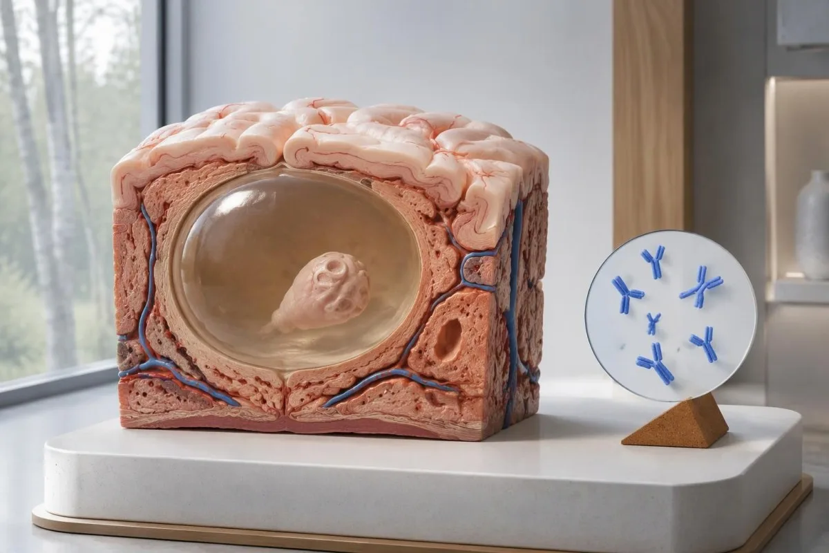
Lo recuèlh IDSA/ASTMH recomanda lo blòt d'immunoelectrotransferéncia ligada a l'enzim, o EITB, coma serologia de confirmacion e avertís explicitament que la sensibilitat es febla en preséncia d'una sola trobalha parenquimatosa o de calcificacions soletas (White et al., 2018). La nauta sensibilitat senhalada en çò de pacients amb mantun quist, una malautiá ventriculara o una malautiá subaracnoïdiana deu pas èsser atribuïda a qualqu'un qu'a 1 pichona anomalia cerebrala.
Imaginatz, sonque per illustrar, qu'un tèst a una sensibilitat de 55% en 100 personas que la malautiá a quist solitari es estada establida independentament. Aperaquí 45 obtendrián un resultat negatiu malgrat aver la malautiá; aquel exemple calculat explica los falses negatius, pas la probabilitat que quin que siá resultat negatiu particular represente una malautiá.
Repetir lo meteis tèst aprèp 2 setmanas pòt pas corregir una responsa febla causada per una carga de malautiá bassa. Aquò es diferent de las infeccions ont lo prélèvement precòç es lo problèma principal, coma s'explica dins nòstre guida suls falses negatius ligats al moment del tèst; demandatz se l'imatjariá o un tèst mai plan caracterizat cambiariá la decision seguenta.
Perqué la neurocisticercòsi calcificada pòt demorar simptomatica?
La neurocisticercòsi calcificada pòt provocar de crisis malgrat d'anticòrs negatius, because the remaining mineralized focus can still be associated with local irritation and episodic surrounding edema. Typical calcifications may measure approximately 1–4 mm, but their small size does not establish whether they are clinically harmless.
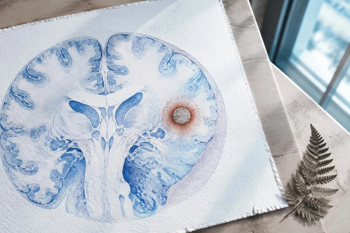
A calcification is generally a remnant rather than a living cyst that antiparasitic medication can eliminate. In the IDSA/ASTMH guideline, calcified-only parenchymal disease is managed symptomatically rather than with antiparasitic drugs; a person with 1 calcified focus and seizures may therefore need antiseizure treatment without needing cyst-killing medication (White et al., 2018).
Surrounding edema can appear intermittently, sometimes long after the original cyst has degenerated. MRI may show this tissue response even when the calcification is more obvious on CT, which is why the 2 scans can tell different but compatible stories rather than one proving that the other is wrong.
Neither a negative antibody result nor a persistent positive result measures seizure risk by itself. The broader lesson resembles other seronegative neurological conditions, discussed in our negative neurological antibody results guide: absence of detectable antibodies is not equivalent to absence of a clinically relevant neurological process.
Quin tèst d’anticòsses contra la cisticercòsi es estat efectivament realizat?
The useful first question is whether the report describes EITB, ELISA, or another antibody method, because these are not interchangeable tests. A negative index below 1 laboratory's cutoff cannot inherit the performance figures of a different reference-laboratory assay, even when both reports use the name cysticercosis IgG.
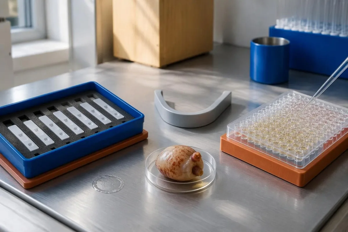
EITB using purified parasite glycoproteins is the preferred serological method in the IDSA/ASTMH guideline, while crude-antigen ELISA methods have recognized sensitivity and specificity limitations (White et al., 2018). Before comparing 2 results, check the method, specimen type, reference interval, and whether the laboratory used a validated confirmatory procedure.
Some laboratories report a qualitative result; others provide an optical-density ratio or numerical index. A value such as 0.8 in a hypothetical assay does not represent 0.8 cysts, an 80% disease probability, or a universal severity score—our resultats qualitatius e quantitatius guide explains that distinction.
Kantesti AI's role is to help readers preserve the laboratory's original wording rather than reinterpret 1 method through another method's thresholds. Our guida dei marcators de laboratòri provides broader report-reading context; for this result, keep the actual assay name available when discussing referral or repeat testing.
Cossí l’istòria d’exposicion modifica la suspicion diagnostica?
Neurocysticercosis follows ingestion of Taenia solium eggs, usually through fecal contamination from a person carrying the intestinal tapeworm. Eating undercooked pork can cause intestinal taeniasis, but brain cysts arise through a different transmission step; symptoms may emerge years after exposure rather than within 1 recent trip.

A vegetarian can develop cysticercosis because eggs can contaminate food or hands independently of meat consumption. Useful history includes past residence, household contacts, sanitation conditions, and possible exposure to a tapeworm carrier; asking only about meals eaten during the last 2 weeks can miss the relevant background.
A negative stool examination does not exclude cysts in the brain. Stool testing addresses intestinal tapeworm carriage, whereas CT and MRI assess nervous-system findings; these are 2 different sites and clinical questions, although public-health teams may recommend evaluating contacts when a carrier could be involved.
Normal eosinophils also cannot exclude neurocysticercosis, and raised eosinophils do not identify the brain as the affected site. Our parasite antibody interpretation guide discusses a separate parasite with similar localization limits; the practical point is to avoid using 1 routine cell count as a substitute for targeted neurological evaluation.
Quand las crisis convulsivas recentas necessitan una avaloracion d’urgéncia?
A first known seizure needs urgent medical assessment regardless of antibody results, and emergency services should be contacted for a first seizure or an ongoing seizure with concerning features. A convulsive seizure lasting 5 minutes, repeated seizures without recovery, breathing difficulty, pregnancy, or significant injury requires emergency care.

Do not wait for the 5-minute mark to seek help after a first known seizure. Keep the person away from hazards, cushion the head if safely possible, do not restrain them or put anything in their mouth, and place them on their side once convulsions stop if they are breathing normally.
Persistent confusion, a severe new headache, fever with neck stiffness, or new weakness increases concern for causes beyond cysticercosis. Sudden one-sided weakness or speech difficulty also demands emergency assessment, as our guida dels signes d'alarma de l'accident vascular cerebral explains; 1 negative infection test cannot rule out stroke, meningitis, or a structural brain problem.
After a suspected seizure, stop driving and avoid swimming alone, heights, or operating machinery until a clinician provides advice consistent with local rules. If a witness can safely record the duration and describe movements or recovery, those observations may be more useful than ordering a second antibody test before the first neurological assessment.
Qu’apòrta la TC al diagnostic de neurocisticercòsi?
Noncontrast CT is particularly useful for detecting calcified cyst remnants, including small approximately 1–4 mm foci that MRI may show less clearly. CT also helps assess urgent structural problems such as hydrocephalus, but 1 isolated calcification is not automatically proof of neurocysticercosis or the cause of a seizure.
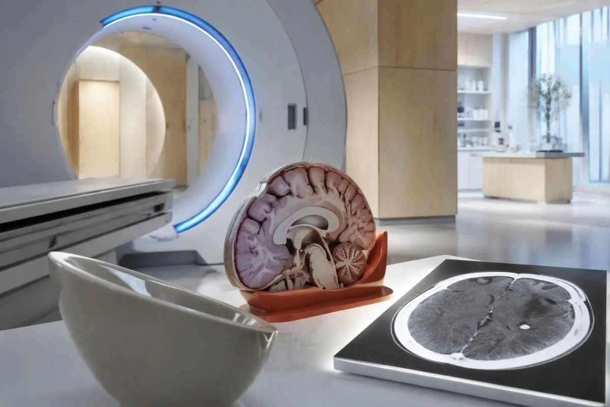
Calcium appears dense on CT, allowing radiologists to identify mineralized remnants and describe their distribution. The IDSA/ASTMH guideline recommends both MRI and noncontrast CT when classifying newly diagnosed neurocysticercosis, because the 2 modalities have complementary strengths rather than being interchangeable alternatives (White et al., 2018).
An incidental calcification needs context: previous granulomatous disease, other causes of mineralization, and scan appearance can influence interpretation. If 1 calcification sits near a suspected seizure focus, concordance with symptoms and EEG may strengthen the explanation, but anatomical proximity alone cannot establish causation.
A laboratory marker used in selected head-injury pathways does not test for brain cysts or replace imaging after an unexplained seizure. Our head-injury CT decisions guide illustrates that narrower setting; ask the clinician whether the present question requires noncontrast CT, contrast imaging, MRI, or a combination of 2 modalities.
Que revela l’IRM que la TC pòt passar per naut?
MRI is better suited to evaluating viable cysts, surrounding edema, and many ventricular or subarachnoid findings. Specialized 3-dimensional sequences such as CISS or FIESTA can improve visualization of cyst membranes within cerebrospinal fluid spaces, where a cyst may otherwise resemble the surrounding fluid.
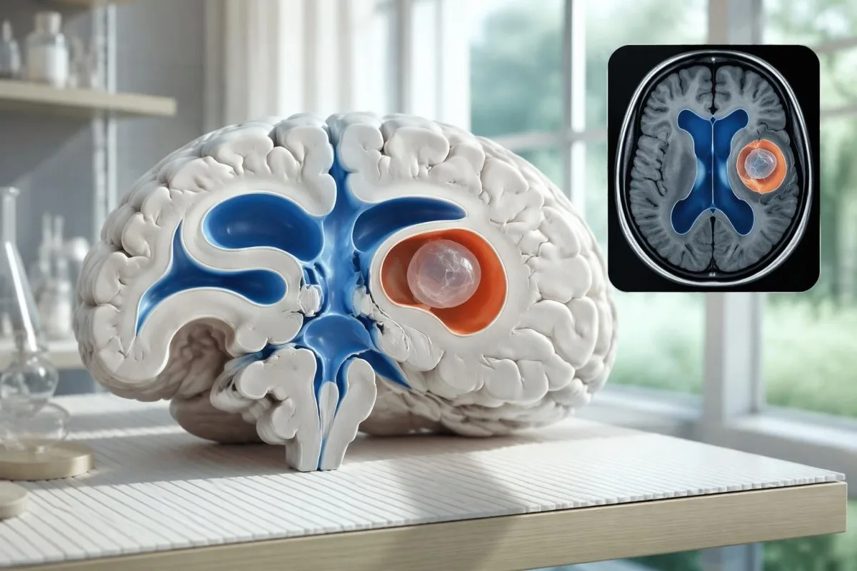
A cyst inside the ventricles can obstruct cerebrospinal fluid flow and cause hydrocephalus even without numerous parenchymal cysts. Headache with vomiting, declining alertness, or new visual symptoms therefore changes urgency; 1 reassuring antibody result should not override symptoms that suggest raised pressure or impaired fluid circulation.
MRI may show a scolex, the small internal structure of the parasite, inside a compatible cyst. This finding can be highly specific when confidently identified, whereas ring enhancement alone has a broader differential; our brain imaging warning signs guide explains why structural abnormalities require interpretation rather than diagnosis from 1 visual feature.
A routine MRI protocol may not answer every cysticercosis question. If ventricular or subarachnoid disease is suspected, discuss whether thin-section 3-dimensional imaging is needed; radiologists choose sequences and contrast according to the clinical question, and a normal scan using 1 protocol does not prove every possible disease compartment has been adequately assessed.
Cossí se diagnostica la neurocisticercòsi quand los tèsts son discordants?
Neurocysticercosis diagnosis combines imaging, clinical findings, exposure information, and supporting tests, rather than requiring a positive antibody result in every case. The revised diagnostic criteria published in 2017 give characteristic neuroimaging findings a central role and distinguish strong evidence from less-specific supportive features (Del Brutto et al., 2017).
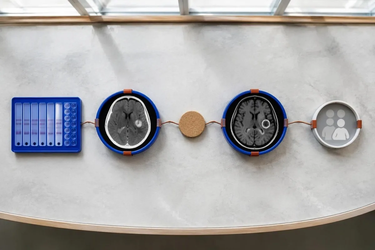
Evidence should be weighted, not tallied. A confidently visualized scolex inside a compatible cyst is different from 1 nonspecific ring-enhancing area, while a positive antibody result is different from direct evidence that a finding is in the brain; our guia detallada de resultats d'anticòrs positifs explains why detection alone cannot establish location.
For a clearly hypothetical probability example, start with a 30% pretest probability and assume a single-cyst assay has 55% sensitivity and 99% specificity. In 1,000 comparable people, about 135 affected and 693 unaffected people would test negative, leaving approximately a 16% probability of disease among negative results—still substantial, although these assumed figures are not a personal risk calculator.
The most useful disagreement to investigate is whether each test is answering the same question. A negative antibody test, a CT calcification, and MRI edema can all be compatible with 1 calcified disease process; unexplained discrepancies should prompt review of the assay and images rather than automatic dismissal of either result.
Qué mai pòt semblar a un quist unic sus l’imatge cerebrala?
A single ring-enhancing brain finding is not specific to neurocysticercosis. Other possibilities include tuberculoma, an abscess, a neoplasm, or an inflammatory process; clinicians use appearance, location, immune status, symptoms, and sometimes follow-up imaging or tissue examination to distinguish these possibilities rather than relying on 1 antibody panel.
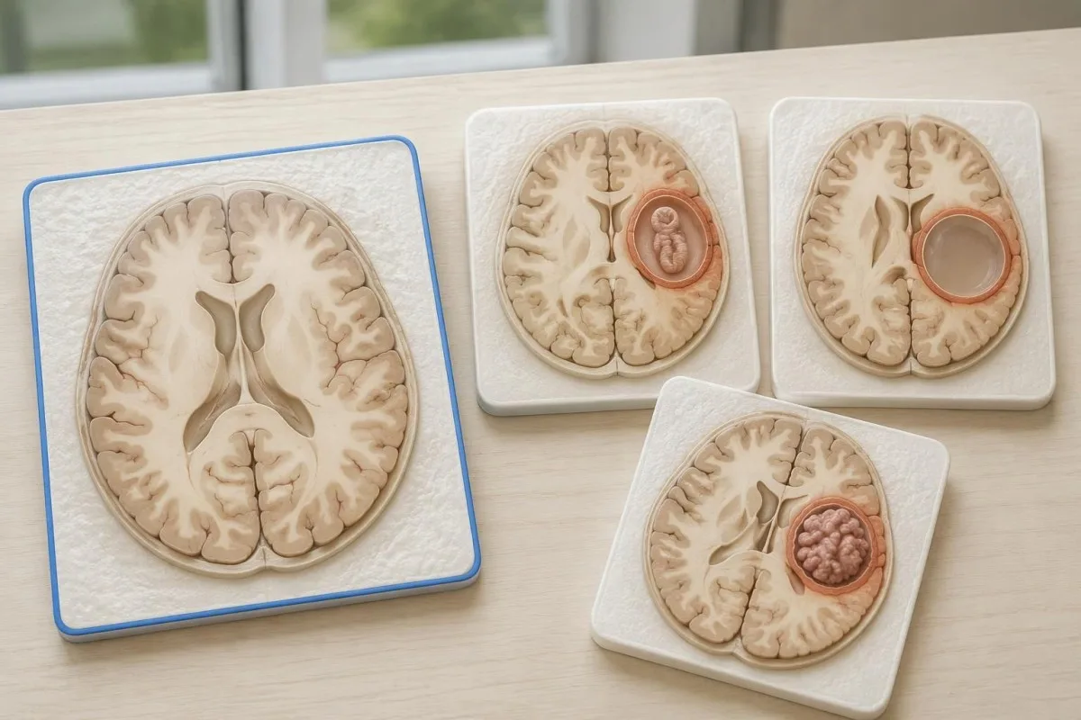
Edema, restricted diffusion, internal structures, and the number of findings all contribute to the differential, but no single nonspecific feature settles every case. Fever and immune suppression may change the suspected cause; a 2 cm ring-enhancing area in a hypothetical immunocompromised person warrants a different investigation from a tiny calcification in an otherwise well person.
Toxoplasmosis is one consideration in selected immunocompromised patients, and a positive IgG result can indicate past exposure rather than prove an active brain process. Our Toxoplasma IgG interpretation guide explains that laboratory distinction; imaging and the patient's immune status determine whether the result is clinically relevant to 1 or more cerebral findings.
New seizures accompanied by rapidly evolving memory problems, psychiatric changes, or abnormal movements can also suggest encephalitis rather than a solitary parasite cyst. Our encephalitis diagnostic pathway describes how MRI, EEG, and cerebrospinal fluid testing answer different questions; a second negative cysticercosis antibody result does not investigate those alternatives.
La recèrca d’antigèn, la PCR o lo LCR pòdon resòlvre l’incertitud?
Antigen tests, PCR, and cerebrospinal fluid analysis can add information in selected cases, particularly when extraparenchymal disease is suspected. None is a universal rescue test after 1 negative antibody result: availability, assay validation, disease compartment, and safety of lumbar puncture determine whether additional testing is appropriate.
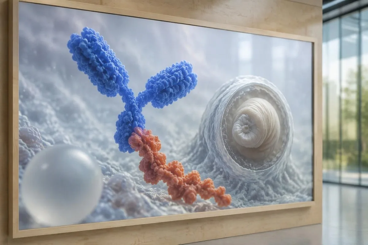
Antibody testing detects the host response, while antigen testing aims to detect parasite-derived material and may better reflect viable disease burden. Antigen detection is often less useful when only 1 small parenchymal cyst or calcifications are present; molecular testing is used in some specialist settings, but performance cannot be generalized across every laboratory or specimen.
Lumbar puncture is not routinely required for an uncomplicated solitary parenchymal finding and can be unsafe when obstructive hydrocephalus or significant mass effect is present. Cerebrospinal fluid tests for other disorders also require disease-specific interpretation, as our CSF band interpretation guide explains; 1 abnormal CSF marker does not identify every possible cause.
Kantesti es un plataforma d’interpretacion d’analisi de sang d’AI, not a service that orders lumbar punctures, identifies cysts on scans, or confirms neurological infections. Our tecnologia d'interpretació AI explains the laboratory-report workflow; when 2 tests disagree, our role is to clarify their stated meanings and limitations, not select an invasive diagnostic procedure.
Perqué lo tractament depend de l’estadi e de la localizacion del quist?
Treatment depends on whether cysts are viable, degenerating, calcified, or located outside the brain tissue, not simply on antibody positivity. Selected viable parenchymal cases receive antiparasitic treatment, whereas calcified-only disease generally receives symptom-directed care; ventricular obstruction may require a procedural approach rather than 1 standard medication regimen.
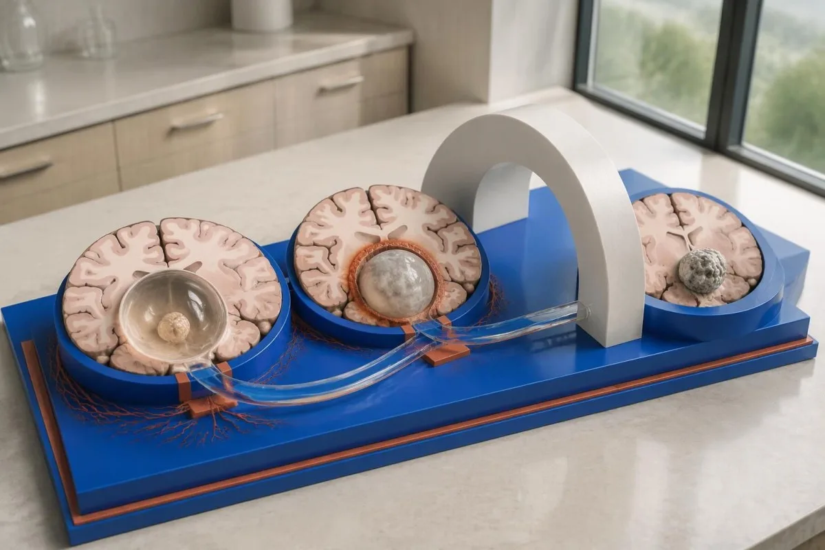
For selected patients with 1–2 viable parenchymal cysts, the IDSA/ASTMH guideline describes albendazole treatment for 10–14 days with appropriate accompanying care. This is a specialist-directed recommendation, not a home-treatment instruction: antiparasitic drugs can provoke an inflammatory response, and untreated hydrocephalus or diffuse cerebral edema changes the order of management (White et al., 2018).
WHO's 2021 guidance focuses on defined presentations of parenchymal neurocysticercosis, while the IDSA/ASTMH guideline also addresses ventricular, subarachnoid, and other forms in detail. Differences in imaging access and disease compartment explain why 1 treatment recommendation cannot be transferred to every patient with an antibody result; the guidelines do not turn negative serology into a rule-out test.
Corticosteroids may accompany antiparasitic therapy, but prolonged use requires additional safety planning, including assessment for relevant latent infections. Our Strongyloides and steroid safety guide explains a separate risk that matters before immune-suppressing treatment; an eye examination is also recommended before antiparasitic therapy to evaluate possible intraocular cysts.
Que deu susvelhar lo seguiment, en mai dels resultats dels anticòsses?
Follow-up should track seizures, neurological symptoms, imaging changes, and medication safety, rather than use antibody disappearance as proof of recovery. For viable parenchymal cysts, the IDSA/ASTMH guideline suggests repeat MRI at least every 6 months until the cystic component resolves, with earlier reassessment when symptoms change.
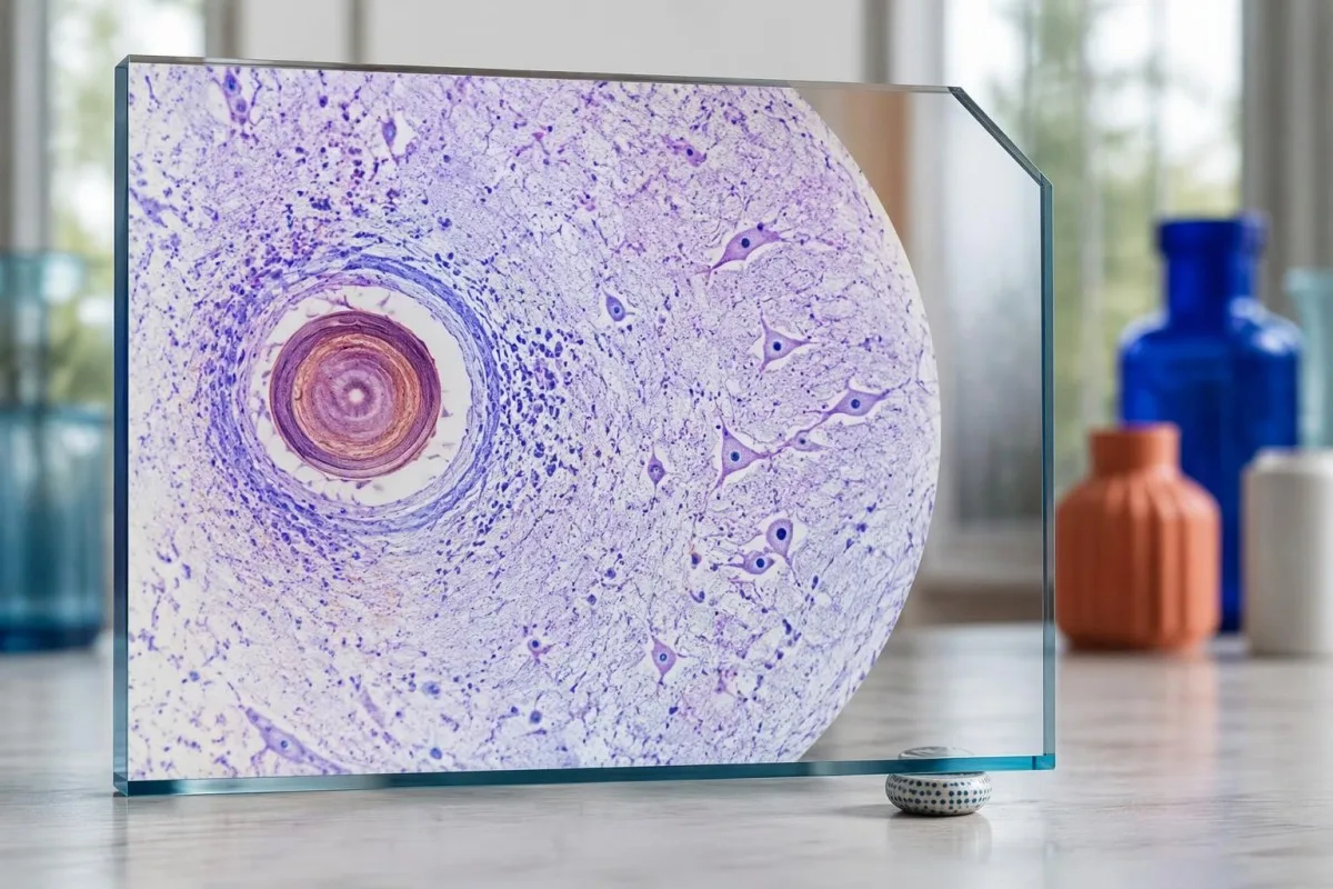
Antibodies may remain detectable after the original disease process has changed, and low-burden disease may never produce a positive result. If 1 new seizure occurs during follow-up, clinicians consider adherence, medication interactions, recurrent edema, and other causes; a rising or falling antibody index alone cannot distinguish among those explanations.
Kantesti AI can help explain reported laboratory results during medication follow-up, but a drug concentration is not a stand-alone measure of seizure control. Our l’interpretacion del taus de levetiracetam guide explains why sample timing and kidney function matter; 2 measurements collected at different times after dosing may not be directly comparable.
Monitoring depends on the chosen drug and treatment duration: prolonged albendazole therapy can require liver-test and blood-count surveillance, while some antiseizure medicines can affect sodium. Our guida sus la seguretat dels electròlits discusses those separate laboratory concerns; do not stop an antiseizure medicine or change its dose because 1 cysticercosis antibody result is negative.
Qué pòt explicar Kantesti, e qué la recèrca pòt pas provar?
AI report interpretation cannot confirm or exclude neurocysticercosis from laboratory results alone. Kantesti es un Outil d’analisi de sang amb IA whose role here is to organize reported laboratory findings; a negative result still needs clinical context, and 1 new seizure takes priority over reviewing an automated explanation.

Prepare a focused review packet with the original antibody report, assay method, CT or MRI report, symptom timeline, and medication list. These 5 information categories reduce the risk of discussing a result without its context; ask specifically whether the imaging suggests viable, degenerating, calcified, ventricular, or subarachnoid disease and which unanswered question the next test would address.
The 2 Kantesti publications listed below are a repository-deposited global analysis report and an educational RDW guide, not clinical evidence validating neurocysticercosis diagnosis. Our guida RDW e CBC acompanhant can support interpretation of a separately ordered blood count, but RDW, MCV, and MCHC do not establish whether a brain cyst is present.
Evidence for the neurological recommendations in this article comes from the cited diagnostic criteria and clinical guidelines, not from general AI benchmarking or laboratory-report volume. Our technical validation scope should be read within its stated boundaries; neither 1 successful report explanation nor a large aggregate dataset substitutes for condition-specific diagnostic validation.
Preguntes més freqüents
Es possible d’aver una neurocisticercòsi amb un test sanguin negatiu?
Òc, la neurocisticercòsi pòt èsser presenta malgrat un tèst sanguin d’anticòrs negatiu, subretot en preséncia d’un quist cerebral unic o d’una malautiá unicament calcificada. Las estimacions publicadas pels laboratòris de referéncia amb l’EITB indican sovent una sensibilitat d’aproximativament 50–60% per un quist unic, fòrça inferiora a sa performància amb mantun quist viable. La TAC, l’IRM, los simptòmas e l’istòria d’exposicion demòran doncas centrals per lo diagnostic. Una crisi epileptica novèla requerís una avaloracion urgenta, puslèu que de se rassolhar pel resultat negatiu.
Quin es lo tèst de neurocisticercòsi mai fisable?
Aucun test de neurocysticercose ne permet à lui seul de répondre à toutes les questions diagnostiques ; l’imagerie cérébrale et une sérologie choisie à bon escient sont complémentaires. L’EITB est la méthode de recherche d’anticorps de confirmation privilégiée dans les recommandations de l’IDSA/ASTMH, mais elle peut ne pas détecter une maladie ne touchant qu’un seul kyste. La TDM sans injection est particulièrement utile pour les calcifications, tandis que l’IRM permet de mieux évaluer de nombreux kystes viables, l’œdème et les anomalies des espaces contenant du liquide. Les cliniciens combinent ces résultats au lieu de considérer un résultat négatif comme définitif.
La TAC o l'IRM es melhor per la neurocisticercòsi?
La TC e l’IRM an d’avalatges diferents dins lo diagnostic de la neurocisticercòsi. La TC sens contrast detecta sovent mai clarament las pichonas calcificacions d’aproximativament 1–4 mm de diamètre, mentre que l’IRM avalora los quists viables, l’edèma perilesional e l’afectacion ventriculara o subaracnoïdiana. De seqüéncias d’IRM especializadas en 3D pòdon ajudar a identificar las membranas dels quists dins los espacis del liquid cefaloraquidian. La recomandacion de l’IDSA/ASTMH preconiza los dos metòdes d’imatge per classificar una malautiá recentament diagnosticada.
Un quist calcificat pòt encara provocar de convulsions?
Un residu de cisticercòsi calcificat pòt contunhar d’èsser associat a de convulsions, quitament se non es pus un quist viable. D’unes fòcus calcificats, sovent d’1–4 mm solament, desvolopan un edèma periferic intermittent que pòt èsser visible a l’IRM. D’anticòrs negatius excluisson pas aqueste mecanisme, e la persisténcia d’anticòrs positius mesura pas lo risc de convulsions. La malautiá unicament calcificada es generalament tractada en foncion dels simptòmas, puslèu qu’amb de medicaments antiparasitaris.
Deuri repetir un tèst d’anticòrs negatiu per la cisticercòsi?
Repetir un tèst d’anticòsses contra la cisticercòsi negatiu es pas automaticament la melhora etapa seguenta. Amb un cist unic de pichona talha o una malautiá calcificada, una responsa d’anticòsses febla pòt demorar negativa quitament se lo meteis assag es repetit aprèp 2 setmanas. Un professional de santat pòt preferir tornar examinar lo metòde original, demandar un assag de confirmacion mai ben caracterizat o donar la prioritat a la TAC e a l’IRM. De novèlas crisis epilepticas o una agravacion dels simptòmas neurologics devon pas esperar que lo tèst siá repetit.
Quand devraiái demandar d’ajuda d’urgéncia per una primièra crisi epilèptica ?
Una primièra crisi coneguda necessita una avaloracion medicala d’urgéncia e cal contactar los servicis d’emergéncia locals, quitament se lo tèst d’anticòsses contra la neurocisticercòsi es negatiu. Una crisi convulsiva que dura 5 minutas, de crises repetidas sens recuperar la consciéncia, una dificultat respiratòria, una grossessa o una nafradura importanta constituïsson una urgéncia. Immobilizetz pas la persona e li metetz pas res dins la boca; un còp las convulsions acabadas, plaçatz-la sul costat se respira normalament. Conduisètz pas fins a çò qu’un professional de santat aja avalorat l’episòdi e explicat las restriccions aplicablas.
Fasètz-vos explicar los resultats analisi de sang
Telecargatz vòstre compte rendut de laboratòri e l’IA de Kantesti explica cada biomarcador en lenga simpla en qualques segondas. Utilizat per mai de 2 milions de personas dins lo mond.
Referéncias
- Klein, T., Mitchell, S., & Weber, H. (2026). Analizator d’analisis de sang amb IA: 2,5M d’analisis analisats | Rapòrt de salut globala 2026. Recèrca medicala per IA de Kantesti. DOI
- Klein, T., Mitchell, S., & Weber, H. (2026). Analisi de sang RDW: guida completa de RDW-CV, MCV e MCHC. Recèrca medicala per IA de Kantesti. DOI
- White AC Jr et al. (2018). Diagnosis and Treatment of Neurocysticercosis: 2017 Clinical Practice Guidelines by the Infectious Diseases Society of America (IDSA) and the American Society of Tropical Medicine and Hygiene (ASTMH). Clinical Infectious Diseases. DOI
- Del Brutto OH et al. (2017). Revised diagnostic criteria for neurocysticercosis. Journal of the Neurological Sciences. DOI
- World Health Organization (2021). WHO guidelines on management of Taenia solium neurocysticercosis. Organizacion Mondiala de la Santat.
- 2M+Tèsts analisats
- 127+Païses
- 100+Lengas
A prepaus de l’equipa medicala


Senhals de confiança E-E-A-T
Experiéncia
Revisión clinica menada pel metge de las practicas d’interpretacion de las analisis.
Expertisa
Fòcus sus la medicina de laboratòri sus cossí los biomarcadors se comportan dins un contèxte clinic.
Autoritat
Revisat medicalament pel Dr Thomas Klein, amb de contribucions de la Dr Sarah Mitchell e del Prof. Dr Hans Weber.
Fisança
Interpretacion basada sus d’evidéncias amb de camins de seguiment clars per reduzir l’alarmisme.





Avís medical
Aqueste article es solament per tòcas educatius e constituís pas de conselh medical. Consultatz totjorn un professional de la sant qualificat per las decisions de diagnostica e de tractament.
Publicat · Contactar l’equipa editoriala
Publicat per Kantesti Ltd, enregistrada en Anglatèrra e País de Galas, n° d’entrepresa. 17090423, Londres, Reialme Unit.
