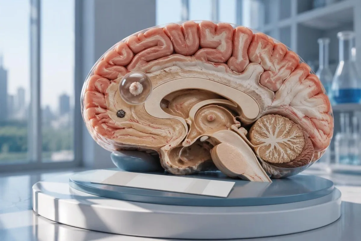
Отрицателният тест за невроцистицеркоза не изключва единична мозъчна киста или калцифицирало заболяване. КТ, ЯМР и спешната оценка на новопоявили се гърчове са по-важни от успокоението, което дава само изследването за антитела.

Основни изводи
- Отрицателни антитела не изключват невроцистицеркоза, особено когато образното изследване показва само 1 засегната област или калцификати.
- EITB в референтна лаборатория може да достигне чувствителност от 98% при повече от 1 жизнеспособна киста, но ефективността е значително по-ниска при единична киста.
- Чувствителност при единична киста често е приблизително 50–60% в публикуваните оценки за EITB; тези стойности не важат за всеки търговски тест за антитела.
- КТ без контраст е особено полезна за малки калцификати, които често са с размер приблизително 1–4 mm.
- ЯМР оценява жизнеспособните кисти, околния оток и вентрикуларното или субарахноидалното засягане; специализирани триизмерни последователности могат да подобрят откриването.
- Първи гърчове изискват спешна медицинска оценка независимо от резултата за антитела. Обадете се на спешна помощ при първи известен гърч или други спешни признаци.
- Пет минути е праг за спешност при продължаващ конвулсивен гърч, а не причина да се изчаква, преди да се потърси помощ.
- Заболяване само с калцификати обикновено не изисква антипаразитно лечение, но може да са необходими лечение на гърчовете и допълнителни образни изследвания.
Може ли отрицателният тест за невроцистицеркоза да я изключи?
A отрицателният тест за невроцистицеркоза не може надеждно да изключи заболяването, особено при 1 мозъчна киста или калцирани остатъци. Изследването за антитела измерва имунния отговор, вместо директно да изследва мозъка; КТ и ЯМР установяват местоположението и стадия на подозрителните находки, а първият гърч изисква спешна оценка.

Резултатът е условен, а не абсолютен. Тестът за антитела срещу цистицеркоза, който се представя добре при хора с няколко жизнеспособни кисти, може да се представи зле при човек само с 1 малка засегната област, така че думата отрицателен означава, че с този тест не се открива отговор — не че в нервната система няма кисти.
Като хипотетичен пример нека разгледаме 34-годишен човек с първи гърч, отрицателен резултат за антитела и единична находка с пръстеновидно контрастно усилване на ЯМР. Резултатът не може да определи дали находката представлява цистицеркоза, друга инфекция или неинфекциозен процес; значението ѝ зависи от образното изследване, анамнезата за експозиция и неврологичната оценка, разгледани заедно.
Кантести е един AI кръвен анализатор която може да помогне за обяснението на съобщени лабораторни находки, но не може да изключи мозъчно заболяване въз основа на 1 отрицателен резултат за антитела. Нашите организация и мисия обясняват фокуса ни върху лабораторната информация; читателите не бива да приемат автоматизираното обяснение на резултат като основание да отложат лечението на гърч.
Защо изследването за антитела може да не открие единична мозъчна киста?
A единична киста може да предизвика отговор на антителата под прага на откриване на теста. Публикуваните оценки на референтните лаборатории за EITB често поставят чувствителността около 50–60% при единична киста, спрямо приблизително 98% при наличие на повече от 1 жизнеспособна киста; разликата отразява тежестта на заболяването и ефективността на теста.
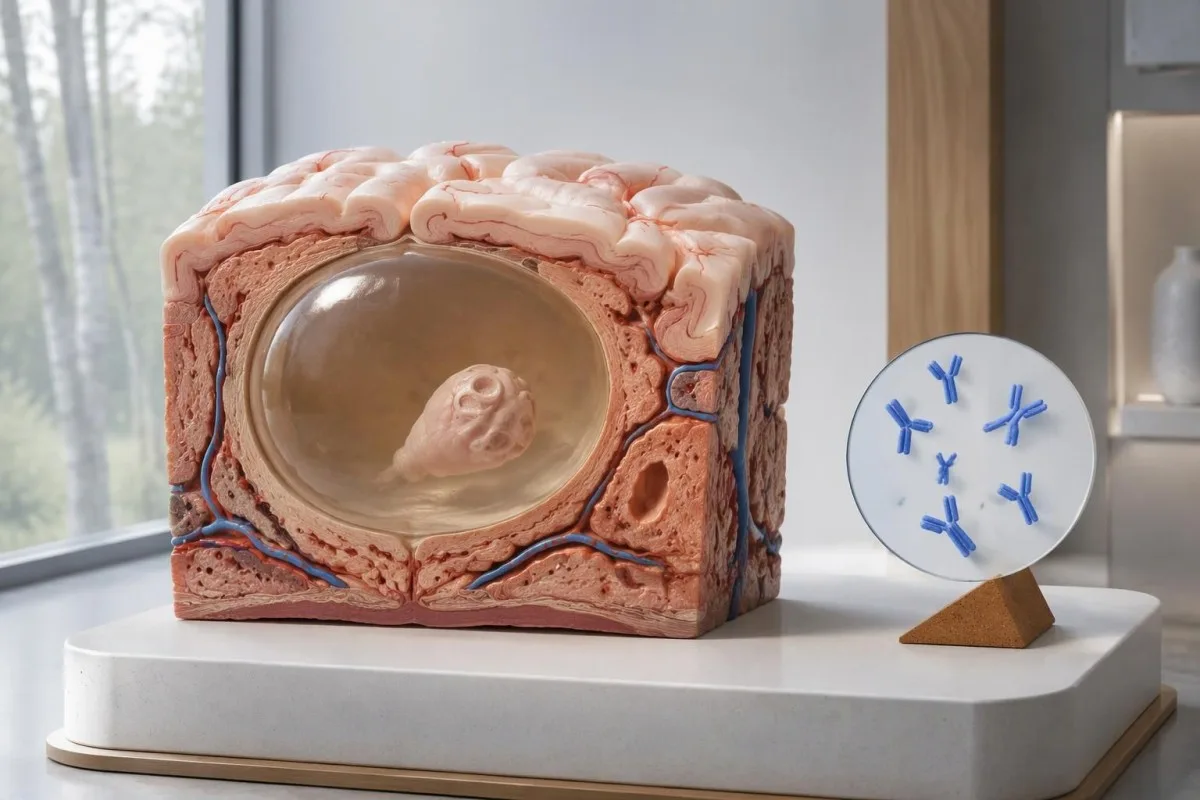
Насоките на IDSA/ASTMH препоръчват ензимно-свързан имуноелектротрансферен блот, или EITB, като потвърдителна серология и изрично предупреждават, че чувствителността е ниска при единична паренхимна находка или само при калцификати (White et al., 2018). Не бива да се пренася върху човек с 1 малка мозъчна аномалия високата чувствителност, докладвана при пациенти с множество кисти, вентрикуларно заболяване или субарахноидно заболяване.
Представете си, само за илюстрация, че даден тест има чувствителност 55% при 100 души, при които заболяване с единична киста вече е било установено по независим начин. Приблизително 45 биха получили отрицателен резултат въпреки наличието на заболяването; този работен пример обяснява пропуснатото откриване, а не вероятността конкретен отрицателен резултат да означава заболяване.
Повтарянето на същия тест след 2 седмици може да не коригира слаб отговор, причинен от ниска тежест на заболяването. Това се различава от инфекциите, при които основният проблем е ранното вземане на пробата, както е обяснено в нашето ръководство за фалшиво отрицателни резултати, свързани с времето на изследването; попитайте дали образното изследване или по-добре характеризиран тест биха променили следващото решение.
Защо калцифицираната невроцистицеркоза може да остане симптоматична?
Калцираната невроцистицеркоза може да причини гърчове въпреки отрицателните резултати за антитела, because the remaining mineralized focus can still be associated with local irritation and episodic surrounding edema. Typical calcifications may measure approximately 1–4 mm, but their small size does not establish whether they are clinically harmless.
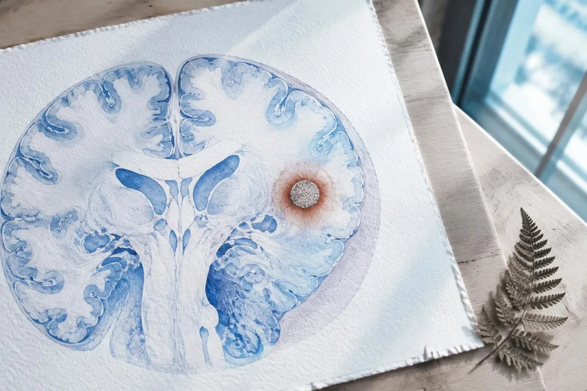
A calcification is generally a remnant rather than a living cyst that antiparasitic medication can eliminate. In the IDSA/ASTMH guideline, calcified-only parenchymal disease is managed symptomatically rather than with antiparasitic drugs; a person with 1 calcified focus and seizures may therefore need antiseizure treatment without needing cyst-killing medication (White et al., 2018).
Surrounding edema can appear intermittently, sometimes long after the original cyst has degenerated. MRI may show this tissue response even when the calcification is more obvious on CT, which is why the 2 scans can tell different but compatible stories rather than one proving that the other is wrong.
Neither a negative antibody result nor a persistent positive result measures seizure risk by itself. The broader lesson resembles other seronegative neurological conditions, discussed in our negative neurological antibody results guide: absence of detectable antibodies is not equivalent to absence of a clinically relevant neurological process.
Кой тест за антитела срещу цистицеркоза е бил извършен в действителност?
The useful first question is whether the report describes EITB, ELISA, or another antibody method, because these are not interchangeable tests. A negative index below 1 laboratory's cutoff cannot inherit the performance figures of a different reference-laboratory assay, even when both reports use the name cysticercosis IgG.
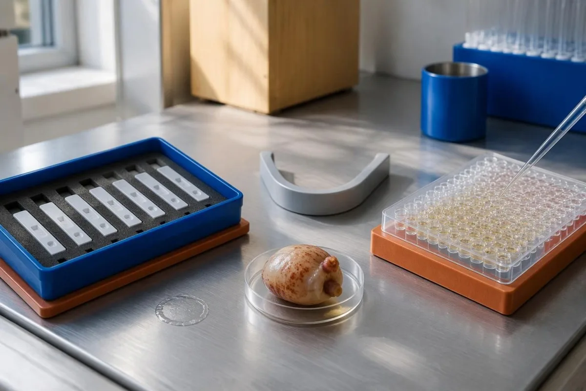
EITB using purified parasite glycoproteins is the preferred serological method in the IDSA/ASTMH guideline, while crude-antigen ELISA methods have recognized sensitivity and specificity limitations (White et al., 2018). Before comparing 2 results, check the method, specimen type, reference interval, and whether the laboratory used a validated confirmatory procedure.
Some laboratories report a qualitative result; others provide an optical-density ratio or numerical index. A value such as 0.8 in a hypothetical assay does not represent 0.8 cysts, an 80% disease probability, or a universal severity score—our качествени и количествени резултати guide explains that distinction.
Kantesti AI's role is to help readers preserve the laboratory's original wording rather than reinterpret 1 method through another method's thresholds. Our наръчник за лабораторни маркери provides broader report-reading context; for this result, keep the actual assay name available when discussing referral or repeat testing.
Как анамнезата за излагане променя диагностичното подозрение?
Neurocysticercosis follows ingestion of Taenia solium eggs, usually through fecal contamination from a person carrying the intestinal tapeworm. Eating undercooked pork can cause intestinal taeniasis, but brain cysts arise through a different transmission step; symptoms may emerge years after exposure rather than within 1 recent trip.

A vegetarian can develop cysticercosis because eggs can contaminate food or hands independently of meat consumption. Useful history includes past residence, household contacts, sanitation conditions, and possible exposure to a tapeworm carrier; asking only about meals eaten during the last 2 weeks can miss the relevant background.
A negative stool examination does not exclude cysts in the brain. Stool testing addresses intestinal tapeworm carriage, whereas CT and MRI assess nervous-system findings; these are 2 different sites and clinical questions, although public-health teams may recommend evaluating contacts when a carrier could be involved.
Normal eosinophils also cannot exclude neurocysticercosis, and raised eosinophils do not identify the brain as the affected site. Our parasite antibody interpretation guide discusses a separate parasite with similar localization limits; the practical point is to avoid using 1 routine cell count as a substitute for targeted neurological evaluation.
Кога новопоявилите се гърчове налагат спешна оценка?
A first known seizure needs urgent medical assessment regardless of antibody results, and emergency services should be contacted for a first seizure or an ongoing seizure with concerning features. A convulsive seizure lasting 5 minutes, repeated seizures without recovery, breathing difficulty, pregnancy, or significant injury requires emergency care.

Do not wait for the 5-minute mark to seek help after a first known seizure. Keep the person away from hazards, cushion the head if safely possible, do not restrain them or put anything in their mouth, and place them on their side once convulsions stop if they are breathing normally.
Persistent confusion, a severe new headache, fever with neck stiffness, or new weakness increases concern for causes beyond cysticercosis. Sudden one-sided weakness or speech difficulty also demands emergency assessment, as our предупредителните признаци на инсулт explains; 1 negative infection test cannot rule out stroke, meningitis, or a structural brain problem.
After a suspected seizure, stop driving and avoid swimming alone, heights, or operating machinery until a clinician provides advice consistent with local rules. If a witness can safely record the duration and describe movements or recovery, those observations may be more useful than ordering a second antibody test before the first neurological assessment.
Какво допринася КТ за диагностицирането на невроцистицеркоза?
Noncontrast CT is particularly useful for detecting calcified cyst remnants, including small approximately 1–4 mm foci that MRI may show less clearly. CT also helps assess urgent structural problems such as hydrocephalus, but 1 isolated calcification is not automatically proof of neurocysticercosis or the cause of a seizure.
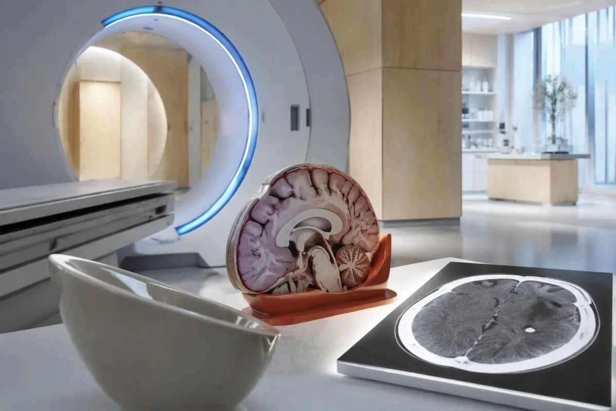
Calcium appears dense on CT, allowing radiologists to identify mineralized remnants and describe their distribution. The IDSA/ASTMH guideline recommends both MRI and noncontrast CT when classifying newly diagnosed neurocysticercosis, because the 2 modalities have complementary strengths rather than being interchangeable alternatives (White et al., 2018).
An incidental calcification needs context: previous granulomatous disease, other causes of mineralization, and scan appearance can influence interpretation. If 1 calcification sits near a suspected seizure focus, concordance with symptoms and EEG may strengthen the explanation, but anatomical proximity alone cannot establish causation.
A laboratory marker used in selected head-injury pathways does not test for brain cysts or replace imaging after an unexplained seizure. Our head-injury CT decisions guide illustrates that narrower setting; ask the clinician whether the present question requires noncontrast CT, contrast imaging, MRI, or a combination of 2 modalities.
Какво разкрива ЯМР, което КТ може да пропусне?
MRI is better suited to evaluating viable cysts, surrounding edema, and many ventricular or subarachnoid findings. Specialized 3-dimensional sequences such as CISS or FIESTA can improve visualization of cyst membranes within cerebrospinal fluid spaces, where a cyst may otherwise resemble the surrounding fluid.
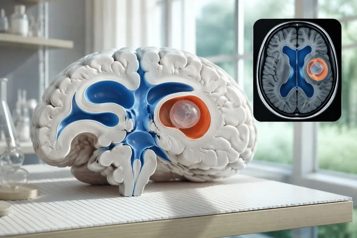
A cyst inside the ventricles can obstruct cerebrospinal fluid flow and cause hydrocephalus even without numerous parenchymal cysts. Headache with vomiting, declining alertness, or new visual symptoms therefore changes urgency; 1 reassuring antibody result should not override symptoms that suggest raised pressure or impaired fluid circulation.
MRI may show a scolex, the small internal structure of the parasite, inside a compatible cyst. This finding can be highly specific when confidently identified, whereas ring enhancement alone has a broader differential; our brain imaging warning signs guide explains why structural abnormalities require interpretation rather than diagnosis from 1 visual feature.
A routine MRI protocol may not answer every cysticercosis question. If ventricular or subarachnoid disease is suspected, discuss whether thin-section 3-dimensional imaging is needed; radiologists choose sequences and contrast according to the clinical question, and a normal scan using 1 protocol does not prove every possible disease compartment has been adequately assessed.
Как се диагностицира невроцистицеркоза, когато резултатите от изследванията си противоречат?
Neurocysticercosis diagnosis combines imaging, clinical findings, exposure information, and supporting tests, rather than requiring a positive antibody result in every case. The revised diagnostic criteria published in 2017 give characteristic neuroimaging findings a central role and distinguish strong evidence from less-specific supportive features (Del Brutto et al., 2017).
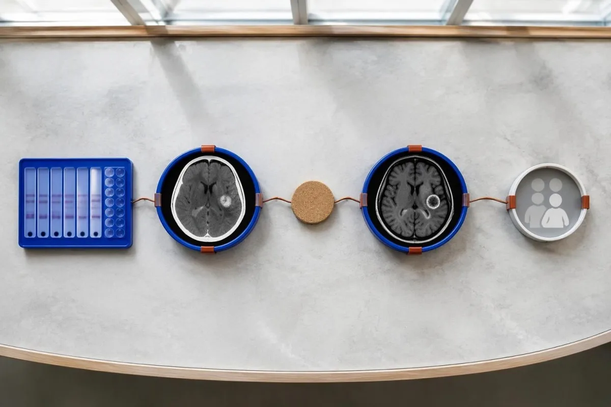
Evidence should be weighted, not tallied. A confidently visualized scolex inside a compatible cyst is different from 1 nonspecific ring-enhancing area, while a positive antibody result is different from direct evidence that a finding is in the brain; our ръководство за положителни резултати от антитела explains why detection alone cannot establish location.
For a clearly hypothetical probability example, start with a 30% pretest probability and assume a single-cyst assay has 55% sensitivity and 99% specificity. In 1,000 comparable people, about 135 affected and 693 unaffected people would test negative, leaving approximately a 16% probability of disease among negative results—still substantial, although these assumed figures are not a personal risk calculator.
The most useful disagreement to investigate is whether each test is answering the same question. A negative antibody test, a CT calcification, and MRI edema can all be compatible with 1 calcified disease process; unexplained discrepancies should prompt review of the assay and images rather than automatic dismissal of either result.
Какво друго може да наподобява единична киста при образно изследване на мозъка?
A single ring-enhancing brain finding is not specific to neurocysticercosis. Other possibilities include tuberculoma, an abscess, a neoplasm, or an inflammatory process; clinicians use appearance, location, immune status, symptoms, and sometimes follow-up imaging or tissue examination to distinguish these possibilities rather than relying on 1 antibody panel.
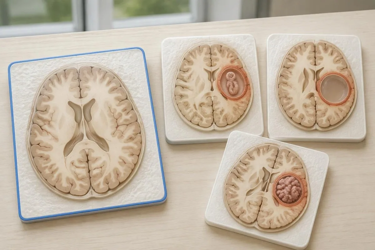
Edema, restricted diffusion, internal structures, and the number of findings all contribute to the differential, but no single nonspecific feature settles every case. Fever and immune suppression may change the suspected cause; a 2 cm ring-enhancing area in a hypothetical immunocompromised person warrants a different investigation from a tiny calcification in an otherwise well person.
Toxoplasmosis is one consideration in selected immunocompromised patients, and a positive IgG result can indicate past exposure rather than prove an active brain process. Our Toxoplasma IgG interpretation guide explains that laboratory distinction; imaging and the patient's immune status determine whether the result is clinically relevant to 1 or more cerebral findings.
New seizures accompanied by rapidly evolving memory problems, psychiatric changes, or abnormal movements can also suggest encephalitis rather than a solitary parasite cyst. Our encephalitis diagnostic pathway describes how MRI, EEG, and cerebrospinal fluid testing answer different questions; a second negative cysticercosis antibody result does not investigate those alternatives.
Могат ли изследването за антиген, PCR или ликворът да разрешат неяснотата?
Antigen tests, PCR, and cerebrospinal fluid analysis can add information in selected cases, particularly when extraparenchymal disease is suspected. None is a universal rescue test after 1 negative antibody result: availability, assay validation, disease compartment, and safety of lumbar puncture determine whether additional testing is appropriate.
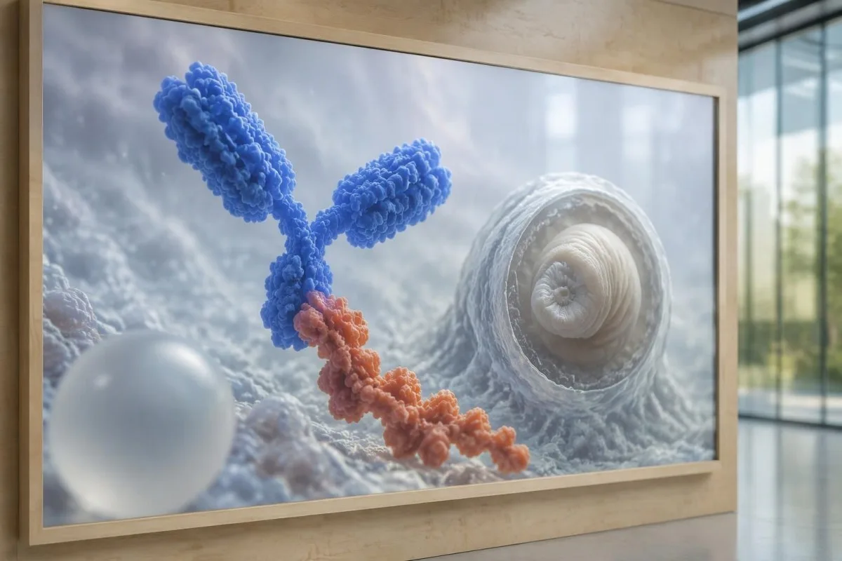
Antibody testing detects the host response, while antigen testing aims to detect parasite-derived material and may better reflect viable disease burden. Antigen detection is often less useful when only 1 small parenchymal cyst or calcifications are present; molecular testing is used in some specialist settings, but performance cannot be generalized across every laboratory or specimen.
Lumbar puncture is not routinely required for an uncomplicated solitary parenchymal finding and can be unsafe when obstructive hydrocephalus or significant mass effect is present. Cerebrospinal fluid tests for other disorders also require disease-specific interpretation, as our CSF band interpretation guide explains; 1 abnormal CSF marker does not identify every possible cause.
Кантести е един Платформа за кръвни изследвания тълкуване с AI, not a service that orders lumbar punctures, identifies cysts on scans, or confirms neurological infections. Our AI технология за интерпретация explains the laboratory-report workflow; when 2 tests disagree, our role is to clarify their stated meanings and limitations, not select an invasive diagnostic procedure.
Защо лечението зависи от стадия и местоположението на кистата?
Treatment depends on whether cysts are viable, degenerating, calcified, or located outside the brain tissue, not simply on antibody positivity. Selected viable parenchymal cases receive antiparasitic treatment, whereas calcified-only disease generally receives symptom-directed care; ventricular obstruction may require a procedural approach rather than 1 standard medication regimen.
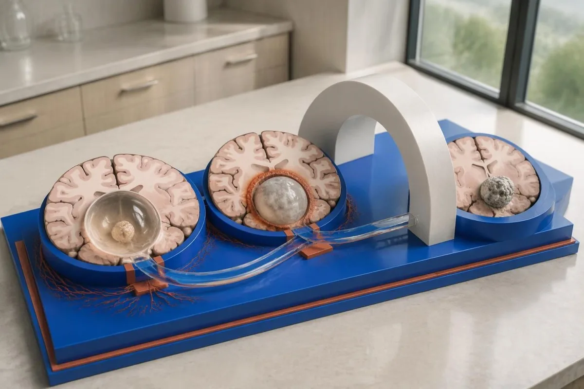
For selected patients with 1–2 viable parenchymal cysts, the IDSA/ASTMH guideline describes albendazole treatment for 10–14 days with appropriate accompanying care. This is a specialist-directed recommendation, not a home-treatment instruction: antiparasitic drugs can provoke an inflammatory response, and untreated hydrocephalus or diffuse cerebral edema changes the order of management (White et al., 2018).
WHO's 2021 guidance focuses on defined presentations of parenchymal neurocysticercosis, while the IDSA/ASTMH guideline also addresses ventricular, subarachnoid, and other forms in detail. Differences in imaging access and disease compartment explain why 1 treatment recommendation cannot be transferred to every patient with an antibody result; the guidelines do not turn negative serology into a rule-out test.
Corticosteroids may accompany antiparasitic therapy, but prolonged use requires additional safety planning, including assessment for relevant latent infections. Our Strongyloides and steroid safety guide explains a separate risk that matters before immune-suppressing treatment; an eye examination is also recommended before antiparasitic therapy to evaluate possible intraocular cysts.
Какво трябва да се проследява при контролни прегледи извън резултатите за антитела?
Follow-up should track seizures, neurological symptoms, imaging changes, and medication safety, rather than use antibody disappearance as proof of recovery. For viable parenchymal cysts, the IDSA/ASTMH guideline suggests repeat MRI at least every 6 months until the cystic component resolves, with earlier reassessment when symptoms change.

Antibodies may remain detectable after the original disease process has changed, and low-burden disease may never produce a positive result. If 1 new seizure occurs during follow-up, clinicians consider adherence, medication interactions, recurrent edema, and other causes; a rising or falling antibody index alone cannot distinguish among those explanations.
Kantesti AI can help explain reported laboratory results during medication follow-up, but a drug concentration is not a stand-alone measure of seizure control. Our интерпретацията на нивото на леветирацетам guide explains why sample timing and kidney function matter; 2 measurements collected at different times after dosing may not be directly comparable.
Monitoring depends on the chosen drug and treatment duration: prolonged albendazole therapy can require liver-test and blood-count surveillance, while some antiseizure medicines can affect sodium. Our ръководство за безопасност на електролитите discusses those separate laboratory concerns; do not stop an antiseizure medicine or change its dose because 1 cysticercosis antibody result is negative.
Какво може да обясни Kantesti и какво не могат да докажат изследванията?
AI report interpretation cannot confirm or exclude neurocysticercosis from laboratory results alone. Kantesti е Инструмент за анализ на кръвни изследвания с AI whose role here is to organize reported laboratory findings; a negative result still needs clinical context, and 1 new seizure takes priority over reviewing an automated explanation.

Prepare a focused review packet with the original antibody report, assay method, CT or MRI report, symptom timeline, and medication list. These 5 information categories reduce the risk of discussing a result without its context; ask specifically whether the imaging suggests viable, degenerating, calcified, ventricular, or subarachnoid disease and which unanswered question the next test would address.
The 2 Kantesti publications listed below are a repository-deposited global analysis report and an educational RDW guide, not clinical evidence validating neurocysticercosis diagnosis. Our RDW и CBC ръководство can support interpretation of a separately ordered blood count, but RDW, MCV, and MCHC do not establish whether a brain cyst is present.
Evidence for the neurological recommendations in this article comes from the cited diagnostic criteria and clinical guidelines, not from general AI benchmarking or laboratory-report volume. Our technical validation scope should be read within its stated boundaries; neither 1 successful report explanation nor a large aggregate dataset substitutes for condition-specific diagnostic validation.
Често задавани въпроси
Възможно ли е да имате невроцистицеркоза при отрицателен кръвен тест?
Да, невроцистицеркозата може да е налице въпреки отрицателния кръвен тест за антитела, особено при 1 мозъчна киста или само калцифицирани лезии. Публикуваните оценки на референтни лаборатории за EITB често показват приблизително 50–60% чувствителност при единична киста — значително по-ниска от тази при множество жизнеспособни кисти. Следователно КТ, ЯМР, симптомите и данните за излагане на заразата остават ключови за диагнозата. При новопоявил се гърч е необходима спешна оценка, а не успокоение въз основа на отрицателния резултат.
Кой е най-надеждният тест за невроцистицеркоза?
Нито един тест за невроцистицеркоза не отговаря на всички диагностични въпроси; образната диагностика на мозъка и подходящо подбраните серологични изследвания се допълват взаимно. EITB е предпочитаният метод за потвърждаване чрез откриване на антитела съгласно насоките на IDSA/ASTMH, но може да не открие заболяване, при което има само 1 киста. КТ без контраст е особено полезна за установяване на калцификати, докато ЯМР позволява по-добра оценка на много жизнеспособни кисти, оток и аномалии в ликворните пространства. Лекарите съпоставят тези находки, вместо да приемат един отрицателен резултат за окончателен.
КТ или ЯМР е по-добро при невроцистицеркоза?
КТ и ЯМР имат различни предимства при диагностицирането на невроцистицеркоза. КТ без контраст често визуализира по-ясно малки калцификати с размер приблизително 1–4 mm, докато ЯМР оценява жизнеспособните кисти, околния оток и засягането на вентрикулите или субарахноидалното пространство. Специализирани 3-мерни ЯМР последователности могат да помогнат за откриването на кистозни мембрани в ликворните пространства. Ръководството на IDSA/ASTMH препоръчва използването и на двата метода при класифициране на новодиагностицирано заболяване.
Може ли калцифицирана киста все още да причинява гърчове?
Калцифициран остатък от цистицеркоза може да продължи да бъде свързан с гърчове, въпреки че вече не представлява жизнеспособна киста. Около някои калцифицирани огнища, често с размер само 1–4 mm, периодично се развива оток, който може да се вижда при ЯМР. Отрицателните антитела не изключват този механизъм, а трайно положителните антитела не са показател за риска от гърчове. При заболяване само с калцификати обикновено се прилага симптоматично лечение, а не противопаразитни лекарства.
Трябва ли да повторя отрицателен тест за антитела срещу цистицеркоза?
Повтарянето на отрицателен тест за антитела срещу цистицеркоза не е автоматично най-добрата следваща стъпка. При 1 малка киста или калцифицирано заболяване слабият антитялов отговор може да остане отрицателен дори ако същият тест се повтори след 2 седмици. Вместо това лекарят може да прегледа първоначалния метод, да назначи по-добре охарактеризиран потвърдителен тест или да даде приоритет на КТ и ЯМР. При новопоявили се гърчове или влошаващи се неврологични симптоми не бива да се чака повторното изследване.
Кога трябва да потърся спешна помощ при първи гърч?
Първи известен гърч налага спешна медицинска оценка и обаждане на местните спешни служби, независимо от отрицателен тест за антитела срещу невроцистицеркоза. Гърч с конвулсии, продължаващ 5 минути, повтарящи се гърчове без възстановяване на съзнанието, затруднено дишане, бременност или значителна травма са спешен случай. Не възпирайте движенията на човека и не поставяйте нищо в устата му; след като конвулсиите спрат, го обърнете настрани, ако диша нормално. Не шофирайте, докато лекар не оцени случилото се и не обясни приложимите ограничения.
Получете обяснение на резултатите от кръвните си изследвания
Качете лабораторния си доклад и AI на Kantesti ще обясни всеки биомаркер на разбираем език за секунди. Използва се от над 2 милиона души по света.
Референции
- Klein, T., Mitchell, S., & Weber, H. (2026). AI анализатор на кръвни изследвания: 2,5M изследвания анализирани | Global Health Report 2026. Медицински изследвания с Kantesti AI. ДОИ
- Klein, T., Mitchell, S., & Weber, H. (2026). Кръвен тест RDW: Пълно ръководство за RDW-CV, MCV и MCHC. Медицински изследвания с Kantesti AI. ДОИ
- White AC Jr et al. (2018). Diagnosis and Treatment of Neurocysticercosis: 2017 Clinical Practice Guidelines by the Infectious Diseases Society of America (IDSA) and the American Society of Tropical Medicine and Hygiene (ASTMH). Clinical Infectious Diseases. ДОИ
- Del Brutto OH et al. (2017). Revised diagnostic criteria for neurocysticercosis. Journal of the Neurological Sciences. ДОИ
- World Health Organization (2021). WHO guidelines on management of Taenia solium neurocysticercosis. Световна здравна организация.
- 2 милиона+Анализирани тестове
- 127+Държави
- 100+Езици
За медицинския екип


Сигнали за доверие E-E-A-T
Опит
Медицински преглед, воден от лекар, на работните процеси за интерпретация на лабораторни резултати.
Експертиза
Фокус в лабораторната медицина върху това как се държат биомаркерите в клиничен контекст.
Авторитетност
Медицинска рецензия от д-р Thomas Klein, с принос от д-р Sarah Mitchell и проф. д-р Hans Weber.
Надеждност
Интерпретация, основана на доказателства, с ясни последващи стъпки за намаляване на тревогата.





Медицински отказ от отговорност
Тази статия е само с образователна цел и не представлява медицински съвет. Винаги се консултирайте с квалифициран медицински специалист за решения относно диагностика и лечение.
Публикувано · Свържете се с редакционния екип
Издава се от Kantesti Ltd, регистрирана в Англия и Уелс, дружество №. 17090423, Лондон, Обединено кралство.
