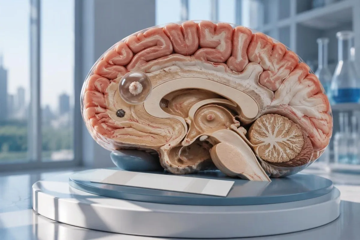
神経嚢虫症検査が陰性でも、脳内の単発嚢胞や石灰化病変は否定できません。新たな発作では、抗体検査だけによる安心材料よりも、CT、MRI、緊急の評価が重要です。

要点
- 抗体陰性 神経嚢虫症を否定するものではありません。特に、画像検査で病変がある領域が1つだけ、または石灰化が見られる場合はそうです。
- 参照検査機関でのEITB 生存嚢胞が1個を超える場合は、感度が98%近くに達することがありますが、嚢胞が1個だけの場合は性能が大幅に低下します。
- 単発嚢胞に対する感度 公表されているEITBの推定値では、およそ50~60%であることが多いものの、これらの数値はすべての市販抗体検査に当てはまるわけではありません。
- 非造影CT 小さな石灰化の検出に特に有用で、その大きさは多くの場合、約1~4 mmです。
- MRI 生存嚢胞、周囲の浮腫、脳室内病変またはくも膜下病変を評価します。特殊な3次元シーケンスにより、検出率が向上することがあります。
- 初回の発作 抗体検査の結果にかかわらず、緊急の医療評価が必要です。初めての発作、またはその他の緊急徴候がある場合は、救急サービスに連絡してください。
- 5分間 は、続いているけいれん発作に対する緊急対応の基準であり、助けを求める前に待つべき時間ではありません。
- 石灰化のみの病変 通常、抗寄生虫薬は必要ありませんが、発作の治療や追加の画像検査が必要となる場合があります。
神経嚢虫症検査が陰性なら、否定できますか?
A 神経嚢虫症検査が陰性でも、神経嚢虫症を確実に否定することはできません, 特に、脳嚢胞が1個、または石灰化した遺残病変がある場合はそうです。抗体検査で測定するのは免疫反応であり、脳を直接調べるわけではありません。CTとMRIで疑わしい所見の位置と病期を確認します。また、初めての発作には緊急の評価が必要です。

結果は条件付きであり、絶対的なものではありません。複数の生存嚢胞がある人で良好な性能を示す嚢虫症抗体検査でも、病変が小さなもの1つだけの人では性能が低い場合があります。したがって、 陰性 とは、その検査法で検出できる反応がないという意味であり、神経系のどこにも嚢胞がないという意味ではありません。
仮の例として、初めての発作があり、抗体検査報告が陰性で、MRIで輪状造影効果を示す単発所見が認められた34歳の人を考えてみましょう。その所見が嚢虫症、別の感染症、または非感染性の病態によるものかは、報告だけでは判断できません。その意味は、画像検査、曝露歴、神経学的評価を総合して判断します。
カンテスティは AI血液検査分析装置 報告された検査所見の解釈に役立ちますが、抗体検査が1回陰性だったというだけで脳疾患を否定することはできません。当 組織と使命 は、当組織が検査情報に重点を置いていることを説明しています。自動生成された報告の解説を根拠に、発作への対応を遅らせてよいと考えないでください。
抗体検査では、脳内の単発嚢胞が見逃されることがあるのはなぜですか?
A 単発の嚢胞では、検査法の検出限界を下回る抗体反応しか生じないことがある。公表されている基準検査施設のEITB感度の推定値では、嚢胞が1個の場合は約50~60%、生存嚢胞が1個を超える場合は約98%とされることがよくあります。この差は、疾患量と検査法の性能を反映しています。
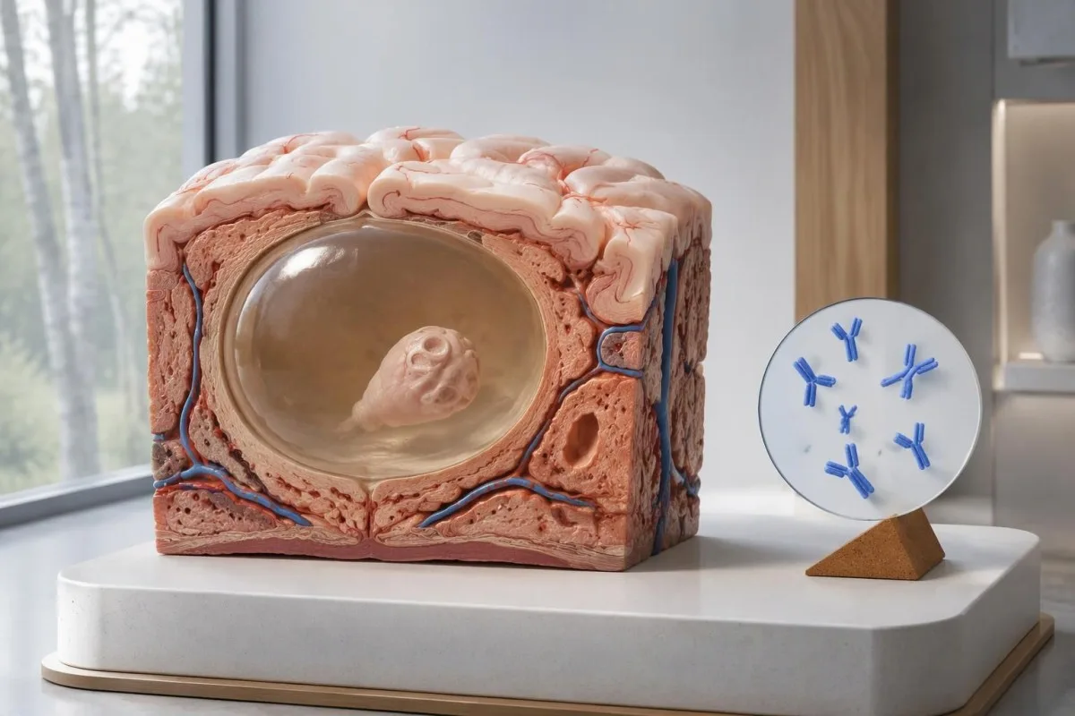
IDSA/ASTMHガイドラインは 酵素結合免疫電気転写ブロット法, (EITB)を確認検査として推奨し、実質内の病変が1つだけの場合や石灰化のみの場合は感度が低いと明確に警告しています(White et al., 2018)。嚢胞が複数ある患者、脳室病変のある患者、またはくも膜下病変のある患者で報告された高い感度を、小さな脳異常が1つある人に当てはめてはいけません。
あくまで説明のために、すでに単発嚢胞性疾患が別の方法で確認された100人を対象とする検査法の感度が55%だと仮定してください。約45人は疾患があるにもかかわらず陰性になるでしょう。この例は見逃しの仕組みを示すものであり、特定の陰性結果が疾患を示す確率を示すものではありません。
2週間後に同じ検査法を繰り返しても、疾患量が少ないことによる弱い反応が改善するとは限りません。これは、初期の検体採取が主な問題となる感染症とは異なります。詳しくは、 時期に関連する偽陰性についてのガイド; をご覧ください。画像検査、または性能特性がより明確な検査法によって次の判断が変わるか、確認してください。
石灰化した神経嚢虫症でも症状が続くことがあるのはなぜですか?
石灰化を伴う神経嚢虫症では、抗体が陰性でも発作が起こることがあります。, because the remaining mineralized focus can still be associated with local irritation and episodic surrounding edema. Typical calcifications may measure approximately 1–4 mm, but their small size does not establish whether they are clinically harmless.
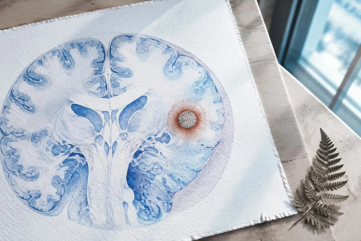
A calcification is generally a remnant rather than a living cyst that antiparasitic medication can eliminate. In the IDSA/ASTMH guideline, calcified-only parenchymal disease is managed symptomatically rather than with antiparasitic drugs; a person with 1 calcified focus and seizures may therefore need antiseizure treatment without needing cyst-killing medication (White et al., 2018).
Surrounding edema can appear intermittently, sometimes long after the original cyst has degenerated. MRI may show this tissue response even when the calcification is more obvious on CT, which is why the 2 scans can tell different but compatible stories rather than one proving that the other is wrong.
Neither a negative antibody result nor a persistent positive result measures seizure risk by itself. The broader lesson resembles other seronegative neurological conditions, discussed in our negative neurological antibody results guide: absence of detectable antibodies is not equivalent to absence of a clinically relevant neurological process.
実際に行われた嚢虫症抗体検査はどれですか?
The useful first question is whether the report describes EITB, ELISA, or another antibody method, because these are not interchangeable tests. A negative index below 1 laboratory's cutoff cannot inherit the performance figures of a different reference-laboratory assay, even when both reports use the name cysticercosis IgG.
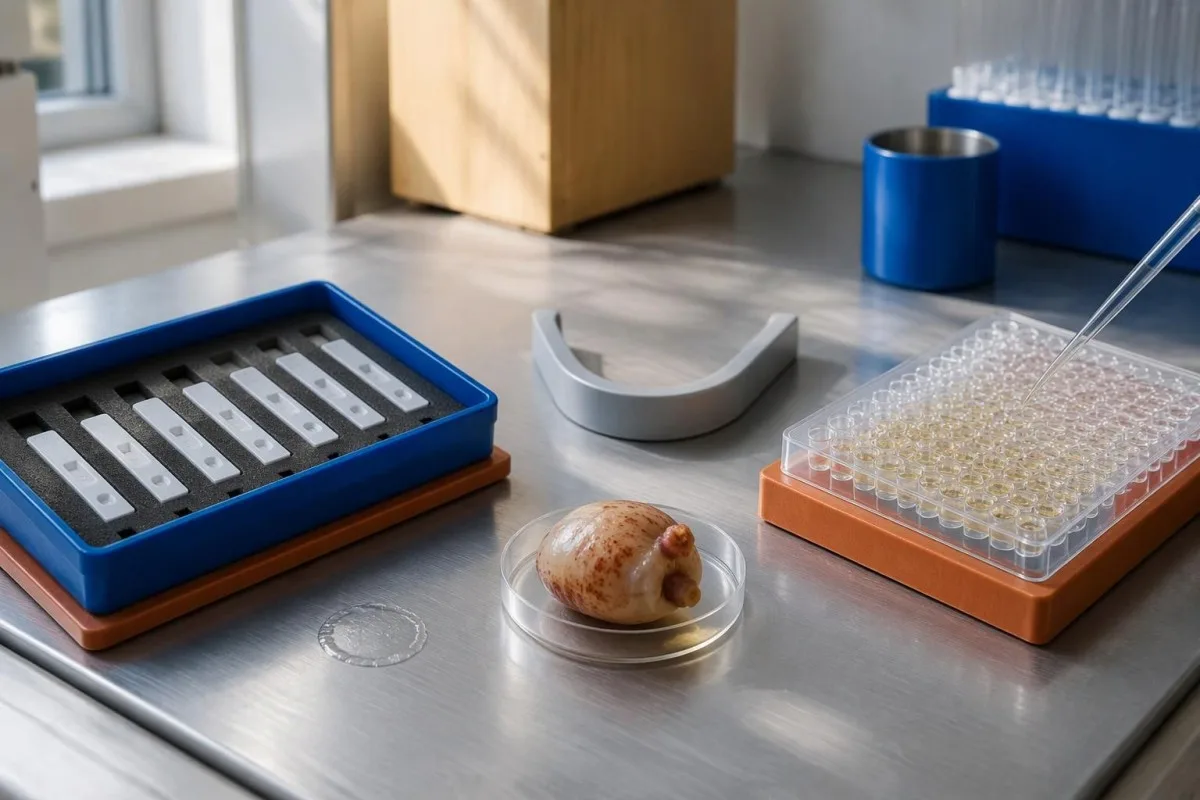
EITB using purified parasite glycoproteins is the preferred serological method in the IDSA/ASTMH guideline, while crude-antigen ELISA methods have recognized sensitivity and specificity limitations (White et al., 2018). Before comparing 2 results, check the method, specimen type, reference interval, and whether the laboratory used a validated confirmatory procedure.
Some laboratories report a qualitative result; others provide an optical-density ratio or numerical index. A value such as 0.8 in a hypothetical assay does not represent 0.8 cysts, an 80% disease probability, or a universal severity score—our 定性および定量結果 guide explains that distinction.
Kantesti AI's role is to help readers preserve the laboratory's original wording rather than reinterpret 1 method through another method's thresholds. Our 検査室マーカーガイド provides broader report-reading context; for this result, keep the actual assay name available when discussing referral or repeat testing.
曝露歴によって診断上の疑いはどのように変わりますか?
Neurocysticercosis follows ingestion of Taenia solium eggs, usually through fecal contamination from a person carrying the intestinal tapeworm. Eating undercooked pork can cause intestinal taeniasis, but brain cysts arise through a different transmission step; symptoms may emerge years after exposure rather than within 1 recent trip.

A vegetarian can develop cysticercosis because eggs can contaminate food or hands independently of meat consumption. Useful history includes past residence, household contacts, sanitation conditions, and possible exposure to a tapeworm carrier; asking only about meals eaten during the last 2 weeks can miss the relevant background.
A negative stool examination does not exclude cysts in the brain. Stool testing addresses intestinal tapeworm carriage, whereas CT and MRI assess nervous-system findings; these are 2 different sites and clinical questions, although public-health teams may recommend evaluating contacts when a carrier could be involved.
Normal eosinophils also cannot exclude neurocysticercosis, and raised eosinophils do not identify the brain as the affected site. Our parasite antibody interpretation guide discusses a separate parasite with similar localization limits; the practical point is to avoid using 1 routine cell count as a substitute for targeted neurological evaluation.
新たな発作では、いつ緊急の評価が必要ですか?
A first known seizure needs urgent medical assessment regardless of antibody results, and emergency services should be contacted for a first seizure or an ongoing seizure with concerning features. A convulsive seizure lasting 5 minutes, repeated seizures without recovery, breathing difficulty, pregnancy, or significant injury requires emergency care.

Do not wait for the 5-minute mark to seek help after a first known seizure. Keep the person away from hazards, cushion the head if safely possible, do not restrain them or put anything in their mouth, and place them on their side once convulsions stop if they are breathing normally.
Persistent confusion, a severe new headache, fever with neck stiffness, or new weakness increases concern for causes beyond cysticercosis. Sudden one-sided weakness or speech difficulty also demands emergency assessment, as our 脳卒中の警告徴候ガイド explains; 1 negative infection test cannot rule out stroke, meningitis, or a structural brain problem.
After a suspected seizure, stop driving and avoid swimming alone, heights, or operating machinery until a clinician provides advice consistent with local rules. If a witness can safely record the duration and describe movements or recovery, those observations may be more useful than ordering a second antibody test before the first neurological assessment.
神経嚢虫症の診断にCTはどのような情報を加えますか?
Noncontrast CT is particularly useful for detecting calcified cyst remnants, including small approximately 1–4 mm foci that MRI may show less clearly. CT also helps assess urgent structural problems such as hydrocephalus, but 1 isolated calcification is not automatically proof of neurocysticercosis or the cause of a seizure.
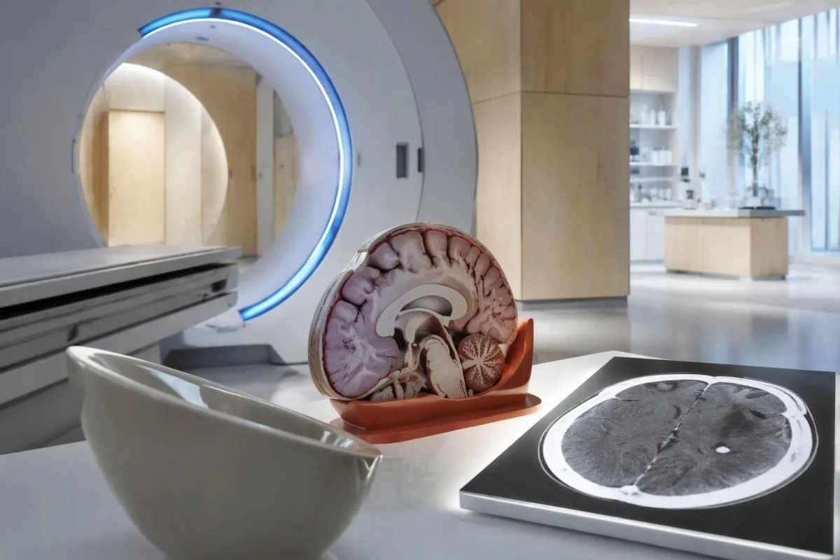
Calcium appears dense on CT, allowing radiologists to identify mineralized remnants and describe their distribution. The IDSA/ASTMH guideline recommends both MRI and noncontrast CT when classifying newly diagnosed neurocysticercosis, because the 2 modalities have complementary strengths rather than being interchangeable alternatives (White et al., 2018).
An incidental calcification needs context: previous granulomatous disease, other causes of mineralization, and scan appearance can influence interpretation. If 1 calcification sits near a suspected seizure focus, concordance with symptoms and EEG may strengthen the explanation, but anatomical proximity alone cannot establish causation.
A laboratory marker used in selected head-injury pathways does not test for brain cysts or replace imaging after an unexplained seizure. Our head-injury CT decisions guide illustrates that narrower setting; ask the clinician whether the present question requires noncontrast CT, contrast imaging, MRI, or a combination of 2 modalities.
CTでは見逃される可能性があるどのような所見をMRIで確認できますか?
MRI is better suited to evaluating viable cysts, surrounding edema, and many ventricular or subarachnoid findings. Specialized 3-dimensional sequences such as CISS or FIESTA can improve visualization of cyst membranes within cerebrospinal fluid spaces, where a cyst may otherwise resemble the surrounding fluid.
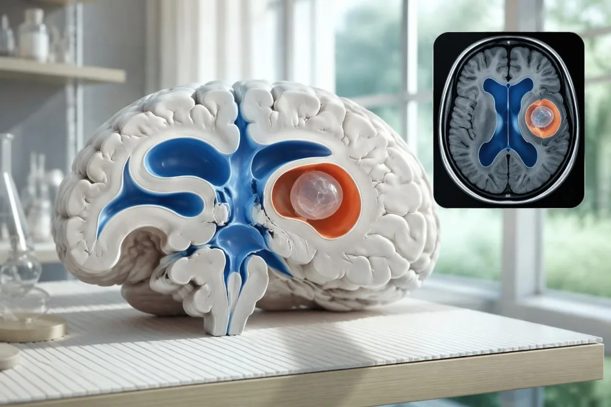
A cyst inside the ventricles can obstruct cerebrospinal fluid flow and cause hydrocephalus even without numerous parenchymal cysts. Headache with vomiting, declining alertness, or new visual symptoms therefore changes urgency; 1 reassuring antibody result should not override symptoms that suggest raised pressure or impaired fluid circulation.
MRI may show a scolex, the small internal structure of the parasite, inside a compatible cyst. This finding can be highly specific when confidently identified, whereas ring enhancement alone has a broader differential; our brain imaging warning signs guide explains why structural abnormalities require interpretation rather than diagnosis from 1 visual feature.
A routine MRI protocol may not answer every cysticercosis question. If ventricular or subarachnoid disease is suspected, discuss whether thin-section 3-dimensional imaging is needed; radiologists choose sequences and contrast according to the clinical question, and a normal scan using 1 protocol does not prove every possible disease compartment has been adequately assessed.
検査結果が一致しない場合、神経嚢虫症はどのように診断されますか?
Neurocysticercosis diagnosis combines imaging, clinical findings, exposure information, and supporting tests, rather than requiring a positive antibody result in every case. The revised diagnostic criteria published in 2017 give characteristic neuroimaging findings a central role and distinguish strong evidence from less-specific supportive features (Del Brutto et al., 2017).
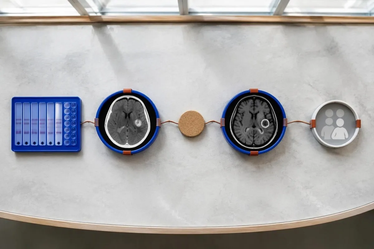
Evidence should be weighted, not tallied. A confidently visualized scolex inside a compatible cyst is different from 1 nonspecific ring-enhancing area, while a positive antibody result is different from direct evidence that a finding is in the brain; our 陽性抗体結果ガイド explains why detection alone cannot establish location.
For a clearly hypothetical probability example, start with a 30% pretest probability and assume a single-cyst assay has 55% sensitivity and 99% specificity. In 1,000 comparable people, about 135 affected and 693 unaffected people would test negative, leaving approximately a 16% probability of disease among negative results—still substantial, although these assumed figures are not a personal risk calculator.
The most useful disagreement to investigate is whether each test is answering the same question. A negative antibody test, a CT calcification, and MRI edema can all be compatible with 1 calcified disease process; unexplained discrepancies should prompt review of the assay and images rather than automatic dismissal of either result.
脳画像検査で単発嚢胞に似て見えるものには、ほかに何がありますか?
A single ring-enhancing brain finding is not specific to neurocysticercosis. Other possibilities include tuberculoma, an abscess, a neoplasm, or an inflammatory process; clinicians use appearance, location, immune status, symptoms, and sometimes follow-up imaging or tissue examination to distinguish these possibilities rather than relying on 1 antibody panel.
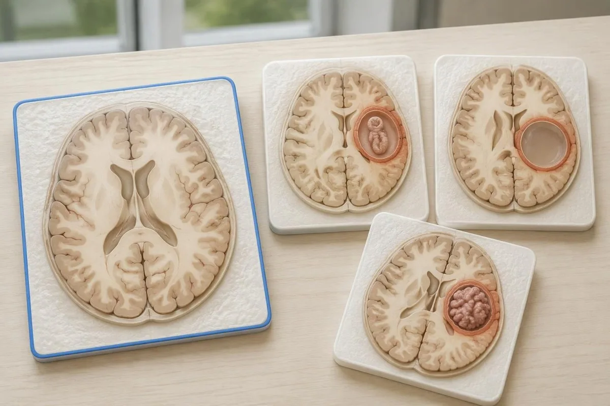
Edema, restricted diffusion, internal structures, and the number of findings all contribute to the differential, but no single nonspecific feature settles every case. Fever and immune suppression may change the suspected cause; a 2 cm ring-enhancing area in a hypothetical immunocompromised person warrants a different investigation from a tiny calcification in an otherwise well person.
Toxoplasmosis is one consideration in selected immunocompromised patients, and a positive IgG result can indicate past exposure rather than prove an active brain process. Our Toxoplasma IgG interpretation guide explains that laboratory distinction; imaging and the patient's immune status determine whether the result is clinically relevant to 1 or more cerebral findings.
New seizures accompanied by rapidly evolving memory problems, psychiatric changes, or abnormal movements can also suggest encephalitis rather than a solitary parasite cyst. Our encephalitis diagnostic pathway describes how MRI, EEG, and cerebrospinal fluid testing answer different questions; a second negative cysticercosis antibody result does not investigate those alternatives.
抗原検査、PCR、またはCSFによって不確実性を解消できますか?
Antigen tests, PCR, and cerebrospinal fluid analysis can add information in selected cases, particularly when extraparenchymal disease is suspected. None is a universal rescue test after 1 negative antibody result: availability, assay validation, disease compartment, and safety of lumbar puncture determine whether additional testing is appropriate.
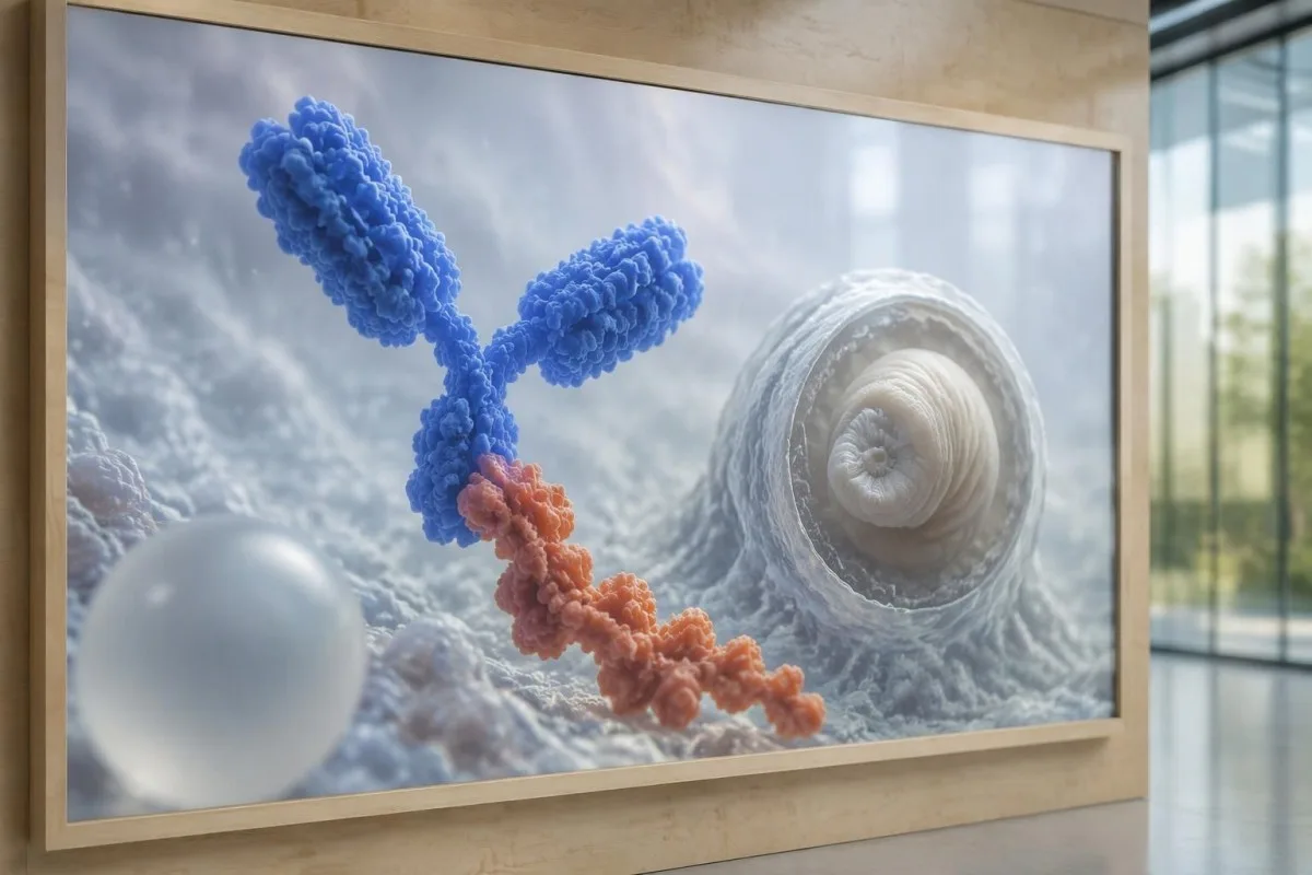
Antibody testing detects the host response, while antigen testing aims to detect parasite-derived material and may better reflect viable disease burden. Antigen detection is often less useful when only 1 small parenchymal cyst or calcifications are present; molecular testing is used in some specialist settings, but performance cannot be generalized across every laboratory or specimen.
Lumbar puncture is not routinely required for an uncomplicated solitary parenchymal finding and can be unsafe when obstructive hydrocephalus or significant mass effect is present. Cerebrospinal fluid tests for other disorders also require disease-specific interpretation, as our CSF band interpretation guide explains; 1 abnormal CSF marker does not identify every possible cause.
カンテスティは AI血液検査の読み方プラットフォーム, not a service that orders lumbar punctures, identifies cysts on scans, or confirms neurological infections. Our AI 解釈テクノロジー explains the laboratory-report workflow; when 2 tests disagree, our role is to clarify their stated meanings and limitations, not select an invasive diagnostic procedure.
治療が嚢胞の病期や位置によって異なるのはなぜですか?
Treatment depends on whether cysts are viable, degenerating, calcified, or located outside the brain tissue, not simply on antibody positivity. Selected viable parenchymal cases receive antiparasitic treatment, whereas calcified-only disease generally receives symptom-directed care; ventricular obstruction may require a procedural approach rather than 1 standard medication regimen.
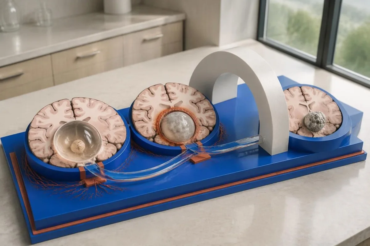
For selected patients with 1–2 viable parenchymal cysts, the IDSA/ASTMH guideline describes albendazole treatment for 10–14 days with appropriate accompanying care. This is a specialist-directed recommendation, not a home-treatment instruction: antiparasitic drugs can provoke an inflammatory response, and untreated hydrocephalus or diffuse cerebral edema changes the order of management (White et al., 2018).
WHO's 2021 guidance focuses on defined presentations of parenchymal neurocysticercosis, while the IDSA/ASTMH guideline also addresses ventricular, subarachnoid, and other forms in detail. Differences in imaging access and disease compartment explain why 1 treatment recommendation cannot be transferred to every patient with an antibody result; the guidelines do not turn negative serology into a rule-out test.
Corticosteroids may accompany antiparasitic therapy, but prolonged use requires additional safety planning, including assessment for relevant latent infections. Our Strongyloides and steroid safety guide explains a separate risk that matters before immune-suppressing treatment; an eye examination is also recommended before antiparasitic therapy to evaluate possible intraocular cysts.
抗体検査の結果以外に、フォローアップでは何を確認すべきですか?
Follow-up should track seizures, neurological symptoms, imaging changes, and medication safety, rather than use antibody disappearance as proof of recovery. For viable parenchymal cysts, the IDSA/ASTMH guideline suggests repeat MRI at least every 6 months until the cystic component resolves, with earlier reassessment when symptoms change.
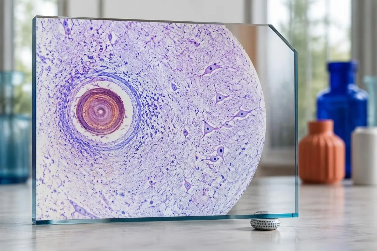
Antibodies may remain detectable after the original disease process has changed, and low-burden disease may never produce a positive result. If 1 new seizure occurs during follow-up, clinicians consider adherence, medication interactions, recurrent edema, and other causes; a rising or falling antibody index alone cannot distinguish among those explanations.
Kantesti AI can help explain reported laboratory results during medication follow-up, but a drug concentration is not a stand-alone measure of seizure control. Our レベチラセタム濃度の解釈 guide explains why sample timing and kidney function matter; 2 measurements collected at different times after dosing may not be directly comparable.
Monitoring depends on the chosen drug and treatment duration: prolonged albendazole therapy can require liver-test and blood-count surveillance, while some antiseizure medicines can affect sodium. Our 電解質安全性ガイド discusses those separate laboratory concerns; do not stop an antiseizure medicine or change its dose because 1 cysticercosis antibody result is negative.
Kantestiで説明できることと、研究では証明できないことは何ですか?
AI report interpretation cannot confirm or exclude neurocysticercosis from laboratory results alone. Kantesti は an AI搭載の血液検査解析ツール whose role here is to organize reported laboratory findings; a negative result still needs clinical context, and 1 new seizure takes priority over reviewing an automated explanation.

Prepare a focused review packet with the original antibody report, assay method, CT or MRI report, symptom timeline, and medication list. These 5 information categories reduce the risk of discussing a result without its context; ask specifically whether the imaging suggests viable, degenerating, calcified, ventricular, or subarachnoid disease and which unanswered question the next test would address.
The 2 Kantesti publications listed below are a repository-deposited global analysis report and an educational RDW guide, not clinical evidence validating neurocysticercosis diagnosis. Our RDW と CBC のガイド can support interpretation of a separately ordered blood count, but RDW, MCV, and MCHC do not establish whether a brain cyst is present.
Evidence for the neurological recommendations in this article comes from the cited diagnostic criteria and clinical guidelines, not from general AI benchmarking or laboratory-report volume. Our technical validation scope should be read within its stated boundaries; neither 1 successful report explanation nor a large aggregate dataset substitutes for condition-specific diagnostic validation.
よくある質問
血液検査が陰性でも神経嚢虫症の可能性はありますか?
はい、血液抗体検査が陰性でも、神経嚢虫症が存在する可能性はあります。特に、脳内の嚢胞が1個だけの場合や、石灰化病変のみの場合が該当します。公表されている基準検査機関でのEITBの感度推定値では、嚢胞が1個の場合は約50~60%とされ、複数の生存嚢胞がある場合の感度を大幅に下回ります。そのため、CT、MRI、症状、曝露歴が引き続き診断の中心となります。新たにけいれん発作が起きた場合は、陰性結果に安心するのではなく、緊急の評価が必要です。
神経嚢虫症の検査で最も信頼性が高いものは何ですか?
神経嚢虫症の検査で、あらゆる診断上の疑問に答えられるものはありません。脳画像検査と、適切に選択した血清学的検査は相補的です。EITBはIDSA/ASTMHガイドラインで推奨される確定診断用の抗体検査法ですが、嚢胞が1個のみの場合は疾患を検出できないことがあります。単純CTは特に石灰化の評価に有用で、MRIは多数の生存嚢胞、浮腫、髄液腔の異常をより詳しく評価できます。臨床医は、単一の陰性結果を確定的なものとみなすのではなく、これらの所見を組み合わせて判断します。
神経嚢虫症にはCTとMRIのどちらが適していますか?
神経嚢虫症の診断では、CTとMRIはそれぞれ異なる強みを持ちます。非造影CTでは、直径約1~4 mmの小さな石灰化をより明瞭に検出できることが多く、一方、MRIでは生存嚢胞、周囲の浮腫、脳室内病変またはくも膜下病変を評価できます。特殊な3次元MRIシーケンスは、脳脊髄液腔内の嚢胞膜の特定に役立つ場合があります。IDSA/ASTMHガイドラインでは、新たに診断された疾患を分類する際、両方の画像診断法を用いることが推奨されています。
石灰化した嚢胞でも発作を引き起こすことはありますか?
石灰化した嚢虫症の残存病変は、もはや生存可能な嚢胞ではなくても、発作との関連が続くことがあります。石灰化巣の一部は、直径がわずか1~4 mm程度であることが多く、間欠的に周囲に浮腫が生じ、MRIで確認できる場合があります。抗体陰性でもこの機序は否定されず、抗体陽性が持続していても発作リスクを示すものではありません。石灰化病変のみの疾患では、一般に駆虫薬ではなく症状に応じた治療が行われます。
陰性の嚢虫症抗体検査を再度受けるべきですか?
嚢虫症抗体検査が陰性だった場合、再検査が必ずしも次の最善の手段とは限りません。小さな嚢胞が1つある場合や石灰化病変がある場合、抗体反応が弱く、同じ検査法で2週間後に再検査しても陰性のままのことがあります。医療従事者は、元の検査法を確認したり、特性がより明らかな確認検査を依頼したり、CTやMRIを優先したりすることがあります。新たな発作や神経症状の悪化がある場合は、再検査を待つべきではありません。
初めての発作を起こしたとき、どのような場合に救急医療を受けるべきですか?
初めて起きた発作が既知のものでも、神経嚢虫症抗体検査が陰性であっても、至急、医療機関で評価を受け、地域の緊急サービスに連絡してください。けいれん発作が5分間続く場合、意識が回復しないまま発作を繰り返す場合、呼吸困難、妊娠中、または重傷を負った場合は緊急事態です。本人を押さえつけたり、口に物を入れたりしないでください。けいれんが止まり、正常に呼吸している場合は、横向きに寝かせてください。医療従事者が発作を評価し、該当する運転制限について説明するまでは、運転しないでください。
血液検査結果の見方を確認しましょう
検査報告書をアップロードすると、KantestiのAIが数秒で各バイオマーカーを平易な言葉で説明します。世界中で200万人以上が利用しています。.
参考文献
- Klein, T., Mitchell, S., & Weber, H. (2026). AI血液検査アナライザー:2.5M件の検査を分析 | グローバルヘルスレポート 2026。. Kantesti AI医学研究。. 掲載日
- Klein, T., Mitchell, S., & Weber, H. (2026). RDW血液検査:RDW-CV、MCV、MCHCの完全ガイド。. Kantesti AI医学研究。. 掲載日
- White AC Jr et al. (2018). Diagnosis and Treatment of Neurocysticercosis: 2017 Clinical Practice Guidelines by the Infectious Diseases Society of America (IDSA) and the American Society of Tropical Medicine and Hygiene (ASTMH). Clinical Infectious Diseases. 掲載日
- Del Brutto OH et al. (2017). Revised diagnostic criteria for neurocysticercosis. Journal of the Neurological Sciences. 掲載日
- World Health Organization (2021). WHO guidelines on management of Taenia solium neurocysticercosis. 世界保健機関。.
- 200万以上分析対象の検査
- 127+国
- 100+言語
医療チームについて


E-E-A-T 信頼性シグナル
経験
医師主導による、検査結果解釈ワークフローの臨床レビュー。.
専門知識
臨床的な文脈においてバイオマーカーがどのように振る舞うかに焦点を当てた検査医学。.
権威
Thomas Klein医師による医学的レビュー。Sarah Mitchell医師およびHans Weber教授の協力。.
信頼性
アラームを減らすための明確なフォローアップ経路を備えた、エビデンスに基づく解釈。.





医療免責事項
この記事は教育目的のみを対象としており、医療助言を構成するものではありません。診断や治療の判断を行う際は、必ず有資格の医療提供者にご相談ください。.
公開 · 編集チームにお問い合わせください
Kantesti Ltdが発行。イングランドおよびウェールズで登録、会社番号. 17090423, 、英国ロンドン。.
