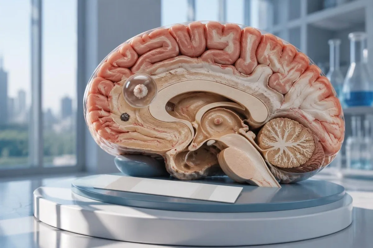
Neurozistizerkosiaren emaitza negatibo batek ez du baztertzen garuneko kiste bakar bat edo gaixotasun kaltzitua izatea. CTak, MRIak eta krisi berrien premiazko ebaluazioak garrantzi handiagoa dute antigorputzen azterketaren emaitzak bakarrik ematen duen lasaitasunak baino.

Ondorio nagusiak
- Antikorpuek negatibo ez dute neurozistizerkosia baztertzen, batez ere irudiek kaltetutako eremu bakarra edo kaltzifikazioak erakusten dituztenean.
- Erreferentzia-laborategiko EITB %ko sentikortasunera irits daiteke kiste bideragarri bat baino gehiago dagoenean, baina errendimendua nabarmen txikiagoa da kiste bakar batekin.
- Kiste bakarraren sentikortasuna maiz –60% ingurukoa da argitaratutako EITBren estimazioetan; kopuru horiek ez zaizkie aplikatzen merkataritzako antigorputz-proba guztiei.
- Kontrasterik gabeko CTa bereziki erabilgarria da kaltzifikazio txikiak detektatzeko; sarritan 1–4 mm ingurukoak izaten dira.
- MRI bat kiste bideragarriak, inguruko edema eta bentrikuluetako edo espazio subaraknoideko gaixotasuna ebaluatzen ditu; hiru dimentsioko sekuentzia espezializatuek detekzioa hobetu dezakete.
- Lehen krisiak antigorputzaren emaitza edozein dela ere, premiazko ebaluazio medikoa behar dute. Deitu larrialdi-zerbitzuetara lehen krisi epileptikoa ezagutzen bada edo bestelako larrialdi-zantzuak badaude.
- Bost minutu da etengabeko konbultsio-krisi baterako larrialdi-atalasea, eta ez laguntza eskatu aurretik itxaroteko arrazoia.
- Kaltzifikazioak bakarrik dituen gaixotasuna oro har, ez da antiparasitarioekin tratatu behar, baina krisien tratamendua eta irudi bidezko azterketa gehiago beharrezkoak izan daitezke.
Neurozistizerkosiaren emaitza negatibo batek bazter dezake?
A neurozistizerkosiaren proba negatiboak ezin du gaixotasuna modu fidagarrian baztertu, bereziki garuneko kiste bakarra edo kaltzifikatutako hondarrak daudenean. Antigorputzen probek erantzun immunologikoa neurtzen dute, garuna zuzenean aztertu beharrean; CTak eta MRIak susmagarrien kokalekua eta estadioa zehazten dituzte, eta lehen krisi epileptikoak premiazko ebaluazioa behar du.

Emaitza baldintzazkoa da, ez erabatekoa. Hainbat kiste bideragarri dituzten pertsonengan emaitza onak dituen zistizerkosiaren antigorputz-probak emaitza txarrak eman ditzake kaltetutako eremu txiki bakarra duen norbaitengan; beraz, hitzak negatiboa esan nahi du ez dela erantzunik detektatu azterketa horren bidez, ez nerbio-sisteman inon kisterik ez dagoenik.
Har dezagun adibide hipotetiko bat: 34 urteko pertsona batek lehen krisia izan du, antigorputzen txostena negatiboa da, eta MRIak eraztun-itxurako lesio bat erakusten du. Txostenak ezin du ebatzi lesio hori zistizerkosia, beste infekzio bat edo prozesu ez-infekzioso bat den; haren esanahia irudi bidezko azterketaren, esposizio-aurrekarien eta ebaluazio neurologikoaren araberakoa da, denak batera hartuta.
Kantesti bat da. AI odol-analisi analizatzailea laborategiko aurkikuntzak azaltzen lagun dezake, baina ezin du garuneko gaixotasuna baztertu antigorputzen emaitza negatibo bakar baten bidez. Gure antolakuntzak eta misioak laborategiko informazioan dugun arreta azaltzen dute; irakurleek ez lukete txosten baten azalpen automatikoa hartu behar krisi epileptikoen arretarako atzerapenik ez dagoela ziurtatzeko.
Zergatik ez du antigorputzen azterketak garuneko kiste bakar bat detektatzen?
A kiste bakar batek azterketaren detekzio-atalasetik beherako antigorputzen erantzuna eragin dezake. Argitaratutako erreferentzia-laborategietako EITB estimazioen arabera, sentikortasuna etik gertu egon ohi da kiste bakarra dagoenean, eta 60%etik behera; alderatuz, gutxi gorabehera 98%ekoa da kiste bideragarri bat baino gehiago dagoenean. Aldea gaixotasunaren kargari eta azterketaren errendimenduari zor zaie.
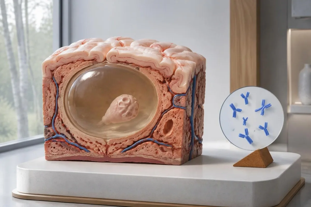
IDSA/ASTMH gidalerroak gomendatzen du entzimekiko lotutako immunoelektrotransferentzia-blota, edo EITB, baieztapen-serologiatzat hartzea, eta berariaz ohartarazten du sentikortasuna eskasa dela parenkimako aurkikuntza bakar bat edo kaltzifikazioak bakarrik daudenean (White et al., 2018). Kiste ugari, bentrikuluetako gaixotasuna edo gaixotasun subaraknoideoa duten pazienteetan jakinarazitako sentikortasun handia ez zaio aplikatu behar garuneko anomalia txiki bakarra duen norbaiti.
Demagun, adibide huts gisa, azterketa batek eko sentikortasuna duela jada modu independentean baieztatu den kiste bakarreko gaixotasuna duten 100 pertsonengan. Gutxi gorabehera 45ek emaitza negatiboa izango lukete gaixotasuna izan arren; adibide honek antzemate-hutsegiteak azaltzen ditu, ez emaitza negatibo jakin batek gaixotasuna adierazteko duen probabilitatea.
Bi asteren buruan azterketa bera errepikatzeak agian ez du zuzenduko gaixotasun-karga txikiak eragindako erantzun ahula. Hori bestelakoa da lehen laginketa goiztiarra izatea arazo nagusi den infekzioetan, gure denborarekin lotutako emaitza faltsu-negatiboen gidan; azaltzen den bezala; galdetu irudi bidezko azterketa batek edo hobeto karakterizatutako azterketa batek hurrengo erabakia aldatuko ote lukeen.
Zergatik izan daiteke neurozistizerkosi kaltzifikatua sintomatikoa?
Neurozistizerkosi kaltzifikatuak krisi epileptikoak eragin ditzake, antigorputzak negatiboak izan arren, because the remaining mineralized focus can still be associated with local irritation and episodic surrounding edema. Typical calcifications may measure approximately 1–4 mm, but their small size does not establish whether they are clinically harmless.
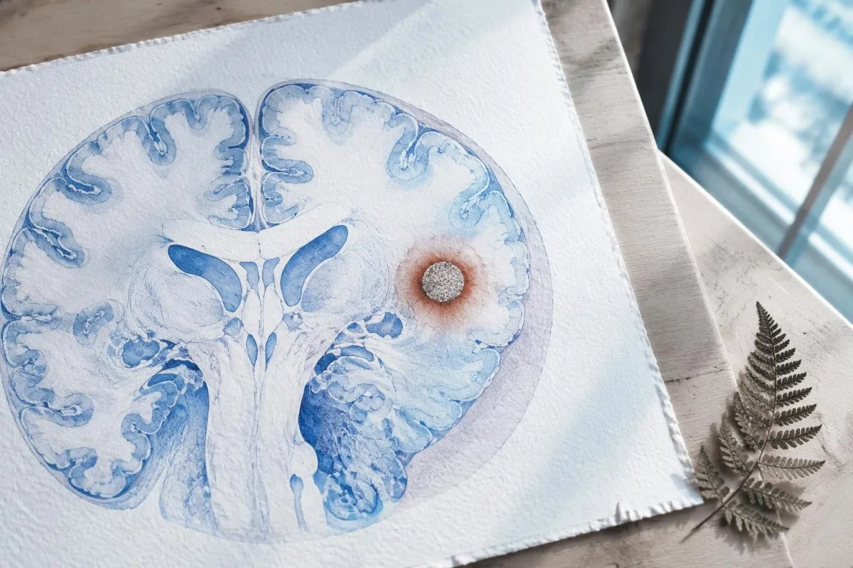
A calcification is generally a remnant rather than a living cyst that antiparasitic medication can eliminate. In the IDSA/ASTMH guideline, calcified-only parenchymal disease is managed symptomatically rather than with antiparasitic drugs; a person with 1 calcified focus and seizures may therefore need antiseizure treatment without needing cyst-killing medication (White et al., 2018).
Surrounding edema can appear intermittently, sometimes long after the original cyst has degenerated. MRI may show this tissue response even when the calcification is more obvious on CT, which is why the 2 scans can tell different but compatible stories rather than one proving that the other is wrong.
Neither a negative antibody result nor a persistent positive result measures seizure risk by itself. The broader lesson resembles other seronegative neurological conditions, discussed in our negative neurological antibody results guide: absence of detectable antibodies is not equivalent to absence of a clinically relevant neurological process.
Zer zistizerkosiaren aurkako antigorputz-proba egin zen benetan?
The useful first question is whether the report describes EITB, ELISA, or another antibody method, because these are not interchangeable tests. A negative index below 1 laboratory's cutoff cannot inherit the performance figures of a different reference-laboratory assay, even when both reports use the name cysticercosis IgG.
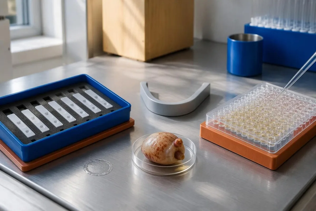
EITB using purified parasite glycoproteins is the preferred serological method in the IDSA/ASTMH guideline, while crude-antigen ELISA methods have recognized sensitivity and specificity limitations (White et al., 2018). Before comparing 2 results, check the method, specimen type, reference interval, and whether the laboratory used a validated confirmatory procedure.
Some laboratories report a qualitative result; others provide an optical-density ratio or numerical index. A value such as 0.8 in a hypothetical assay does not represent 0.8 cysts, an 80% disease probability, or a universal severity score—our emaitza kualitatiboak eta kuantitatiboak guide explains that distinction.
Kantesti AI's role is to help readers preserve the laboratory's original wording rather than reinterpret 1 method through another method's thresholds. Our laborategiko markatzaileen gida provides broader report-reading context; for this result, keep the actual assay name available when discussing referral or repeat testing.
Nola aldatzen du esposizio-aurrekariak diagnostiko-susmoa?
Neurocysticercosis follows ingestion of Taenia solium eggs, usually through fecal contamination from a person carrying the intestinal tapeworm. Eating undercooked pork can cause intestinal taeniasis, but brain cysts arise through a different transmission step; symptoms may emerge years after exposure rather than within 1 recent trip.

A vegetarian can develop cysticercosis because eggs can contaminate food or hands independently of meat consumption. Useful history includes past residence, household contacts, sanitation conditions, and possible exposure to a tapeworm carrier; asking only about meals eaten during the last 2 weeks can miss the relevant background.
A negative stool examination does not exclude cysts in the brain. Stool testing addresses intestinal tapeworm carriage, whereas CT and MRI assess nervous-system findings; these are 2 different sites and clinical questions, although public-health teams may recommend evaluating contacts when a carrier could be involved.
Normal eosinophils also cannot exclude neurocysticercosis, and raised eosinophils do not identify the brain as the affected site. Our parasite antibody interpretation guide discusses a separate parasite with similar localization limits; the practical point is to avoid using 1 routine cell count as a substitute for targeted neurological evaluation.
Noiz behar dute krisi berriek larrialdiko ebaluazioa?
A first known seizure needs urgent medical assessment regardless of antibody results, and emergency services should be contacted for a first seizure or an ongoing seizure with concerning features. A convulsive seizure lasting 5 minutes, repeated seizures without recovery, breathing difficulty, pregnancy, or significant injury requires emergency care.

Do not wait for the 5-minute mark to seek help after a first known seizure. Keep the person away from hazards, cushion the head if safely possible, do not restrain them or put anything in their mouth, and place them on their side once convulsions stop if they are breathing normally.
Persistent confusion, a severe new headache, fever with neck stiffness, or new weakness increases concern for causes beyond cysticercosis. Sudden one-sided weakness or speech difficulty also demands emergency assessment, as our iktusaren abisu-seinaleen gida explains; 1 negative infection test cannot rule out stroke, meningitis, or a structural brain problem.
After a suspected seizure, stop driving and avoid swimming alone, heights, or operating machinery until a clinician provides advice consistent with local rules. If a witness can safely record the duration and describe movements or recovery, those observations may be more useful than ordering a second antibody test before the first neurological assessment.
Zer gehitzen dio CTak neurozistizerkosiaren diagnostikoari?
Noncontrast CT is particularly useful for detecting calcified cyst remnants, including small approximately 1–4 mm foci that MRI may show less clearly. CT also helps assess urgent structural problems such as hydrocephalus, but 1 isolated calcification is not automatically proof of neurocysticercosis or the cause of a seizure.
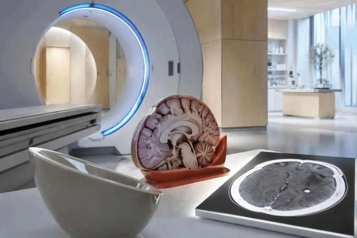
Calcium appears dense on CT, allowing radiologists to identify mineralized remnants and describe their distribution. The IDSA/ASTMH guideline recommends both MRI and noncontrast CT when classifying newly diagnosed neurocysticercosis, because the 2 modalities have complementary strengths rather than being interchangeable alternatives (White et al., 2018).
An incidental calcification needs context: previous granulomatous disease, other causes of mineralization, and scan appearance can influence interpretation. If 1 calcification sits near a suspected seizure focus, concordance with symptoms and EEG may strengthen the explanation, but anatomical proximity alone cannot establish causation.
A laboratory marker used in selected head-injury pathways does not test for brain cysts or replace imaging after an unexplained seizure. Our head-injury CT decisions guide illustrates that narrower setting; ask the clinician whether the present question requires noncontrast CT, contrast imaging, MRI, or a combination of 2 modalities.
Zer erakusten du MRIak CTak gal dezakeena?
MRI is better suited to evaluating viable cysts, surrounding edema, and many ventricular or subarachnoid findings. Specialized 3-dimensional sequences such as CISS or FIESTA can improve visualization of cyst membranes within cerebrospinal fluid spaces, where a cyst may otherwise resemble the surrounding fluid.
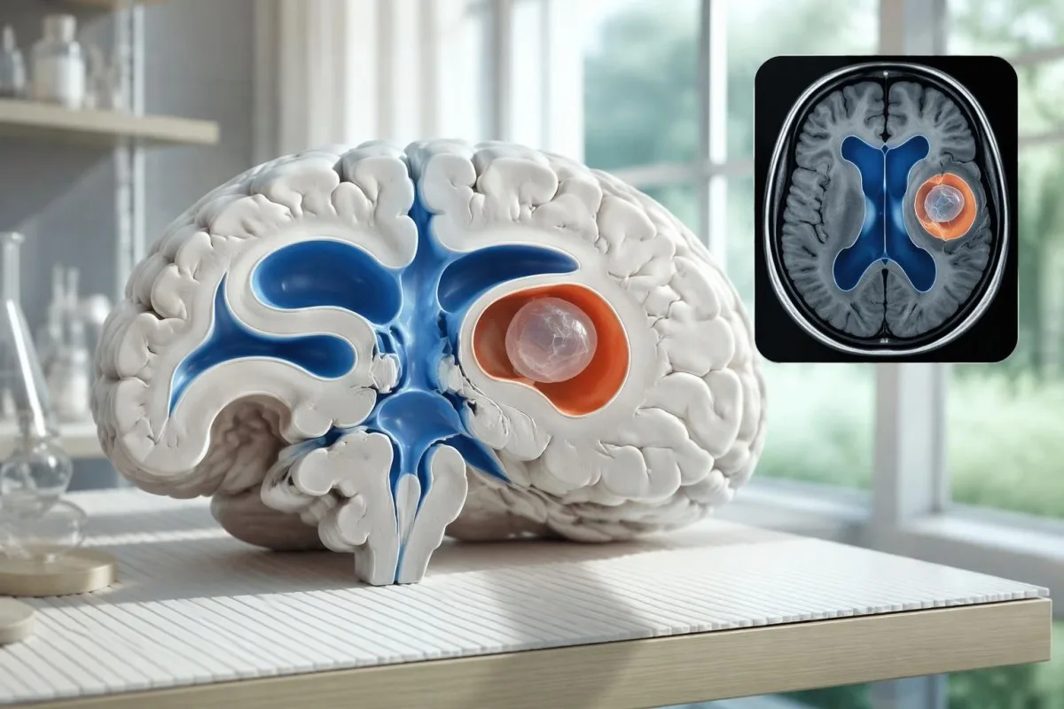
A cyst inside the ventricles can obstruct cerebrospinal fluid flow and cause hydrocephalus even without numerous parenchymal cysts. Headache with vomiting, declining alertness, or new visual symptoms therefore changes urgency; 1 reassuring antibody result should not override symptoms that suggest raised pressure or impaired fluid circulation.
MRI may show a scolex, the small internal structure of the parasite, inside a compatible cyst. This finding can be highly specific when confidently identified, whereas ring enhancement alone has a broader differential; our brain imaging warning signs guide explains why structural abnormalities require interpretation rather than diagnosis from 1 visual feature.
A routine MRI protocol may not answer every cysticercosis question. If ventricular or subarachnoid disease is suspected, discuss whether thin-section 3-dimensional imaging is needed; radiologists choose sequences and contrast according to the clinical question, and a normal scan using 1 protocol does not prove every possible disease compartment has been adequately assessed.
Nola diagnostikatzen da neurozistizerkosia proben emaitzak bat ez datozenean?
Neurocysticercosis diagnosis combines imaging, clinical findings, exposure information, and supporting tests, rather than requiring a positive antibody result in every case. The revised diagnostic criteria published in 2017 give characteristic neuroimaging findings a central role and distinguish strong evidence from less-specific supportive features (Del Brutto et al., 2017).
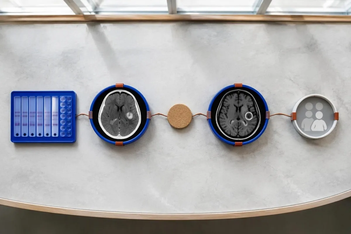
Evidence should be weighted, not tallied. A confidently visualized scolex inside a compatible cyst is different from 1 nonspecific ring-enhancing area, while a positive antibody result is different from direct evidence that a finding is in the brain; our emaitza antigorputz positiboaren gida explains why detection alone cannot establish location.
For a clearly hypothetical probability example, start with a 30% pretest probability and assume a single-cyst assay has 55% sensitivity and 99% specificity. In 1,000 comparable people, about 135 affected and 693 unaffected people would test negative, leaving approximately a 16% probability of disease among negative results—still substantial, although these assumed figures are not a personal risk calculator.
The most useful disagreement to investigate is whether each test is answering the same question. A negative antibody test, a CT calcification, and MRI edema can all be compatible with 1 calcified disease process; unexplained discrepancies should prompt review of the assay and images rather than automatic dismissal of either result.
Zer beste gauza izan daiteke garuneko irudietan kiste bakar baten antzekoa?
A single ring-enhancing brain finding is not specific to neurocysticercosis. Other possibilities include tuberculoma, an abscess, a neoplasm, or an inflammatory process; clinicians use appearance, location, immune status, symptoms, and sometimes follow-up imaging or tissue examination to distinguish these possibilities rather than relying on 1 antibody panel.
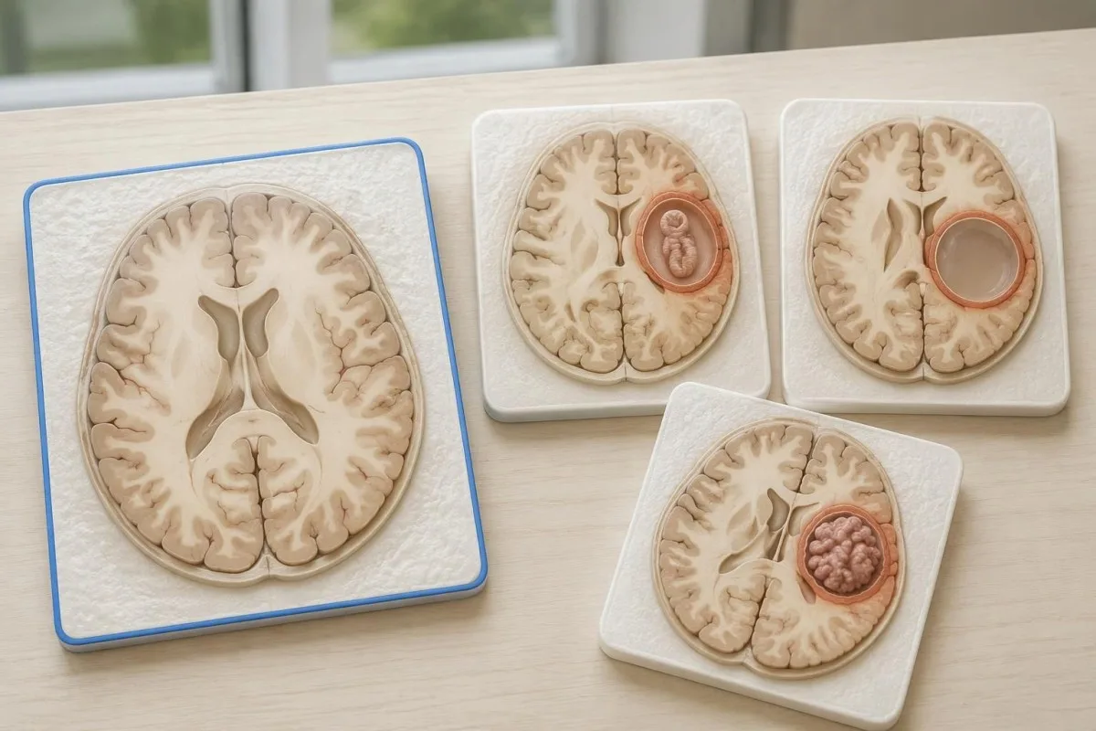
Edema, restricted diffusion, internal structures, and the number of findings all contribute to the differential, but no single nonspecific feature settles every case. Fever and immune suppression may change the suspected cause; a 2 cm ring-enhancing area in a hypothetical immunocompromised person warrants a different investigation from a tiny calcification in an otherwise well person.
Toxoplasmosis is one consideration in selected immunocompromised patients, and a positive IgG result can indicate past exposure rather than prove an active brain process. Our Toxoplasma IgG interpretation guide explains that laboratory distinction; imaging and the patient's immune status determine whether the result is clinically relevant to 1 or more cerebral findings.
New seizures accompanied by rapidly evolving memory problems, psychiatric changes, or abnormal movements can also suggest encephalitis rather than a solitary parasite cyst. Our encephalitis diagnostic pathway describes how MRI, EEG, and cerebrospinal fluid testing answer different questions; a second negative cysticercosis antibody result does not investigate those alternatives.
Antigenoen probek, PCRak edo LZRak argi dezakete ziurgabetasuna?
Antigen tests, PCR, and cerebrospinal fluid analysis can add information in selected cases, particularly when extraparenchymal disease is suspected. None is a universal rescue test after 1 negative antibody result: availability, assay validation, disease compartment, and safety of lumbar puncture determine whether additional testing is appropriate.
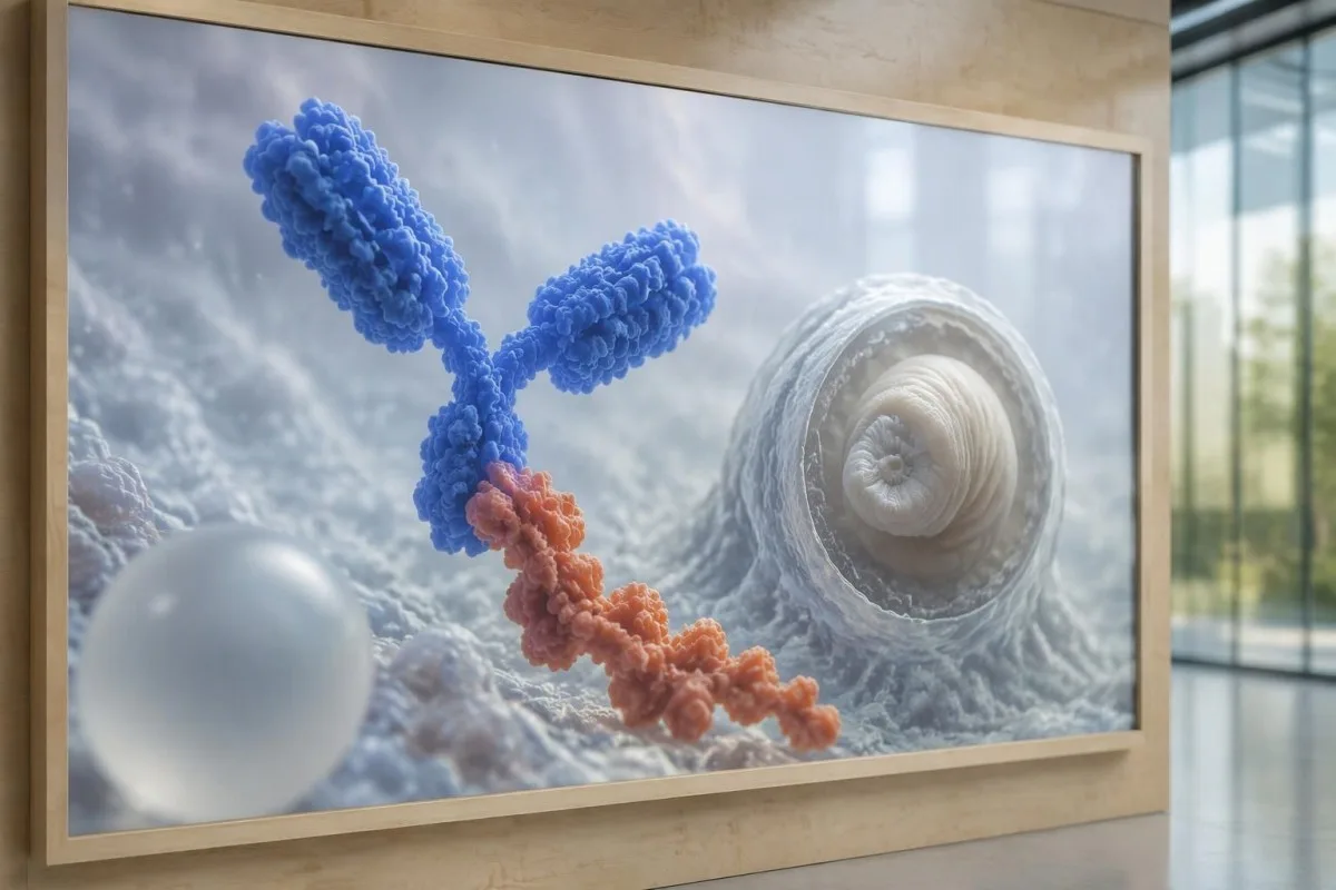
Antibody testing detects the host response, while antigen testing aims to detect parasite-derived material and may better reflect viable disease burden. Antigen detection is often less useful when only 1 small parenchymal cyst or calcifications are present; molecular testing is used in some specialist settings, but performance cannot be generalized across every laboratory or specimen.
Lumbar puncture is not routinely required for an uncomplicated solitary parenchymal finding and can be unsafe when obstructive hydrocephalus or significant mass effect is present. Cerebrospinal fluid tests for other disorders also require disease-specific interpretation, as our CSF band interpretation guide explains; 1 abnormal CSF marker does not identify every possible cause.
Kantesti bat da. AI odol-analisien interpretazio-plataforma, not a service that orders lumbar punctures, identifies cysts on scans, or confirms neurological infections. Our AI interpretazio teknologia explains the laboratory-report workflow; when 2 tests disagree, our role is to clarify their stated meanings and limitations, not select an invasive diagnostic procedure.
Zergatik dago tratamendua kistearen fasearen eta kokapenaren mende?
Treatment depends on whether cysts are viable, degenerating, calcified, or located outside the brain tissue, not simply on antibody positivity. Selected viable parenchymal cases receive antiparasitic treatment, whereas calcified-only disease generally receives symptom-directed care; ventricular obstruction may require a procedural approach rather than 1 standard medication regimen.
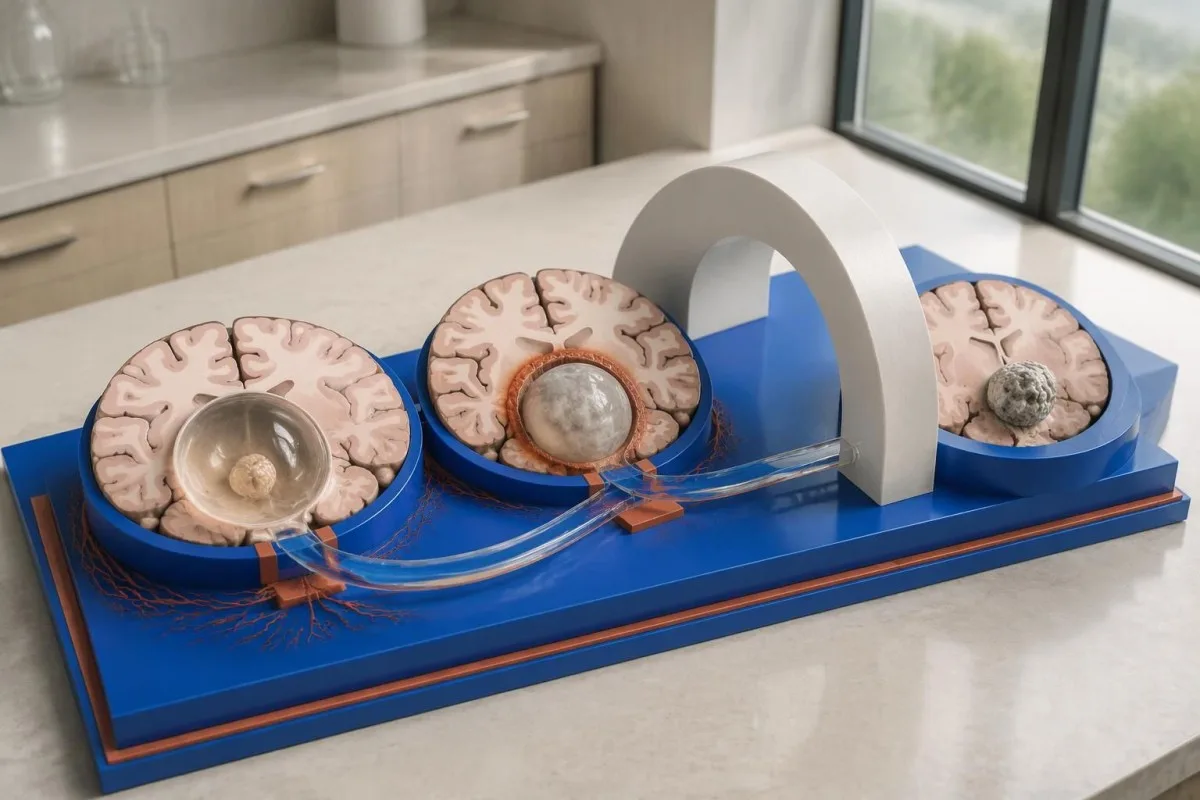
For selected patients with 1–2 viable parenchymal cysts, the IDSA/ASTMH guideline describes albendazole treatment for 10–14 days with appropriate accompanying care. This is a specialist-directed recommendation, not a home-treatment instruction: antiparasitic drugs can provoke an inflammatory response, and untreated hydrocephalus or diffuse cerebral edema changes the order of management (White et al., 2018).
WHO's 2021 guidance focuses on defined presentations of parenchymal neurocysticercosis, while the IDSA/ASTMH guideline also addresses ventricular, subarachnoid, and other forms in detail. Differences in imaging access and disease compartment explain why 1 treatment recommendation cannot be transferred to every patient with an antibody result; the guidelines do not turn negative serology into a rule-out test.
Corticosteroids may accompany antiparasitic therapy, but prolonged use requires additional safety planning, including assessment for relevant latent infections. Our Strongyloides and steroid safety guide explains a separate risk that matters before immune-suppressing treatment; an eye examination is also recommended before antiparasitic therapy to evaluate possible intraocular cysts.
Zer kontrolatu behar da jarraipenean, antigorputzen emaitzez gain?
Follow-up should track seizures, neurological symptoms, imaging changes, and medication safety, rather than use antibody disappearance as proof of recovery. For viable parenchymal cysts, the IDSA/ASTMH guideline suggests repeat MRI at least every 6 months until the cystic component resolves, with earlier reassessment when symptoms change.
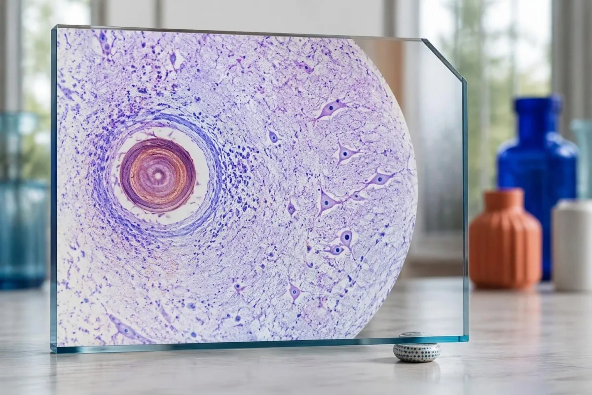
Antibodies may remain detectable after the original disease process has changed, and low-burden disease may never produce a positive result. If 1 new seizure occurs during follow-up, clinicians consider adherence, medication interactions, recurrent edema, and other causes; a rising or falling antibody index alone cannot distinguish among those explanations.
Kantesti AI can help explain reported laboratory results during medication follow-up, but a drug concentration is not a stand-alone measure of seizure control. Our levetiracetam-mailaren interpretazioak guide explains why sample timing and kidney function matter; 2 measurements collected at different times after dosing may not be directly comparable.
Monitoring depends on the chosen drug and treatment duration: prolonged albendazole therapy can require liver-test and blood-count surveillance, while some antiseizure medicines can affect sodium. Our elektrolitoen segurtasun-gidak discusses those separate laboratory concerns; do not stop an antiseizure medicine or change its dose because 1 cysticercosis antibody result is negative.
Zer azal dezake Kantestik, eta zer ezin dute ikerketek frogatu?
AI report interpretation cannot confirm or exclude neurocysticercosis from laboratory results alone. Kantesti da AI bidezko odol-analisien analisi-tresna whose role here is to organize reported laboratory findings; a negative result still needs clinical context, and 1 new seizure takes priority over reviewing an automated explanation.

Prepare a focused review packet with the original antibody report, assay method, CT or MRI report, symptom timeline, and medication list. These 5 information categories reduce the risk of discussing a result without its context; ask specifically whether the imaging suggests viable, degenerating, calcified, ventricular, or subarachnoid disease and which unanswered question the next test would address.
The 2 Kantesti publications listed below are a repository-deposited global analysis report and an educational RDW guide, not clinical evidence validating neurocysticercosis diagnosis. Our RDW eta CBC gidak can support interpretation of a separately ordered blood count, but RDW, MCV, and MCHC do not establish whether a brain cyst is present.
Evidence for the neurological recommendations in this article comes from the cited diagnostic criteria and clinical guidelines, not from general AI benchmarking or laboratory-report volume. Our technical validation scope should be read within its stated boundaries; neither 1 successful report explanation nor a large aggregate dataset substitutes for condition-specific diagnostic validation.
Ohiko galderak
Izan al daiteke neuroziztizerkosia izatea odol-analisiaren emaitza negatiboa izanda?
Bai, neurozistizerkosia egon daiteke odoleko antigorputzen proba negatiboa izan arren, bereziki garuneko kiste bakarra dagoenean edo gaixotasuna kiste kaltzifikatuek bakarrik eragindakoa denean. Argitaratutako erreferentzia-laborategietako EITB sentikortasun-estimazioek maiz etik 60%erainokoa adierazten dute kiste bakar baterako, eta emaitza hori nabarmen txikiagoa da kiste bideragarri anitz daudenean lortzen dena baino. Beraz, CTak, MRIak, sintomek eta esposizio-aurrekariek funtsezkoak izaten jarraitzen dute diagnostikorako. Krisi epileptiko berri batek berehalako ebaluazioa behar du; emaitza negatiboak ez du lasaitzeko arrazoia izan behar.
Zein da neurozistizerkosia diagnostikatzeko proba fidagarriena?
Neurozistizerkosiaren test bakar batek ere ez ditu diagnostikoari buruzko galdera guztiak erantzuten; garuneko irudi-probak eta behar bezala hautatutako serologia osagarriak dira. EITB da antigorputzak baieztatzeko metodo hobetsia IDSA/ASTMHren gidalerroan, baina kiste bakarra dagoen gaixotasuna hauteman gabe utz dezake. Kontrasterik gabeko CT bereziki erabilgarria da kaltzifikazioak ikusteko; MRI, berriz, hobea da kiste bizigarri asko, edema eta likido-espazioetako anomaliak ebaluatzeko. Klinikariek aurkikuntza horiek bateratzen dituzte, emaitza negatibo bakar bat behin betikotzat hartu beharrean.
CT edo MRIa hobea da neurozistizerkosirako?
CTk eta MRIk indargune desberdinak dituzte neurozistizerkosiaren diagnostikoan. Kontrasterik gabeko CT bidez, askotan, 1–4 mm inguruko kaltzifikazio txikiak hobeto antzematen dira; MRIk, berriz, kiste bideragarriak, inguruko edema eta gaixotasun bentrikularra edo azpiaraknoideoa ebaluatzen ditu. Hiru dimentsioko MRI sekuentzia espezializatuek likido zefalorrakideoaren barrunbeetako kiste-mintzak identifikatzen lagun dezakete. IDSA/ASTMH gidalerroak bi modalitateak erabiltzea gomendatzen du diagnostikatu berri den gaixotasuna sailkatzean.
Kiste kaltzifikatu batek krisi epileptikoak eragin ditzake oraindik?
Kaltzifikatutako zistizerkosi-hondar batek krisiekin lotuta jarrai dezake, jada kiste bideragarria ez izan arren. Kaltzifikatutako foku batzuek, sarritan 1–4 mm-ko diametroa baino ez dutenek, aldizkako edema ingurukoa garatzen dute, MRI bidez ikus daitekeena. Antigorputzen emaitza negatiboek ez dute mekanismo hori baztertzen, eta antigorputzen emaitza positibo iraunkorrek ez dute krisi-arriskua neurtzen. Oro har, kaltzifikazio hutseko gaixotasuna sintometara bideratutako tratamenduarekin kudeatzen da, botika antiparasitarioak eman beharrean.
Errepikatu behar al dut zistizerkosiaren aurkako antigorputzen proba negatiboa?
Zistizerkosiaren aurkako antigorputzen proba negatiboa errepikatzea ez da automatikoki hurrengo urratsik onena. Kiste txiki 1 edo gaixotasun kaltzifikatua dagoenean, antigorputzen erantzun ahulak negatibo izaten jarrai dezake, baita proba bera 2 aste geroago errepikatuta ere. Horren ordez, klinikari batek jatorrizko metodoa berrikus dezake, hobeto karakterizatutako proba baieztatzaile bat eska dezake edo CTari eta MRIari lehentasuna eman. Krisi epileptiko berriak edo sintoma neurologikoak okertzea ez dira itxaron behar proba errepikatu arte.
Noiz eskatu behar dut larrialdietako laguntza lehen krisi epileptiko bat izanez gero?
Lehen aldiz ezagutzen den krisi epileptiko batek premiazko ebaluazio medikoa egitea eta tokiko larrialdi-zerbitzuekin harremanetan jartzea eskatzen du, neurozistizerkosiaren aurkako antigorputzen testaren emaitza negatiboa izan arren. 5 minutuko krisi konbultsiboa, errekuperatu gabe errepikatzen diren krisiak, arnasteko zailtasuna, haurdunaldia edo lesio larria larrialdi-egoerak dira. Ez eutsi pertsonari eta ez sartu ezer ahoan; konbultsioak gelditu ondoren, jarri alboz, normaltasunez arnasten ari bada. Ez gidatu mediku batek gertakaria ebaluatu eta aplikatu beharreko murrizketak azaldu arte.
Jaso odol-analisien emaitzak nola irakurri
Kargatu zure laborategiko txostena, eta Kantesti-ren AIak biomarkatzaile bakoitza hizkera arruntean azalduko du segundo gutxitan. Mundu osoan 2 milioi pertsona baino gehiagok erabiltzen dute.
Erreferentziak
- Klein, T., Mitchell, S., & Weber, H. (2026). AI Odol Analisien Analisatzailea: 2,5M proba aztertuak | Osasun Globalaren Txostena 2026. Kantesti AI Ikerketa Medikoa. DOI
- Klein, T., Mitchell, S., & Weber, H. (2026). RDW odol-analisia: RDW-CV, MCV eta MCHCren gida osoa. Kantesti AI Ikerketa Medikoa. DOI
- White AC Jr et al. (2018). Diagnosis and Treatment of Neurocysticercosis: 2017 Clinical Practice Guidelines by the Infectious Diseases Society of America (IDSA) and the American Society of Tropical Medicine and Hygiene (ASTMH). Gaixotasun Infekzioso Klinikoak. DOI
- Del Brutto OH et al. (2017). Revised diagnostic criteria for neurocysticercosis. Journal of the Neurological Sciences. DOI
- World Health Organization (2021). WHO guidelines on management of Taenia solium neurocysticercosis. Munduko Osasun Erakundea.
- 2M+Aztertutako probak
- 127+Herrialdeak
- 100+Hizkuntzak
Medikuntza-taldeari buruz


E-E-A-T Konfiantza-seinaleak
Esperientzia
Medikuek gidatutako berrikuspen klinikoa laborategiko interpretazioaren lan-fluxuei buruz.
Espezializazioa
Laborategiko medikuntzaren ikuspegia biomarkatzaileek testuinguru klinikoan nola jokatzen duten aztertzean.
Autoritatea
Thomas Klein doktoreak medikoki berrikusia, Sarah Mitchell doktorearen eta Hans Weber irakasle doktorearen ekarpenekin.
Fidagarritasuna
Ebidentzian oinarritutako interpretazioa, alarma murrizteko jarraipen-bide argiekin.





Ohar medikoa
Artikulu hau hezkuntza-helburuetarako da soilik eta ez du mediku-aholkurik ematen. Diagnostiko- eta tratamendu-erabakietarako, beti kontsultatu osasun-profesional kualifikatu bati.
Argitaratua · Jarri harremanetan erredakzio-taldearekin
Kantesti Ltd-k argitaratua, Ingalaterran eta Galesen erregistratua, enpresa zk. 17090423, Londres, Erresuma Batua.
