
Negatívny test na neurocysticerkózu nevylučuje prítomnosť jednej mozgovej cysty ani kalcifikované ochorenie. CT, MRI a urgentné vyšetrenie pri nových záchvatoch sú dôležitejšie než upokojenie založené iba na testovaní protilátok.

Kľúčové poznatky
- Negatívne protilátky nevylučujú neurocysticerkózu, najmä keď zobrazovacie vyšetrenie ukazuje iba 1 postihnutú oblasť alebo kalcifikácie.
- EITB v referenčnom laboratóriu môže dosahovať citlivosť blížiacu sa k 98% pri viac než 1 životaschopnej cyste, ale pri jednej cyste je jeho výkonnosť podstatne nižšia.
- Citlivosť pri jednej cyste je podľa publikovaných odhadov pre EITB často približne 50–60%; tieto údaje sa nevzťahujú na každý komerčný test na protilátky.
- Nekontrastné CT je obzvlášť užitočné na zobrazenie malých kalcifikácií, ktoré majú často priemer približne 1–4 mm.
- MRI hodnotí životaschopné cysty, okolitý edém a ochorenie komôr alebo subarachnoidálneho priestoru; špecializované trojrozmerné sekvencie môžu zlepšiť detekciu.
- Prvé záchvaty vyžadujú urgentné lekárske vyšetrenie bez ohľadu na výsledok testu na protilátky. Pri prvom známom záchvate alebo iných príznakoch núdzového stavu volajte záchrannú službu.
- Päť minút je hranica, pri ktorej sa pretrvávajúci kŕčový záchvat považuje za urgentný stav, nie dôvod čakať s vyhľadaním pomoci.
- Iba kalcifikované ložiská si vo všeobecnosti nevyžadujú antiparazitárnu liečbu, no liečba záchvatov a ďalšie zobrazovacie vyšetrenia môžu byť stále potrebné.
Môže negatívny test na neurocysticerkózu vylúčiť toto ochorenie?
A negatívny test na neurocysticerkózu nedokáže spoľahlivo vylúčiť ochorenie, najmä pri 1 mozgovej cyste alebo kalcifikovaných pozostatkoch. Testovanie protilátok meria imunitnú odpoveď, a nie priamo stav mozgu; CT a MRI určujú umiestnenie a štádium podozrivých nálezov, zatiaľ čo prvý záchvat si vyžaduje urgentné vyšetrenie.

Výsledok je podmienený, nie absolútny. Test na protilátky proti cysticerkóze, ktorý dosahuje dobré výsledky u ľudí s viacerými životaschopnými cystami, môže mať slabú výkonnosť u niekoho, kto má len 1 malé postihnuté ložisko, takže slovo negatívny znamená, že daným testom nebola zistená žiadna odpoveď — nie že sa v nervovom systéme nikde nenachádzajú cysty.
Ako hypotetický príklad uvažujme 34-ročnú osobu s prvým záchvatom, negatívnym nálezom protilátok a solitárnym prstencovito sa sýtiacim ložiskom na MRI. Výsledok nedokáže určiť, či tento nález predstavuje cysticerkózu, inú infekciu alebo neinfekčný proces; jeho význam závisí od spoločného posúdenia snímky, anamnézy expozície a neurologického vyšetrenia.
Kantesti je Analyzátor krvných testov s umelou inteligenciou ktoré môže pomôcť vysvetliť uvádzané laboratórne nálezy, nedokáže však na základe 1 negatívneho výsledku testu na protilátky vylúčiť ochorenie mozgu. Naše zameranie a poslanie vysvetľujú naše zameranie na laboratórne informácie; čitatelia by nemali považovať automatizované vysvetlenie výsledkov za dôvod na odklad starostlivosti pri záchvate.
Prečo môže testovanie protilátok prehliadnuť jednu mozgovú cystu?
A jediná cysta môže vyvolať tvorbu protilátok, ktorých hladina je nižšia než prah detekcie testu. Odhady citlivosti EITB vykonané v referenčných laboratóriách sa pri jedinej cyste často pohybujú okolo 50–60%, zatiaľ čo pri viac než 1 životaschopnej cyste dosahujú približne 98%; rozdiel odráža rozsah ochorenia a výkonnosť testu.
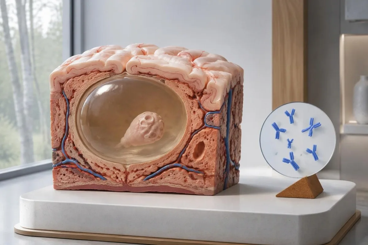
Smernica IDSA/ASTMH odporúča enzýmom viazaný imunotransferový blot, čiže EITB, ako konfirmačné sérologické vyšetrenie a výslovne upozorňuje, že jeho citlivosť je pri jedinom parenchýmovom náleze alebo iba kalcifikáciách nízka (White et al., 2018). Vysokú citlivosť uvádzanú u pacientov s viacerými cystami, komorovým alebo subarachnoidálnym ochorením nemožno prenášať na osobu s 1 malou mozgovou abnormalitou.
Predstavme si čisto na ilustráciu, že test má citlivosť 55 % u 100 ľudí, u ktorých bolo ochorenie s jedinou cystou potvrdené nezávisle od testu. Približne 45 z nich by malo negatívny výsledok napriek tomu, že ochorenie majú; tento názorný príklad vysvetľuje falošne negatívne výsledky, nie pravdepodobnosť, že konkrétny negatívny výsledok znamená ochorenie.
Opakovanie toho istého testu po 2 týždňoch nemusí napraviť slabú odpoveď spôsobenú malým rozsahom ochorenia. To sa líši od infekcií, pri ktorých je hlavným problémom príliš skorý odber vzorky, ako vysvetľuje náš sprievodca falošne negatívnymi výsledkami súvisiacimi s načasovaním; opýtajte sa, či by zobrazovacie vyšetrenie alebo lepšie charakterizovaný test zmenili ďalší postup.
Prečo môže byť kalcifikovaná neurocysticerkóza naďalej symptomatická?
Kalcifikovaná neurocysticerkóza môže spôsobovať záchvaty aj pri negatívnom náleze protilátok, because the remaining mineralized focus can still be associated with local irritation and episodic surrounding edema. Typical calcifications may measure approximately 1–4 mm, but their small size does not establish whether they are clinically harmless.
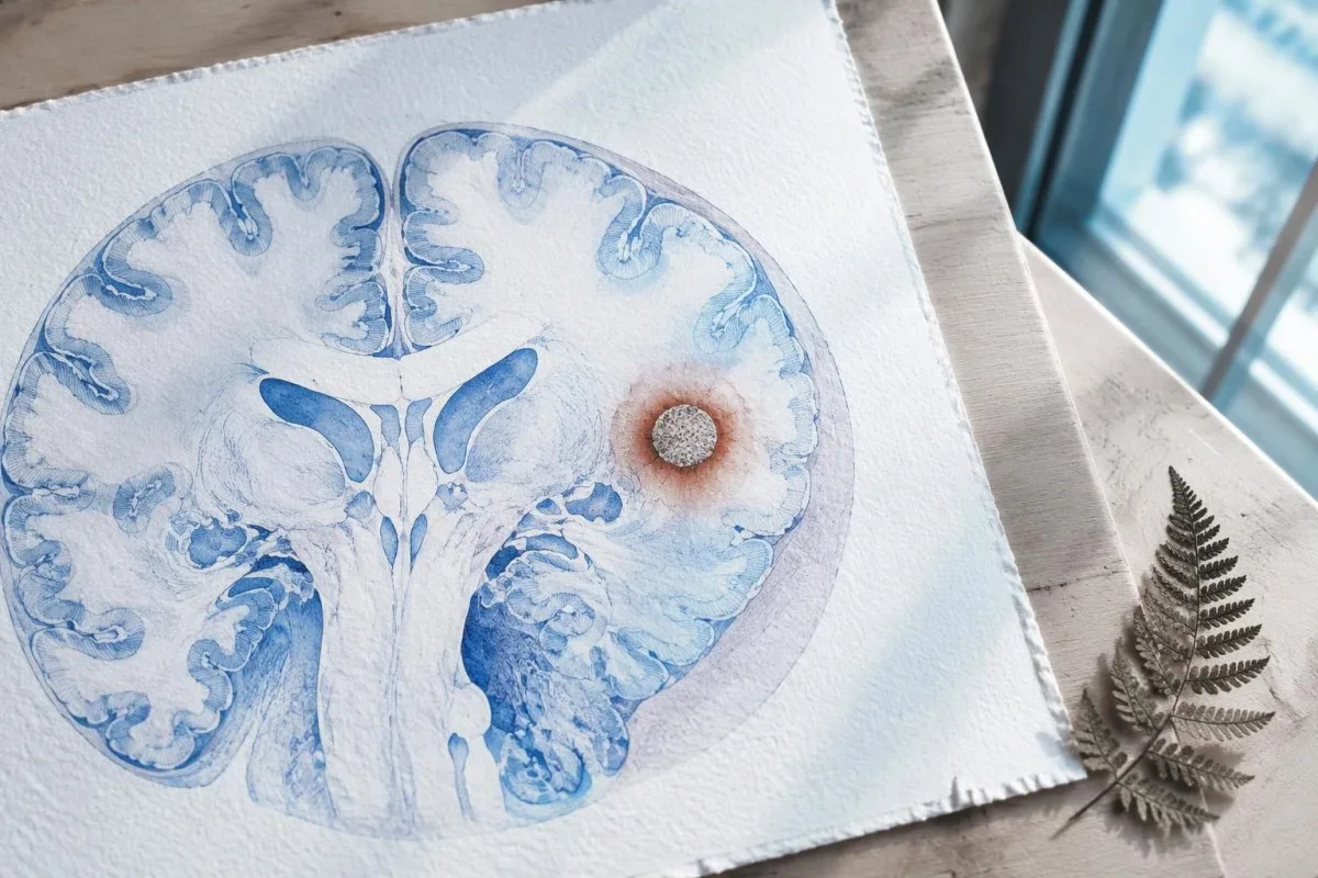
A calcification is generally a remnant rather than a living cyst that antiparasitic medication can eliminate. In the IDSA/ASTMH guideline, calcified-only parenchymal disease is managed symptomatically rather than with antiparasitic drugs; a person with 1 calcified focus and seizures may therefore need antiseizure treatment without needing cyst-killing medication (White et al., 2018).
Surrounding edema can appear intermittently, sometimes long after the original cyst has degenerated. MRI may show this tissue response even when the calcification is more obvious on CT, which is why the 2 scans can tell different but compatible stories rather than one proving that the other is wrong.
Neither a negative antibody result nor a persistent positive result measures seizure risk by itself. The broader lesson resembles other seronegative neurological conditions, discussed in our negative neurological antibody results guide: absence of detectable antibodies is not equivalent to absence of a clinically relevant neurological process.
Ktorý test na protilátky proti cysticerkóze bol vlastne vykonaný?
The useful first question is whether the report describes EITB, ELISA, or another antibody method, because these are not interchangeable tests. A negative index below 1 laboratory's cutoff cannot inherit the performance figures of a different reference-laboratory assay, even when both reports use the name cysticercosis IgG.
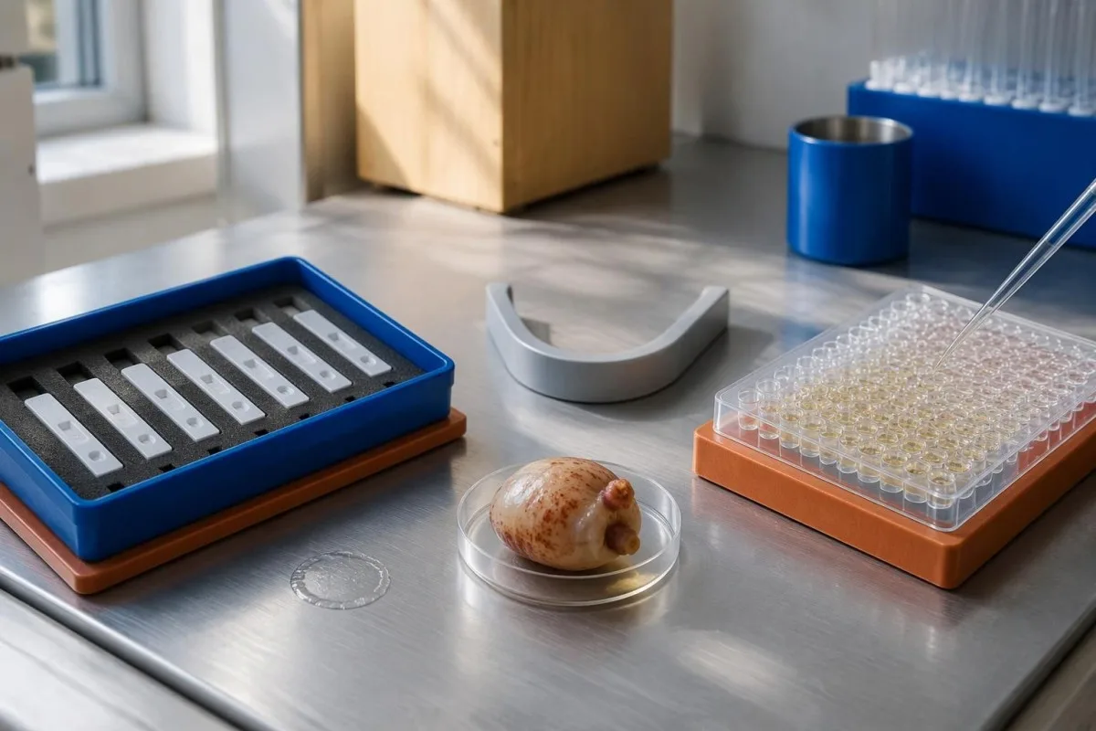
EITB using purified parasite glycoproteins is the preferred serological method in the IDSA/ASTMH guideline, while crude-antigen ELISA methods have recognized sensitivity and specificity limitations (White et al., 2018). Before comparing 2 results, check the method, specimen type, reference interval, and whether the laboratory used a validated confirmatory procedure.
Some laboratories report a qualitative result; others provide an optical-density ratio or numerical index. A value such as 0.8 in a hypothetical assay does not represent 0.8 cysts, an 80% disease probability, or a universal severity score—our kvalitatívne a kvantitatívne výsledky guide explains that distinction.
Kantesti AI's role is to help readers preserve the laboratory's original wording rather than reinterpret 1 method through another method's thresholds. Our sprievodca laboratórnymi značkami provides broader report-reading context; for this result, keep the actual assay name available when discussing referral or repeat testing.
Ako anamnéza expozície mení diagnostické podozrenie?
Neurocysticercosis follows ingestion of Taenia solium eggs, usually through fecal contamination from a person carrying the intestinal tapeworm. Eating undercooked pork can cause intestinal taeniasis, but brain cysts arise through a different transmission step; symptoms may emerge years after exposure rather than within 1 recent trip.

A vegetarian can develop cysticercosis because eggs can contaminate food or hands independently of meat consumption. Useful history includes past residence, household contacts, sanitation conditions, and possible exposure to a tapeworm carrier; asking only about meals eaten during the last 2 weeks can miss the relevant background.
A negative stool examination does not exclude cysts in the brain. Stool testing addresses intestinal tapeworm carriage, whereas CT and MRI assess nervous-system findings; these are 2 different sites and clinical questions, although public-health teams may recommend evaluating contacts when a carrier could be involved.
Normal eosinophils also cannot exclude neurocysticercosis, and raised eosinophils do not identify the brain as the affected site. Our parasite antibody interpretation guide discusses a separate parasite with similar localization limits; the practical point is to avoid using 1 routine cell count as a substitute for targeted neurological evaluation.
Kedy si nové záchvaty vyžadujú urgentné vyšetrenie?
A first known seizure needs urgent medical assessment regardless of antibody results, and emergency services should be contacted for a first seizure or an ongoing seizure with concerning features. A convulsive seizure lasting 5 minutes, repeated seizures without recovery, breathing difficulty, pregnancy, or significant injury requires emergency care.

Do not wait for the 5-minute mark to seek help after a first known seizure. Keep the person away from hazards, cushion the head if safely possible, do not restrain them or put anything in their mouth, and place them on their side once convulsions stop if they are breathing normally.
Persistent confusion, a severe new headache, fever with neck stiffness, or new weakness increases concern for causes beyond cysticercosis. Sudden one-sided weakness or speech difficulty also demands emergency assessment, as our varovných príznakov cievnej mozgovej príhody explains; 1 negative infection test cannot rule out stroke, meningitis, or a structural brain problem.
After a suspected seizure, stop driving and avoid swimming alone, heights, or operating machinery until a clinician provides advice consistent with local rules. If a witness can safely record the duration and describe movements or recovery, those observations may be more useful than ordering a second antibody test before the first neurological assessment.
Čo prináša CT do diagnostiky neurocysticerkózy?
Noncontrast CT is particularly useful for detecting calcified cyst remnants, including small approximately 1–4 mm foci that MRI may show less clearly. CT also helps assess urgent structural problems such as hydrocephalus, but 1 isolated calcification is not automatically proof of neurocysticercosis or the cause of a seizure.
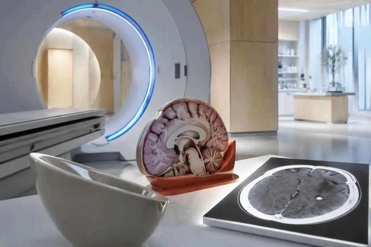
Calcium appears dense on CT, allowing radiologists to identify mineralized remnants and describe their distribution. The IDSA/ASTMH guideline recommends both MRI and noncontrast CT when classifying newly diagnosed neurocysticercosis, because the 2 modalities have complementary strengths rather than being interchangeable alternatives (White et al., 2018).
An incidental calcification needs context: previous granulomatous disease, other causes of mineralization, and scan appearance can influence interpretation. If 1 calcification sits near a suspected seizure focus, concordance with symptoms and EEG may strengthen the explanation, but anatomical proximity alone cannot establish causation.
A laboratory marker used in selected head-injury pathways does not test for brain cysts or replace imaging after an unexplained seizure. Our head-injury CT decisions guide illustrates that narrower setting; ask the clinician whether the present question requires noncontrast CT, contrast imaging, MRI, or a combination of 2 modalities.
Čo odhalí MRI, čo môže CT prehliadnuť?
MRI is better suited to evaluating viable cysts, surrounding edema, and many ventricular or subarachnoid findings. Specialized 3-dimensional sequences such as CISS or FIESTA can improve visualization of cyst membranes within cerebrospinal fluid spaces, where a cyst may otherwise resemble the surrounding fluid.
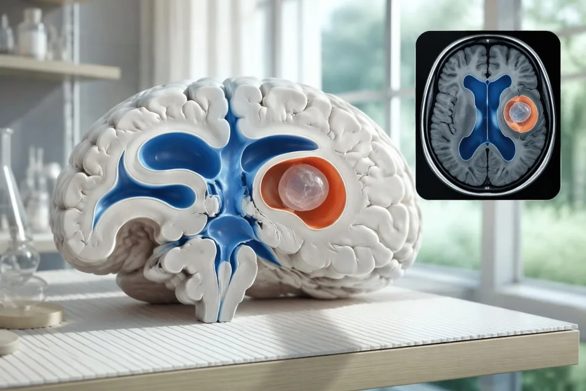
A cyst inside the ventricles can obstruct cerebrospinal fluid flow and cause hydrocephalus even without numerous parenchymal cysts. Headache with vomiting, declining alertness, or new visual symptoms therefore changes urgency; 1 reassuring antibody result should not override symptoms that suggest raised pressure or impaired fluid circulation.
MRI may show a scolex, the small internal structure of the parasite, inside a compatible cyst. This finding can be highly specific when confidently identified, whereas ring enhancement alone has a broader differential; our brain imaging warning signs guide explains why structural abnormalities require interpretation rather than diagnosis from 1 visual feature.
A routine MRI protocol may not answer every cysticercosis question. If ventricular or subarachnoid disease is suspected, discuss whether thin-section 3-dimensional imaging is needed; radiologists choose sequences and contrast according to the clinical question, and a normal scan using 1 protocol does not prove every possible disease compartment has been adequately assessed.
Ako sa diagnostikuje neurocysticerkóza, keď si výsledky testov odporujú?
Neurocysticercosis diagnosis combines imaging, clinical findings, exposure information, and supporting tests, rather than requiring a positive antibody result in every case. The revised diagnostic criteria published in 2017 give characteristic neuroimaging findings a central role and distinguish strong evidence from less-specific supportive features (Del Brutto et al., 2017).
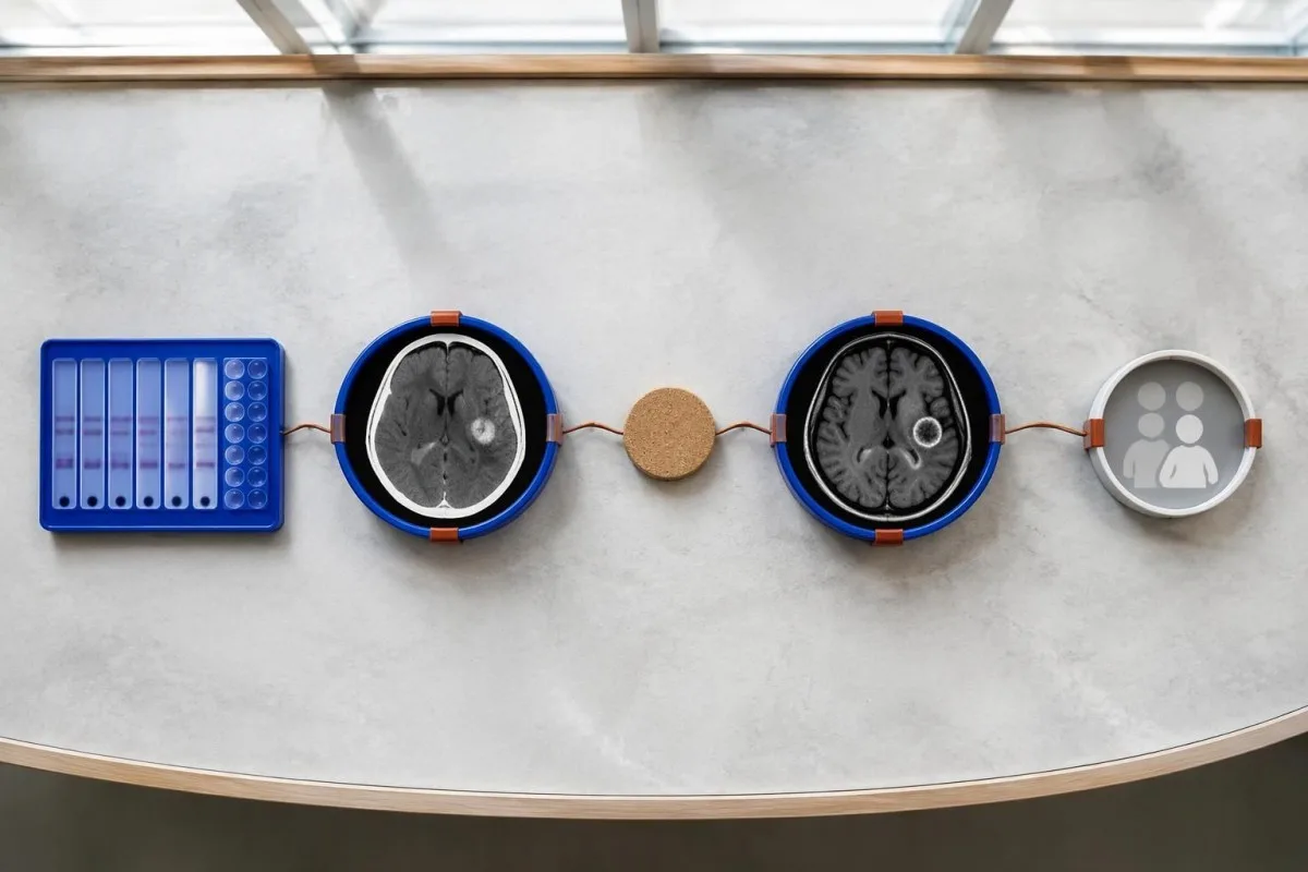
Evidence should be weighted, not tallied. A confidently visualized scolex inside a compatible cyst is different from 1 nonspecific ring-enhancing area, while a positive antibody result is different from direct evidence that a finding is in the brain; our sprievodca k pozitívnym výsledkom protilátok explains why detection alone cannot establish location.
For a clearly hypothetical probability example, start with a 30% pretest probability and assume a single-cyst assay has 55% sensitivity and 99% specificity. In 1,000 comparable people, about 135 affected and 693 unaffected people would test negative, leaving approximately a 16% probability of disease among negative results—still substantial, although these assumed figures are not a personal risk calculator.
The most useful disagreement to investigate is whether each test is answering the same question. A negative antibody test, a CT calcification, and MRI edema can all be compatible with 1 calcified disease process; unexplained discrepancies should prompt review of the assay and images rather than automatic dismissal of either result.
Čo ďalšie môže na zobrazení mozgu pripomínať jednu cystu?
A single ring-enhancing brain finding is not specific to neurocysticercosis. Other possibilities include tuberculoma, an abscess, a neoplasm, or an inflammatory process; clinicians use appearance, location, immune status, symptoms, and sometimes follow-up imaging or tissue examination to distinguish these possibilities rather than relying on 1 antibody panel.
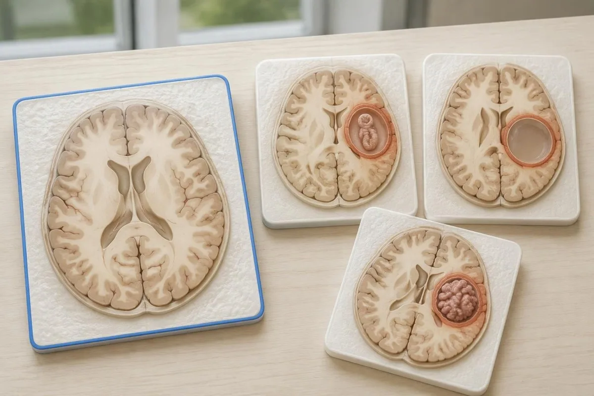
Edema, restricted diffusion, internal structures, and the number of findings all contribute to the differential, but no single nonspecific feature settles every case. Fever and immune suppression may change the suspected cause; a 2 cm ring-enhancing area in a hypothetical immunocompromised person warrants a different investigation from a tiny calcification in an otherwise well person.
Toxoplasmosis is one consideration in selected immunocompromised patients, and a positive IgG result can indicate past exposure rather than prove an active brain process. Our Toxoplasma IgG interpretation guide explains that laboratory distinction; imaging and the patient's immune status determine whether the result is clinically relevant to 1 or more cerebral findings.
New seizures accompanied by rapidly evolving memory problems, psychiatric changes, or abnormal movements can also suggest encephalitis rather than a solitary parasite cyst. Our encephalitis diagnostic pathway describes how MRI, EEG, and cerebrospinal fluid testing answer different questions; a second negative cysticercosis antibody result does not investigate those alternatives.
Môžu testovanie antigénov, PCR alebo vyšetrenie CSF vyriešiť neistotu?
Antigen tests, PCR, and cerebrospinal fluid analysis can add information in selected cases, particularly when extraparenchymal disease is suspected. None is a universal rescue test after 1 negative antibody result: availability, assay validation, disease compartment, and safety of lumbar puncture determine whether additional testing is appropriate.
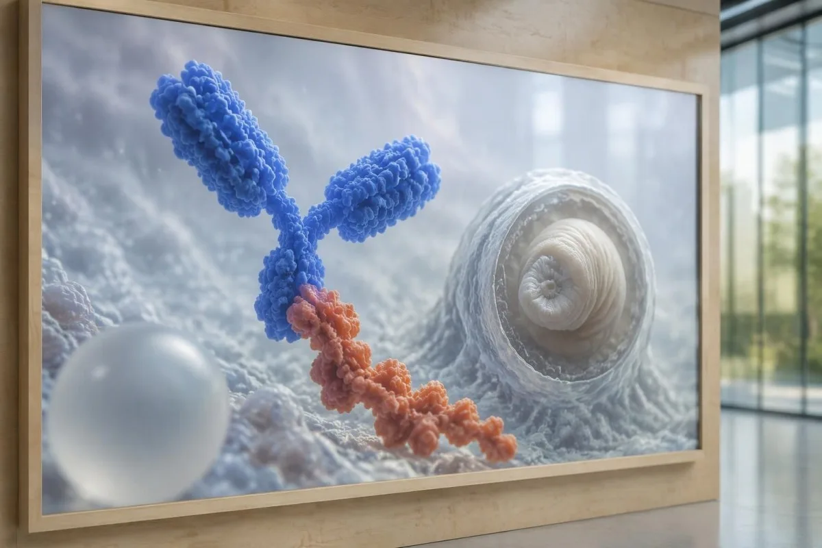
Antibody testing detects the host response, while antigen testing aims to detect parasite-derived material and may better reflect viable disease burden. Antigen detection is often less useful when only 1 small parenchymal cyst or calcifications are present; molecular testing is used in some specialist settings, but performance cannot be generalized across every laboratory or specimen.
Lumbar puncture is not routinely required for an uncomplicated solitary parenchymal finding and can be unsafe when obstructive hydrocephalus or significant mass effect is present. Cerebrospinal fluid tests for other disorders also require disease-specific interpretation, as our CSF band interpretation guide explains; 1 abnormal CSF marker does not identify every possible cause.
Kantesti je platforma na interpretáciu krvného testu AI, not a service that orders lumbar punctures, identifies cysts on scans, or confirms neurological infections. Our technológia interpretácie AI explains the laboratory-report workflow; when 2 tests disagree, our role is to clarify their stated meanings and limitations, not select an invasive diagnostic procedure.
Prečo liečba závisí od štádia a umiestnenia cysty?
Treatment depends on whether cysts are viable, degenerating, calcified, or located outside the brain tissue, not simply on antibody positivity. Selected viable parenchymal cases receive antiparasitic treatment, whereas calcified-only disease generally receives symptom-directed care; ventricular obstruction may require a procedural approach rather than 1 standard medication regimen.
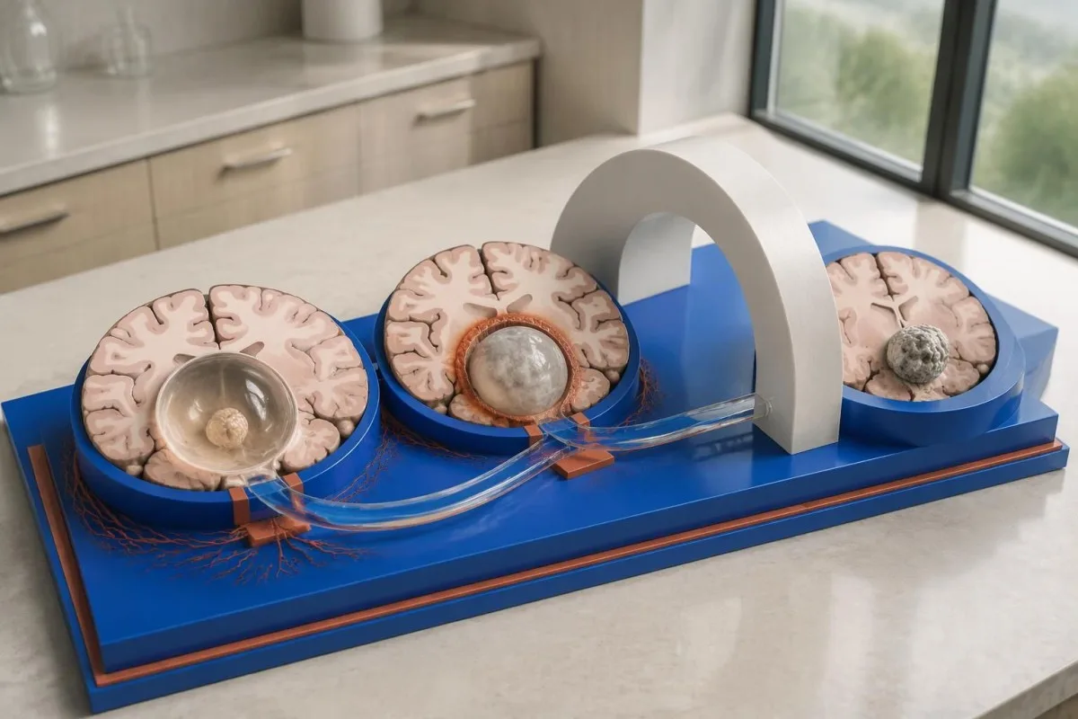
For selected patients with 1–2 viable parenchymal cysts, the IDSA/ASTMH guideline describes albendazole treatment for 10–14 days with appropriate accompanying care. This is a specialist-directed recommendation, not a home-treatment instruction: antiparasitic drugs can provoke an inflammatory response, and untreated hydrocephalus or diffuse cerebral edema changes the order of management (White et al., 2018).
WHO's 2021 guidance focuses on defined presentations of parenchymal neurocysticercosis, while the IDSA/ASTMH guideline also addresses ventricular, subarachnoid, and other forms in detail. Differences in imaging access and disease compartment explain why 1 treatment recommendation cannot be transferred to every patient with an antibody result; the guidelines do not turn negative serology into a rule-out test.
Corticosteroids may accompany antiparasitic therapy, but prolonged use requires additional safety planning, including assessment for relevant latent infections. Our Strongyloides and steroid safety guide explains a separate risk that matters before immune-suppressing treatment; an eye examination is also recommended before antiparasitic therapy to evaluate possible intraocular cysts.
Čo má sledovať následné vyšetrenie okrem výsledkov testov na protilátky?
Follow-up should track seizures, neurological symptoms, imaging changes, and medication safety, rather than use antibody disappearance as proof of recovery. For viable parenchymal cysts, the IDSA/ASTMH guideline suggests repeat MRI at least every 6 months until the cystic component resolves, with earlier reassessment when symptoms change.
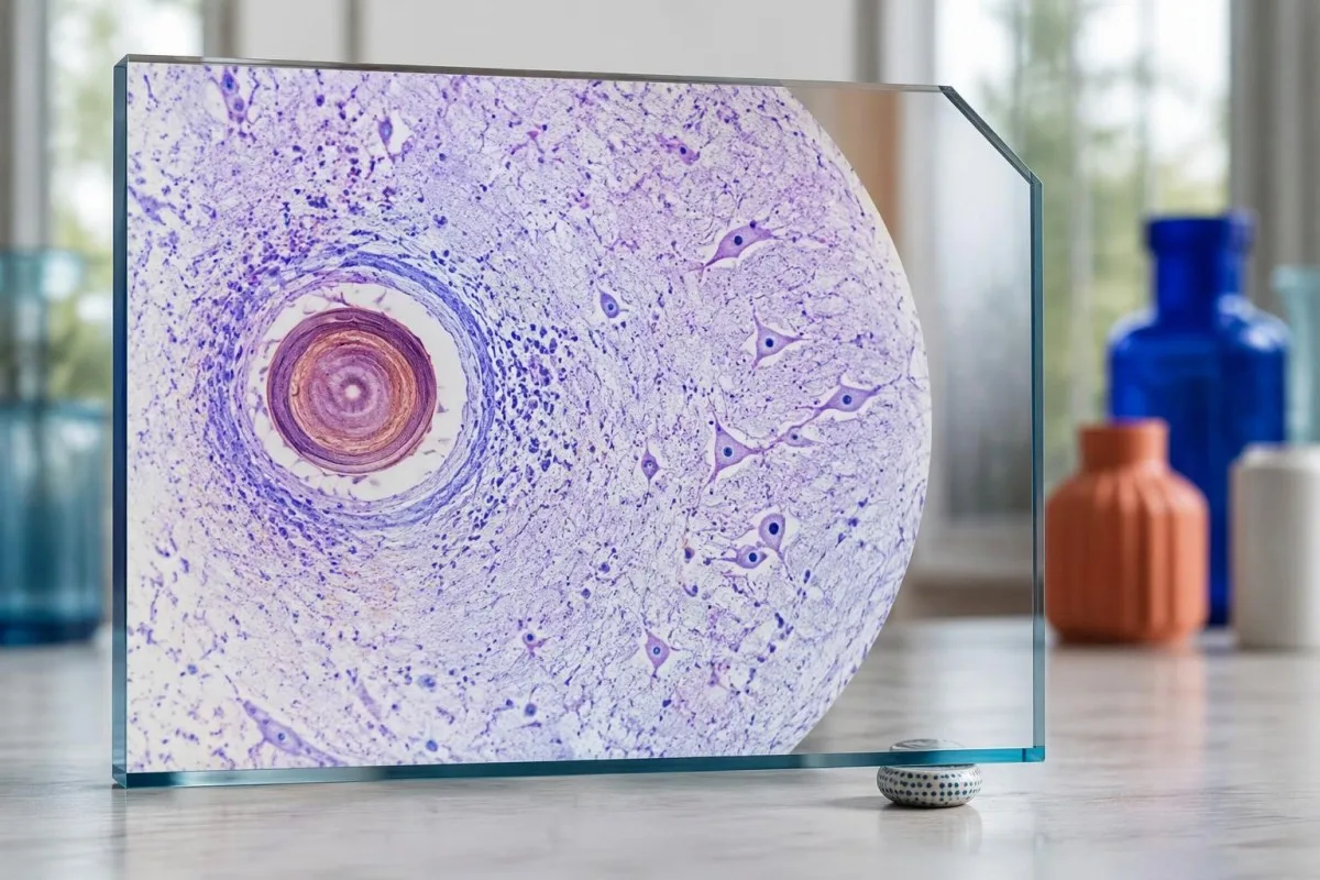
Antibodies may remain detectable after the original disease process has changed, and low-burden disease may never produce a positive result. If 1 new seizure occurs during follow-up, clinicians consider adherence, medication interactions, recurrent edema, and other causes; a rising or falling antibody index alone cannot distinguish among those explanations.
Kantesti AI can help explain reported laboratory results during medication follow-up, but a drug concentration is not a stand-alone measure of seizure control. Our interpretácia hladiny levetiracetamu guide explains why sample timing and kidney function matter; 2 measurements collected at different times after dosing may not be directly comparable.
Monitoring depends on the chosen drug and treatment duration: prolonged albendazole therapy can require liver-test and blood-count surveillance, while some antiseizure medicines can affect sodium. Our sprievodca bezpečnosťou elektrolytov discusses those separate laboratory concerns; do not stop an antiseizure medicine or change its dose because 1 cysticercosis antibody result is negative.
Čo môže vysvetliť Kantesti a čo výskum nedokáže potvrdiť?
AI report interpretation cannot confirm or exclude neurocysticercosis from laboratory results alone. Kantesti je an Nástroj na analýzu krvných testov poháňaný AI whose role here is to organize reported laboratory findings; a negative result still needs clinical context, and 1 new seizure takes priority over reviewing an automated explanation.

Prepare a focused review packet with the original antibody report, assay method, CT or MRI report, symptom timeline, and medication list. These 5 information categories reduce the risk of discussing a result without its context; ask specifically whether the imaging suggests viable, degenerating, calcified, ventricular, or subarachnoid disease and which unanswered question the next test would address.
The 2 Kantesti publications listed below are a repository-deposited global analysis report and an educational RDW guide, not clinical evidence validating neurocysticercosis diagnosis. Our príručka RDW a CBC can support interpretation of a separately ordered blood count, but RDW, MCV, and MCHC do not establish whether a brain cyst is present.
Evidence for the neurological recommendations in this article comes from the cited diagnostic criteria and clinical guidelines, not from general AI benchmarking or laboratory-report volume. Our technical validation scope should be read within its stated boundaries; neither 1 successful report explanation nor a large aggregate dataset substitutes for condition-specific diagnostic validation.
Často kladené otázky
Môže sa pri negatívnom krvnom teste vyskytnúť neurocysticerkóza?
Áno, neurocysticerkóza môže byť prítomná aj napriek negatívnemu krvnému testu na protilátky, najmä pri jednej mozgovej cyste alebo pri ochorení s výlučne kalcifikovanými cystami. Publikované odhady citlivosti testu EITB v referenčných laboratóriách často uvádzajú približne 50–60% pri jednej cyste, čo je podstatne menej než pri viacerých životaschopných cystách. CT, MRI, príznaky a anamnéza expozície preto zostávajú pri diagnostike kľúčové. Nový záchvat si vyžaduje urgentné vyšetrenie; negatívny výsledok nie je dôvodom na upokojenie.
Ktorý test na neurocysticerkózu je najspoľahlivejší?
Žiadny jednotlivý test na neurocysticerkózu neodpovedá na všetky diagnostické otázky; zobrazovacie vyšetrenia mozgu a vhodne zvolené sérologické testy sa vzájomne dopĺňajú. EITB je podľa usmernení IDSA/ASTMH preferovanou potvrdzujúcou metódou na detekciu protilátok, no môže nerozpoznať ochorenie, pri ktorom je prítomná iba 1 cysta. Natívne CT je obzvlášť užitočné pri zisťovaní kalcifikácií, zatiaľ čo MRI lepšie hodnotí mnohé životaschopné cysty, edém a abnormality v priestoroch s mozgovomiechovým mokom. Lekári tieto nálezy vyhodnocujú spoločne a nepovažujú jediný negatívny výsledok za definitívny.
Je pri neurocysticerkóze lepšie CT alebo MRI?
CT a MRI majú pri diagnostike neurocysticerkózy odlišné prednosti. Natívne CT často zreteľnejšie zachytí drobné kalcifikácie s priemerom približne 1–4 mm, zatiaľ čo MRI umožňuje posúdiť životaschopné cysty, okolitý edém a postihnutie komôr alebo subarachnoidálneho priestoru. Špecializované trojrozmerné sekvencie MRI môžu pomôcť identifikovať cystové membrány v priestoroch s mozgovomiechovým mokom. Odporúčania IDSA/ASTMH navrhujú pri klasifikácii novodiagnostikovaného ochorenia použiť obidve zobrazovacie metódy.
Môže kalcifikovaná cysta stále spôsobovať záchvaty?
Kalcifikovaný zvyšok cysticerkózy môže aj naďalej súvisieť so záchvatmi, hoci už nejde o životaschopnú cystu. Okolo niektorých kalcifikovaných ložísk, často s priemerom iba 1–4 mm, sa môže občas vyvinúť okolitý edém viditeľný na MRI. Negatívne protilátky tento mechanizmus nevylučujú a pretrvávajúca pozitivita protilátok nevyjadruje riziko záchvatov. Pri ochorení s výlučne kalcifikovanými ložiskami sa spravidla volí symptomatická liečba namiesto antiparazitárnej medikácie.
Mám zopakovať negatívny test na protilátky proti cysticerkóze?
Opakovanie negatívneho testu na protilátky proti cysticerkóze nie je automaticky najlepším ďalším krokom. Pri 1 malej cyste alebo kalcifikovanom ochorení môže byť protilátková odpoveď slabá a výsledok môže zostať negatívny, aj keď sa rovnaký test zopakuje po 2 týždňoch. Lekár môže namiesto toho prehodnotiť pôvodnú metódu, požiadať o lepšie charakterizovaný konfirmačný test alebo uprednostniť CT a MRI. Pri nových záchvatoch alebo zhoršujúcich sa neurologických príznakoch sa nemá čakať na opakovanie testu.
Kedy mám pri prvom epileptickom záchvate vyhľadať urgentnú pomoc?
Prvý známy záchvat si vyžaduje urgentné lekárske vyšetrenie a kontaktovanie miestnej záchrannej služby bez ohľadu na negatívny výsledok testu na protilátky proti neurocysticerkóze. Kŕčový záchvat trvajúci 5 minút, opakované záchvaty bez nadobudnutia vedomia, ťažkosti s dýchaním, tehotenstvo alebo závažné poranenie predstavujú stav vyžadujúci neodkladnú pomoc. Osobu nedržte nasilu ani jej nič nevkladajte do úst; keď kŕče ustanú a osoba normálne dýcha, uložte ju na bok. Nešoférujte, kým lekár nevyhodnotí udalosť a nevysvetlí vám príslušné obmedzenia.
Získajte vysvetlenie výsledkov krvných testov
Nahrajte svoj laboratórny nález a AI nástroja Kantesti vám v priebehu niekoľkých sekúnd vysvetlí každý biomarker jednoduchým jazykom. Používa ho viac než 2 milióny ľudí na celom svete.
Referencie
- Klein, T., Mitchell, S., & Weber, H. (2026). AI analýza krvi: 2,5 mil. analyzovaných testov | Global Health Report 2026. Kantesti AI lekársky výskum. DOI
- Klein, T., Mitchell, S., & Weber, H. (2026). Krvný test RDW: kompletný sprievodca hodnotami RDW-CV, MCV a MCHC. Kantesti AI lekársky výskum. DOI
- White AC Jr et al. (2018). Diagnosis and Treatment of Neurocysticercosis: 2017 Clinical Practice Guidelines by the Infectious Diseases Society of America (IDSA) and the American Society of Tropical Medicine and Hygiene (ASTMH). Clinical Infectious Diseases. DOI
- Del Brutto OH et al. (2017). Revised diagnostic criteria for neurocysticercosis. Journal of the Neurological Sciences. DOI
- World Health Organization (2021). WHO guidelines on management of Taenia solium neurocysticercosis. Svetová zdravotnícka organizácia.
- 2 milióny+Analyzované testy
- 127+Krajiny
- 100+Jazyky
O zdravotníckom tíme


Signály dôvery E-E-A-T Trust Signals
Skúsenosti
Klinické hodnotenie vedené lekárom pracovných postupov interpretácie laboratórnych výsledkov.
Odbornosť
Laboratórna medicína so zameraním na to, ako sa biomarkery správajú v klinickom kontexte.
Autoritatívnosť
Lekársky recenzoval Dr. Thomas Klein, s príspevkami Dr. Sarah Mitchell a prof. Dr. Hansa Webera.
Dôveryhodnosť
Interpretácia založená na dôkazoch s jasnými následnými krokmi na zníženie poplachu.





Zdravotnícke upozornenie
Tento článok je určený len na vzdelávacie účely a nepredstavuje lekárske poradenstvo. Pri rozhodnutiach o diagnostike a liečbe sa vždy poraďte s kvalifikovaným poskytovateľom zdravotnej starostlivosti.
Publikované · Kontaktujte redakčný tím
Vydáva Kantesti Ltd, registrovaná v Anglicku a Walese, číslo spoločnosti. 17090423, Londýn, Spojené kráľovstvo.
