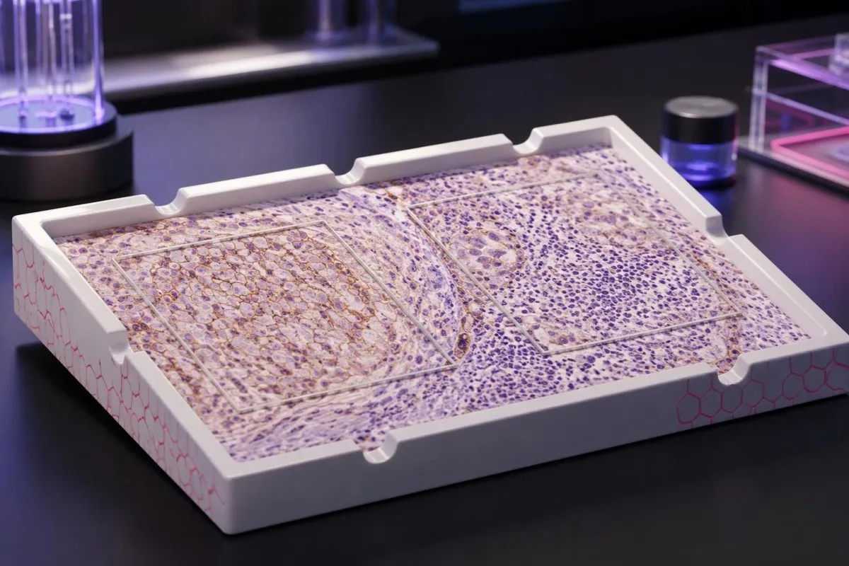
PD-L1スコアは組織サンプルにおける染色の程度を示すものであり、治療が成功する個人的な可能性を示すものではありません。スコアリング方法、がんの診断、提案される薬剤の併用によって、その結果から判断できる内容が決まります。

要点
- PD-L1検査結果 はがん組織におけるタンパク質の染色を測定するもので、通常の血液検査結果でも、がん検診でもありません。
- TPS は、条件を満たす膜染色が認められる生存腫瘍細胞の割合で、0%から100%で報告されます。
- CPS は、条件を満たすPD-L1陽性の腫瘍細胞、リンパ球、マクロファージを数え、生存腫瘍細胞数で割って100を掛けた値です。報告されるスコアの上限は100です。
- スコアが異なれば 変換できません:TPS 10%は、免疫細胞の染色状況によって、CPS 10、CPS 25、または別の値に対応する可能性があります。
- 閾値 たとえばTPS 1%、TPS 50%、CPS 1、CPS 10などは、特定のがんや治療の状況に適用されるものであり、すべての免疫療法に当てはまるわけではありません。
- 陽性結果は 治療選択の参考にはなりますが、効果を保証するものではありません。スコアが50でも、奏効確率が50%という意味ではありません。
- 陰性の結果 免疫療法が必ずしも除外されるわけではありません。PD-L1陽性を要件としない併用療法やバイオマーカーに基づく治療もあるためです。
- 検体の適格性 は重要です。一般的に用いられるスコアリング法のいくつかでは、生存腫瘍細胞が少なくとも100個必要ですが、検査室は適応症ごとの規則を適用する必要があります。
PD-L1検査結果から実際に何がわかるのでしょうか?
PD-L1検査結果 は、がん組織におけるタンパク質の染色を示します。 TPS 染色された腫瘍細胞の割合を数えます。 CPS は、選択された染色免疫細胞も含めます。いずれのスコアも、それだけで治療を決定するものではありません。関連するカットオフ値は、がんの種類、病期、検査法、治療レジメンによって異なり、陽性・陰性の結果が効果や治療除外を保証するものではありません。
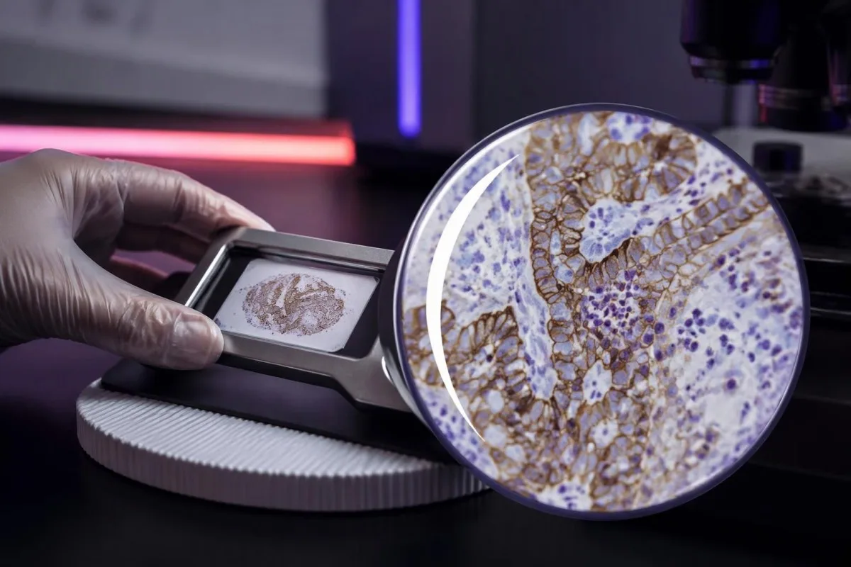
PD-L1、すなわちプログラム細胞死リガンド1は、免疫細胞上のPD-1受容体に結合して、その活性を低下させることがあります。この相互作用を標的とする薬剤は、抗がん免疫活性を回復させる可能性がありますが、1つのタンパク質を測定しても、免疫細胞ががんを認識し、制御するかどうかを決めるすべての要因を捉えることはできません。
組織のPD-L1検査結果は通常、 免疫組織化学, によって得られます。これは抗体を用いて、保存された試料内でのタンパク質の位置を明らかにする方法です。報告される数値は選択された組織切片上の細胞を示すもので、全身に存在するすべてのがん細胞を対象とするわけではありません。また、異なる部位から採取した2つの試料では、結果が異なる場合があります。
カンテスティは AI血液検査分析装置, であり、組織病理診断や腫瘍治療の判断に代わるものではありません。当社の 組織および臨床スコープ は、検査結果に関する教育的支援と、病理検査報告書の原本、画像診断、検討中の治療レジメンを併せて確認する必要がある判断とを区別しています。
数値を解釈する前に、がんの診断名、スコアがCPSかTPSか、使用された検査法、検討中の治療という4つの情報を確認してください。「PD-L1陽性」とだけ記載された報告書には、臨床的に重要な情報が不足しています。スコアリング法と数値を確認してください。
PD-L1 TPSスコアの意味:腫瘍細胞の割合
腫瘍細胞割合スコア, 、 または TPS, は、PD-L1の適格な部分的または完全な膜染色が認められる生存腫瘍細胞の割合です。TPSは 0%~100%; の範囲で、がん細胞の隣に強く染色された免疫細胞があっても、分子には含めません。
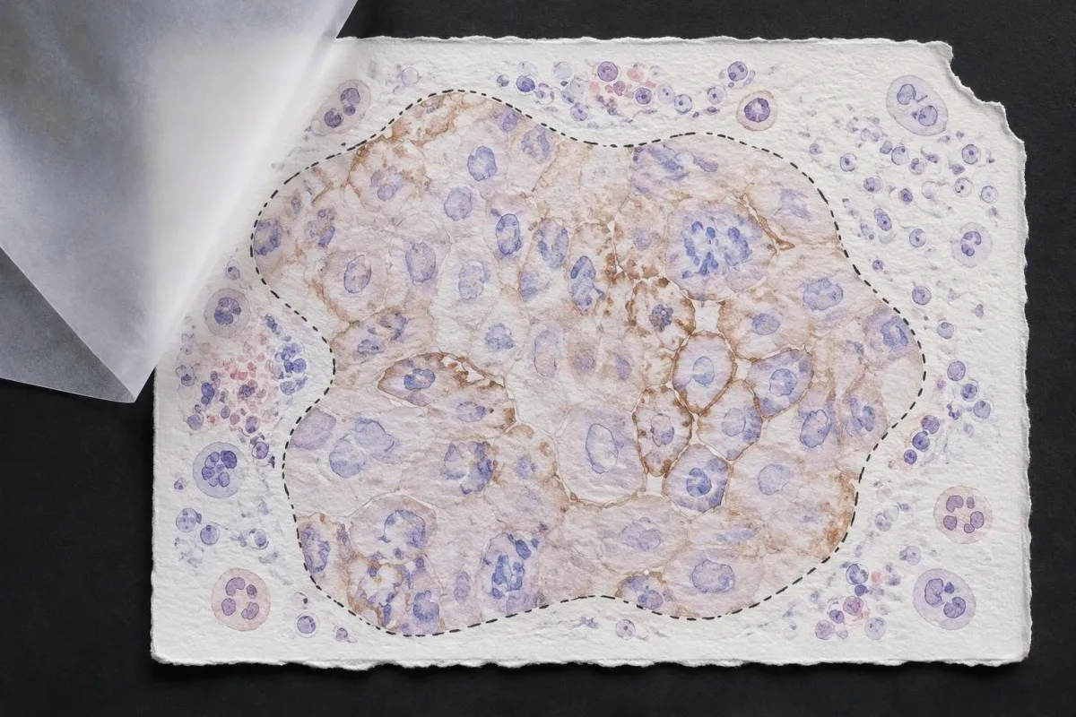
TPS = PD-L1陽性の生存腫瘍細胞数 ÷ 生存腫瘍細胞総数 × 100%。 仮想的な組織視野に生存腫瘍細胞が200個含まれる場合、適格な陽性細胞が80個でTPS 40%となります。近くの染色リンパ球はこの計算を変えませんが、別のスコアリング法には影響する可能性があります。
Membrane location matters more than visual darkness alone. For commonly used TPS methods, weak qualifying membrane staining can count, whereas staining limited to tumor-cell cytoplasm does not establish a positive tumor cell; TPS 1% therefore describes the fraction of qualifying cells, not a required intensity grade.
Cutoffs such as TPS 1% そして TPS 50% were used to define populations in particular lung-cancer trials and treatment pathways. They are not boundaries between harmless and dangerous cancer, and crossing 50% does not create an abrupt biological switch from treatment resistance to guaranteed sensitivity.
Read the exact reporting format: TPS <1%, TPS 1–49%, and TPS ≥50% provide different detail from a single positive flag. Our explanation of 定性検査結果と定量検査結果 covers why a category cannot replace a number, although PD-L1 uses tissue-specific—not blood-reference-range—rules.
PD-L1 CPSスコアの意味:免疫細胞が数えられる理由
Combined positive score, 、 または CPS, includes qualifying PD-L1-positive tumor cells, lymphocytes, and macrophages. CPS divides their combined count by the number of viable tumor cells and multiplies by 100; the denominator does not become all cells simply because immune cells enter the numerator.
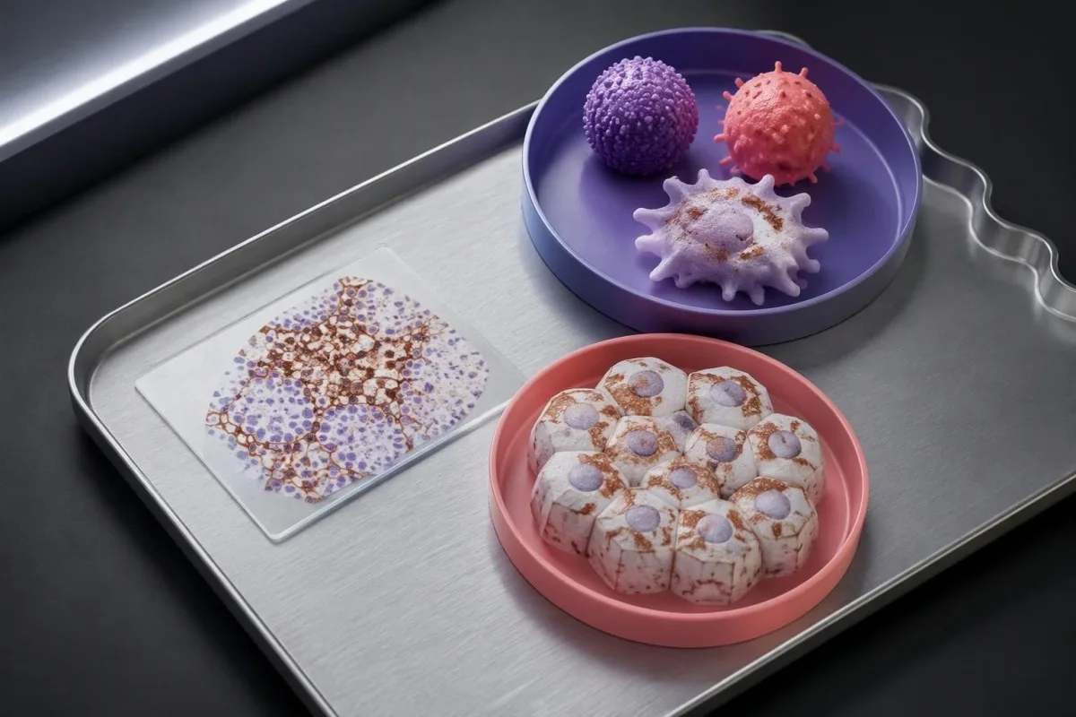
CPS = positive tumor cells plus qualifying positive lymphocytes and macrophages ÷ viable tumor cells × 100. In a hypothetical sample with 500 viable tumor cells, 50 positive tumor cells, and 75 qualifying positive immune cells, CPS is 25; the tumor-cell-only TPS would be 10%.
The reported CPS is capped at 100, even if the uncapped arithmetic would exceed 100 because many qualifying immune cells surround relatively few tumor cells. CPS is a unitless score, not a percentage of all cells, a blood concentration, or a probability that immunotherapy will work.
Not every stained immune cell belongs in the CPS count. The pathologist applies the assay manual's rules for eligible lymphocytes and macrophages associated with the tumor; neutrophils, unrelated inflammatory areas, and nonviable tissue are not simply added because a slide contains visible staining.
A CPS of 20 can arise from more than 1 cellular pattern: abundant tumor-cell staining, substantial immune-cell staining, or both. This denominator issue resembles the distinction between cell counts and percentages, but a circulating monocyte count cannot supply the tissue information needed to calculate CPS.
CPSとTPSを置き換えたり換算したりできない理由
CPS and TPS are not interchangeable because they count different cells and were validated in different treatment settings. A TPS of 10% cannot be converted reliably into CPS 10, and a CPS of 20 does not establish that 20% of tumor cells stain.
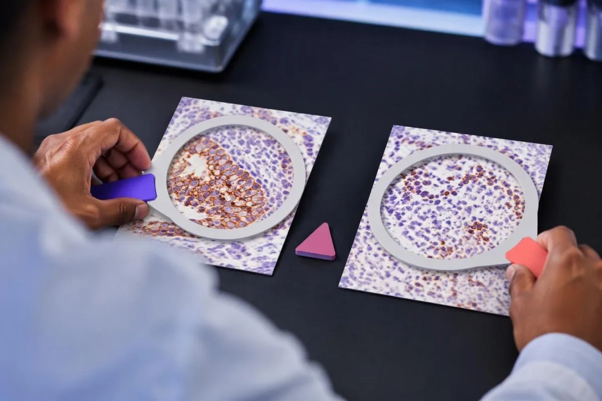
Consider 2 hypothetical specimens with 500 viable tumor cells and 50 positive tumor cells each. The first has no qualifying positive immune cells and gives TPS 10% with CPS 10; the second contains 75 qualifying positive immune cells and gives the same TPS 10% with CPS 25.
The reverse inference fails too. A hypothetical CPS 20 could result from 20 positive tumor cells among 100 tumor cells, or from 5 positive tumor cells plus 15 qualifying positive immune cells; those examples have TPS 20% and TPS 5%, respectively, despite identical CPS values.
アン immune-cell score, sometimes abbreviated IC, is another system rather than a third spelling of CPS. Some IC methods measure the proportion of tumor area occupied by stained immune cells, so an IC result of 1% must be interpreted using its own assay and indication—not a TPS threshold.
Kantesti's educational explanation should preserve all 3 scoring labels rather than flatten them into positive or negative. When using AI report explanations safely, check that the original score, assay, and specimen remain visible; an AI-generated conversion without the underlying tissue counts is not clinically dependable.
スコア以外に病理報告書のどの詳細が重要ですか?
A usable PD-L1 report should identify the specimen, assay, scoring method, numerical result, and adequacy assessment. Several commonly used protocols require at least 100 viable tumor cells, but minimum counts and acceptable specimen types depend on the particular test and cancer indication.

Antibody identifiers such as 22C3, 28-8, SP263, and SP142 describe different testing assays or antibody clones, not different PD-L1 proteins. Some assays show substantial agreement in selected settings, but laboratories should not assume universal equivalence across tumor types, scoring methods, immune-cell assessments, and treatment requirements.
The specimen date and site can change the interpretation of 1 reported number. A small primary-site sample taken before treatment may not represent a later metastatic deposit; ask whether the report describes the current treatment-setting specimen or archived tissue that the oncology team considers suitable.
Insufficient tissue is not PD-L1 negative. A sample with too few viable tumor cells, failed controls, extensive tissue change, or unsuitable processing may be unreportable; the practical next step is laboratory clarification, additional sections, or another suitable specimen—not assigning TPS 0% or CPS 0.
Comparisons between 2 reports need more than matching dates. Our test timing and comparability guide explains the general problem of comparing unlike measurements; for PD-L1, the extra checks are anatomical site, prior treatment, fixation, tissue adequacy, assay, and scoring system.
がんの種類によって陽性の意味はどのように変わりますか?
PD-L1 positivity is indication-specific, not a universal diagnosis. TPS thresholds such as 1% or 50% are familiar in non-small-cell lung cancer, while CPS thresholds such as 1, 5, or 10 appear in selected other cancer settings; the drug, stage, and regimen must accompany the cutoff.

For recurrent or metastatic head-and-neck squamous-cell carcinoma, KEYNOTE-048 evaluated CPS-defined populations. Pembrolizumab alone improved median overall survival in the CPS ≥20 group to 14.9 months versus 10.7 months with the comparator regimen, showing why a CPS threshold cannot be borrowed from a lung-cancer TPS report (Burtness et al., 2019).
Selected established pembrolizumab pathways use CPS ≥10 for unresectable locally advanced or metastatic triple-negative breast cancer with chemotherapy, and CPS ≥1 for certain persistent, recurrent, or metastatic cervical-cancer regimens. Those examples do not establish eligibility for every earlier-stage setting, drug combination, country, or funding pathway.
Gastric and gastroesophageal-junction cancers require particular care because treatment selection may also involve HER2 そして MSI/MMR, with PD-L1 requirements differing across regimens and jurisdictions. A serum marker described in our CA 72-4 interpretation guide cannot replace the tissue biomarker panel or define CPS.
Cancer diagnosis and treatment selection are 2 separate steps. Our persistent cough assessment guide addresses when lung symptoms need investigation; once cancer is confirmed, ask the oncology team to match the PD-L1 result to the current local product information and clinical pathway.
治療レジメンがカットオフ値と同じくらい重要な理由
Immunotherapy alone and immunotherapy combinations can have different PD-L1 requirements. A TPS below 1% may rule out one PD-L1-selected monotherapy pathway without ruling out an appropriate chemotherapy–immunotherapy combination; these are different clinical decisions, not contradictory interpretations of the same result.
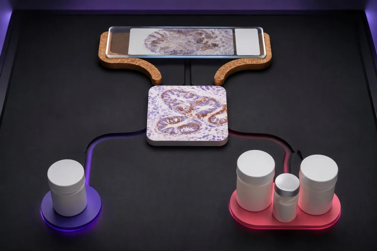
KEYNOTE-024 enrolled previously untreated advanced non-small-cell lung cancer with TPS ≥50% and no sensitizing EGFR mutation or ALK rearrangement. Median progression-free survival was 10.3 months with pembrolizumab versus 6.0 months with chemotherapy, supporting that specific high-TPS monotherapy setting—not every cancer with a score above 50 (Reck et al., 2016).
では、 KEYNOTE-189, pembrolizumab plus pemetrexed and platinum chemotherapy improved outcomes in previously untreated metastatic nonsquamous non-small-cell lung cancer without EGFR or ALK alterations. Twelve-month overall survival was 69.2% versus 49.4%, and benefit was observed across PD-L1 categories, including TPS <1% (Gandhi et al., 2018).
Combination treatment may address rapid disease progression differently from a single agent, but adding drugs also adds toxicity and monitoring needs. If a proposed regimen includes fluorouracil or capecitabine, 化学療法前のDPYD検査 concerns treatment safety; it does not measure the PD-L1 pathway or predict the same benefit.
カンテスティは AI血液検査の読み方プラットフォーム that helps explain supporting laboratory measurements; it does not select between 1-drug and multidrug cancer regimens. Similarly, UGT1A1 and irinotecan safety address metabolism-related toxicity, illustrating why each biomarker should be matched to the specific medicine under discussion.
PD-L1陽性の結果があれば、免疫療法の効果は保証されますか?
A positive PD-L1 result does not guarantee a response. In KEYNOTE-024's selected high-expression lung-cancer population, the objective response rate with pembrolizumab was 44.8%—substantial benefit for many participants, but clearly not a response in every person with TPS ≥50%.
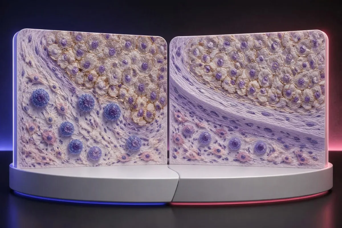
The 44.8% response rate in KEYNOTE-024 describes a trial population under defined conditions, not an individual's calculated chance of success (Reck et al., 2016). Objective response also means measurable tumor shrinkage by trial criteria; stable disease and survival benefit are different outcomes that should not be collapsed into that single percentage.
PD-L1 is only 1 part of a broader immune environment. A cancer can show substantial staining while lacking effective immune-cell access, recognizable targets, or intact antigen presentation; these mechanisms help explain why expression enriches for benefit in some settings without functioning as a deterministic on–off switch.
Higher does not always mean better across different cancers and combinations. A CPS 30 in one disease cannot be ranked against TPS 60% in another, and the same CPS threshold may have different predictive value when used for monotherapy versus a combination that already provides benefit through other mechanisms.
Some melanoma immunotherapy regimens do not require a positive PD-L1 test, so a low score should not be interpreted using lung-cancer monotherapy rules. Our melanoma warning-sign guide addresses recognition and assessment, while treatment benefit must be discussed using disease-specific trial evidence and the person's overall condition.
PD-L1陰性でも免疫療法を受けられる場合はありますか?
PD-L1-negative cancers can still have appropriate immunotherapy options. TPS <1% or CPS <1 may exclude a particular PD-L1-selected regimen, but some combinations, earlier-stage pathways, and treatments selected by other biomarkers do not require PD-L1 positivity.
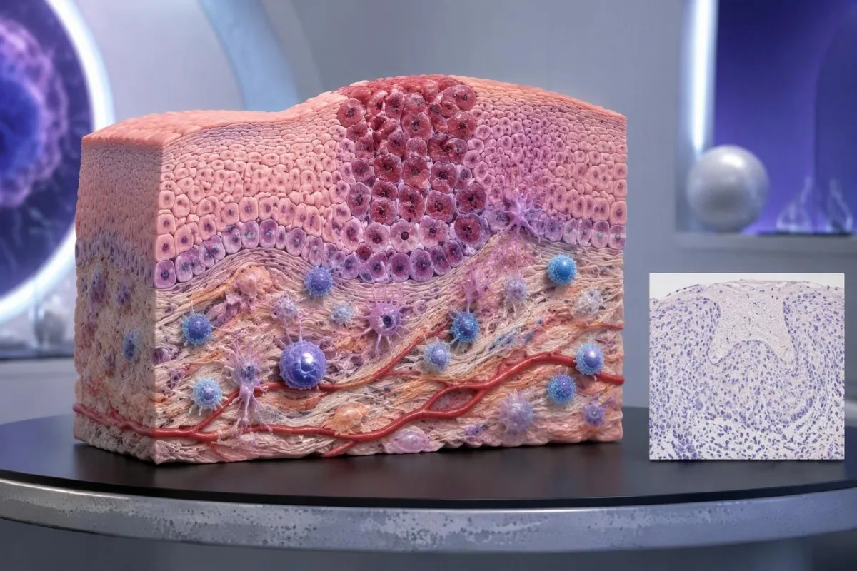
PD-L1-negative should first be translated into the laboratory's actual definition. TPS <1%, CPS <1, and an assay-specific IC-negative category are not identical measurements; an unreportable or inadequate sample is a fourth situation and should not be grouped with a technically valid negative result.
アン MSI-high or mismatch-repair-deficient cancer may support particular immunotherapy pathways without PD-L1 selection, depending on the disease, stage, prior treatment, and local authorization. Tumor MMR testing and リンチ症候群の遺伝学的検査 answer related but different questions: an abnormal tumor result does not automatically prove an inherited syndrome.
Other genetic findings may point toward targeted treatment or different planning rather than immunotherapy. A result discussed in our BRCA variant interpretation guide does not turn CPS 0 into CPS 10; clinicians combine distinct biomarkers without pretending they measure the same biological feature.
No PD-L1 requirement is not the same as no selection criteria. A regimen can still require a particular histology, stage, treatment history, organ function, or absence of an actionable driver alteration; ask whether 1 negative score changes only the proposed option or the broader treatment plan.
検体採取や境界域のスコアを理由に再評価が必要となるのはどのような場合ですか?
Sampling variation can change PD-L1 classification, especially near a treatment threshold. A CPS of 9 versus 10, or TPS 0.8% versus 1%, may prompt pathology review when the distinction would change an otherwise suitable treatment—but repeating every result is not automatically useful.
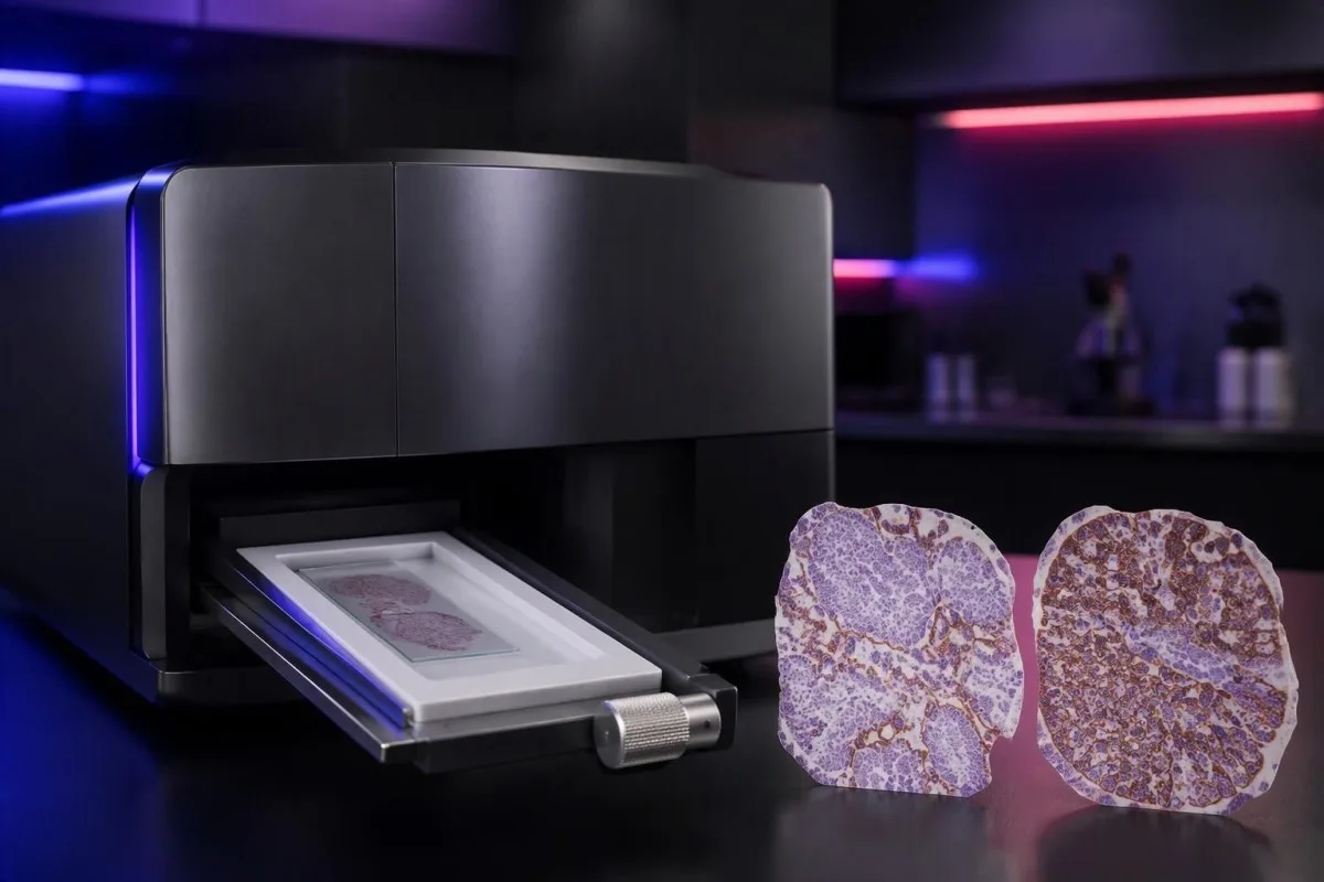
PD-L1 staining can be heterogeneous within 1 tissue block and between primary and metastatic sites. A small tissue examination may miss a highly expressing area or overrepresent it; the pathologist assesses the available viable tissue rather than assuming a single selected field describes the whole cancer.
Pretreatment and post-treatment specimens are not always equivalent. Chemotherapy, radiotherapy, and changes in the immune environment can alter expression, while decalcification of some bone specimens can impair antigen detection; ask whether a lower result reflects biology, processing limitations, or simply a different sampled site.
Borderline results require the actual assay rules, not homemade averaging. If 2 reports show CPS 8 and CPS 12, calculating CPS 10 does not create a newly validated result; the laboratory and oncology team should decide which specimen and assessment are appropriate for the treatment indication.
Kantesti cannot verify viable-cell counts, staining controls, or tissue suitability from a score alone. Our 臨床的な検証と監督 should be read in that limited context; when another tissue procedure is considered, its risks and likely impact on treatment need to be weighed against the uncertainty in the existing result.
PD-L1とあわせて確認すべき他のバイオマーカーは何ですか?
Other biomarkers can take priority over PD-L1, depending on the cancer. In advanced non-small-cell lung cancer, molecular alterations such as EGFR mutations and ALK rearrangements affect treatment planning; in selected other cancers, HER2, MSI/MMR, or additional molecular findings may change the preferred pathway.

A high TPS does not cancel an actionable molecular finding. The pivotal KEYNOTE-024 そして KEYNOTE-189 populations excluded relevant EGFR or ALK alterations, so their results should not be applied unchanged to those groups; treatment sequence also matters because some targeted therapies and checkpoint inhibitors can interact through toxicity.
Tumor mutational burden, often measured in mutations per megabase, is a different biomarker from PD-L1. A threshold such as 10 mutations/Mb has been used in a specific tissue-agnostic treatment context, but assay differences, cancer type, authorization, and clinical setting prevent it from becoming a universal immunotherapy rule.
A liquid molecular test may find circulating tumor DNA, but it does not automatically replace tissue PD-L1 immunohistochemistry. The 2 tests measure different things—genetic material versus protein location within cells—and a negative liquid result may also reflect limited DNA shedding rather than absence of a tumor alteration.
Kantesti AI distinguishes the role of supporting laboratory markers from tissue-based treatment biomarkers in our 血液バイオマーカーの参照ガイド. A blood-cell ratio, LDH level, or serum tumor-marker concentration should not be used to manufacture TPS 50%, estimate CPS 10, or bypass an indicated molecular workup.
免疫療法を検討する場合、どのような安全性確認が重要ですか?
PD-L1 scores predict neither treatment safety nor immune-related toxicity. A TPS of 90% does not make checkpoint treatment safer than TPS 1%; the team assesses medical history, organ function, medications, and the particular regimen separately from the tissue biomarker.
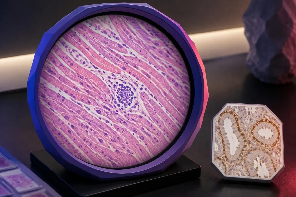
Checkpoint inhibitors can cause immune-related effects involving the thyroid, liver, lungs, bowel, kidneys, heart, and other organs. Baseline testing commonly includes liver chemistry, creatinine, thyroid measurements, and a complete blood count, with monitoring schedules varying by regimen; 1 normal baseline panel does not prevent later toxicity.
New chest pain, palpitations, fainting, or breathlessness during treatment requires prompt assessment because myocarditis, although uncommon, can be serious. Our myocarditis test interpretation guide explains why troponin supports evaluation but cannot determine the cause by itself; severe symptoms warrant emergency care rather than waiting for the next scheduled panel.
Kidney changes need interpretation against the person's own baseline and treatment exposures. A hypothetical creatinine rise from 0.8 to 1.6 mg/dL is a doubling even if the absolute value looks only modestly elevated; our kidney function testing guide explains supporting measurements, not an immunotherapy-specific treatment decision.
Pre-existing autoimmune disease, organ transplantation, ongoing immunosuppression, or previous severe immune-related toxicity can complicate treatment planning even when CPS is 100. Report new persistent diarrhea, worsening cough, jaundice, unusual weakness, or marked thirst promptly; neither a high PD-L1 score nor previously reassuring blood results justify delaying symptom assessment.
腫瘍科の診察で何を尋ねるべきですか?
Bring the complete PD-L1 pathology report and ask which exact treatment decision it informs. Four useful starting questions are: Which score was used? Which cutoff applies? Is the specimen adequate? What options remain if the result is below the cutoff?

A hypothetical report reading 22C3, CPS 12, adequate tissue is still incomplete as a treatment answer until the cancer, stage, and regimen are specified. Ask whether CPS ≥10 is relevant to the proposed option, whether other biomarkers are pending, and whether a combination would be considered under different selection rules.
Request explanations of 3 outcomes separately: tumor shrinkage, duration of disease control, and survival. A trial's response rate is not the same as its survival benefit, while median survival is a group midpoint—not an individual deadline—and the balance between rapid control and treatment toxicity may change the preferred regimen.
カンテスティは AIラボ検査解釈サービス for supporting laboratory understanding, not independent immunotherapy eligibility assessment. If blood monitoring is planned, our ベースライン検査準備ガイド can help organize previous values, units, medication lists, and collection dates so that the oncology team can compare results meaningfully.
Keep tissue scores separate from serial blood monitoring in 2 clearly labeled records. Our 薬剤安全性トレンドガイド explains why changes over time need context; our 責任あるAIの原則 also make clear that automated explanations should not trigger treatment changes without clinical review.
PD-L1に関する研究論文とエビデンスの限界
PD-L1 interpretation should be grounded in cancer-specific trials and validated pathology methods. The 3 clinical references below address high-TPS lung-cancer monotherapy, lung-cancer combination treatment, and CPS-selected head-and-neck treatment; the 2 separate Zenodo publications concern other laboratory topics and do not establish PD-L1 thresholds.

KEYNOTE-024 supports a defined TPS ≥50% advanced lung-cancer population, whereas KEYNOTE-189 supports a particular combination across PD-L1 categories (Reck et al., 2016; Gandhi et al., 2018). Those different study designs explain why evidence for one regimen cannot be substituted for evidence supporting another simply because both drugs affect the same immune pathway.
KEYNOTE-048 assessed CPS-defined recurrent or metastatic head-and-neck cancer populations, including CPS ≥1 and CPS ≥20 groups (Burtness et al., 2019). When reading a trial, check the histology, stage, previous treatment, comparator, and endpoint; a subgroup survival finding does not automatically quantify an individual's chance of measurable shrinkage.
Kantesti's 2 Zenodo publications listed below are educational resources on complement/ANA testing and Nipah-virus diagnostics, not PD-L1 clinical trials or assay-validation studies. The 補体とANAのガイド concerns different laboratory questions, and a DOI identifies a deposited publication without independently establishing clinical validity.
Use the original pathology result and current local treatment information to resolve eligibility—not a generalized positive/negative table or an AI-generated score conversion. Our 医療コンテンツ基準 emphasize source relevance and uncertainty; ask your oncology team to document the 1 specific regimen, applicable threshold, and remaining alternatives.
よくある質問
PD-L1陽性とはどういう意味ですか?
PD-L1陽性とは、組織検査の結果が、該当する状況においてTPS ≥1%やCPS ≥1など、所定の測定法およびスコア判定の基準を満たしていることを意味します。基準値は、がんの種類、治療レジメン、および地域の治療要件によって異なります。陽性であっても免疫療法が奏効することを証明するものではなく、報告書には陽性の判定だけでなく、数値スコアを記載する必要があります。
CPSとTPSの違いは何ですか?
TPSは、基準を満たすPD-L1の膜染色が認められる生存腫瘍細胞の割合を測定します。一方、CPSは、基準を満たす陽性腫瘍細胞、リンパ球、マクロファージの数を生存腫瘍細胞数で割り、100を掛けて算出します。TPSは0%から100%で報告され、CPSの報告値は100を上限とします。CPSはTPSでは数えない免疫細胞の染色に依存するため、両スコアを相互に換算することはできません。
CPS 10は、免疫療法が効く確率が10%という意味ですか?
CPS 10は、免疫療法に反応する確率が10%であることを意味しません。CPSは生存腫瘍細胞を分母とする組織染色の計算値であり、個人の治療反応率を示すものではありません。CPS ≥10は、がんの種類に応じた特定の治療レジメンの選択を支持しますが、期待される有益性を推定するには、がんの診断名、病期、その他のバイオマーカー、および提案されている治療のエビデンスも必要です。
PD-L1陰性でも免疫療法を受けられますか?
PD-L1陰性の結果が、すべての免疫療法の選択肢を除外するわけではありません。TPSが1%未満でも、PD-L1陽性を必要としない化学療法と免疫療法の併用療法や、その他のバイオマーカーに基づいて選択される治療経路があります。適応の可否は、がんの種類、病期、分子学的所見、治療歴、医学的適合性、地域での承認状況によって異なります。また、検体量が不十分な場合は、有効な陰性結果として扱うべきではありません。
TPS 50%はPD-L1高発現とみなされますか?
TPS ≥50%は、特定の非小細胞肺がんの状況において、一般にPD-L1高発現と表現されます。このカットオフ値は、感受性EGFR変異またはALK再構成を有さない、特定の進行肺がん患者集団を対象としたKEYNOTE-024で使用されました。TPS 50%はCPS 50と同等ではなく、あらゆるがん、病期、免疫療法レジメンに共通するカットオフ値でもありません。
PD-L1は血液検査ですか?また、再検査は必要ですか?
CPSおよびTPSの臨床的なPD-L1スコアリングでは、通常、一般的な血液検体ではなく、免疫組織化学で検査した保存癌組織を用います。組織が不十分な場合、結果がCPS 10など治療方針を変えるカットオフ値に近い場合、または別の検体のほうが適している場合は、再評価が有用なことがあります。再検討、追加切片の作製、または別の検体の採取によって、関連する労力や処置上のリスクを正当化するほど治療方針が変わる可能性があるかどうかは、腫瘍診療チームと病理チームが判断すべきです。
血液検査結果の見方を確認しましょう
検査報告書をアップロードすると、KantestiのAIが数秒で各バイオマーカーを平易な言葉で説明します。世界中で200万人以上が利用しています。.
参考文献
- Klein, T., Mitchell, S., & Weber, H. (2026). C3 C4補体血液検査とANA力価ガイド。. Kantesti AI医学研究。. 掲載日
- Klein, T., Mitchell, S., & Weber, H. (2026). ニパウイルス血液検査:早期検出・診断ガイド2026。. Kantesti AI医学研究。. 掲載日
- Reck M et al. (2016). Pembrolizumab versus Chemotherapy for PD-L1–Positive Non–Small-Cell Lung Cancer. New England Journal of Medicine。. 掲載日
- Gandhi L et al. (2018). Pembrolizumab plus Chemotherapy in Metastatic Non–Small-Cell Lung Cancer. New England Journal of Medicine。. 掲載日
- Burtness B et al. (2019). Pembrolizumab alone or with chemotherapy versus cetuximab with chemotherapy for recurrent or metastatic squamous cell carcinoma of the head and neck (KEYNOTE-048): a randomised, open-label, phase 3 study. The Lancet. 掲載日
- 200万以上分析対象の検査
- 127+国
- 100+言語
医療チームについて


E-E-A-T 信頼性シグナル
経験
医師主導による、検査結果解釈ワークフローの臨床レビュー。.
専門知識
臨床的な文脈においてバイオマーカーがどのように振る舞うかに焦点を当てた検査医学。.
権威
Thomas Klein医師による医学的レビュー。Sarah Mitchell医師およびHans Weber教授の協力。.
信頼性
アラームを減らすための明確なフォローアップ経路を備えた、エビデンスに基づく解釈。.






医療免責事項
この記事は教育目的のみを対象としており、医療助言を構成するものではありません。診断や治療の判断を行う際は、必ず有資格の医療提供者にご相談ください。.
公開 · 編集チームにお問い合わせください
Kantesti Ltdが発行。イングランドおよびウェールズで登録、会社番号. 17090423, 、英国ロンドン。.
