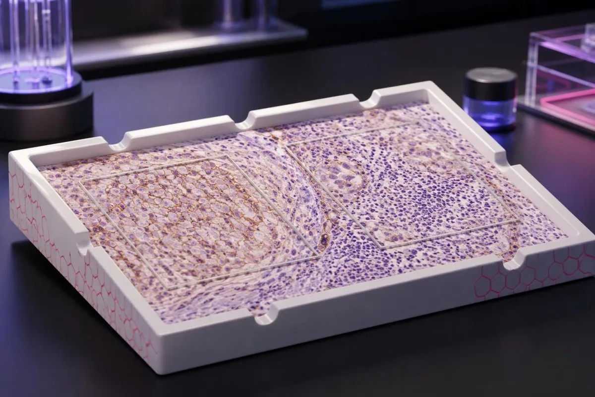
PD-L1-skårer beskriver farging i en vevsprøve – ikke din personlige sjanse for å lykkes med behandlingen. Skåringsmetoden, kreftdiagnosen og den foreslåtte legemiddelkombinasjonen avgjør hva resultatet kan si noe om.

Viktige punkter
- PD-L1-testresultater måler proteinfarging i kreftvev; de er ikke rutinemessige blodprøveresultater eller en kreftscreeningstest.
- TPS er prosentandelen levedyktige tumorceller med kvalifiserende membranfarging, rapportert fra 0 % til 100 %.
- CPS teller kvalifiserende PD-L1-positive tumorceller, lymfocytter og makrofager, deler på antallet levedyktige tumorceller og multipliserer med 100; den rapporterte skåren er begrenset til maksimalt 100.
- Ulike skårer kan ikke omregnes: TPS 10% kan forekomme sammen med CPS 10, CPS 25 eller en annen verdi, avhengig av fargingen av immunceller.
- Terskler som TPS 1%, TPS 50%, CPS 1 og CPS 10 gjelder for bestemte krefttyper og behandlingssituasjoner – ikke for all immunterapi.
- Positive resultater kan bidra til å velge behandling, men garanterer ikke effekt; en skår på 50 betyr ikke 50% sannsynlighet for respons.
- Negative resultater utelukker ikke alltid immunterapi, fordi enkelte kombinasjoner og biomarkørdefinerte behandlinger ikke krever PD-L1-positivitet.
- Prøvens egnethet er viktig: flere vanlig brukte skåringsprotokoller krever minst 100 levedyktige tumorceller, men laboratoriet må følge reglene som gjelder for den aktuelle indikasjonen.
Hva forteller PD-L1-testresultater egentlig?
PD-L1-testresultater beskriver proteinfarging i kreftvev. TPS teller prosentandelen fargede tumorceller; CPS inkluderer også utvalgte fargede immunceller. Ingen av skårene avgjør behandlingen alene: krefttype, stadium, analyse og behandlingsregime bestemmer hvilken grenseverdi som er relevant, og positive eller negative resultater garanterer ikke effekt eller at behandling kan utelukkes.
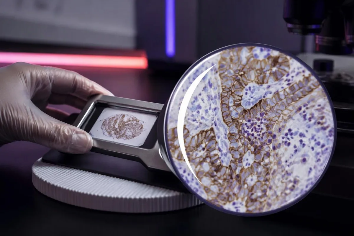
PD-L1, eller programmert celledød-ligand 1, kan binde seg til PD-1-reseptoren på immunceller og redusere aktiviteten deres. Legemidler som retter seg mot denne interaksjonen, kan gjenopprette immunaktivitet mot kreft, men måling av 1 protein fanger ikke opp alle faktorene som avgjør om immunceller gjenkjenner og kontrollerer en kreftsykdom.
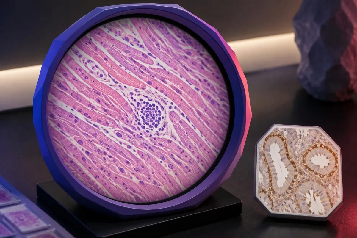
Et PD-L1-resultat fra vev produseres vanligvis ved hjelp av immunhistokjemi, som bruker et antistoff til å vise hvor proteinet befinner seg i en bevart prøve. Det rapporterte tallet beskriver celler i utvalgte vevssnitt – ikke hver eneste kreftcelle i hele kroppen – og 2 prøver fra ulike steder kan gi forskjellige resultater.
Kantesti er en AI-blodprøveanalysator, ikke en erstatning for vevspatologi eller en behandlingsbeslutning innen onkologi. Våre organisasjon og kliniske omfang skiller pedagogisk støtte for laboratorieresultater fra beslutninger som krever at den opprinnelige patologirapporten, bildediagnostikken og det foreslåtte behandlingsregimet vurderes samlet.
Begynn med 4 opplysninger før du tolker tallet: kreftdiagnosen, om skåren er CPS eller TPS, hvilken analyse som er brukt, og hvilken behandling som vurderes. En rapport som bare sier PD-L1-positiv, etterlater et klinisk viktig informasjonsgap; be om skåringsmetoden og det numeriske resultatet.
Betydningen av PD-L1 TPS-skår: prosentandelen tumorceller
Tumorandelsskår, eller TPS, er prosentandelen levedyktige tumorceller som viser kvalifiserende delvis eller fullstendig PD-L1-farging av cellemembranen. TPS varierer fra 0% til 100%; immunceller inngår ikke i telleren, selv når de farges kraftig ved siden av kreftcellene.
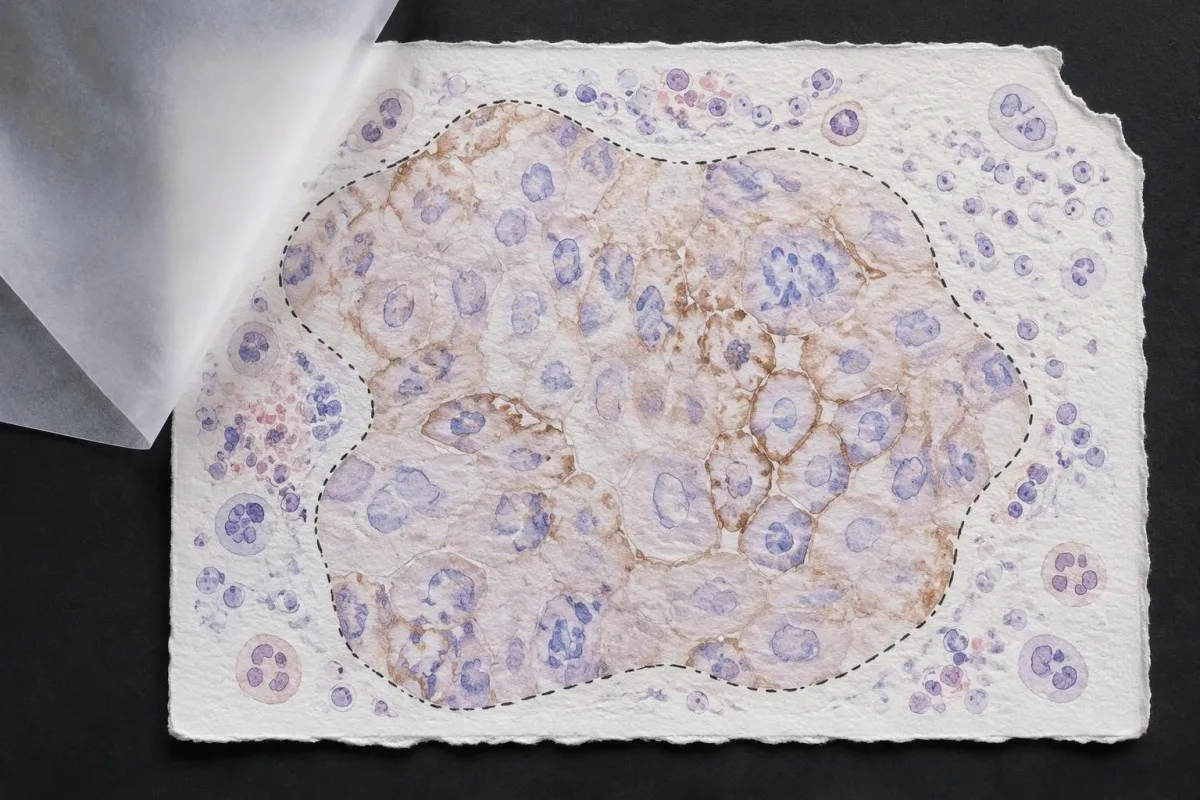
TPS = PD-L1-positive levedyktige tumorceller ÷ alle levedyktige tumorceller × 100%. I et hypotetisk vevsfelt med 200 levedyktige tumorceller gir 80 kvalifiserende positive celler TPS 40%; fargede lymfocytter i nærheten endrer ikke denne beregningen, selv om de kan påvirke et annet skåringssystem.
Membrane location matters more than visual darkness alone. For commonly used TPS methods, weak qualifying membrane staining can count, whereas staining limited to tumor-cell cytoplasm does not establish a positive tumor cell; TPS 1% therefore describes the fraction of qualifying cells, not a required intensity grade.
Cutoffs such as TPS 1% og TPS 50% were used to define populations in particular lung-cancer trials and treatment pathways. They are not boundaries between harmless and dangerous cancer, and crossing 50% does not create an abrupt biological switch from treatment resistance to guaranteed sensitivity.
Read the exact reporting format: TPS <1%, TPS 1–49%, and TPS ≥50% provide different detail from a single positive flag. Our explanation of kvalitative versus kvantitative resultater covers why a category cannot replace a number, although PD-L1 uses tissue-specific—not blood-reference-range—rules.
Betydningen av PD-L1 CPS-skår: hvorfor immunceller teller
Combined positive score, eller CPS, includes qualifying PD-L1-positive tumor cells, lymphocytes, and macrophages. CPS divides their combined count by the number of viable tumor cells and multiplies by 100; the denominator does not become all cells simply because immune cells enter the numerator.
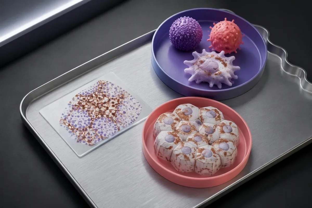
CPS = positive tumor cells plus qualifying positive lymphocytes and macrophages ÷ viable tumor cells × 100. In a hypothetical sample with 500 viable tumor cells, 50 positive tumor cells, and 75 qualifying positive immune cells, CPS is 25; the tumor-cell-only TPS would be 10%.
The reported CPS is capped at 100, even if the uncapped arithmetic would exceed 100 because many qualifying immune cells surround relatively few tumor cells. CPS is a unitless score, not a percentage of all cells, a blood concentration, or a probability that immunotherapy will work.
Not every stained immune cell belongs in the CPS count. The pathologist applies the assay manual's rules for eligible lymphocytes and macrophages associated with the tumor; neutrophils, unrelated inflammatory areas, and nonviable tissue are not simply added because a slide contains visible staining.
A CPS of 20 can arise from more than 1 cellular pattern: abundant tumor-cell staining, substantial immune-cell staining, or both. This denominator issue resembles the distinction between cell counts and percentages, but a circulating monocyte count cannot supply the tissue information needed to calculate CPS.
Hvorfor CPS og TPS ikke kan erstatte eller omregnes til hverandre
CPS and TPS are not interchangeable because they count different cells and were validated in different treatment settings. A TPS of 10% cannot be converted reliably into CPS 10, and a CPS of 20 does not establish that 20% of tumor cells stain.
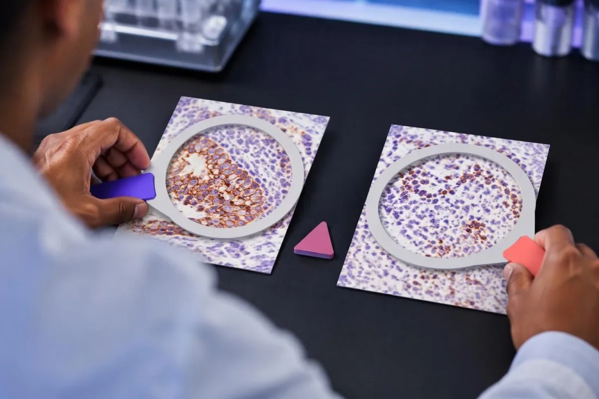
Consider 2 hypothetical specimens with 500 viable tumor cells and 50 positive tumor cells each. The first has no qualifying positive immune cells and gives TPS 10% with CPS 10; the second contains 75 qualifying positive immune cells and gives the same TPS 10% with CPS 25.
The reverse inference fails too. A hypothetical CPS 20 could result from 20 positive tumor cells among 100 tumor cells, or from 5 positive tumor cells plus 15 qualifying positive immune cells; those examples have TPS 20% and TPS 5%, respectively, despite identical CPS values.
En immune-cell score, sometimes abbreviated IC, is another system rather than a third spelling of CPS. Some IC methods measure the proportion of tumor area occupied by stained immune cells, so an IC result of 1% must be interpreted using its own assay and indication—not a TPS threshold.
Kantesti's educational explanation should preserve all 3 scoring labels rather than flatten them into positive or negative. When using AI report explanations safely, check that the original score, assay, and specimen remain visible; an AI-generated conversion without the underlying tissue counts is not clinically dependable.
Hvilke detaljer i patologirapporten er viktige utover skåren?
A usable PD-L1 report should identify the specimen, assay, scoring method, numerical result, and adequacy assessment. Several commonly used protocols require at least 100 viable tumor cells, but minimum counts and acceptable specimen types depend on the particular test and cancer indication.

Antibody identifiers such as 22C3, 28-8, SP263, and SP142 describe different testing assays or antibody clones, not different PD-L1 proteins. Some assays show substantial agreement in selected settings, but laboratories should not assume universal equivalence across tumor types, scoring methods, immune-cell assessments, and treatment requirements.
The specimen date and site can change the interpretation of 1 reported number. A small primary-site sample taken before treatment may not represent a later metastatic deposit; ask whether the report describes the current treatment-setting specimen or archived tissue that the oncology team considers suitable.
Insufficient tissue is not PD-L1 negative. A sample with too few viable tumor cells, failed controls, extensive tissue change, or unsuitable processing may be unreportable; the practical next step is laboratory clarification, additional sections, or another suitable specimen—not assigning TPS 0% or CPS 0.
Comparisons between 2 reports need more than matching dates. Our test timing and comparability guide explains the general problem of comparing unlike measurements; for PD-L1, the extra checks are anatomical site, prior treatment, fixation, tissue adequacy, assay, and scoring system.
Hvordan endrer krefttypen betydningen av positivitet?
PD-L1 positivity is indication-specific, not a universal diagnosis. TPS thresholds such as 1% or 50% are familiar in non-small-cell lung cancer, while CPS thresholds such as 1, 5, or 10 appear in selected other cancer settings; the drug, stage, and regimen must accompany the cutoff.

For recurrent or metastatic head-and-neck squamous-cell carcinoma, KEYNOTE-048 evaluated CPS-defined populations. Pembrolizumab alone improved median overall survival in the CPS ≥20 group to 14.9 months versus 10.7 months with the comparator regimen, showing why a CPS threshold cannot be borrowed from a lung-cancer TPS report (Burtness et al., 2019).
Selected established pembrolizumab pathways use CPS ≥10 for unresectable locally advanced or metastatic triple-negative breast cancer with chemotherapy, and CPS ≥1 for certain persistent, recurrent, or metastatic cervical-cancer regimens. Those examples do not establish eligibility for every earlier-stage setting, drug combination, country, or funding pathway.
Gastric and gastroesophageal-junction cancers require particular care because treatment selection may also involve HER2 og MSI/MMR, with PD-L1 requirements differing across regimens and jurisdictions. A serum marker described in our CA 72-4 interpretation guide cannot replace the tissue biomarker panel or define CPS.
Cancer diagnosis and treatment selection are 2 separate steps. Our persistent cough assessment guide addresses when lung symptoms need investigation; once cancer is confirmed, ask the oncology team to match the PD-L1 result to the current local product information and clinical pathway.
Hvorfor behandlingsregimet er like viktig som grenseverdien
Immunotherapy alone and immunotherapy combinations can have different PD-L1 requirements. A TPS below 1% may rule out one PD-L1-selected monotherapy pathway without ruling out an appropriate chemotherapy–immunotherapy combination; these are different clinical decisions, not contradictory interpretations of the same result.
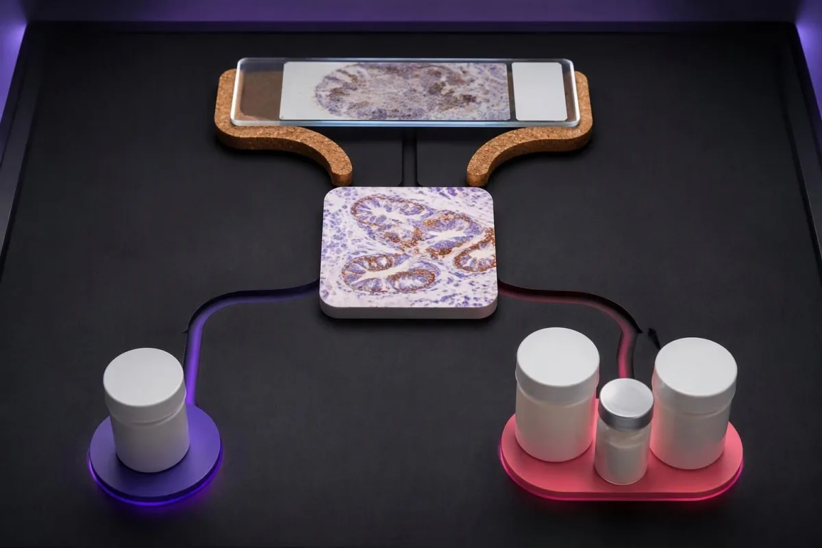
KEYNOTE-024 enrolled previously untreated advanced non-small-cell lung cancer with TPS ≥50% and no sensitizing EGFR mutation or ALK rearrangement. Median progression-free survival was 10.3 months with pembrolizumab versus 6.0 months with chemotherapy, supporting that specific high-TPS monotherapy setting—not every cancer with a score above 50 (Reck et al., 2016).
I KEYNOTE-189, pembrolizumab plus pemetrexed and platinum chemotherapy improved outcomes in previously untreated metastatic nonsquamous non-small-cell lung cancer without EGFR or ALK alterations. Twelve-month overall survival was 69.2% versus 49.4%, and benefit was observed across PD-L1 categories, including TPS <1% (Gandhi et al., 2018).
Combination treatment may address rapid disease progression differently from a single agent, but adding drugs also adds toxicity and monitoring needs. If a proposed regimen includes fluorouracil or capecitabine, DPYD-testing før kjemoterapi concerns treatment safety; it does not measure the PD-L1 pathway or predict the same benefit.
Kantesti er en AI-blodprøvetolkningsplattform that helps explain supporting laboratory measurements; it does not select between 1-drug and multidrug cancer regimens. Similarly, UGT1A1 and irinotecan safety address metabolism-related toxicity, illustrating why each biomarker should be matched to the specific medicine under discussion.
Garanterer et positivt PD-L1-resultat effekt av immunterapi?
A positive PD-L1 result does not guarantee a response. In KEYNOTE-024's selected high-expression lung-cancer population, the objective response rate with pembrolizumab was 44.8%—substantial benefit for many participants, but clearly not a response in every person with TPS ≥50%.
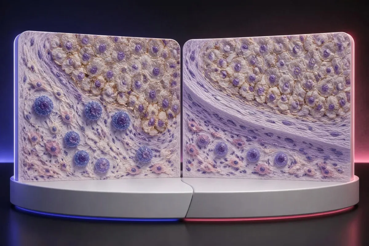
The 44.8% response rate in KEYNOTE-024 describes a trial population under defined conditions, not an individual's calculated chance of success (Reck et al., 2016). Objective response also means measurable tumor shrinkage by trial criteria; stable disease and survival benefit are different outcomes that should not be collapsed into that single percentage.
PD-L1 is only 1 part of a broader immune environment. A cancer can show substantial staining while lacking effective immune-cell access, recognizable targets, or intact antigen presentation; these mechanisms help explain why expression enriches for benefit in some settings without functioning as a deterministic on–off switch.
Higher does not always mean better across different cancers and combinations. A CPS 30 in one disease cannot be ranked against TPS 60% in another, and the same CPS threshold may have different predictive value when used for monotherapy versus a combination that already provides benefit through other mechanisms.
Some melanoma immunotherapy regimens do not require a positive PD-L1 test, so a low score should not be interpreted using lung-cancer monotherapy rules. Our melanoma warning-sign guide addresses recognition and assessment, while treatment benefit must be discussed using disease-specific trial evidence and the person's overall condition.
Kan immunterapi fortsatt tilbys når PD-L1 er negativ?
PD-L1-negative cancers can still have appropriate immunotherapy options. TPS <1% or CPS <1 may exclude a particular PD-L1-selected regimen, but some combinations, earlier-stage pathways, and treatments selected by other biomarkers do not require PD-L1 positivity.
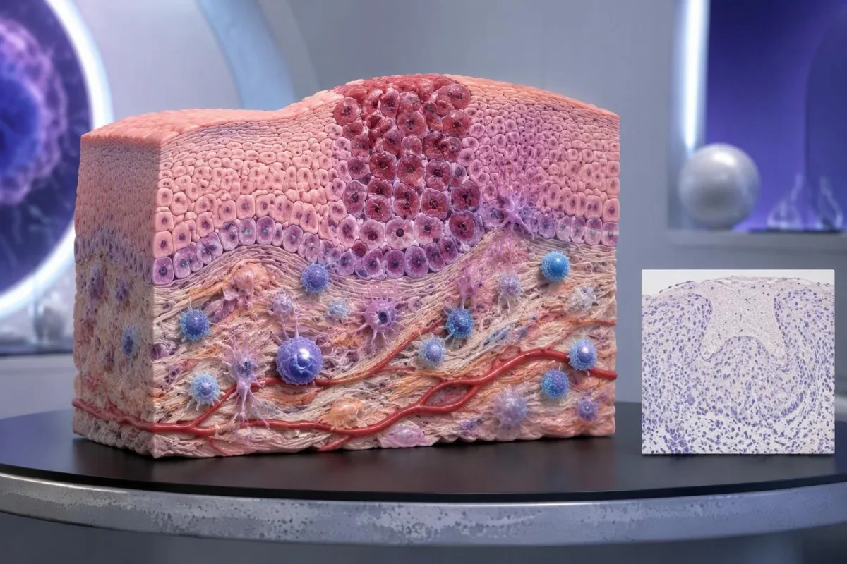
PD-L1-negative should first be translated into the laboratory's actual definition. TPS <1%, CPS <1, and an assay-specific IC-negative category are not identical measurements; an unreportable or inadequate sample is a fourth situation and should not be grouped with a technically valid negative result.
En MSI-high or mismatch-repair-deficient cancer may support particular immunotherapy pathways without PD-L1 selection, depending on the disease, stage, prior treatment, and local authorization. Tumor MMR testing and Genetisk testing for Lynch syndrom answer related but different questions: an abnormal tumor result does not automatically prove an inherited syndrome.
Other genetic findings may point toward targeted treatment or different planning rather than immunotherapy. A result discussed in our BRCA variant interpretation guide does not turn CPS 0 into CPS 10; clinicians combine distinct biomarkers without pretending they measure the same biological feature.
No PD-L1 requirement is not the same as no selection criteria. A regimen can still require a particular histology, stage, treatment history, organ function, or absence of an actionable driver alteration; ask whether 1 negative score changes only the proposed option or the broader treatment plan.
Når kan prøvetaking eller en grenseverdi nær grensen begrunne en ny vurdering?
Sampling variation can change PD-L1 classification, especially near a treatment threshold. A CPS of 9 versus 10, or TPS 0.8% versus 1%, may prompt pathology review when the distinction would change an otherwise suitable treatment—but repeating every result is not automatically useful.
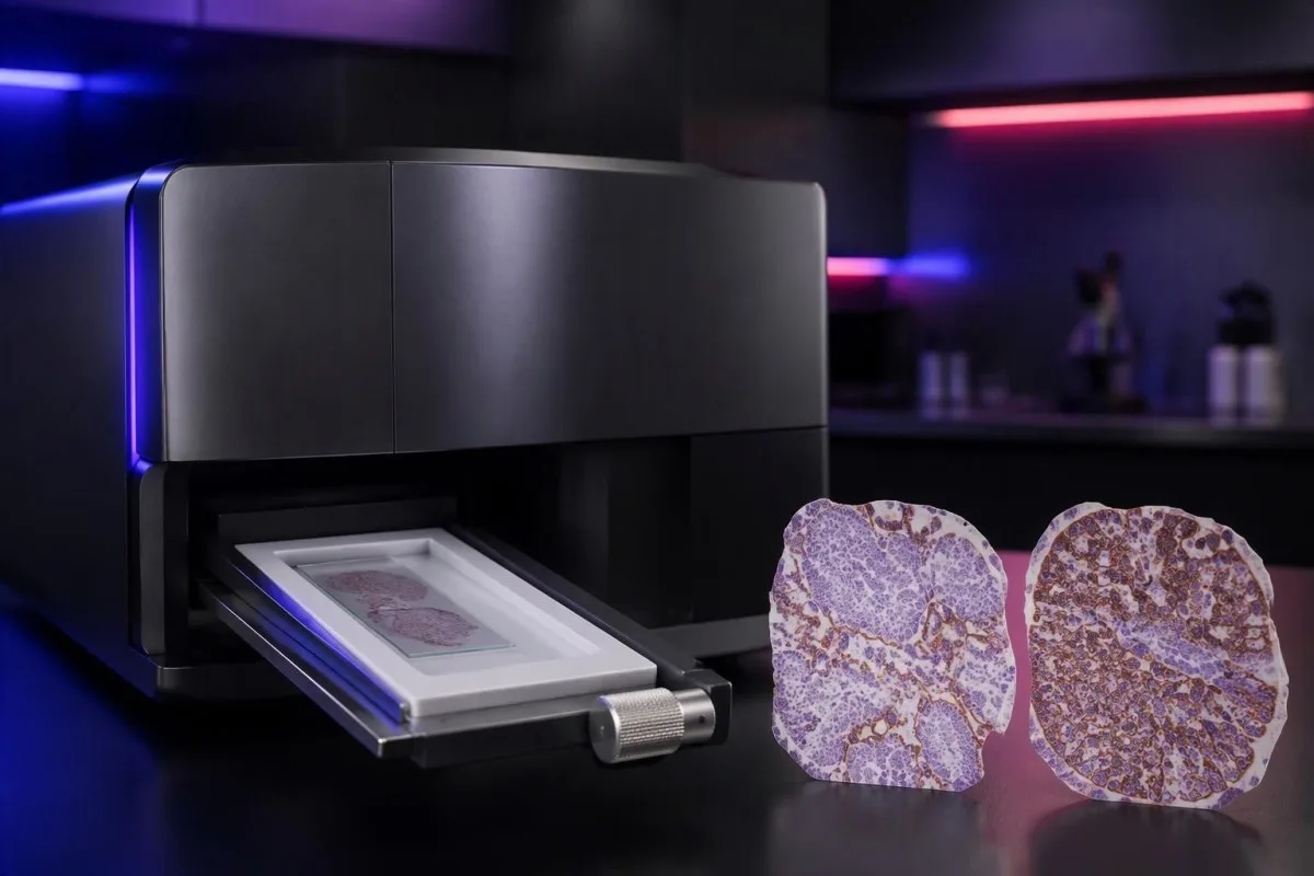
PD-L1 staining can be heterogeneous within 1 tissue block and between primary and metastatic sites. A small tissue examination may miss a highly expressing area or overrepresent it; the pathologist assesses the available viable tissue rather than assuming a single selected field describes the whole cancer.
Pretreatment and post-treatment specimens are not always equivalent. Chemotherapy, radiotherapy, and changes in the immune environment can alter expression, while decalcification of some bone specimens can impair antigen detection; ask whether a lower result reflects biology, processing limitations, or simply a different sampled site.
Borderline results require the actual assay rules, not homemade averaging. If 2 reports show CPS 8 and CPS 12, calculating CPS 10 does not create a newly validated result; the laboratory and oncology team should decide which specimen and assessment are appropriate for the treatment indication.
Kantesti cannot verify viable-cell counts, staining controls, or tissue suitability from a score alone. Our kliniske validering og tilsyn should be read in that limited context; when another tissue procedure is considered, its risks and likely impact on treatment need to be weighed against the uncertainty in the existing result.
Hvilke andre biomarkører bør vurderes sammen med PD-L1?
Other biomarkers can take priority over PD-L1, depending on the cancer. In advanced non-small-cell lung cancer, molecular alterations such as EGFR mutations and ALK rearrangements affect treatment planning; in selected other cancers, HER2, MSI/MMR, or additional molecular findings may change the preferred pathway.

A high TPS does not cancel an actionable molecular finding. The pivotal KEYNOTE-024 og KEYNOTE-189 populations excluded relevant EGFR or ALK alterations, so their results should not be applied unchanged to those groups; treatment sequence also matters because some targeted therapies and checkpoint inhibitors can interact through toxicity.
Tumor mutational burden, often measured in mutations per megabase, is a different biomarker from PD-L1. A threshold such as 10 mutations/Mb has been used in a specific tissue-agnostic treatment context, but assay differences, cancer type, authorization, and clinical setting prevent it from becoming a universal immunotherapy rule.
A liquid molecular test may find circulating tumor DNA, but it does not automatically replace tissue PD-L1 immunohistochemistry. The 2 tests measure different things—genetic material versus protein location within cells—and a negative liquid result may also reflect limited DNA shedding rather than absence of a tumor alteration.
Kantesti AI distinguishes the role of supporting laboratory markers from tissue-based treatment biomarkers in our referanseguide for blodbiomarkører. A blood-cell ratio, LDH level, or serum tumor-marker concentration should not be used to manufacture TPS 50%, estimate CPS 10, or bypass an indicated molecular workup.
Hvilke sikkerhetskontroller er viktige hvis immunterapi vurderes?
PD-L1 scores predict neither treatment safety nor immune-related toxicity. A TPS of 90% does not make checkpoint treatment safer than TPS 1%; the team assesses medical history, organ function, medications, and the particular regimen separately from the tissue biomarker.
Checkpoint inhibitors can cause immune-related effects involving the thyroid, liver, lungs, bowel, kidneys, heart, and other organs. Baseline testing commonly includes liver chemistry, creatinine, thyroid measurements, and a complete blood count, with monitoring schedules varying by regimen; 1 normal baseline panel does not prevent later toxicity.
New chest pain, palpitations, fainting, or breathlessness during treatment requires prompt assessment because myocarditis, although uncommon, can be serious. Our myocarditis test interpretation guide explains why troponin supports evaluation but cannot determine the cause by itself; severe symptoms warrant emergency care rather than waiting for the next scheduled panel.
Kidney changes need interpretation against the person's own baseline and treatment exposures. A hypothetical creatinine rise from 0.8 to 1.6 mg/dL is a doubling even if the absolute value looks only modestly elevated; our kidney function testing guide explains supporting measurements, not an immunotherapy-specific treatment decision.
Pre-existing autoimmune disease, organ transplantation, ongoing immunosuppression, or previous severe immune-related toxicity can complicate treatment planning even when CPS is 100. Report new persistent diarrhea, worsening cough, jaundice, unusual weakness, or marked thirst promptly; neither a high PD-L1 score nor previously reassuring blood results justify delaying symptom assessment.
Hva bør du spørre om under onkologtimen?
Ta med complete PD-L1 pathology report and ask which exact treatment decision it informs. Four useful starting questions are: Which score was used? Which cutoff applies? Is the specimen adequate? What options remain if the result is below the cutoff?

A hypothetical report reading 22C3, CPS 12, adequate tissue is still incomplete as a treatment answer until the cancer, stage, and regimen are specified. Ask whether CPS ≥10 is relevant to the proposed option, whether other biomarkers are pending, and whether a combination would be considered under different selection rules.
Request explanations of 3 outcomes separately: tumor shrinkage, duration of disease control, and survival. A trial's response rate is not the same as its survival benefit, while median survival is a group midpoint—not an individual deadline—and the balance between rapid control and treatment toxicity may change the preferred regimen.
Kantesti er en Tjeneste for tolkning av laboratorieprøver med AI for supporting laboratory understanding, not independent immunotherapy eligibility assessment. If blood monitoring is planned, our veiledning for forberedelse til grunnlinjetesting can help organize previous values, units, medication lists, and collection dates so that the oncology team can compare results meaningfully.
Keep tissue scores separate from serial blood monitoring in 2 clearly labeled records. Our veileder for medikamentsikkerhetstrender explains why changes over time need context; our prinsipper for ansvarlig AI also make clear that automated explanations should not trigger treatment changes without clinical review.
Forskningspublikasjoner og begrensningene ved PD-L1-dokumentasjonen
PD-L1 interpretation should be grounded in cancer-specific trials and validated pathology methods. The 3 clinical references below address high-TPS lung-cancer monotherapy, lung-cancer combination treatment, and CPS-selected head-and-neck treatment; the 2 separate Zenodo publications concern other laboratory topics and do not establish PD-L1 thresholds.

KEYNOTE-024 supports a defined TPS ≥50% advanced lung-cancer population, whereas KEYNOTE-189 supports a particular combination across PD-L1 categories (Reck et al., 2016; Gandhi et al., 2018). Those different study designs explain why evidence for one regimen cannot be substituted for evidence supporting another simply because both drugs affect the same immune pathway.
KEYNOTE-048 assessed CPS-defined recurrent or metastatic head-and-neck cancer populations, including CPS ≥1 and CPS ≥20 groups (Burtness et al., 2019). When reading a trial, check the histology, stage, previous treatment, comparator, and endpoint; a subgroup survival finding does not automatically quantify an individual's chance of measurable shrinkage.
Kantesti's 2 Zenodo publications listed below are educational resources on complement/ANA testing and Nipah-virus diagnostics, not PD-L1 clinical trials or assay-validation studies. The komplement og ANA-veiledning concerns different laboratory questions, and a DOI identifies a deposited publication without independently establishing clinical validity.
Use the original pathology result and current local treatment information to resolve eligibility—not a generalized positive/negative table or an AI-generated score conversion. Our standarder for medisinsk innhold emphasize source relevance and uncertainty; ask your oncology team to document the 1 specific regimen, applicable threshold, and remaining alternatives.
Ofte stilte spørsmål
Hva betyr PD-L1-positiv?
PD-L1-positiv betyr at et vevsprøveresultat oppfyller en spesifisert test- og skåringsterskel, for eksempel TPS ≥1 % eller CPS ≥1 i en relevant situasjon. Terskelen avhenger av krefttypen, behandlingsregimet og lokale behandlingskrav. Positivitet beviser ikke at immunterapi vil virke, og rapporten bør angi den numeriske skåren, ikke bare markere resultatet som positivt.
Hva er forskjellen mellom CPS og TPS?
TPS måler prosentandelen levedyktige tumorceller med kvalifiserende PD-L1-farging av cellemembranen, mens CPS inkluderer kvalifiserende positive tumorceller, lymfocytter og makrofager, delt på antallet levedyktige tumorceller og multiplisert med 100. TPS rapporteres fra 0 % til 100 %, og rapportert CPS er begrenset til 100. Skårene kan ikke konverteres, fordi CPS avhenger av farging av immunceller som TPS ikke teller med.
Betyr CPS 10 en 10% sjanse for at immunterapi virker?
CPS 10 betyr ikke en 10% sannsynlighet for respons på immunterapi. CPS er en vevsfargingsberegning der levedyktige tumorceller utgjør nevneren, ikke en sannsynlighet for behandlingsrespons hos enkeltpasienter. CPS ≥10 støtter valg av bestemte kreftspesifikke behandlingsregimer, men for å anslå sannsynlig nytte må man også ta hensyn til kreftdiagnosen, stadiet, andre biomarkører og dokumentasjonen for den foreslåtte behandlingen.
Kan jeg få immunterapi hvis PD-L1 er negativt?
Et PD-L1-negativt resultat utelukker ikke alle alternativer innen immunterapi. Noen kombinasjoner av kjemoterapi og immunterapi, samt andre biomarkørstyrte behandlingsforløp, krever ikke PD-L1-positivitet, selv når TPS er under 1 %. Kvalifikasjonen avhenger fortsatt av krefttype, stadium, molekylære funn, behandlingshistorikk, medisinsk egnethet og lokal godkjenning; en utilstrekkelig vevsprøve bør ikke behandles som et gyldig negativt resultat.
Regnes TPS på 50 % som høy PD-L1-ekspresjon?
TPS ≥50 % beskrives ofte som høy PD-L1-ekspresjon i utvalgte sammenhenger ved ikke-småcellet lungekreft. Denne terskelen ble brukt i KEYNOTE-024, som undersøkte en bestemt populasjon med avansert lungekreft uten sensibiliserende EGFR-mutasjoner eller ALK-rearrangementer. TPS 50 % er ikke det samme som CPS 50 og er ikke en universell terskel for alle krefttyper, stadier eller immunterapiregimer.
Er PD-L1 en blodprøve, og bør den gjentas?
Klinisk PD-L1-skåring for CPS og TPS gjøres vanligvis på bevart kreftvev undersøkt med immunhistokjemi, ikke på en rutinemessig blodprøve. En ny vurdering kan være nyttig hvis vevsmaterialet er utilstrekkelig, resultatet ligger nær en grenseverdi som kan endre behandlingsvalg, for eksempel CPS 10, eller en annen prøve er mer egnet. Onkologi- og patologiteamene bør avgjøre om en ny gjennomgang, flere snitt eller en annen prøve sannsynligvis vil endre behandlingen nok til å rettferdiggjøre innsatsen og de tilhørende prosedyremessige risikoene.
Få forklart blodprøveresultatene dine
Last opp laboratorierapporten din, så forklarer AI-en til Kantesti hver biomarkør på et lettfattelig språk i løpet av sekunder. Brukt av mer enn 2 millioner mennesker verden over.
References
- Klein, T., Mitchell, S., & Weber, H. (2026). Veiledning om C3-, C4-komplementblodprøver og ANA-titer. Kantesti AI medisinsk forskning. DOI
- Klein, T., Mitchell, S., & Weber, H. (2026). Blodprøve for Nipah-virus: veiledning om tidlig påvisning og diagnostisering 2026. Kantesti AI medisinsk forskning. DOI
- Reck M et al. (2016). Pembrolizumab versus Chemotherapy for PD-L1–Positive Non–Small-Cell Lung Cancer. New England Journal of Medicine. DOI
- Gandhi L et al. (2018). Pembrolizumab plus Chemotherapy in Metastatic Non–Small-Cell Lung Cancer. New England Journal of Medicine. DOI
- Burtness B et al. (2019). Pembrolizumab alone or with chemotherapy versus cetuximab with chemotherapy for recurrent or metastatic squamous cell carcinoma of the head and neck (KEYNOTE-048): a randomised, open-label, phase 3 study. The Lancet. DOI
- 2M+Analyserte prøver
- 127+Countries
- 100+Språk
Om det medisinske teamet


E-E-A-T tillitssignaler
Experience
Klinisk gjennomgang ledet av lege av arbeidsflyter for laboratorietolkning.
Expertise
Fagområde laboratoriemedisin med fokus på hvordan biomarkører oppfører seg i klinisk kontekst.
Authoritativeness
Medisinsk gjennomgått av Dr. Thomas Klein, med bidrag fra Dr. Sarah Mitchell og Prof. Dr. Hans Weber.
Trustworthiness
Evidensbasert tolkning med tydelige oppfølgingsveier for å redusere uro.





Medisinsk ansvarsfraskrivelse
Denne artikkelen er kun for informasjonsformål og utgjør ikke medisinsk rådgivning. Rådfør deg alltid med en kvalifisert helsepersonell for beslutninger om diagnose og behandling.
Publisert · Kontakt redaksjonen
Publisert av Kantesti Ltd, registrert i England og Wales, selskapsnr. 17090423, London, Storbritannia.
