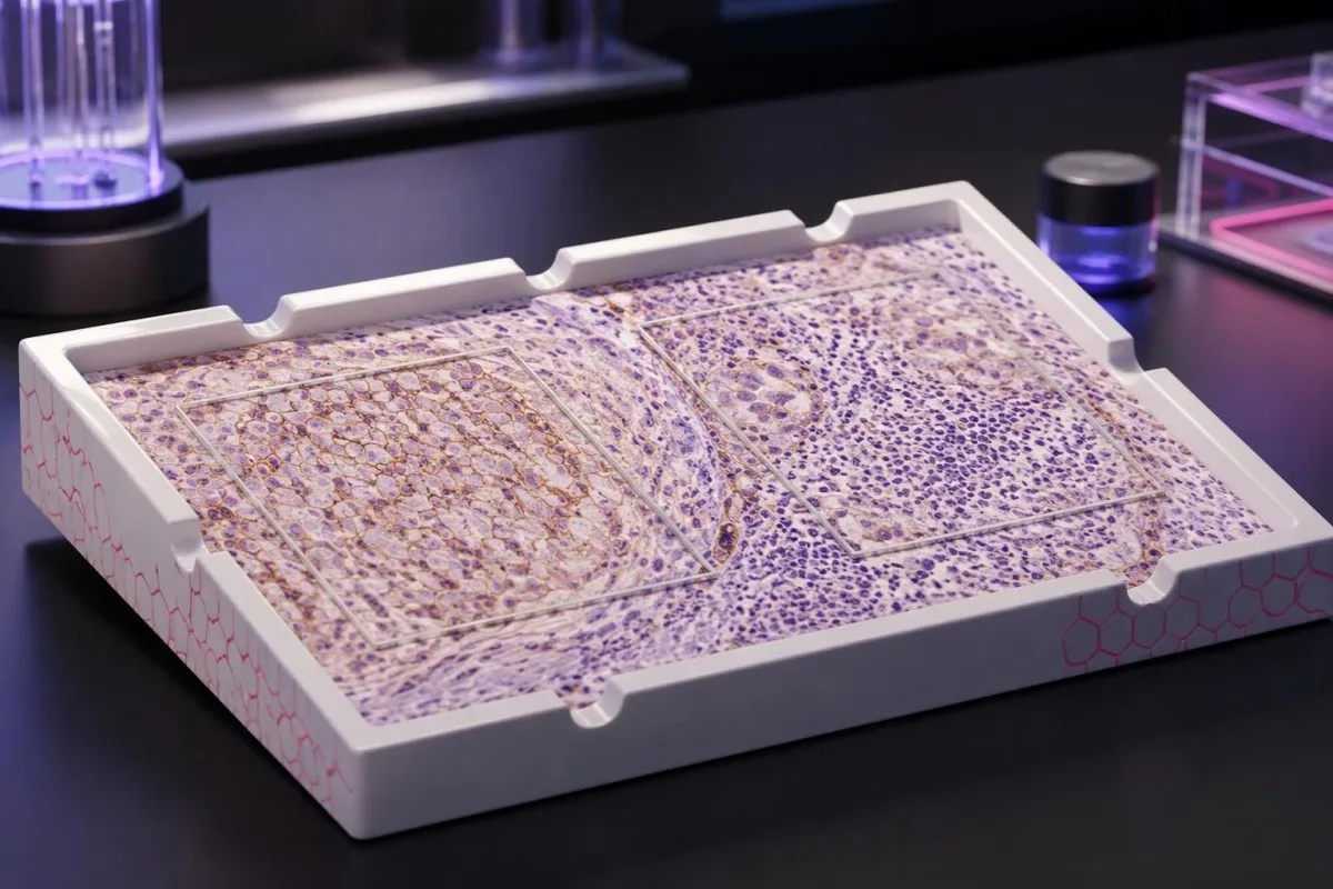
PD-L1-pisteet kuvaavat kudosnäytteen värjäytymistä – eivät henkilökohtaista mahdollisuuttasi hyötyä hoidosta. Pisteytysmenetelmä, syöpädiagnoosi ja ehdotettu lääkeyhdistelmä määrittävät, mitä tuloksen perusteella voidaan päätellä.

Keskeiset huomiot
- PD-L1-testitulokset mittaavat proteiinin värjäytymistä syöpäkudoksessa; ne eivät ole rutiininomaisia verikoetuloksia eivätkä syöpäseulontatesti.
- TPS tarkoittaa niiden elinkelpoisten kasvainsolujen prosenttiosuutta, joissa todetaan hyväksyttävää kalvovärjäytymistä; tulos ilmoitetaan välillä 0–100 %.
- CPS laskee hyväksyttävää PD-L1-positiivista värjäytymistä osoittavat kasvainsolut, lymfosyytit ja makrofagit, jakaa määrän elinkelpoisten kasvainsolujen määrällä ja kertoo tuloksen sadalla; ilmoitettava pistemäärä voi olla enintään 100.
- Eri pisteytykset ei voida muuntaa: TPS 10% voi vastata CPS-arvoa 10, CPS-arvoa 25 tai jotakin muuta arvoa immuunisolujen värjäytymisestä riippuen.
- Kynnysarvot kuten TPS 1 %, TPS 50 %, CPS 1 ja CPS 10 koskevat tiettyjä syöpiä ja hoitotilanteita – eivät kaikkia immunoterapioita.
- Positiiviset tulokset voi auttaa hoidon valinnassa, mutta ei takaa hyötyä; arvo 50 ei tarkoita 50 prosentin todennäköisyyttä hoitovasteeseen.
- Negatiiviset tulokset eivät aina sulje pois immunoterapiaa, koska joissakin yhdistelmähoidoissa ja biomarkkerin perusteella valittavissa hoidoissa PD-L1-positiivisuutta ei edellytetä.
- Näytteen riittävyys on tärkeää: useat yleisesti käytetyt pisteytysprotokollat edellyttävät vähintään 100 elinkykyistä kasvainsolua, mutta laboratorion on noudatettava käyttöaihekohtaisia sääntöjään.
Mitä PD-L1-testitulokset oikeastaan kertovat?
PD-L1-testitulokset kuvaavat proteiinin värjäytymistä syöpäkudoksessa. TPS laskee värjäytyneiden kasvainsolujen prosenttiosuuden; CPS sisältää myös tietyt värjäytyneet immuunisolut. Kumpikaan pistemäärä ei yksin ratkaise hoitoa: merkityksellisen raja-arvon määrittävät syöpätyyppi, vaihe, määritysmenetelmä ja hoito-ohjelma, eivätkä positiiviset tai negatiiviset tulokset takaa hyötyä tai sulje sitä pois.
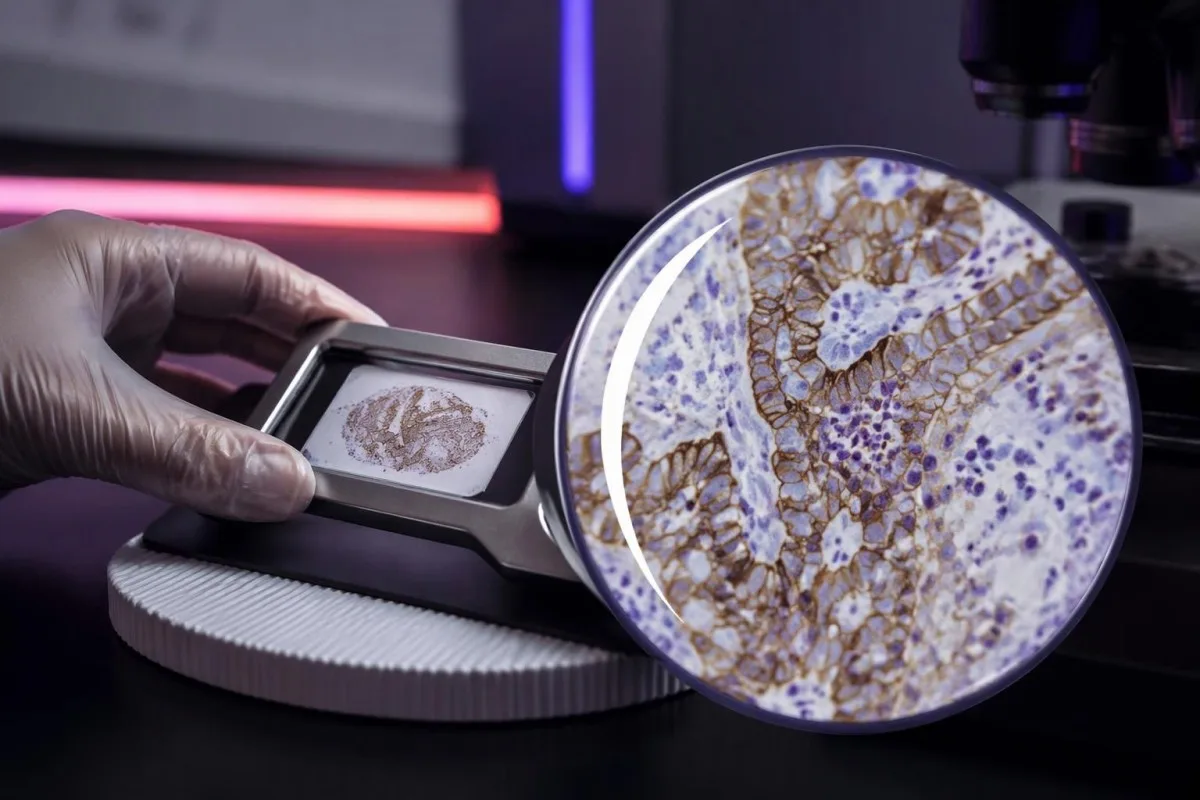
PD-L1, eli ohjelmoituun solukuolemaan liittyvä ligandi 1 voi sitoutua immuunisolujen PD-1-reseptoriin ja vähentää niiden aktiivisuutta. Tähän vuorovaikutukseen kohdistuvat lääkkeet voivat palauttaa syövänvastaisen immuunitoiminnan, mutta yhden proteiinin mittaaminen ei kata kaikkia tekijöitä, jotka määrittävät, tunnistavatko ja hallitsevatko immuunisolut syöpää.
Kudoksen PD-L1-tulos saadaan yleensä immunohistokemialla, jossa vasta-aineen avulla osoitetaan proteiinin sijainti säilytetyssä näytteessä. Ilmoitettu luku kuvaa valituissa kudosleikkeissä olevia soluja – ei kaikkia syöpäsoluja koko kehossa – ja kahdesta eri kohdasta otetusta näytteestä voi saada erilaiset tulokset.
Kantesti on Tekoälyinen verikoeanalysaattori, ei kudospatologian tai syövän hoitopäätöksen korvaajana. Meidän organisaatiomme ja kliininen soveltamisala erottavat laboratoriotulosten tulkintaa tukevan koulutuksellisen tuen päätöksistä, jotka edellyttävät alkuperäisen patologiaraportin, kuvantamistutkimusten ja suunnitellun hoito-ohjelman tarkastelua yhdessä.
Selvitä ensin neljä asiaa ennen luvun tulkitsemista: syöpädiagnoosi, onko pistemäärä CPS vai TPS, käytetty määritysmenetelmä ja harkittava hoito. Pelkkä maininta PD-L1-positiivisuudesta jättää kliinisesti merkittävän aukon; pyydä pisteytysmenetelmä ja numeerinen tulos.
PD-L1 TPS -pisteytyksen merkitys: kasvainsolujen prosenttiosuus
Kasvaimen osuuspistemäärä, tai TPS, eli TPS on niiden elinkykyisten kasvainsolujen prosenttiosuus, joiden kalvolla näkyy määritelmän mukainen osittainen tai täydellinen PD-L1-värjäytyminen. TPS vaihtelee välillä 0–100 %; immuunisoluja ei lasketa sen osoittajaan, vaikka ne värjäytyisivät voimakkaasti syöpäsolujen vieressä.
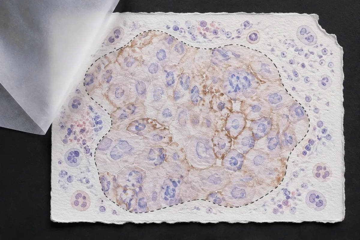
TPS = PD-L1-positiiviset elinkykyiset kasvainsolut ÷ kaikki elinkykyiset kasvainsolut × 100 %. Kuvitteellisessa kudoskentässä, jossa on 200 elinkykyistä kasvainsolua, 80 määritelmän mukaista positiivista solua tuottaa TPS-arvon 40%; viereiset värjäytyneet lymfosyytit eivät muuta tätä laskelmaa, vaikka ne voisivat vaikuttaa toiseen pisteytysjärjestelmään.
Membrane location matters more than visual darkness alone. For commonly used TPS methods, weak qualifying membrane staining can count, whereas staining limited to tumor-cell cytoplasm does not establish a positive tumor cell; TPS 1% therefore describes the fraction of qualifying cells, not a required intensity grade.
Cutoffs such as TPS 1% ja TPS 50% were used to define populations in particular lung-cancer trials and treatment pathways. They are not boundaries between harmless and dangerous cancer, and crossing 50% does not create an abrupt biological switch from treatment resistance to guaranteed sensitivity.
Read the exact reporting format: TPS <1%, TPS 1–49%, and TPS ≥50% provide different detail from a single positive flag. Our explanation of laadulliset versus määrälliset tulokset covers why a category cannot replace a number, although PD-L1 uses tissue-specific—not blood-reference-range—rules.
PD-L1 CPS -pisteytyksen merkitys: miksi myös immuunisolut lasketaan
Combined positive score, tai CPS, includes qualifying PD-L1-positive tumor cells, lymphocytes, and macrophages. CPS divides their combined count by the number of viable tumor cells and multiplies by 100; the denominator does not become all cells simply because immune cells enter the numerator.
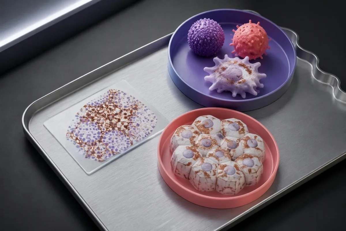
CPS = positive tumor cells plus qualifying positive lymphocytes and macrophages ÷ viable tumor cells × 100. In a hypothetical sample with 500 viable tumor cells, 50 positive tumor cells, and 75 qualifying positive immune cells, CPS is 25; the tumor-cell-only TPS would be 10%.
The reported CPS is capped at 100, even if the uncapped arithmetic would exceed 100 because many qualifying immune cells surround relatively few tumor cells. CPS is a unitless score, not a percentage of all cells, a blood concentration, or a probability that immunotherapy will work.
Not every stained immune cell belongs in the CPS count. The pathologist applies the assay manual's rules for eligible lymphocytes and macrophages associated with the tumor; neutrophils, unrelated inflammatory areas, and nonviable tissue are not simply added because a slide contains visible staining.
A CPS of 20 can arise from more than 1 cellular pattern: abundant tumor-cell staining, substantial immune-cell staining, or both. This denominator issue resembles the distinction between cell counts and percentages, but a circulating monocyte count cannot supply the tissue information needed to calculate CPS.
Miksi CPS- ja TPS-pisteytyksiä ei voi korvata toisillaan tai muuntaa toisikseen
CPS and TPS are not interchangeable because they count different cells and were validated in different treatment settings. A TPS of 10% cannot be converted reliably into CPS 10, and a CPS of 20 does not establish that 20% of tumor cells stain.
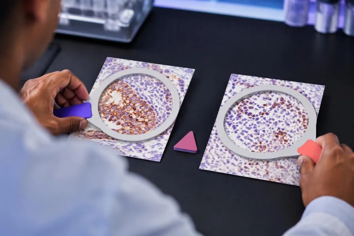
Consider 2 hypothetical specimens with 500 viable tumor cells and 50 positive tumor cells each. The first has no qualifying positive immune cells and gives TPS 10% with CPS 10; the second contains 75 qualifying positive immune cells and gives the same TPS 10% with CPS 25.
The reverse inference fails too. A hypothetical CPS 20 could result from 20 positive tumor cells among 100 tumor cells, or from 5 positive tumor cells plus 15 qualifying positive immune cells; those examples have TPS 20% and TPS 5%, respectively, despite identical CPS values.
An immune-cell score, sometimes abbreviated IC, is another system rather than a third spelling of CPS. Some IC methods measure the proportion of tumor area occupied by stained immune cells, so an IC result of 1% must be interpreted using its own assay and indication—not a TPS threshold.
Kantesti's educational explanation should preserve all 3 scoring labels rather than flatten them into positive or negative. When using AI report explanations safely, check that the original score, assay, and specimen remain visible; an AI-generated conversion without the underlying tissue counts is not clinically dependable.
Mitkä patologian lausunnon tiedot ovat pisteytyksen lisäksi tärkeitä?
A usable PD-L1 report should identify the specimen, assay, scoring method, numerical result, and adequacy assessment. Several commonly used protocols require at least 100 viable tumor cells, but minimum counts and acceptable specimen types depend on the particular test and cancer indication.
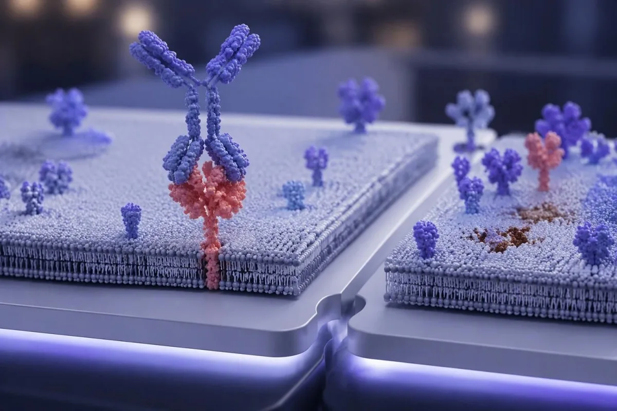
Antibody identifiers such as 22C3, 28-8, SP263, and SP142 describe different testing assays or antibody clones, not different PD-L1 proteins. Some assays show substantial agreement in selected settings, but laboratories should not assume universal equivalence across tumor types, scoring methods, immune-cell assessments, and treatment requirements.
The specimen date and site can change the interpretation of 1 reported number. A small primary-site sample taken before treatment may not represent a later metastatic deposit; ask whether the report describes the current treatment-setting specimen or archived tissue that the oncology team considers suitable.
Insufficient tissue is not PD-L1 negative. A sample with too few viable tumor cells, failed controls, extensive tissue change, or unsuitable processing may be unreportable; the practical next step is laboratory clarification, additional sections, or another suitable specimen—not assigning TPS 0% or CPS 0.
Comparisons between 2 reports need more than matching dates. Our test timing and comparability guide explains the general problem of comparing unlike measurements; for PD-L1, the extra checks are anatomical site, prior treatment, fixation, tissue adequacy, assay, and scoring system.
Miten syöpätyyppi vaikuttaa positiivisen tuloksen merkitykseen?
PD-L1 positivity is indication-specific, not a universal diagnosis. TPS thresholds such as 1% or 50% are familiar in non-small-cell lung cancer, while CPS thresholds such as 1, 5, or 10 appear in selected other cancer settings; the drug, stage, and regimen must accompany the cutoff.

For recurrent or metastatic head-and-neck squamous-cell carcinoma, KEYNOTE-048 evaluated CPS-defined populations. Pembrolizumab alone improved median overall survival in the CPS ≥20 group to 14.9 months versus 10.7 months with the comparator regimen, showing why a CPS threshold cannot be borrowed from a lung-cancer TPS report (Burtness et al., 2019).
Selected established pembrolizumab pathways use CPS ≥10 for unresectable locally advanced or metastatic triple-negative breast cancer with chemotherapy, and CPS ≥1 for certain persistent, recurrent, or metastatic cervical-cancer regimens. Those examples do not establish eligibility for every earlier-stage setting, drug combination, country, or funding pathway.
Gastric and gastroesophageal-junction cancers require particular care because treatment selection may also involve HER2 ja MSI/MMR, with PD-L1 requirements differing across regimens and jurisdictions. A serum marker described in our CA 72-4 interpretation guide cannot replace the tissue biomarker panel or define CPS.
Cancer diagnosis and treatment selection are 2 separate steps. Our persistent cough assessment guide addresses when lung symptoms need investigation; once cancer is confirmed, ask the oncology team to match the PD-L1 result to the current local product information and clinical pathway.
Miksi hoito-ohjelma on yhtä tärkeä kuin raja-arvo
Immunotherapy alone and immunotherapy combinations can have different PD-L1 requirements. A TPS below 1% may rule out one PD-L1-selected monotherapy pathway without ruling out an appropriate chemotherapy–immunotherapy combination; these are different clinical decisions, not contradictory interpretations of the same result.
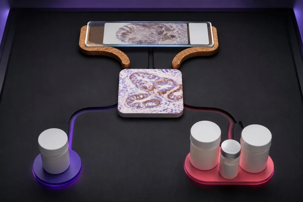
KEYNOTE-024 enrolled previously untreated advanced non-small-cell lung cancer with TPS ≥50% and no sensitizing EGFR mutation or ALK rearrangement. Median progression-free survival was 10.3 months with pembrolizumab versus 6.0 months with chemotherapy, supporting that specific high-TPS monotherapy setting—not every cancer with a score above 50 (Reck et al., 2016).
, ESR on usein selvästi koholla, joskus KEYNOTE-189, pembrolizumab plus pemetrexed and platinum chemotherapy improved outcomes in previously untreated metastatic nonsquamous non-small-cell lung cancer without EGFR or ALK alterations. Twelve-month overall survival was 69.2% versus 49.4%, and benefit was observed across PD-L1 categories, including TPS <1% (Gandhi et al., 2018).
Combination treatment may address rapid disease progression differently from a single agent, but adding drugs also adds toxicity and monitoring needs. If a proposed regimen includes fluorouracil or capecitabine, DPYD-testaus ennen solunsalpaajahoitoa concerns treatment safety; it does not measure the PD-L1 pathway or predict the same benefit.
Kantesti on AI verikoe-tulkinta-alusta that helps explain supporting laboratory measurements; it does not select between 1-drug and multidrug cancer regimens. Similarly, UGT1A1 and irinotecan safety address metabolism-related toxicity, illustrating why each biomarker should be matched to the specific medicine under discussion.
Takaako positiivinen PD-L1-tulos immunoterapiasta saatavan hyödyn?
A positive PD-L1 result does not guarantee a response. In KEYNOTE-024's selected high-expression lung-cancer population, the objective response rate with pembrolizumab was 44.8%—substantial benefit for many participants, but clearly not a response in every person with TPS ≥50%.
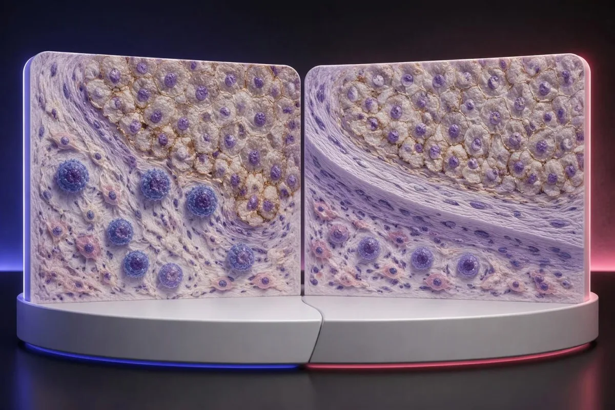
The 44.8% response rate in KEYNOTE-024 describes a trial population under defined conditions, not an individual's calculated chance of success (Reck et al., 2016). Objective response also means measurable tumor shrinkage by trial criteria; stable disease and survival benefit are different outcomes that should not be collapsed into that single percentage.
PD-L1 is only 1 part of a broader immune environment. A cancer can show substantial staining while lacking effective immune-cell access, recognizable targets, or intact antigen presentation; these mechanisms help explain why expression enriches for benefit in some settings without functioning as a deterministic on–off switch.
Higher does not always mean better across different cancers and combinations. A CPS 30 in one disease cannot be ranked against TPS 60% in another, and the same CPS threshold may have different predictive value when used for monotherapy versus a combination that already provides benefit through other mechanisms.
Some melanoma immunotherapy regimens do not require a positive PD-L1 test, so a low score should not be interpreted using lung-cancer monotherapy rules. Our melanoma warning-sign guide addresses recognition and assessment, while treatment benefit must be discussed using disease-specific trial evidence and the person's overall condition.
Voidaanko immunoterapiaa silti tarjota, kun PD-L1-tulos on negatiivinen?
PD-L1-negative cancers can still have appropriate immunotherapy options. TPS <1% or CPS <1 may exclude a particular PD-L1-selected regimen, but some combinations, earlier-stage pathways, and treatments selected by other biomarkers do not require PD-L1 positivity.
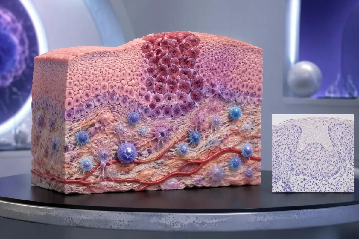
PD-L1-negative should first be translated into the laboratory's actual definition. TPS <1%, CPS <1, and an assay-specific IC-negative category are not identical measurements; an unreportable or inadequate sample is a fourth situation and should not be grouped with a technically valid negative result.
An MSI-high or mismatch-repair-deficient cancer may support particular immunotherapy pathways without PD-L1 selection, depending on the disease, stage, prior treatment, and local authorization. Tumor MMR testing and Lynchin oireyhtymän geneettinen testaus answer related but different questions: an abnormal tumor result does not automatically prove an inherited syndrome.
Other genetic findings may point toward targeted treatment or different planning rather than immunotherapy. A result discussed in our BRCA variant interpretation guide does not turn CPS 0 into CPS 10; clinicians combine distinct biomarkers without pretending they measure the same biological feature.
No PD-L1 requirement is not the same as no selection criteria. A regimen can still require a particular histology, stage, treatment history, organ function, or absence of an actionable driver alteration; ask whether 1 negative score changes only the proposed option or the broader treatment plan.
Milloin näytteenotto tai rajatapaustulos voi olla peruste uudelleenarvioinnille?
Sampling variation can change PD-L1 classification, especially near a treatment threshold. A CPS of 9 versus 10, or TPS 0.8% versus 1%, may prompt pathology review when the distinction would change an otherwise suitable treatment—but repeating every result is not automatically useful.
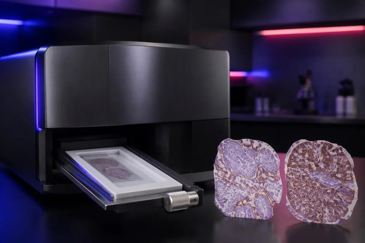
PD-L1 staining can be heterogeneous within 1 tissue block and between primary and metastatic sites. A small tissue examination may miss a highly expressing area or overrepresent it; the pathologist assesses the available viable tissue rather than assuming a single selected field describes the whole cancer.
Pretreatment and post-treatment specimens are not always equivalent. Chemotherapy, radiotherapy, and changes in the immune environment can alter expression, while decalcification of some bone specimens can impair antigen detection; ask whether a lower result reflects biology, processing limitations, or simply a different sampled site.
Borderline results require the actual assay rules, not homemade averaging. If 2 reports show CPS 8 and CPS 12, calculating CPS 10 does not create a newly validated result; the laboratory and oncology team should decide which specimen and assessment are appropriate for the treatment indication.
Kantesti cannot verify viable-cell counts, staining controls, or tissue suitability from a score alone. Our kliininen validointi ja valvonta should be read in that limited context; when another tissue procedure is considered, its risks and likely impact on treatment need to be weighed against the uncertainty in the existing result.
Mitkä muut biomarkkerit ovat tärkeitä PD-L1:n lisäksi?
Other biomarkers can take priority over PD-L1, depending on the cancer. In advanced non-small-cell lung cancer, molecular alterations such as EGFR mutations and ALK rearrangements affect treatment planning; in selected other cancers, HER2, MSI/MMR, or additional molecular findings may change the preferred pathway.
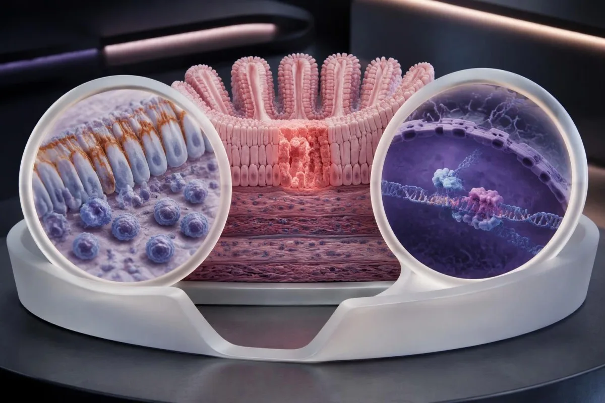
A high TPS does not cancel an actionable molecular finding. The pivotal KEYNOTE-024 ja KEYNOTE-189 populations excluded relevant EGFR or ALK alterations, so their results should not be applied unchanged to those groups; treatment sequence also matters because some targeted therapies and checkpoint inhibitors can interact through toxicity.
Tumor mutational burden, often measured in mutations per megabase, is a different biomarker from PD-L1. A threshold such as 10 mutations/Mb has been used in a specific tissue-agnostic treatment context, but assay differences, cancer type, authorization, and clinical setting prevent it from becoming a universal immunotherapy rule.
A liquid molecular test may find circulating tumor DNA, but it does not automatically replace tissue PD-L1 immunohistochemistry. The 2 tests measure different things—genetic material versus protein location within cells—and a negative liquid result may also reflect limited DNA shedding rather than absence of a tumor alteration.
Kantesti AI distinguishes the role of supporting laboratory markers from tissue-based treatment biomarkers in our veribiomarkkereiden viiteopas. A blood-cell ratio, LDH level, or serum tumor-marker concentration should not be used to manufacture TPS 50%, estimate CPS 10, or bypass an indicated molecular workup.
Mitkä turvallisuustarkistukset ovat tärkeitä immunoterapiaa harkittaessa?
PD-L1 scores predict neither treatment safety nor immune-related toxicity. A TPS of 90% does not make checkpoint treatment safer than TPS 1%; the team assesses medical history, organ function, medications, and the particular regimen separately from the tissue biomarker.
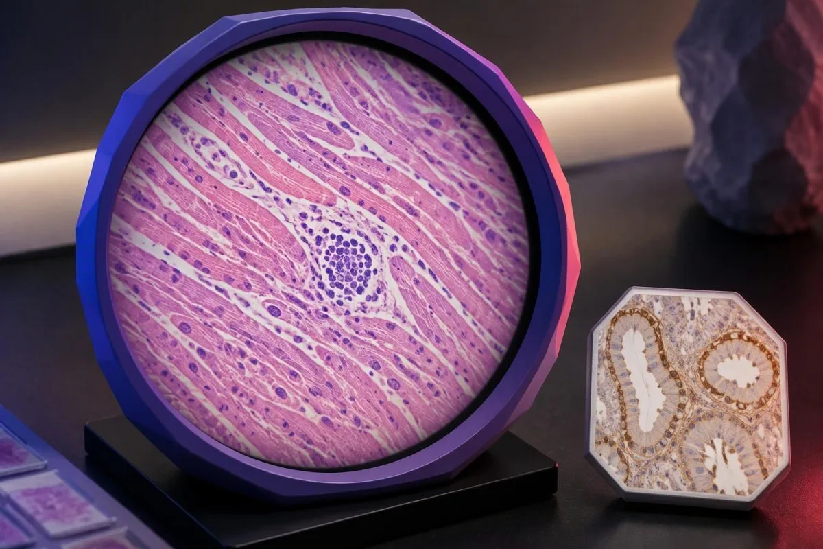
Checkpoint inhibitors can cause immune-related effects involving the thyroid, liver, lungs, bowel, kidneys, heart, and other organs. Baseline testing commonly includes liver chemistry, creatinine, thyroid measurements, and a complete blood count, with monitoring schedules varying by regimen; 1 normal baseline panel does not prevent later toxicity.
New chest pain, palpitations, fainting, or breathlessness during treatment requires prompt assessment because myocarditis, although uncommon, can be serious. Our myocarditis test interpretation guide explains why troponin supports evaluation but cannot determine the cause by itself; severe symptoms warrant emergency care rather than waiting for the next scheduled panel.
Kidney changes need interpretation against the person's own baseline and treatment exposures. A hypothetical creatinine rise from 0.8 to 1.6 mg/dL is a doubling even if the absolute value looks only modestly elevated; our kidney function testing guide explains supporting measurements, not an immunotherapy-specific treatment decision.
Pre-existing autoimmune disease, organ transplantation, ongoing immunosuppression, or previous severe immune-related toxicity can complicate treatment planning even when CPS is 100. Report new persistent diarrhea, worsening cough, jaundice, unusual weakness, or marked thirst promptly; neither a high PD-L1 score nor previously reassuring blood results justify delaying symptom assessment.
Mitä sinun kannattaa kysyä onkologin vastaanotolla?
Tuo complete PD-L1 pathology report and ask which exact treatment decision it informs. Four useful starting questions are: Which score was used? Which cutoff applies? Is the specimen adequate? What options remain if the result is below the cutoff?

A hypothetical report reading 22C3, CPS 12, adequate tissue is still incomplete as a treatment answer until the cancer, stage, and regimen are specified. Ask whether CPS ≥10 is relevant to the proposed option, whether other biomarkers are pending, and whether a combination would be considered under different selection rules.
Request explanations of 3 outcomes separately: tumor shrinkage, duration of disease control, and survival. A trial's response rate is not the same as its survival benefit, while median survival is a group midpoint—not an individual deadline—and the balance between rapid control and treatment toxicity may change the preferred regimen.
Kantesti on AI-laboratoriotestien tulkintapalvelu for supporting laboratory understanding, not independent immunotherapy eligibility assessment. If blood monitoring is planned, our lähtötason testaukseen valmistautumisen oppaamme can help organize previous values, units, medication lists, and collection dates so that the oncology team can compare results meaningfully.
Keep tissue scores separate from serial blood monitoring in 2 clearly labeled records. Our lääketurvatrendiopas explains why changes over time need context; our vastuullisen tekoälyn periaatteemme also make clear that automated explanations should not trigger treatment changes without clinical review.
Tutkimusjulkaisut ja PD-L1-näytön rajoitukset
PD-L1 interpretation should be grounded in cancer-specific trials and validated pathology methods. The 3 clinical references below address high-TPS lung-cancer monotherapy, lung-cancer combination treatment, and CPS-selected head-and-neck treatment; the 2 separate Zenodo publications concern other laboratory topics and do not establish PD-L1 thresholds.

KEYNOTE-024 supports a defined TPS ≥50% advanced lung-cancer population, whereas KEYNOTE-189 supports a particular combination across PD-L1 categories (Reck et al., 2016; Gandhi et al., 2018). Those different study designs explain why evidence for one regimen cannot be substituted for evidence supporting another simply because both drugs affect the same immune pathway.
KEYNOTE-048 assessed CPS-defined recurrent or metastatic head-and-neck cancer populations, including CPS ≥1 and CPS ≥20 groups (Burtness et al., 2019). When reading a trial, check the histology, stage, previous treatment, comparator, and endpoint; a subgroup survival finding does not automatically quantify an individual's chance of measurable shrinkage.
Kantesti's 2 Zenodo publications listed below are educational resources on complement/ANA testing and Nipah-virus diagnostics, not PD-L1 clinical trials or assay-validation studies. The komplementti- ja ANA-ohje concerns different laboratory questions, and a DOI identifies a deposited publication without independently establishing clinical validity.
Use the original pathology result and current local treatment information to resolve eligibility—not a generalized positive/negative table or an AI-generated score conversion. Our lääketieteellisen sisällön standardit emphasize source relevance and uncertainty; ask your oncology team to document the 1 specific regimen, applicable threshold, and remaining alternatives.
Usein kysytyt kysymykset
Mitä PD-L1-positiivinen tarkoittaa?
PD-L1-positiivisuus tarkoittaa, että kudosnäyte täyttää tietyssä tutkimusmenetelmässä ja pisteytyksessä asetetun raja-arvon, kuten TPS ≥1 % tai CPS ≥1 soveltuvassa tilanteessa. Raja-arvo riippuu syöpätyypistä, hoito-ohjelmasta ja paikallisista hoitovaatimuksista. Positiivisuus ei takaa immunoterapian tehoa, ja lausunnossa tulisi ilmoittaa numeerinen pistemäärä pelkän positiivisuusmerkinnän sijaan.
Mitä eroa on CPS:llä ja TPS:llä?
TPS mittaa niiden elävien kasvainsolujen prosenttiosuuden, joissa on hyväksyttävä PD-L1-kalvovärjäytyminen, kun taas CPS lasketaan jakamalla hyväksyttävän värjäytymisen saaneiden positiivisten kasvainsolujen, lymfosyyttien ja makrofagien määrä elävien kasvainsolujen määrällä ja kertomalla tulos sadalla. TPS ilmoitetaan välillä 0–100 %, ja ilmoitettava CPS on enintään 100. Pisteytyksiä ei voi muuntaa toisikseen, koska CPS perustuu immuunisolujen värjäytymiseen, jota TPS ei laske mukaan.
Tarkoittaako CPS 10, että immunoterapia tehoaa 10% todennäköisyydellä?
CPS 10 ei tarkoita 10%:n todennäköisyyttä saada vastetta immunoterapiasta. CPS on kudosvärjäykseen perustuva laskelma, jonka nimittäjänä ovat elinkelpoiset kasvainsolut; se ei ilmaise yksittäisen potilaan hoitovasteen todennäköisyyttä. CPS ≥10 tukee tiettyihin syöpäkohtaisiin hoito-ohjelmiin valintaa, mutta hoidon todennäköisen hyödyn arviointi edellyttää myös syöpädiagnoosia, taudin vaihetta, muita biomarkkereita ja näyttöä ehdotetun hoidon tehosta.
Voinko saada immunoterapiaa, jos PD-L1 on negatiivinen?
PD-L1-negatiivinen tulos ei sulje pois kaikkia immunoterapiahoitovaihtoehtoja. Jotkin kemoterapian ja immunoterapian yhdistelmähoidot sekä muut biomarkkereihin perustuvat hoitopolut eivät edellytä PD-L1-positiivisuutta, vaikka TPS-arvo olisi alle 1 %. Hoitoon soveltuvuus riippuu edelleen syöpätyypistä, taudin vaiheesta, molekyylilöydöksistä, aiemmista hoidoista, potilaan terveydentilasta ja paikallisista myyntiluvista; riittämätöntä kudosnäytettä ei pidä tulkita päteväksi negatiiviseksi tulokseksi.
Pidetäänkö TPS-arvoa 50 % korkeana PD-L1-tasona?
TPS ≥50 %:a kuvataan usein korkeaksi PD-L1-ilmentymäksi tietyissä ei-pienisoluisen keuhkosyövän tilanteissa. Kynnysarvoa käytettiin KEYNOTE-024-tutkimuksessa, jossa tutkittiin tiettyä pitkälle edennyttä keuhkosyöpäpotilaiden ryhmää, jolla ei ollut herkistäviä EGFR-mutaatioita tai ALK-uudelleenjärjestymiä. TPS 50 % ei vastaa arvoa CPS 50, eikä se ole yleispätevä kynnysarvo kaikille syöville, taudin vaiheille tai immunoterapiaohjelmille.
Onko PD-L1 verikoe, ja pitäisikö se uusia?
PD-L1-pisteytyksessä CPS- ja TPS-menetelmillä käytetään yleensä säilynyttä syöpäkudosta, joka tutkitaan immunohistokemiallisesti, ei tavanomaista verinäytettä. Uusinta-arvioinnista voi olla hyötyä, jos kudosnäyte ei ole riittävä, tulos on lähellä hoitopäätökseen vaikuttavaa raja-arvoa, kuten CPS-arvoa 10, tai jokin muu näyte soveltuu paremmin. Onkologian ja patologian tiimien tulee päättää, onko uusinta-arviointi, lisäleikkeet tai toinen näyte todennäköisesti niin merkityksellinen hoidon kannalta, että se oikeuttaa siihen liittyvän työmäärän ja toimenpideriskit.
Saat verikoetulokset selitettyinä
Lataa laboratoriolausuntosi, niin Kantesti:n tekoäly selittää jokaisen biomarkkerin selkokielellä sekunneissa. Yli 2 miljoonaa käyttäjää maailmanlaajuisesti.
Viitteet
- Klein, T., Mitchell, S. & Weber, H. (2026). C3- ja C4-komplementtiverikoe ja ANA-tiitteriopas. Kantesti AI Medical Research. DOI-koodi
- Klein, T., Mitchell, S. & Weber, H. (2026). Nipah-viruksen verikoe: varhaisen havaitsemisen ja diagnosoinnin opas 2026. Kantesti AI Medical Research. DOI-koodi
- Reck M et al. (2016). Pembrolizumab versus Chemotherapy for PD-L1–Positive Non–Small-Cell Lung Cancer. New England Journal of Medicine. DOI-koodi
- Gandhi L et al. (2018). Pembrolizumab plus Chemotherapy in Metastatic Non–Small-Cell Lung Cancer. New England Journal of Medicine. DOI-koodi
- Burtness B et al. (2019). Pembrolizumab alone or with chemotherapy versus cetuximab with chemotherapy for recurrent or metastatic squamous cell carcinoma of the head and neck (KEYNOTE-048): a randomised, open-label, phase 3 study. The Lancet. DOI-koodi
- 2 kk+Analysoidut testit
- 127+Maat
- 100+Kielet
Tietoa lääketieteellisestä tiimistä


E-E-A-T-luottamussignaalit
Kokea
Lääkärin johtama kliininen arviointi laboratoriotulkinnan työnkuluista.
Asiantuntemus
Laboratoriolääketiede keskittyy siihen, miten biomarkkerit käyttäytyvät kliinisessä kontekstissa.
Auktoriteetti
Lääketieteellisen tarkastuksen on tehnyt tohtori Thomas Klein, ja siihen ovat osallistuneet tohtori Sarah Mitchell ja professori Hans Weber.
Luotettavuus
Näyttöön perustuva tulkinta selkeillä jatkopoluilla, jotka vähentävät hälytystä.





Lääketieteellinen vastuuvapauslauseke
Tämä artikkeli on tarkoitettu vain koulutustarkoituksiin eikä se muodosta lääketieteellistä neuvontaa. Ota aina yhteyttä pätevään terveydenhuollon ammattilaiseen diagnoosi- ja hoitopäätöksiä varten.
Julkaistu · Ota yhteyttä toimitukseen
Julkaisija: Kantesti Ltd, rekisteröity Englannissa ja Walesissa, yrityksen nro. 17090423, Lontoo, Yhdistynyt kuningaskunta.
