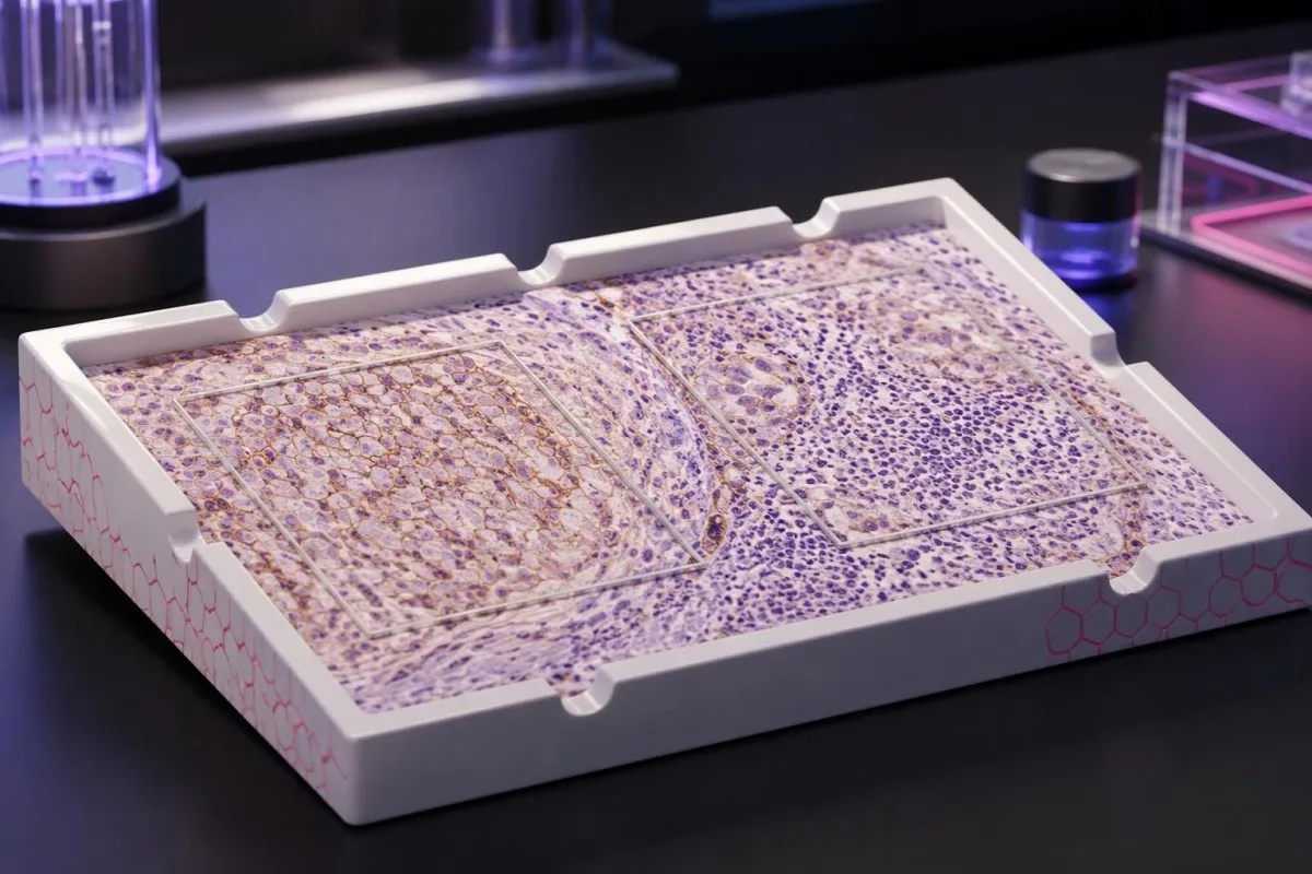
Резултатите од PD-L1 го опишуваат боењето во примерок од ткиво — не вашите лични шанси за успех на лекувањето. Методот на бодирање, дијагнозата на ракот и предложената комбинација на лекови одредуваат што може да потврди резултатот.

Клучни заклучоци
- Резултати од тестот за PD-L1 мерат боење на протеини во канцерозното ткиво; тие не се рутински резултати од крвна слика ниту тест за скрининг на рак.
- TPS е процентот на одржливи туморски клетки со соодветно мембранско боење, прикажан од 0% до 100%.
- CPS ги брои соодветните PD-L1-позитивни туморски клетки, лимфоцити и макрофаги, го дели бројот со бројот на одржливи туморски клетки и го множи со 100; прикажаниот резултат е ограничен на 100.
- Различни резултати не може да се претвори: TPS 10% може да одговара на CPS 10, CPS 25 или друга вредност, зависно од боењето на имунолошките клетки.
- Прагови како TPS 1%, TPS 50%, CPS 1 и CPS 10 важат за одредени видови рак и начини на лекување — не за секоја имунотерапија.
- Позитивните резултати може да помогнат при изборот на третман, но не гарантираат корист; резултат од 50 не значи 50% веројатност за одговор.
- Негативни резултати не ја исклучуваат секогаш имунотерапијата, бидејќи за некои комбинации и третмани дефинирани според биомаркери не е потребен позитивен PD-L1 резултат.
- Соодветност на примерокот е важна: неколку често користени протоколи за оценување бараат најмалку 100 витални туморски клетки, но лабораторијата мора да ги применува правилата специфични за индикацијата.
Што всушност ви кажуваат резултатите од тестот за PD-L1?
Резултати од тестот за PD-L1 го опишуваат боењето на протеините во канцерогеното ткиво. TPS го пресметува процентот на обоени туморски клетки; CPS ги вклучува и избраните обоени имунолошки клетки. Ниту еден од резултатите самостојно не го одредува третманот: видот на ракот, стадиумот, тестот и терапевтскиот режим го одредуваат релевантниот праг, а позитивните или негативните резултати не гарантираат корист ниту ја исклучуваат.
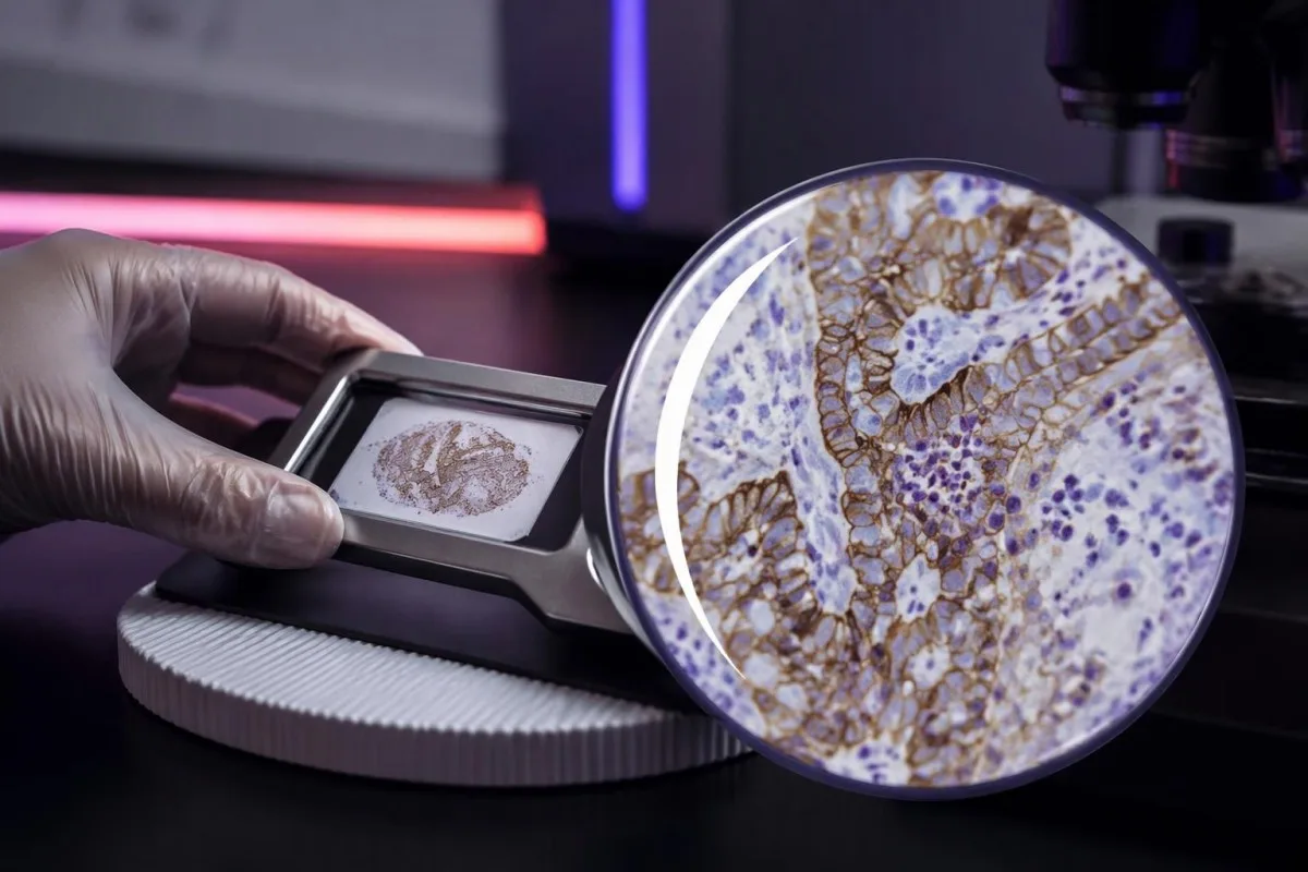
PD-L1, или лиганд 1 на програмираната клеточна смрт, може да се врзе за рецепторот PD-1 на имунолошките клетки и да ја намали нивната активност. Лековите насочени кон оваа интеракција можат да ја обноват имуноактивноста против ракот, но мерењето на 1 протеин не ги опфаќа сите фактори што одредуваат дали имунолошките клетки ќе го препознаат и контролираат ракот.
Резултатот за PD-L1 во ткиво обично се добива со имунохистохемија, при која се користи антитело за да се покаже местоположбата на протеинот во зачуван примерок. Пријавениот број ги опишува клетките на избрани ткивни пресеци — не секоја клетка на ракот низ целото тело — а 2 примерока од различни места можат да дадат различни резултати.
Кантести е анализатор на крвна слика со вештачка интелигенција, а не замена за ткивна патологија или одлука за онколошки третман. Нашите организација и клинички обем прават разлика меѓу едукативна поддршка за лабораториски резултати и одлуки за кои заедно се потребни оригиналниот патолошки извештај, снимките и предложениот терапевтски режим.
Пред толкувањето на бројката, започнете со 4 детали: дијагнозата на ракот, дали резултатот е CPS или TPS, користениот тест и третманот што се разгледува. Извештај во кој стои само дека PD-L1 е позитивен остава клинички значаен недостиг на информации; побарајте го методот на оценување и нумеричкиот резултат.
Значење на TPS-резултатот за PD-L1: процентот на туморски клетки
Туморски пропорциски скор, или TPS, е процентот на витални туморски клетки што покажуваат соодветно делумно или целосно мембранско боење за PD-L1. TPS се движи од 0% до 100%; имунолошките клетки не се вклучуваат во неговиот броител, дури и кога силно се обоени покрај клетките на ракот.
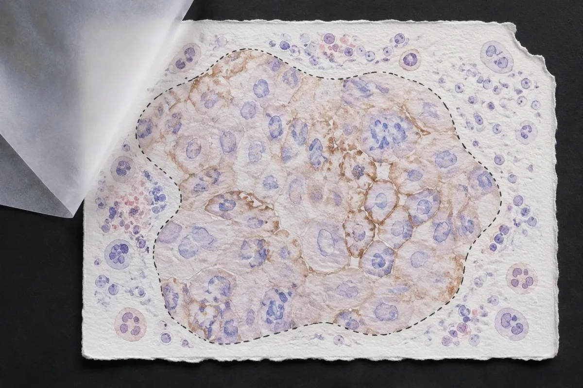
TPS = витални туморски клетки позитивни на PD-L1 ÷ сите витални туморски клетки × 100%. Во хипотетичко ткивно поле што содржи 200 витални туморски клетки, 80 соодветно позитивни клетки даваат TPS 40%; обоените лимфоцити во близина не ја менуваат таа пресметка, иако може да влијаат врз друг систем за оценување.
Membrane location matters more than visual darkness alone. For commonly used TPS methods, weak qualifying membrane staining can count, whereas staining limited to tumor-cell cytoplasm does not establish a positive tumor cell; TPS 1% therefore describes the fraction of qualifying cells, not a required intensity grade.
Cutoffs such as TPS 1% и TPS 50% were used to define populations in particular lung-cancer trials and treatment pathways. They are not boundaries between harmless and dangerous cancer, and crossing 50% does not create an abrupt biological switch from treatment resistance to guaranteed sensitivity.
Read the exact reporting format: TPS <1%, TPS 1–49%, and TPS ≥50% provide different detail from a single positive flag. Our explanation of квалитативни наспроти квантитативни резултати covers why a category cannot replace a number, although PD-L1 uses tissue-specific—not blood-reference-range—rules.
Значење на CPS-резултатот за PD-L1: зошто се бројат имуните клетки
Combined positive score, или CPS, includes qualifying PD-L1-positive tumor cells, lymphocytes, and macrophages. CPS divides their combined count by the number of viable tumor cells and multiplies by 100; the denominator does not become all cells simply because immune cells enter the numerator.
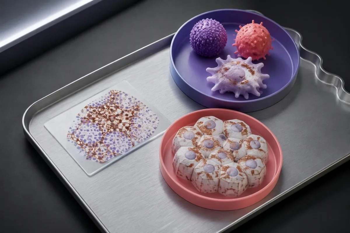
CPS = positive tumor cells plus qualifying positive lymphocytes and macrophages ÷ viable tumor cells × 100. In a hypothetical sample with 500 viable tumor cells, 50 positive tumor cells, and 75 qualifying positive immune cells, CPS is 25; the tumor-cell-only TPS would be 10%.
The reported CPS is capped at 100, even if the uncapped arithmetic would exceed 100 because many qualifying immune cells surround relatively few tumor cells. CPS is a unitless score, not a percentage of all cells, a blood concentration, or a probability that immunotherapy will work.
Not every stained immune cell belongs in the CPS count. The pathologist applies the assay manual's rules for eligible lymphocytes and macrophages associated with the tumor; neutrophils, unrelated inflammatory areas, and nonviable tissue are not simply added because a slide contains visible staining.
A CPS of 20 can arise from more than 1 cellular pattern: abundant tumor-cell staining, substantial immune-cell staining, or both. This denominator issue resembles the distinction between cell counts and percentages, but a circulating monocyte count cannot supply the tissue information needed to calculate CPS.
Зошто CPS и TPS не можат да се заменуваат или претвораат еден во друг
CPS and TPS are not interchangeable because they count different cells and were validated in different treatment settings. A TPS of 10% cannot be converted reliably into CPS 10, and a CPS of 20 does not establish that 20% of tumor cells stain.
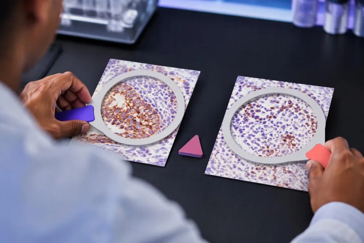
Consider 2 hypothetical specimens with 500 viable tumor cells and 50 positive tumor cells each. The first has no qualifying positive immune cells and gives TPS 10% with CPS 10; the second contains 75 qualifying positive immune cells and gives the same TPS 10% with CPS 25.
The reverse inference fails too. A hypothetical CPS 20 could result from 20 positive tumor cells among 100 tumor cells, or from 5 positive tumor cells plus 15 qualifying positive immune cells; those examples have TPS 20% and TPS 5%, respectively, despite identical CPS values.
Ан immune-cell score, sometimes abbreviated IC, is another system rather than a third spelling of CPS. Some IC methods measure the proportion of tumor area occupied by stained immune cells, so an IC result of 1% must be interpreted using its own assay and indication—not a TPS threshold.
Kantesti's educational explanation should preserve all 3 scoring labels rather than flatten them into positive or negative. When using AI report explanations safely, check that the original score, assay, and specimen remain visible; an AI-generated conversion without the underlying tissue counts is not clinically dependable.
Кои детали во патолошкиот извештај се важни надвор од резултатот?
A usable PD-L1 report should identify the specimen, assay, scoring method, numerical result, and adequacy assessment. Several commonly used protocols require at least 100 viable tumor cells, but minimum counts and acceptable specimen types depend on the particular test and cancer indication.
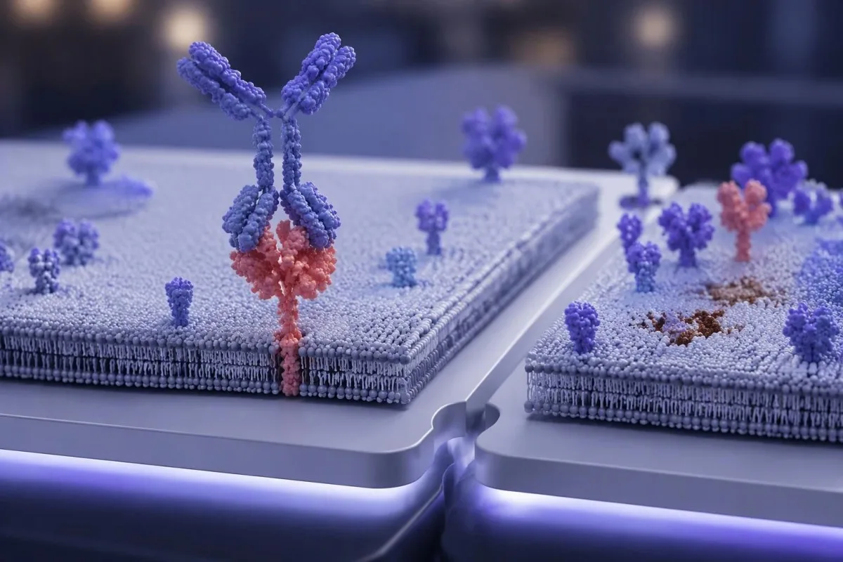
Antibody identifiers such as 22C3, 28-8, SP263, and SP142 describe different testing assays or antibody clones, not different PD-L1 proteins. Some assays show substantial agreement in selected settings, but laboratories should not assume universal equivalence across tumor types, scoring methods, immune-cell assessments, and treatment requirements.
The specimen date and site can change the interpretation of 1 reported number. A small primary-site sample taken before treatment may not represent a later metastatic deposit; ask whether the report describes the current treatment-setting specimen or archived tissue that the oncology team considers suitable.
Insufficient tissue is not PD-L1 negative. A sample with too few viable tumor cells, failed controls, extensive tissue change, or unsuitable processing may be unreportable; the practical next step is laboratory clarification, additional sections, or another suitable specimen—not assigning TPS 0% or CPS 0.
Comparisons between 2 reports need more than matching dates. Our test timing and comparability guide explains the general problem of comparing unlike measurements; for PD-L1, the extra checks are anatomical site, prior treatment, fixation, tissue adequacy, assay, and scoring system.
Како видот на ракот го менува значењето на позитивниот резултат?
PD-L1 positivity is indication-specific, not a universal diagnosis. TPS thresholds such as 1% or 50% are familiar in non-small-cell lung cancer, while CPS thresholds such as 1, 5, or 10 appear in selected other cancer settings; the drug, stage, and regimen must accompany the cutoff.

For recurrent or metastatic head-and-neck squamous-cell carcinoma, KEYNOTE-048 evaluated CPS-defined populations. Pembrolizumab alone improved median overall survival in the CPS ≥20 group to 14.9 months versus 10.7 months with the comparator regimen, showing why a CPS threshold cannot be borrowed from a lung-cancer TPS report (Burtness et al., 2019).
Selected established pembrolizumab pathways use CPS ≥10 for unresectable locally advanced or metastatic triple-negative breast cancer with chemotherapy, and CPS ≥1 for certain persistent, recurrent, or metastatic cervical-cancer regimens. Those examples do not establish eligibility for every earlier-stage setting, drug combination, country, or funding pathway.
Gastric and gastroesophageal-junction cancers require particular care because treatment selection may also involve HER2 и MSI/MMR, with PD-L1 requirements differing across regimens and jurisdictions. A serum marker described in our CA 72-4 interpretation guide cannot replace the tissue biomarker panel or define CPS.
Cancer diagnosis and treatment selection are 2 separate steps. Our persistent cough assessment guide addresses when lung symptoms need investigation; once cancer is confirmed, ask the oncology team to match the PD-L1 result to the current local product information and clinical pathway.
Зошто режимот на лекување е исто толку важен колку и граничната вредност
Immunotherapy alone and immunotherapy combinations can have different PD-L1 requirements. A TPS below 1% may rule out one PD-L1-selected monotherapy pathway without ruling out an appropriate chemotherapy–immunotherapy combination; these are different clinical decisions, not contradictory interpretations of the same result.
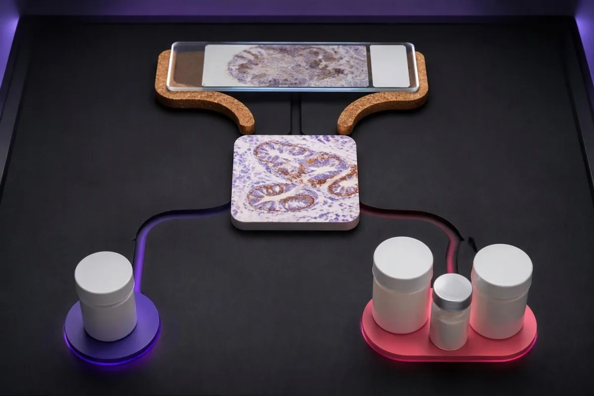
KEYNOTE-024 enrolled previously untreated advanced non-small-cell lung cancer with TPS ≥50% and no sensitizing EGFR mutation or ALK rearrangement. Median progression-free survival was 10.3 months with pembrolizumab versus 6.0 months with chemotherapy, supporting that specific high-TPS monotherapy setting—not every cancer with a score above 50 (Reck et al., 2016).
Во KEYNOTE-189, pembrolizumab plus pemetrexed and platinum chemotherapy improved outcomes in previously untreated metastatic nonsquamous non-small-cell lung cancer without EGFR or ALK alterations. Twelve-month overall survival was 69.2% versus 49.4%, and benefit was observed across PD-L1 categories, including TPS <1% (Gandhi et al., 2018).
Combination treatment may address rapid disease progression differently from a single agent, but adding drugs also adds toxicity and monitoring needs. If a proposed regimen includes fluorouracil or capecitabine, тестирањето на DPYD пред хемотерапија concerns treatment safety; it does not measure the PD-L1 pathway or predict the same benefit.
Кантести е AI платформа за толкување крвна слика that helps explain supporting laboratory measurements; it does not select between 1-drug and multidrug cancer regimens. Similarly, UGT1A1 and irinotecan safety address metabolism-related toxicity, illustrating why each biomarker should be matched to the specific medicine under discussion.
Дали позитивниот резултат за PD-L1 гарантира корист од имунотерапијата?
A positive PD-L1 result does not guarantee a response. In KEYNOTE-024's selected high-expression lung-cancer population, the objective response rate with pembrolizumab was 44.8%—substantial benefit for many participants, but clearly not a response in every person with TPS ≥50%.
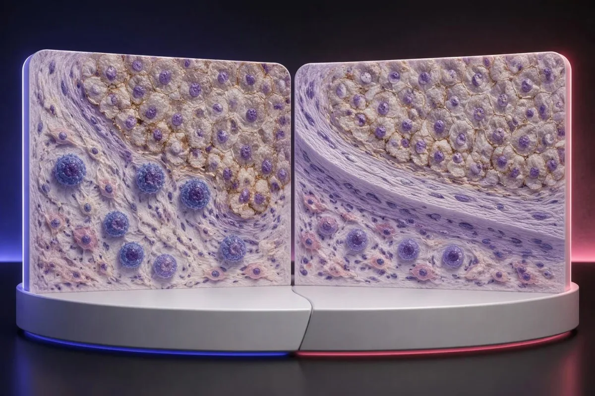
The 44.8% response rate in KEYNOTE-024 describes a trial population under defined conditions, not an individual's calculated chance of success (Reck et al., 2016). Objective response also means measurable tumor shrinkage by trial criteria; stable disease and survival benefit are different outcomes that should not be collapsed into that single percentage.
PD-L1 is only 1 part of a broader immune environment. A cancer can show substantial staining while lacking effective immune-cell access, recognizable targets, or intact antigen presentation; these mechanisms help explain why expression enriches for benefit in some settings without functioning as a deterministic on–off switch.
Higher does not always mean better across different cancers and combinations. A CPS 30 in one disease cannot be ranked against TPS 60% in another, and the same CPS threshold may have different predictive value when used for monotherapy versus a combination that already provides benefit through other mechanisms.
Some melanoma immunotherapy regimens do not require a positive PD-L1 test, so a low score should not be interpreted using lung-cancer monotherapy rules. Our melanoma warning-sign guide addresses recognition and assessment, while treatment benefit must be discussed using disease-specific trial evidence and the person's overall condition.
Може ли да се понуди имунотерапија кога резултатот за PD-L1 е негативен?
PD-L1-negative cancers can still have appropriate immunotherapy options. TPS <1% or CPS <1 may exclude a particular PD-L1-selected regimen, but some combinations, earlier-stage pathways, and treatments selected by other biomarkers do not require PD-L1 positivity.
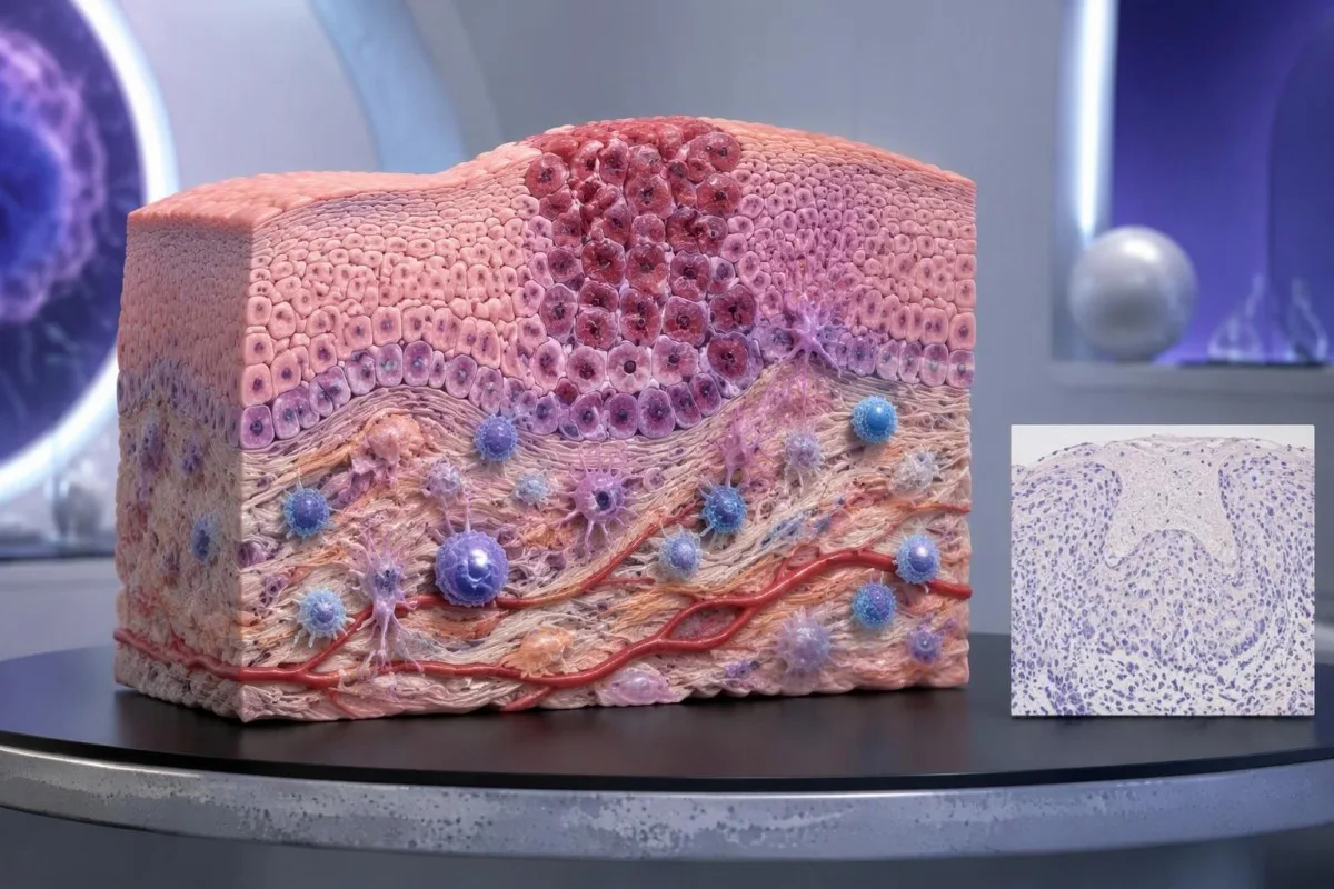
PD-L1-negative should first be translated into the laboratory's actual definition. TPS <1%, CPS <1, and an assay-specific IC-negative category are not identical measurements; an unreportable or inadequate sample is a fourth situation and should not be grouped with a technically valid negative result.
Ан MSI-high or mismatch-repair-deficient cancer may support particular immunotherapy pathways without PD-L1 selection, depending on the disease, stage, prior treatment, and local authorization. Tumor MMR testing and Генетското тестирање за синдром Линч answer related but different questions: an abnormal tumor result does not automatically prove an inherited syndrome.
Other genetic findings may point toward targeted treatment or different planning rather than immunotherapy. A result discussed in our BRCA variant interpretation guide does not turn CPS 0 into CPS 10; clinicians combine distinct biomarkers without pretending they measure the same biological feature.
No PD-L1 requirement is not the same as no selection criteria. A regimen can still require a particular histology, stage, treatment history, organ function, or absence of an actionable driver alteration; ask whether 1 negative score changes only the proposed option or the broader treatment plan.
Кога земањето примерок или граничен резултат може да оправдаат преиспитување?
Sampling variation can change PD-L1 classification, especially near a treatment threshold. A CPS of 9 versus 10, or TPS 0.8% versus 1%, may prompt pathology review when the distinction would change an otherwise suitable treatment—but repeating every result is not automatically useful.
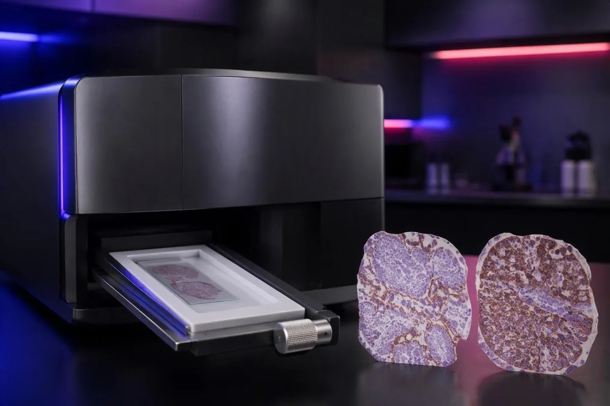
PD-L1 staining can be heterogeneous within 1 tissue block and between primary and metastatic sites. A small tissue examination may miss a highly expressing area or overrepresent it; the pathologist assesses the available viable tissue rather than assuming a single selected field describes the whole cancer.
Pretreatment and post-treatment specimens are not always equivalent. Chemotherapy, radiotherapy, and changes in the immune environment can alter expression, while decalcification of some bone specimens can impair antigen detection; ask whether a lower result reflects biology, processing limitations, or simply a different sampled site.
Borderline results require the actual assay rules, not homemade averaging. If 2 reports show CPS 8 and CPS 12, calculating CPS 10 does not create a newly validated result; the laboratory and oncology team should decide which specimen and assessment are appropriate for the treatment indication.
Kantesti cannot verify viable-cell counts, staining controls, or tissue suitability from a score alone. Our клиничка валидација и надзор should be read in that limited context; when another tissue procedure is considered, its risks and likely impact on treatment need to be weighed against the uncertainty in the existing result.
Кои други биомаркери треба да се разгледаат заедно со PD-L1?
Other biomarkers can take priority over PD-L1, depending on the cancer. In advanced non-small-cell lung cancer, molecular alterations such as EGFR mutations and ALK rearrangements affect treatment planning; in selected other cancers, HER2, MSI/MMR, or additional molecular findings may change the preferred pathway.

A high TPS does not cancel an actionable molecular finding. The pivotal KEYNOTE-024 и KEYNOTE-189 populations excluded relevant EGFR or ALK alterations, so their results should not be applied unchanged to those groups; treatment sequence also matters because some targeted therapies and checkpoint inhibitors can interact through toxicity.
Tumor mutational burden, often measured in mutations per megabase, is a different biomarker from PD-L1. A threshold such as 10 mutations/Mb has been used in a specific tissue-agnostic treatment context, but assay differences, cancer type, authorization, and clinical setting prevent it from becoming a universal immunotherapy rule.
A liquid molecular test may find circulating tumor DNA, but it does not automatically replace tissue PD-L1 immunohistochemistry. The 2 tests measure different things—genetic material versus protein location within cells—and a negative liquid result may also reflect limited DNA shedding rather than absence of a tumor alteration.
Kantesti AI distinguishes the role of supporting laboratory markers from tissue-based treatment biomarkers in our водич за референтни вредности за крвни биомаркери. A blood-cell ratio, LDH level, or serum tumor-marker concentration should not be used to manufacture TPS 50%, estimate CPS 10, or bypass an indicated molecular workup.
Кои безбедносни проверки се важни ако се разгледува имунотерапија?
PD-L1 scores predict neither treatment safety nor immune-related toxicity. A TPS of 90% does not make checkpoint treatment safer than TPS 1%; the team assesses medical history, organ function, medications, and the particular regimen separately from the tissue biomarker.
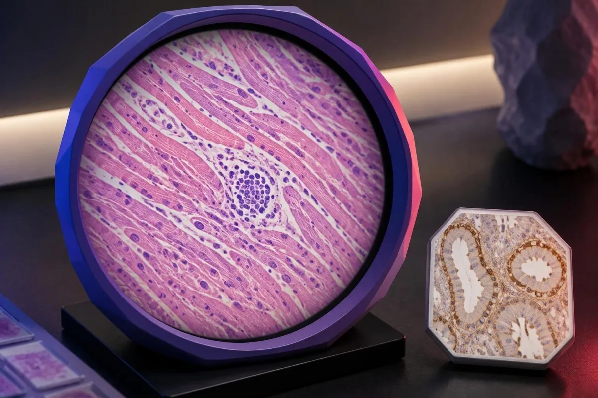
Checkpoint inhibitors can cause immune-related effects involving the thyroid, liver, lungs, bowel, kidneys, heart, and other organs. Baseline testing commonly includes liver chemistry, creatinine, thyroid measurements, and a complete blood count, with monitoring schedules varying by regimen; 1 normal baseline panel does not prevent later toxicity.
New chest pain, palpitations, fainting, or breathlessness during treatment requires prompt assessment because myocarditis, although uncommon, can be serious. Our myocarditis test interpretation guide explains why troponin supports evaluation but cannot determine the cause by itself; severe symptoms warrant emergency care rather than waiting for the next scheduled panel.
Kidney changes need interpretation against the person's own baseline and treatment exposures. A hypothetical creatinine rise from 0.8 to 1.6 mg/dL is a doubling even if the absolute value looks only modestly elevated; our kidney function testing guide explains supporting measurements, not an immunotherapy-specific treatment decision.
Pre-existing autoimmune disease, organ transplantation, ongoing immunosuppression, or previous severe immune-related toxicity can complicate treatment planning even when CPS is 100. Report new persistent diarrhea, worsening cough, jaundice, unusual weakness, or marked thirst promptly; neither a high PD-L1 score nor previously reassuring blood results justify delaying symptom assessment.
Што треба да прашате на онколошкиот преглед?
Донесете го complete PD-L1 pathology report and ask which exact treatment decision it informs. Four useful starting questions are: Which score was used? Which cutoff applies? Is the specimen adequate? What options remain if the result is below the cutoff?

A hypothetical report reading 22C3, CPS 12, adequate tissue is still incomplete as a treatment answer until the cancer, stage, and regimen are specified. Ask whether CPS ≥10 is relevant to the proposed option, whether other biomarkers are pending, and whether a combination would be considered under different selection rules.
Request explanations of 3 outcomes separately: tumor shrinkage, duration of disease control, and survival. A trial's response rate is not the same as its survival benefit, while median survival is a group midpoint—not an individual deadline—and the balance between rapid control and treatment toxicity may change the preferred regimen.
Кантести е услугата за интерпретација на тестови на AI for supporting laboratory understanding, not independent immunotherapy eligibility assessment. If blood monitoring is planned, our водич за подготовка за основно тестирање can help organize previous values, units, medication lists, and collection dates so that the oncology team can compare results meaningfully.
Keep tissue scores separate from serial blood monitoring in 2 clearly labeled records. Our Водич за трендови на безбедност на лекови explains why changes over time need context; our принципи за одговорна вештачка интелигенција also make clear that automated explanations should not trigger treatment changes without clinical review.
Истражувачки публикации и ограничувањата на доказите за PD-L1
PD-L1 interpretation should be grounded in cancer-specific trials and validated pathology methods. The 3 clinical references below address high-TPS lung-cancer monotherapy, lung-cancer combination treatment, and CPS-selected head-and-neck treatment; the 2 separate Zenodo publications concern other laboratory topics and do not establish PD-L1 thresholds.

KEYNOTE-024 supports a defined TPS ≥50% advanced lung-cancer population, whereas KEYNOTE-189 supports a particular combination across PD-L1 categories (Reck et al., 2016; Gandhi et al., 2018). Those different study designs explain why evidence for one regimen cannot be substituted for evidence supporting another simply because both drugs affect the same immune pathway.
KEYNOTE-048 assessed CPS-defined recurrent or metastatic head-and-neck cancer populations, including CPS ≥1 and CPS ≥20 groups (Burtness et al., 2019). When reading a trial, check the histology, stage, previous treatment, comparator, and endpoint; a subgroup survival finding does not automatically quantify an individual's chance of measurable shrinkage.
Kantesti's 2 Zenodo publications listed below are educational resources on complement/ANA testing and Nipah-virus diagnostics, not PD-L1 clinical trials or assay-validation studies. The водичот за комплемент и ANA concerns different laboratory questions, and a DOI identifies a deposited publication without independently establishing clinical validity.
Use the original pathology result and current local treatment information to resolve eligibility—not a generalized positive/negative table or an AI-generated score conversion. Our стандарди за медицинска содржина emphasize source relevance and uncertainty; ask your oncology team to document the 1 specific regimen, applicable threshold, and remaining alternatives.
Најчесто поставувани прашања
Што значи PD-L1 позитивен?
PD-L1 позитивно значи дека резултатот од ткивниот примерок го исполнува прагот на конкретниот тест и системот за оценување, како што е TPS ≥1% или CPS ≥1 во соодветен контекст. Прагот зависи од типот на ракот, терапевтскиот режим и локалните барања за лекување. Позитивниот резултат не докажува дека имунотерапијата ќе делува, а во извештајот треба да се наведе нумеричката вредност, а не само ознаката за позитивен резултат.
Која е разликата помеѓу CPS и TPS?
TPS го мери процентот на одржливи туморски клетки со квалификувачко мембранско боење за PD-L1, додека CPS ги вклучува квалификувачките позитивни туморски клетки, лимфоцити и макрофаги, поделени со бројот на одржливи туморски клетки и помножени со 100. TPS се пријавува од 0% до 100%, а пријавениот CPS е ограничен на 100. Резултатите не можат да се претворат еден во друг бидејќи CPS зависи од боењето на имунолошките клетки, кое TPS не го брои.
Дали CPS 10 значи 10% шанса имунотерапијата да делува?
CPS 10 не значи 10% шанса за одговор на имунотерапија. CPS е пресметка врз основа на боење на ткивото, чиј именител се вијабилните туморски клетки, а не веројатноста за одговор на третманот кај поединец. CPS ≥10 е основа за избор на одредени режими специфични за ракот, но за процена на веројатната корист се потребни и дијагнозата на ракот, стадиумот, другите биомаркери и доказите за предложениот третман.
Може ли да примам имунотерапија ако PD-L1 е негативен?
Негативниот резултат за PD-L1 не ја исклучува секоја опција за имунотерапија. За некои комбинации на хемотерапија и имунотерапија, како и за други патеки на лекување избрани според биомаркери, не е потребна PD-L1-позитивност, дури и кога TPS е под 1%. Подобноста сепак зависи од видот и стадиумот на ракот, молекуларните наоди, историјата на лекување, медицинската соодветност и локалното одобрение; несоодветен примерок од ткиво не треба да се смета за валиден негативен резултат.
Дали TPS од 50% се смета за висока експресија на PD-L1?
TPS ≥50% вообичаено се опишува како висока експресија на PD-L1 во одредени случаи на недребноклеточен карцином на белите дробови. Овој праг беше користен во KEYNOTE-024, во која беше испитувана специфична група пациенти со напреднат карцином на белите дробови, без сензитизирачки мутации на EGFR или преуредувања на ALK. TPS 50% не е исто што и CPS 50 и не е универзален праг за секој вид рак, стадиум или режим на имунотерапија.
Дали PD-L1 е крвен тест и дали треба да се повтори?
Клиничкото оценување на PD-L1 за CPS и TPS обично се врши на зачувано канцерско ткиво испитано со имунохистохемија, а не на рутински примерок од крв. Повторната процена може да помогне ако ткивото е недоволно, резултатот е близу до гранична вредност што може да ја промени одлуката, како што е CPS 10, или ако друг примерок е посоодветен. Онколошкиот и патолошкиот тим треба да одлучат дали преглед, дополнителни исечоци или друг примерок веројатно ќе ја променат грижата доволно за да го оправдаат поврзаниот напор и процедуралните ризици.
Дознајте како да ги читате крвните резултати
Поставете го вашиот лабораториски извештај и AI на Kantesti ќе го објасни секој биомаркер на разбирлив јазик за неколку секунди. Го користат повеќе од 2 милиони луѓе ширум светот.
Референци
- Клајн, Т., Мичел, С., & Вебер, Х. (2026). Водич за тест на крвта за C3 C4 комплемент и титар на ANA. Kantesti AI за медицински истражувања. ДОИ
- Клајн, Т., Мичел, С., & Вебер, Х. (2026). Тест на крвта за вирусот Нипах: водич за рано откривање и дијагностицирање 2026. Kantesti AI за медицински истражувања. ДОИ
- Reck M et al. (2016). Pembrolizumab versus Chemotherapy for PD-L1–Positive Non–Small-Cell Lung Cancer. New England Journal of Medicine. ДОИ
- Gandhi L et al. (2018). Pembrolizumab plus Chemotherapy in Metastatic Non–Small-Cell Lung Cancer. New England Journal of Medicine. ДОИ
- Burtness B et al. (2019). Pembrolizumab alone or with chemotherapy versus cetuximab with chemotherapy for recurrent or metastatic squamous cell carcinoma of the head and neck (KEYNOTE-048): a randomised, open-label, phase 3 study. The Lancet. ДОИ
- 2 милиони+Анализирани тестови
- 127+Земји
- 100+Јазици
За медицинскиот тим


Сигнали за доверба E-E-A-T
Искуство
Клиничка ревизија водена од лекар за работните текови на толкување на лабораториски резултати.
Експертиза
Фокус на лабораториска медицина за тоа како биомаркерите се однесуваат во клинички контекст.
Авторитивност
Медицински рецензирано од д-р Thomas Klein, со придонес од д-р Sarah Mitchell и проф. д-р Hans Weber.
Доверливост
Толкување засновано на докази со јасни патеки за следење за да се намали алармирањето.






Медицинско одрекување од одговорност
Овој напис е само за едукативни цели и не претставува медицински совет. Секогаш консултирајте се со квалификуван здравствен работник за одлуки за дијагноза и третман.
Објавено · Контактирајте го уредничкиот тим
Објавено од Kantesti Ltd, регистрирана во Англија и Велс, регистарски број на компанијата. 17090423, Лондон, Обединето Кралство.
