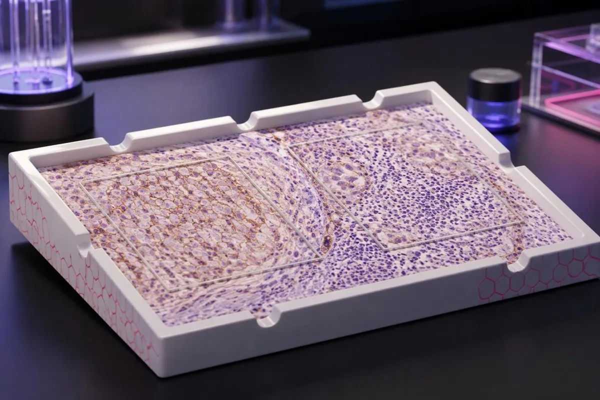
PD-L1-poentaroj priskribas la makuliĝon en histospecimeno — ne vian personan ŝancon, ke la kuracado sukcesos. La poentadmetodo, kancera diagnozo kaj proponita kombino de medikamentoj determinas, kion la rezulto povas pravigi.

Ĉefaj konkludoj
- Rezultoj de PD-L1-testado mezuras proteinan makuliĝon en kancera histo; ili ne estas rutinaj rezultoj de sangoanalizo aŭ kancera ekzameno.
- TPS estas la procento de vivkapablaj tumorĉeloj kun kvalifikiĝanta membrana makuliĝo, raportata de 0% ĝis 100%.
- CPS kalkulas la kvalifikiĝantajn PD-L1-pozitivajn tumorĉelojn, limfocitojn kaj makrofagojn, dividas la sumon per la nombro de vivkapablaj tumorĉeloj kaj multiplikas per 100; la raportata poentaro estas limigita al 100.
- Malsamaj poentaroj ne povas esti konvertita: TPS 10% povus akompani CPS 10, CPS 25 aŭ alian valoron depende de la makuliĝo de imunĉeloj.
- Sojloj kiel TPS 1%, TPS 50%, CPS 1 kaj CPS 10 validas por specifaj kanceroj kaj kuracaj situacioj—ne por ĉiu imunoterapio.
- Pozitivaj rezultoj povas subteni elekton de kuracado, sed ne garantias utilon; poentaro de 50 ne estas 50%-a probableco de respondo.
- Negativaj rezultoj ne ĉiam ekskludas imunoterapion, ĉar iuj kombinaĵoj kaj biomarkil-difinitaj kuracadoj ne postulas PD-L1-pozitivecon.
- Adekvateco de la specimeno gravas: pluraj ofte uzataj poentadaj protokoloj postulas almenaŭ 100 vivkapablajn tumorĉelojn, sed la laboratorio devas apliki siajn regulojn specifajn por la indiko.
Kion efektive diras al vi la rezultoj de PD-L1-testado?
Rezultoj de PD-L1-testado priskribas la proteinan makuliĝon en kancera histo. TPS kalkulas la procenton de makuliĝintaj tumorĉeloj; CPS inkluzivas ankaŭ elektitajn makuliĝintajn imunĉelojn. Neniu el la poentaroj sola decidas la kuracadon: la kancertipo, stadio, analizo kaj kuraca reĝimo determinas la koncernan limvaloron, kaj pozitivaj aŭ negativaj rezultoj ne garantias utilon aŭ ekskludon.
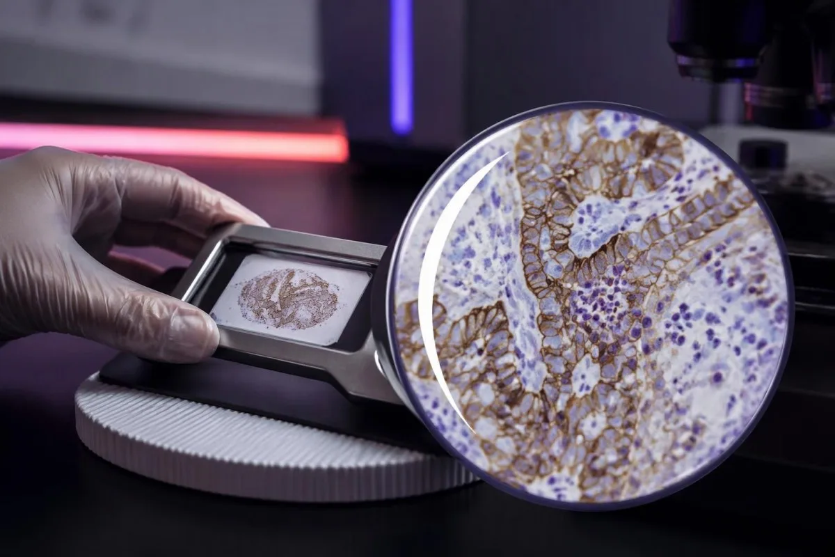
PD-L1, aŭ programita ĉela mortigando 1, povas ligiĝi al la PD-1-receptoro ĉe imunĉeloj kaj redukti ilian aktivecon. Medikamentoj celantaj ĉi tiun interagon povas restarigi kontraŭkanceran imunaktivecon, sed mezurado de 1 proteino ne kaptas ĉiun faktoron, kiu determinas ĉu imunĉeloj rekonas kaj regas kanceron.
Hista PD-L1-rezulto estas kutime produktata per imunohistokemio, kiu uzas antikorpon por montri la lokon de la proteino en konservita specimeno. La raportita nombro priskribas ĉelojn en elektitaj histosekcioj—ne ĉiun kancerĉelon tra la tuta korpo—kaj 2 specimenoj el malsamaj lokoj povas doni malsamajn rezultojn.
Kantesti estas AI-sangotesta analizilo, ne anstataŭaĵo de histopatologio aŭ onkologia kuraca decido. Niaj organizo kaj klinika amplekso distingas edukajn klarigojn pri laboratoriaj rezultoj disde decidoj, kiuj postulas kune la originalan histopatologian raporton, bildigojn kaj proponitan kuracan reĝimon.
Komencu per 4 detaloj antaŭ ol interpreti la nombron: la kancerdiagnozo, ĉu la poentaro estas CPS aŭ TPS, la uzita analizo kaj la kuracado konsiderata. Raporto, kiu diras nur PD-L1-pozitiva, lasas klinike gravan mankon; petu la poentadan metodon kaj la nombran rezulton.
Signifo de la PD-L1-TPS-poentaro: la procento de tumorĉeloj
Tumora proporcia poentaro, aŭ TPS, estas la procento de vivkapablaj tumorĉeloj, kiuj montras kriterikvalifikan partan aŭ kompletan PD-L1-makuliĝon de la membrano. TPS varias de 0% ĝis 100%; imunĉeloj estas ekskluditaj el ĝia numeratoro, eĉ kiam ili forte makuliĝas apud la kancerĉeloj.
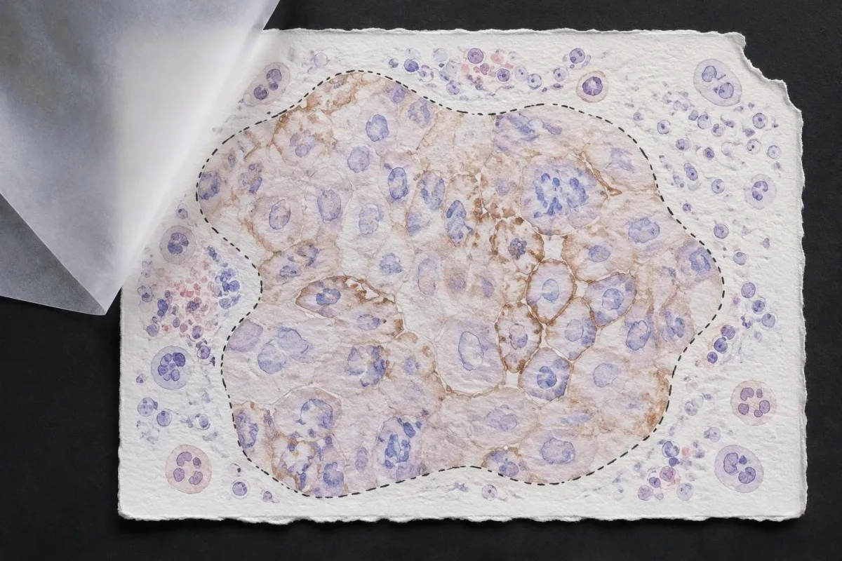
TPS = PD-L1-pozitivaj vivkapablaj tumorĉeloj ÷ ĉiuj vivkapablaj tumorĉeloj × 100%. En hipoteza histokampo enhavanta 200 vivkapablajn tumorĉelojn, 80 kriterikvalifikaj pozitivaj ĉeloj donas TPS 40%; proksimaj makuliĝintaj limfocitoj ne ŝanĝas tiun kalkulon, kvankam ili povus influi alian poentadsistemon.
Membrane location matters more than visual darkness alone. For commonly used TPS methods, weak qualifying membrane staining can count, whereas staining limited to tumor-cell cytoplasm does not establish a positive tumor cell; TPS 1% therefore describes the fraction of qualifying cells, not a required intensity grade.
Cutoffs such as TPS 1% kaj TPS 50% were used to define populations in particular lung-cancer trials and treatment pathways. They are not boundaries between harmless and dangerous cancer, and crossing 50% does not create an abrupt biological switch from treatment resistance to guaranteed sensitivity.
Read the exact reporting format: TPS <1%, TPS 1–49%, and TPS ≥50% provide different detail from a single positive flag. Our explanation of kvalita kontraŭ kvanta rezultoj covers why a category cannot replace a number, although PD-L1 uses tissue-specific—not blood-reference-range—rules.
Signifo de la PD-L1-CPS-poentaro: kial imunĉeloj gravas
Combined positive score, aŭ CPS, includes qualifying PD-L1-positive tumor cells, lymphocytes, and macrophages. CPS divides their combined count by the number of viable tumor cells and multiplies by 100; the denominator does not become all cells simply because immune cells enter the numerator.
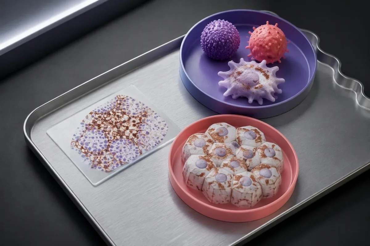
CPS = positive tumor cells plus qualifying positive lymphocytes and macrophages ÷ viable tumor cells × 100. In a hypothetical sample with 500 viable tumor cells, 50 positive tumor cells, and 75 qualifying positive immune cells, CPS is 25; the tumor-cell-only TPS would be 10%.
The reported CPS is capped at 100, even if the uncapped arithmetic would exceed 100 because many qualifying immune cells surround relatively few tumor cells. CPS is a unitless score, not a percentage of all cells, a blood concentration, or a probability that immunotherapy will work.
Not every stained immune cell belongs in the CPS count. The pathologist applies the assay manual's rules for eligible lymphocytes and macrophages associated with the tumor; neutrophils, unrelated inflammatory areas, and nonviable tissue are not simply added because a slide contains visible staining.
A CPS of 20 can arise from more than 1 cellular pattern: abundant tumor-cell staining, substantial immune-cell staining, or both. This denominator issue resembles the distinction between cell counts and percentages, but a circulating monocyte count cannot supply the tissue information needed to calculate CPS.
Kial CPS kaj TPS ne estas interŝanĝeblaj kaj ne povas esti konvertitaj
CPS and TPS are not interchangeable because they count different cells and were validated in different treatment settings. A TPS of 10% cannot be converted reliably into CPS 10, and a CPS of 20 does not establish that 20% of tumor cells stain.
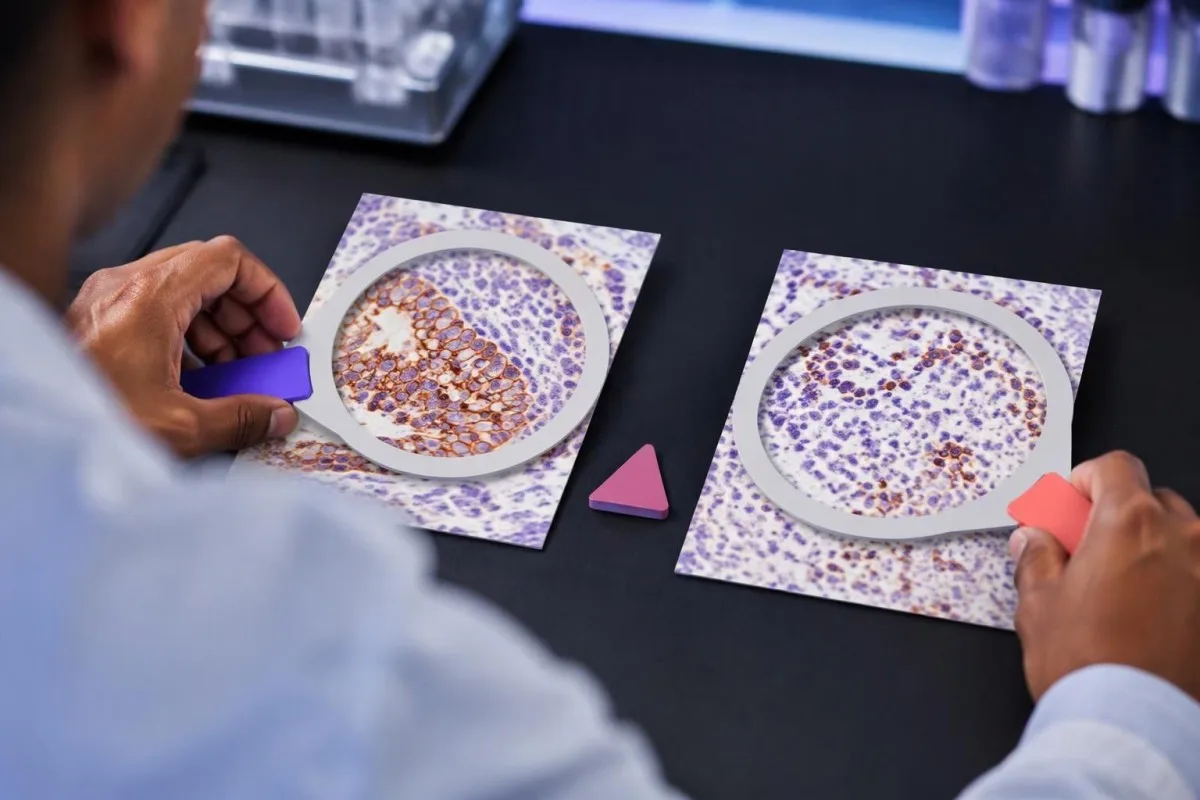
Consider 2 hypothetical specimens with 500 viable tumor cells and 50 positive tumor cells each. The first has no qualifying positive immune cells and gives TPS 10% with CPS 10; the second contains 75 qualifying positive immune cells and gives the same TPS 10% with CPS 25.
The reverse inference fails too. A hypothetical CPS 20 could result from 20 positive tumor cells among 100 tumor cells, or from 5 positive tumor cells plus 15 qualifying positive immune cells; those examples have TPS 20% and TPS 5%, respectively, despite identical CPS values.
Unu immune-cell score, sometimes abbreviated IC, is another system rather than a third spelling of CPS. Some IC methods measure the proportion of tumor area occupied by stained immune cells, so an IC result of 1% must be interpreted using its own assay and indication—not a TPS threshold.
Kantesti's educational explanation should preserve all 3 scoring labels rather than flatten them into positive or negative. When using AI report explanations safely, check that the original score, assay, and specimen remain visible; an AI-generated conversion without the underlying tissue counts is not clinically dependable.
Kiuj detaloj en la patologia raporto gravas krom la poentaro?
A usable PD-L1 report should identify the specimen, assay, scoring method, numerical result, and adequacy assessment. Several commonly used protocols require at least 100 viable tumor cells, but minimum counts and acceptable specimen types depend on the particular test and cancer indication.

Antibody identifiers such as 22C3, 28-8, SP263, and SP142 describe different testing assays or antibody clones, not different PD-L1 proteins. Some assays show substantial agreement in selected settings, but laboratories should not assume universal equivalence across tumor types, scoring methods, immune-cell assessments, and treatment requirements.
The specimen date and site can change the interpretation of 1 reported number. A small primary-site sample taken before treatment may not represent a later metastatic deposit; ask whether the report describes the current treatment-setting specimen or archived tissue that the oncology team considers suitable.
Insufficient tissue is not PD-L1 negative. A sample with too few viable tumor cells, failed controls, extensive tissue change, or unsuitable processing may be unreportable; the practical next step is laboratory clarification, additional sections, or another suitable specimen—not assigning TPS 0% or CPS 0.
Comparisons between 2 reports need more than matching dates. Our test timing and comparability guide explains the general problem of comparing unlike measurements; for PD-L1, the extra checks are anatomical site, prior treatment, fixation, tissue adequacy, assay, and scoring system.
Kiel la tipo de kancero ŝanĝas la signifon de pozitiva rezulto?
PD-L1 positivity is indication-specific, not a universal diagnosis. TPS thresholds such as 1% or 50% are familiar in non-small-cell lung cancer, while CPS thresholds such as 1, 5, or 10 appear in selected other cancer settings; the drug, stage, and regimen must accompany the cutoff.

For recurrent or metastatic head-and-neck squamous-cell carcinoma, KEYNOTE-048 evaluated CPS-defined populations. Pembrolizumab alone improved median overall survival in the CPS ≥20 group to 14.9 months versus 10.7 months with the comparator regimen, showing why a CPS threshold cannot be borrowed from a lung-cancer TPS report (Burtness et al., 2019).
Selected established pembrolizumab pathways use CPS ≥10 for unresectable locally advanced or metastatic triple-negative breast cancer with chemotherapy, and CPS ≥1 for certain persistent, recurrent, or metastatic cervical-cancer regimens. Those examples do not establish eligibility for every earlier-stage setting, drug combination, country, or funding pathway.
Gastric and gastroesophageal-junction cancers require particular care because treatment selection may also involve HER2 kaj MSI/MMR, with PD-L1 requirements differing across regimens and jurisdictions. A serum marker described in our CA 72-4 interpretation guide cannot replace the tissue biomarker panel or define CPS.
Cancer diagnosis and treatment selection are 2 separate steps. Our persistent cough assessment guide addresses when lung symptoms need investigation; once cancer is confirmed, ask the oncology team to match the PD-L1 result to the current local product information and clinical pathway.
Kial la kuracskemo gravas same kiel la sojlo
Immunotherapy alone and immunotherapy combinations can have different PD-L1 requirements. A TPS below 1% may rule out one PD-L1-selected monotherapy pathway without ruling out an appropriate chemotherapy–immunotherapy combination; these are different clinical decisions, not contradictory interpretations of the same result.
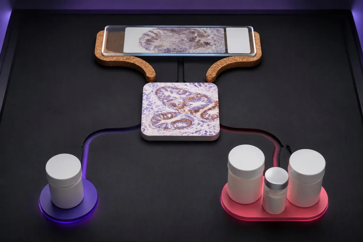
KEYNOTE-024 enrolled previously untreated advanced non-small-cell lung cancer with TPS ≥50% and no sensitizing EGFR mutation or ALK rearrangement. Median progression-free survival was 10.3 months with pembrolizumab versus 6.0 months with chemotherapy, supporting that specific high-TPS monotherapy setting—not every cancer with a score above 50 (Reck et al., 2016).
En KEYNOTE-189, pembrolizumab plus pemetrexed and platinum chemotherapy improved outcomes in previously untreated metastatic nonsquamous non-small-cell lung cancer without EGFR or ALK alterations. Twelve-month overall survival was 69.2% versus 49.4%, and benefit was observed across PD-L1 categories, including TPS <1% (Gandhi et al., 2018).
Combination treatment may address rapid disease progression differently from a single agent, but adding drugs also adds toxicity and monitoring needs. If a proposed regimen includes fluorouracil or capecitabine, testado de DPYD antaŭ kemioterapio concerns treatment safety; it does not measure the PD-L1 pathway or predict the same benefit.
Kantesti estas AI sangotesta interpretada platformo that helps explain supporting laboratory measurements; it does not select between 1-drug and multidrug cancer regimens. Similarly, UGT1A1 and irinotecan safety address metabolism-related toxicity, illustrating why each biomarker should be matched to the specific medicine under discussion.
Ĉu pozitiva PD-L1-rezulto garantias utilon de imunoterapio?
A positive PD-L1 result does not guarantee a response. In KEYNOTE-024's selected high-expression lung-cancer population, the objective response rate with pembrolizumab was 44.8%—substantial benefit for many participants, but clearly not a response in every person with TPS ≥50%.
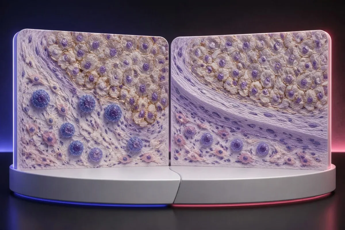
The 44.8% response rate in KEYNOTE-024 describes a trial population under defined conditions, not an individual's calculated chance of success (Reck et al., 2016). Objective response also means measurable tumor shrinkage by trial criteria; stable disease and survival benefit are different outcomes that should not be collapsed into that single percentage.
PD-L1 is only 1 part of a broader immune environment. A cancer can show substantial staining while lacking effective immune-cell access, recognizable targets, or intact antigen presentation; these mechanisms help explain why expression enriches for benefit in some settings without functioning as a deterministic on–off switch.
Higher does not always mean better across different cancers and combinations. A CPS 30 in one disease cannot be ranked against TPS 60% in another, and the same CPS threshold may have different predictive value when used for monotherapy versus a combination that already provides benefit through other mechanisms.
Some melanoma immunotherapy regimens do not require a positive PD-L1 test, so a low score should not be interpreted using lung-cancer monotherapy rules. Our melanoma warning-sign guide addresses recognition and assessment, while treatment benefit must be discussed using disease-specific trial evidence and the person's overall condition.
Ĉu imunoterapio ankoraŭ povas esti proponata kiam PD-L1 estas negativa?
PD-L1-negative cancers can still have appropriate immunotherapy options. TPS <1% or CPS <1 may exclude a particular PD-L1-selected regimen, but some combinations, earlier-stage pathways, and treatments selected by other biomarkers do not require PD-L1 positivity.
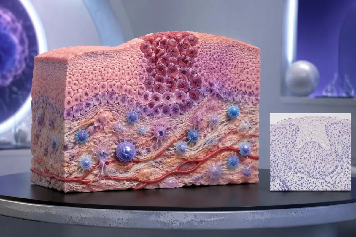
PD-L1-negative should first be translated into the laboratory's actual definition. TPS <1%, CPS <1, and an assay-specific IC-negative category are not identical measurements; an unreportable or inadequate sample is a fourth situation and should not be grouped with a technically valid negative result.
Unu MSI-high or mismatch-repair-deficient cancer may support particular immunotherapy pathways without PD-L1 selection, depending on the disease, stage, prior treatment, and local authorization. Tumor MMR testing and Genetika testado pri Lynch-sindromo answer related but different questions: an abnormal tumor result does not automatically prove an inherited syndrome.
Other genetic findings may point toward targeted treatment or different planning rather than immunotherapy. A result discussed in our BRCA variant interpretation guide does not turn CPS 0 into CPS 10; clinicians combine distinct biomarkers without pretending they measure the same biological feature.
No PD-L1 requirement is not the same as no selection criteria. A regimen can still require a particular histology, stage, treatment history, organ function, or absence of an actionable driver alteration; ask whether 1 negative score changes only the proposed option or the broader treatment plan.
Kiam specimenado aŭ limvalora poentaro povas pravigi revizion?
Sampling variation can change PD-L1 classification, especially near a treatment threshold. A CPS of 9 versus 10, or TPS 0.8% versus 1%, may prompt pathology review when the distinction would change an otherwise suitable treatment—but repeating every result is not automatically useful.
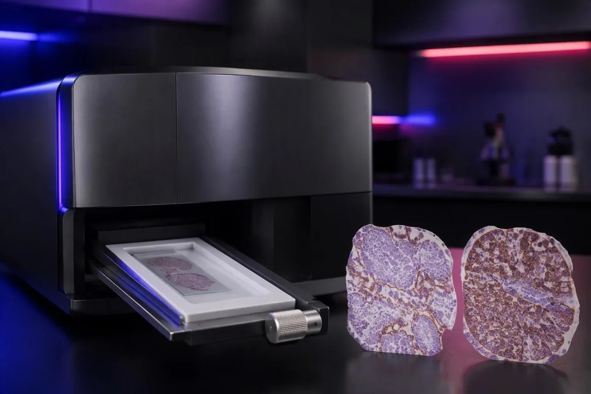
PD-L1 staining can be heterogeneous within 1 tissue block and between primary and metastatic sites. A small tissue examination may miss a highly expressing area or overrepresent it; the pathologist assesses the available viable tissue rather than assuming a single selected field describes the whole cancer.
Pretreatment and post-treatment specimens are not always equivalent. Chemotherapy, radiotherapy, and changes in the immune environment can alter expression, while decalcification of some bone specimens can impair antigen detection; ask whether a lower result reflects biology, processing limitations, or simply a different sampled site.
Borderline results require the actual assay rules, not homemade averaging. If 2 reports show CPS 8 and CPS 12, calculating CPS 10 does not create a newly validated result; the laboratory and oncology team should decide which specimen and assessment are appropriate for the treatment indication.
Kantesti cannot verify viable-cell counts, staining controls, or tissue suitability from a score alone. Our klinika validigo kaj superrigardo should be read in that limited context; when another tissue procedure is considered, its risks and likely impact on treatment need to be weighed against the uncertainty in the existing result.
Kiuj aliaj biosignoj estu konsiderataj kune kun PD-L1?
Other biomarkers can take priority over PD-L1, depending on the cancer. In advanced non-small-cell lung cancer, molecular alterations such as EGFR mutations and ALK rearrangements affect treatment planning; in selected other cancers, HER2, MSI/MMR, or additional molecular findings may change the preferred pathway.

A high TPS does not cancel an actionable molecular finding. The pivotal KEYNOTE-024 kaj KEYNOTE-189 populations excluded relevant EGFR or ALK alterations, so their results should not be applied unchanged to those groups; treatment sequence also matters because some targeted therapies and checkpoint inhibitors can interact through toxicity.
Tumor mutational burden, often measured in mutations per megabase, is a different biomarker from PD-L1. A threshold such as 10 mutations/Mb has been used in a specific tissue-agnostic treatment context, but assay differences, cancer type, authorization, and clinical setting prevent it from becoming a universal immunotherapy rule.
A liquid molecular test may find circulating tumor DNA, but it does not automatically replace tissue PD-L1 immunohistochemistry. The 2 tests measure different things—genetic material versus protein location within cells—and a negative liquid result may also reflect limited DNA shedding rather than absence of a tumor alteration.
Kantesti AI distinguishes the role of supporting laboratory markers from tissue-based treatment biomarkers in our sangobiomarkila referenca gvidilo. A blood-cell ratio, LDH level, or serum tumor-marker concentration should not be used to manufacture TPS 50%, estimate CPS 10, or bypass an indicated molecular workup.
Kiujn sekurecajn kontrolojn gravas fari, se oni konsideras imunoterapion?
PD-L1 scores predict neither treatment safety nor immune-related toxicity. A TPS of 90% does not make checkpoint treatment safer than TPS 1%; the team assesses medical history, organ function, medications, and the particular regimen separately from the tissue biomarker.
Checkpoint inhibitors can cause immune-related effects involving the thyroid, liver, lungs, bowel, kidneys, heart, and other organs. Baseline testing commonly includes liver chemistry, creatinine, thyroid measurements, and a complete blood count, with monitoring schedules varying by regimen; 1 normal baseline panel does not prevent later toxicity.
New chest pain, palpitations, fainting, or breathlessness during treatment requires prompt assessment because myocarditis, although uncommon, can be serious. Our myocarditis test interpretation guide explains why troponin supports evaluation but cannot determine the cause by itself; severe symptoms warrant emergency care rather than waiting for the next scheduled panel.
Kidney changes need interpretation against the person's own baseline and treatment exposures. A hypothetical creatinine rise from 0.8 to 1.6 mg/dL is a doubling even if the absolute value looks only modestly elevated; our kidney function testing guide explains supporting measurements, not an immunotherapy-specific treatment decision.
Pre-existing autoimmune disease, organ transplantation, ongoing immunosuppression, or previous severe immune-related toxicity can complicate treatment planning even when CPS is 100. Report new persistent diarrhea, worsening cough, jaundice, unusual weakness, or marked thirst promptly; neither a high PD-L1 score nor previously reassuring blood results justify delaying symptom assessment.
Kion vi demandu ĉe via onkologia konsulto?
Alportu la complete PD-L1 pathology report and ask which exact treatment decision it informs. Four useful starting questions are: Which score was used? Which cutoff applies? Is the specimen adequate? What options remain if the result is below the cutoff?

A hypothetical report reading 22C3, CPS 12, adequate tissue is still incomplete as a treatment answer until the cancer, stage, and regimen are specified. Ask whether CPS ≥10 is relevant to the proposed option, whether other biomarkers are pending, and whether a combination would be considered under different selection rules.
Request explanations of 3 outcomes separately: tumor shrinkage, duration of disease control, and survival. A trial's response rate is not the same as its survival benefit, while median survival is a group midpoint—not an individual deadline—and the balance between rapid control and treatment toxicity may change the preferred regimen.
Kantesti estas AI-labora testa interpretservo for supporting laboratory understanding, not independent immunotherapy eligibility assessment. If blood monitoring is planned, our gvidilo pri preparado por bazlinia testado can help organize previous values, units, medication lists, and collection dates so that the oncology team can compare results meaningfully.
Keep tissue scores separate from serial blood monitoring in 2 clearly labeled records. Our gvidilo pri medikamenta sekureca tendenco explains why changes over time need context; our principoj pri respondeca AI also make clear that automated explanations should not trigger treatment changes without clinical review.
Esploraj publikaĵoj kaj la limoj de la pruvoj pri PD-L1
PD-L1 interpretation should be grounded in cancer-specific trials and validated pathology methods. The 3 clinical references below address high-TPS lung-cancer monotherapy, lung-cancer combination treatment, and CPS-selected head-and-neck treatment; the 2 separate Zenodo publications concern other laboratory topics and do not establish PD-L1 thresholds.

KEYNOTE-024 supports a defined TPS ≥50% advanced lung-cancer population, whereas KEYNOTE-189 supports a particular combination across PD-L1 categories (Reck et al., 2016; Gandhi et al., 2018). Those different study designs explain why evidence for one regimen cannot be substituted for evidence supporting another simply because both drugs affect the same immune pathway.
KEYNOTE-048 assessed CPS-defined recurrent or metastatic head-and-neck cancer populations, including CPS ≥1 and CPS ≥20 groups (Burtness et al., 2019). When reading a trial, check the histology, stage, previous treatment, comparator, and endpoint; a subgroup survival finding does not automatically quantify an individual's chance of measurable shrinkage.
Kantesti's 2 Zenodo publications listed below are educational resources on complement/ANA testing and Nipah-virus diagnostics, not PD-L1 clinical trials or assay-validation studies. The gvidilo pri komplemento kaj ANA concerns different laboratory questions, and a DOI identifies a deposited publication without independently establishing clinical validity.
Use the original pathology result and current local treatment information to resolve eligibility—not a generalized positive/negative table or an AI-generated score conversion. Our normoj pri medicina enhavo emphasize source relevance and uncertainty; ask your oncology team to document the 1 specific regimen, applicable threshold, and remaining alternatives.
Oftaj demandoj
Kion signifas PD-L1-pozitiva?
PD-L1 pozitiva signifas, ke histologia rezulto plenumas specifan sojlon de analizo kaj poentado, ekzemple TPS ≥1% aŭ CPS ≥1 en koncerna klinika situacio. La sojlo dependas de la tipo de kancero, la terapireĝimo kaj la lokaj terapiaj postuloj. Pozitiveco ne pruvas, ke imunoterapio efikos, kaj la raporto devas indiki la nombran poentaron, ne nur marki la rezulton kiel pozitivan.
Kio estas la diferenco inter CPS kaj TPS?
TPS mezuras la procenton de vivkapablaj tumorĉeloj kun kvalifika membrana makuliĝo por PD-L1, dum CPS inkluzivas kvalifikajn pozitivajn tumorĉelojn, limfocitojn kaj makrofagojn, dividitajn per la nombro de vivkapablaj tumorĉeloj kaj multiplikitajn per 100. TPS estas raportata de 0% ĝis 100%, kaj la raportata CPS estas limigita al 100. La poentaroj ne estas konverteblaj, ĉar CPS dependas de makuliĝo de imunĉeloj, kiun TPS ne enkalkulas.
Ĉu CPS 10 signifas 10% da ŝanco, ke imunoterapio funkcios?
CPS 10 ne signifas ŝancon de 10% respondi al imunoterapio. CPS estas histomakula kalkulo, kies denominatoro estas vivkapablaj tumorĉeloj, ne individua probableco respondi al kuracado. CPS ≥10 subtenas la elekton de certaj kontraŭkanceraj terapiaj reĝimoj, sed por taksi la probablan utilon necesas ankaŭ la diagnozo de kancero, la stadio, aliaj biosignoj kaj la pruvoj pri la proponita kuracado.
Ĉu mi povas ricevi imunoterapion se PD-L1 estas negativa?
Negativa rezulto pri PD-L1 ne ekskludas ĉiun imunterapian eblon. Iuj kombinaĵoj de kemioterapio kaj imunoterapio, kaj aliaj vojoj elektitaj laŭ biomarkiloj, ne postulas PD-L1-pozitivecon, eĉ kiam la TPS estas sub 1 %. Kvalifikiĝo ankoraŭ dependas de la tipo kaj stadio de la kancero, molekulaj rezultoj, antaŭa kuracado, medicina taŭgeco kaj loka aprobo; nesufiĉa histoprovaĵo ne estu traktata kiel valida negativa rezulto.
Ĉu TPS 50% estas konsiderata alta PD-L1-esprimo?
TPS ≥50% estas kutime priskribita kiel alta PD-L1-esprimo en elektitaj situacioj de ne-malgrandĉela pulmokancero. La sojlo estis uzata en KEYNOTE-024, kiu studis specifan progresintan pulmonkanceran populacion sen sensivigaj EGFR-mutacioj aŭ ALK-rearanĝoj. TPS 50% ne egalas al CPS 50 kaj ne estas universala sojlo por ĉiu kancero, stadio aŭ imunoterapia reĝimo.
Ĉu PD-L1 estas sangokontrolo, kaj ĉu ĝi devas esti ripetita?
La klinika PD-L1-poentado por CPS kaj TPS kutime uzas konservitan kanceran histon ekzamenitan per imunohistokemio, ne rutinan sangospecimenon. Ripeta taksado povas helpi, se la histo estas nesufiĉa, la rezulto estas proksima al sojlo, kiu ŝanĝus la decidon, kiel CPS 10, aŭ se alia specimeno pli taŭgas. La onkologia kaj patologia teamoj devas decidi, ĉu revizio, pliaj histosekcioj aŭ alia specimeno verŝajne ŝanĝos la kuracadon sufiĉe por pravigi la rilatan penon kaj procedurajn riskojn.
Klarigu viajn rezultojn de sangoanalizo
Alŝutu vian laboratorian raporton kaj la AI de Kantesti klarigas ĉiun biomarkilon per simpla lingvaĵo ene de sekundoj. Uzata de pli ol 2 milionoj da homoj tutmonde.
Referencoj
- Klein, T., Mitchell, S., & Weber, H. (2026). Gvidilo pri C3 C4-komplementa sangotesto kaj ANA-titro. Kantesti AI-esplorado pri medicino. DOI
- Klein, T., Mitchell, S., & Weber, H. (2026). Sangotesto por Nipah-viruso: Gvidilo pri frua detekto kaj diagnozo 2026. Kantesti AI-esplorado pri medicino. DOI
- Reck M et al. (2016). Pembrolizumab versus Chemotherapy for PD-L1–Positive Non–Small-Cell Lung Cancer. New England Journal of Medicine. DOI
- Gandhi L et al. (2018). Pembrolizumab plus Chemotherapy in Metastatic Non–Small-Cell Lung Cancer. New England Journal of Medicine. DOI
- Burtness B et al. (2019). Pembrolizumab alone or with chemotherapy versus cetuximab with chemotherapy for recurrent or metastatic squamous cell carcinoma of the head and neck (KEYNOTE-048): a randomised, open-label, phase 3 study. The Lancet. DOI
- 2M+Analizitaj testoj
- 127+Landoj
- 100+Lingvoj
Pri la medicina teamo


Signaloj de fido E-E-A-T
Sperto
Kuracista gvidata klinika revizio de laboratoriaj interpretaj laborfluoj.
Kompetenteco
Laboratoria medicino fokusiĝas pri kiel biomarkiloj kondutas en klinika kunteksto.
Aŭtoritateco
Medicine recenzita de D-ro Thomas Klein, kun kontribuoj de D-rino Sarah Mitchell kaj Prof. D-ro Hans Weber.
Fidindeco
Evident-bazita interpretado kun klaraj sekvaj vojoj por redukti alarmon.






Medicina malrespondeca deklaro
Ĉi tiu artikolo estas nur por edukaj celoj kaj ne konsistigas medicinan konsilon. Ĉiam konsultu kvalifikitan sanprovizanton por diagnozaj kaj traktadaj decidoj.
Publikigita · Kontaktu la redakcian teamon
Eldonita de Kantesti Ltd, registrita en Anglio kaj Kimrio, kompanio n-ro. 17090423, Londono, Unuiĝinta Reĝlando.
