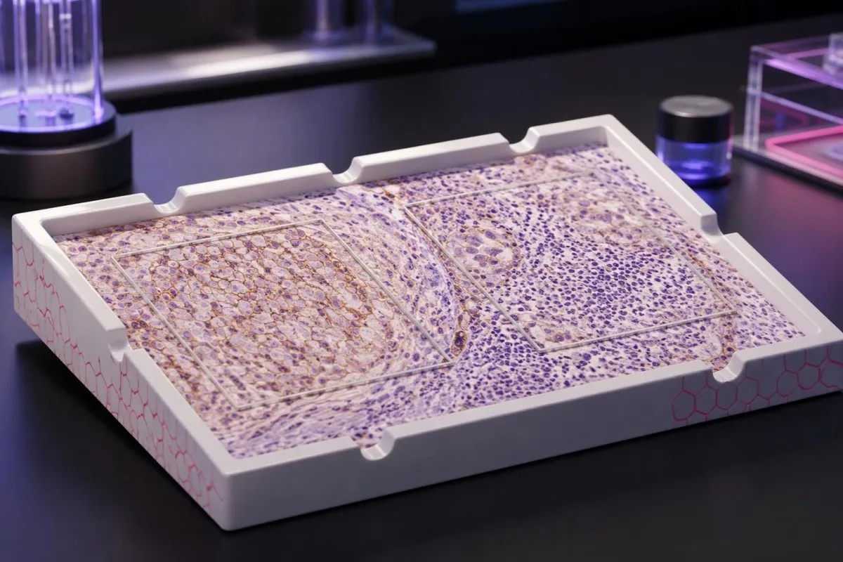
Показники PD-L1 описують забарвлення зразка тканини, а не вашу особисту ймовірність успіху лікування. Метод оцінювання, діагноз раку та запропонована комбінація препаратів визначають, що можна встановити за результатом.

Ключові висновки
- Результати тесту на PD-L1 вимірюють забарвлення білка в тканині раку; це не рутинні результати аналізу крові й не скринінговий тест на рак.
- TPS це відсоток життєздатних пухлинних клітин із відповідним мембранним забарвленням; результат указують у межах від 0% до 100%.
- CPS враховує кількість позитивних за PD-L1 пухлинних клітин, лімфоцитів і макрофагів, ділить її на кількість життєздатних пухлинних клітин і множить на 100; максимальне значення показника — 100.
- Різні показники неможливо перетворити: TPS 10% може відповідати CPS 10, CPS 25 або іншому значенню залежно від забарвлення імунних клітин.
- Порогові значення як-от TPS 1%, TPS 50%, CPS 1 і CPS 10, застосовуються до конкретних типів раку та умов лікування, а не до кожної імунотерапії.
- Позитивні результати можуть допомогти обрати лікування, але не гарантують користі; оцінка 50 не означає ймовірність відповіді 50%.
- Негативні результати не завжди виключають імунотерапію, оскільки для деяких комбінацій і методів лікування, що визначаються біомаркерами, позитивний статус PD-L1 не є обов’язковим.
- Достатність зразка має значення: у кількох поширених протоколах оцінювання вимагається щонайменше 100 життєздатних пухлинних клітин, але лабораторія має дотримуватися правил, установлених для конкретного показання.
Що саме показують результати тесту на PD-L1?
Результати тесту на PD-L1 описують забарвлення білка в раковій тканині. TPS підраховує відсоток забарвлених пухлинних клітин; CPS також включає певні забарвлені імунні клітини. Жодна з оцінок сама по собі не визначає лікування: відповідне порогове значення залежить від типу раку, стадії, тесту й схеми лікування, а позитивний чи негативний результат не гарантує користі й не виключає її.
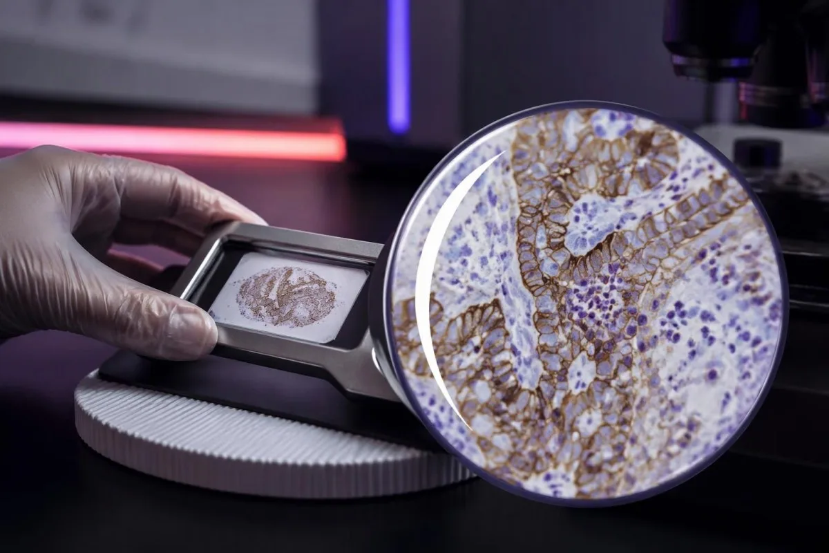
PD-L1, або ліганд програмованої загибелі клітин 1, може зв’язуватися з рецептором PD-1 на імунних клітинах і знижувати їхню активність. Препарати, що впливають на цю взаємодію, можуть відновити протипухлинну імунну активність, однак вимірювання 1 білка не охоплює всіх чинників, від яких залежить, чи розпізнають імунні клітини рак і контролюватимуть його.
Результат дослідження PD-L1 у тканині зазвичай отримують за допомогою імуногістохімії, під час якої антитіло виявляє розташування білка в збереженому зразку. Наведене число описує клітини на вибраних зрізах тканини, а не всі ракові клітини в організмі; результати дослідження 2 зразків із різних ділянок можуть відрізнятися.
Кантесті – це Аналізатор крові зі штучним інтелектом, а не заміна дослідження тканини патологоанатомом чи рішення онколога щодо лікування. Наші організація та клінічна сфера відрізняють освітню підтримку під час тлумачення лабораторних результатів від ухвалення рішень, для яких потрібні оригінальний патогістологічний висновок, дані візуалізації та запропонована схема лікування разом.
Перш ніж тлумачити число, з’ясуйте 4 деталі: діагноз раку, чи є оцінка CPS або TPS, який тест використано та яке лікування розглядається. У висновку, де зазначено лише позитивний PD-L1, бракує клінічно важливої інформації; попросіть уточнити метод оцінювання та числовий результат.
Значення показника TPS PD-L1: відсоток пухлинних клітин
Показник частки пухлинних клітин, або TPS, це відсоток життєздатних пухлинних клітин із відповідним частковим або повним мембранним забарвленням PD-L1. Значення TPS становить від 0% до 100%; імунні клітини не включають до чисельника, навіть якщо вони сильно забарвлені поруч із раковими клітинами.
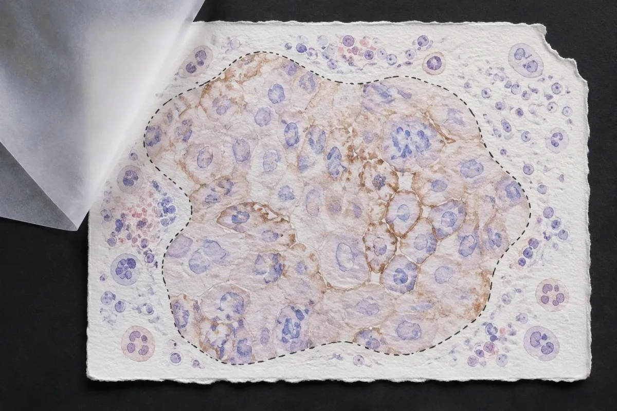
TPS = позитивні щодо PD-L1 життєздатні пухлинні клітини ÷ усі життєздатні пухлинні клітини × 100%. У гіпотетичній ділянці тканини, що містить 200 життєздатних пухлинних клітин, 80 клітин із відповідним позитивним забарвленням дають TPS 40%; забарвлені лімфоцити поруч не змінюють цього розрахунку, хоча можуть вплинути на іншу систему оцінювання.
Membrane location matters more than visual darkness alone. For commonly used TPS methods, weak qualifying membrane staining can count, whereas staining limited to tumor-cell cytoplasm does not establish a positive tumor cell; TPS 1% therefore describes the fraction of qualifying cells, not a required intensity grade.
Cutoffs such as TPS 1% і TPS 50% were used to define populations in particular lung-cancer trials and treatment pathways. They are not boundaries between harmless and dangerous cancer, and crossing 50% does not create an abrupt biological switch from treatment resistance to guaranteed sensitivity.
Read the exact reporting format: TPS <1%, TPS 1–49%, and TPS ≥50% provide different detail from a single positive flag. Our explanation of якісні проти кількісних результатів covers why a category cannot replace a number, although PD-L1 uses tissue-specific—not blood-reference-range—rules.
Значення показника CPS PD-L1: чому враховують імунні клітини
Combined positive score, або CPS, includes qualifying PD-L1-positive tumor cells, lymphocytes, and macrophages. CPS divides their combined count by the number of viable tumor cells and multiplies by 100; the denominator does not become all cells simply because immune cells enter the numerator.
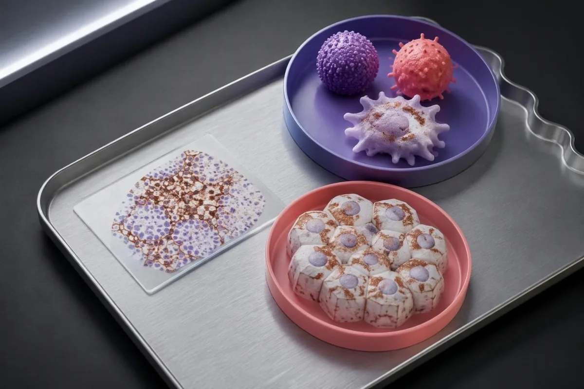
CPS = positive tumor cells plus qualifying positive lymphocytes and macrophages ÷ viable tumor cells × 100. In a hypothetical sample with 500 viable tumor cells, 50 positive tumor cells, and 75 qualifying positive immune cells, CPS is 25; the tumor-cell-only TPS would be 10%.
The reported CPS is capped at 100, even if the uncapped arithmetic would exceed 100 because many qualifying immune cells surround relatively few tumor cells. CPS is a unitless score, not a percentage of all cells, a blood concentration, or a probability that immunotherapy will work.
Not every stained immune cell belongs in the CPS count. The pathologist applies the assay manual's rules for eligible lymphocytes and macrophages associated with the tumor; neutrophils, unrelated inflammatory areas, and nonviable tissue are not simply added because a slide contains visible staining.
A CPS of 20 can arise from more than 1 cellular pattern: abundant tumor-cell staining, substantial immune-cell staining, or both. This denominator issue resembles the distinction between cell counts and percentages, but a circulating monocyte count cannot supply the tissue information needed to calculate CPS.
Чому показники CPS і TPS не можна замінювати один одним або перераховувати
CPS and TPS are not interchangeable because they count different cells and were validated in different treatment settings. A TPS of 10% cannot be converted reliably into CPS 10, and a CPS of 20 does not establish that 20% of tumor cells stain.
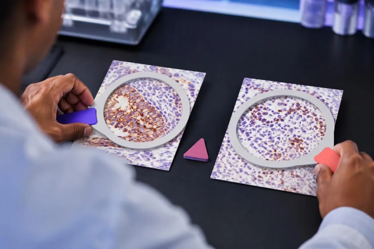
Consider 2 hypothetical specimens with 500 viable tumor cells and 50 positive tumor cells each. The first has no qualifying positive immune cells and gives TPS 10% with CPS 10; the second contains 75 qualifying positive immune cells and gives the same TPS 10% with CPS 25.
The reverse inference fails too. A hypothetical CPS 20 could result from 20 positive tumor cells among 100 tumor cells, or from 5 positive tumor cells plus 15 qualifying positive immune cells; those examples have TPS 20% and TPS 5%, respectively, despite identical CPS values.
Ан immune-cell score, sometimes abbreviated IC, is another system rather than a third spelling of CPS. Some IC methods measure the proportion of tumor area occupied by stained immune cells, so an IC result of 1% must be interpreted using its own assay and indication—not a TPS threshold.
Kantesti's educational explanation should preserve all 3 scoring labels rather than flatten them into positive or negative. When using AI report explanations safely, check that the original score, assay, and specimen remain visible; an AI-generated conversion without the underlying tissue counts is not clinically dependable.
Які деталі патоморфологічного висновку важливі, окрім показника?
A usable PD-L1 report should identify the specimen, assay, scoring method, numerical result, and adequacy assessment. Several commonly used protocols require at least 100 viable tumor cells, but minimum counts and acceptable specimen types depend on the particular test and cancer indication.
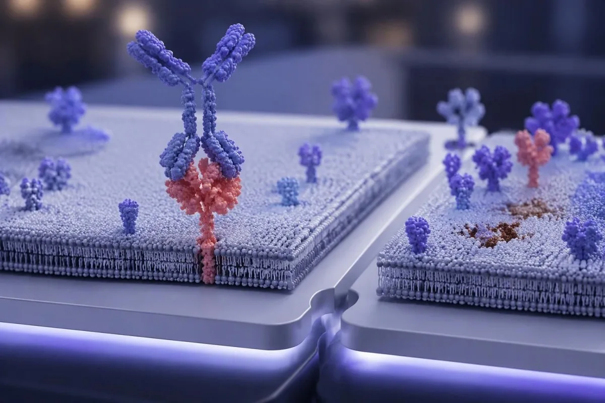
Antibody identifiers such as 22C3, 28-8, SP263, and SP142 describe different testing assays or antibody clones, not different PD-L1 proteins. Some assays show substantial agreement in selected settings, but laboratories should not assume universal equivalence across tumor types, scoring methods, immune-cell assessments, and treatment requirements.
The specimen date and site can change the interpretation of 1 reported number. A small primary-site sample taken before treatment may not represent a later metastatic deposit; ask whether the report describes the current treatment-setting specimen or archived tissue that the oncology team considers suitable.
Insufficient tissue is not PD-L1 negative. A sample with too few viable tumor cells, failed controls, extensive tissue change, or unsuitable processing may be unreportable; the practical next step is laboratory clarification, additional sections, or another suitable specimen—not assigning TPS 0% or CPS 0.
Comparisons between 2 reports need more than matching dates. Our test timing and comparability guide explains the general problem of comparing unlike measurements; for PD-L1, the extra checks are anatomical site, prior treatment, fixation, tissue adequacy, assay, and scoring system.
Як тип раку змінює значення позитивного результату?
PD-L1 positivity is indication-specific, not a universal diagnosis. TPS thresholds such as 1% or 50% are familiar in non-small-cell lung cancer, while CPS thresholds such as 1, 5, or 10 appear in selected other cancer settings; the drug, stage, and regimen must accompany the cutoff.

For recurrent or metastatic head-and-neck squamous-cell carcinoma, KEYNOTE-048 evaluated CPS-defined populations. Pembrolizumab alone improved median overall survival in the CPS ≥20 group to 14.9 months versus 10.7 months with the comparator regimen, showing why a CPS threshold cannot be borrowed from a lung-cancer TPS report (Burtness et al., 2019).
Selected established pembrolizumab pathways use CPS ≥10 for unresectable locally advanced or metastatic triple-negative breast cancer with chemotherapy, and CPS ≥1 for certain persistent, recurrent, or metastatic cervical-cancer regimens. Those examples do not establish eligibility for every earlier-stage setting, drug combination, country, or funding pathway.
Gastric and gastroesophageal-junction cancers require particular care because treatment selection may also involve HER2 і MSI/MMR, with PD-L1 requirements differing across regimens and jurisdictions. A serum marker described in our CA 72-4 interpretation guide cannot replace the tissue biomarker panel or define CPS.
Cancer diagnosis and treatment selection are 2 separate steps. Our persistent cough assessment guide addresses when lung symptoms need investigation; once cancer is confirmed, ask the oncology team to match the PD-L1 result to the current local product information and clinical pathway.
Чому схема лікування не менш важлива, ніж порогове значення
Immunotherapy alone and immunotherapy combinations can have different PD-L1 requirements. A TPS below 1% may rule out one PD-L1-selected monotherapy pathway without ruling out an appropriate chemotherapy–immunotherapy combination; these are different clinical decisions, not contradictory interpretations of the same result.
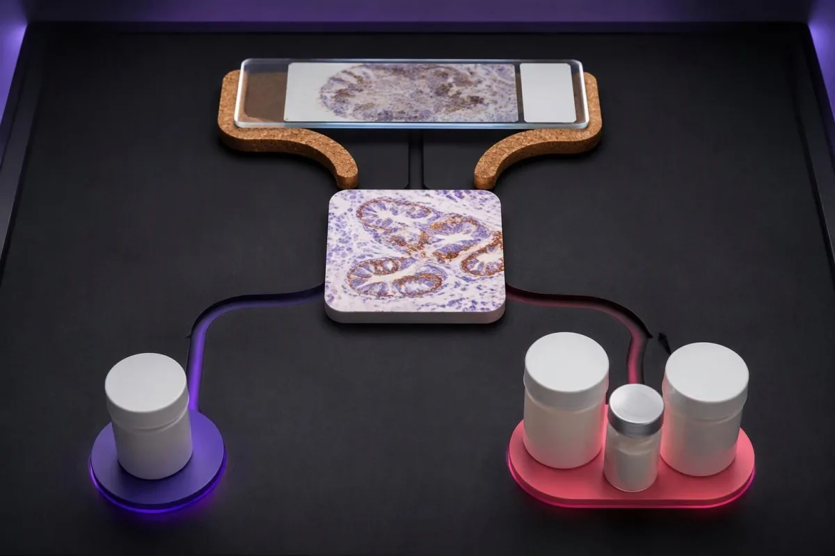
KEYNOTE-024 enrolled previously untreated advanced non-small-cell lung cancer with TPS ≥50% and no sensitizing EGFR mutation or ALK rearrangement. Median progression-free survival was 10.3 months with pembrolizumab versus 6.0 months with chemotherapy, supporting that specific high-TPS monotherapy setting—not every cancer with a score above 50 (Reck et al., 2016).
У KEYNOTE-189, pembrolizumab plus pemetrexed and platinum chemotherapy improved outcomes in previously untreated metastatic nonsquamous non-small-cell lung cancer without EGFR or ALK alterations. Twelve-month overall survival was 69.2% versus 49.4%, and benefit was observed across PD-L1 categories, including TPS <1% (Gandhi et al., 2018).
Combination treatment may address rapid disease progression differently from a single agent, but adding drugs also adds toxicity and monitoring needs. If a proposed regimen includes fluorouracil or capecitabine, тестування DPYD перед хіміотерапією concerns treatment safety; it does not measure the PD-L1 pathway or predict the same benefit.
Кантесті – це Платформа для розшифровки аналізу крові AI that helps explain supporting laboratory measurements; it does not select between 1-drug and multidrug cancer regimens. Similarly, UGT1A1 and irinotecan safety address metabolism-related toxicity, illustrating why each biomarker should be matched to the specific medicine under discussion.
Чи гарантує позитивний результат PD-L1 користь від імунотерапії?
A positive PD-L1 result does not guarantee a response. In KEYNOTE-024's selected high-expression lung-cancer population, the objective response rate with pembrolizumab was 44.8%—substantial benefit for many participants, but clearly not a response in every person with TPS ≥50%.
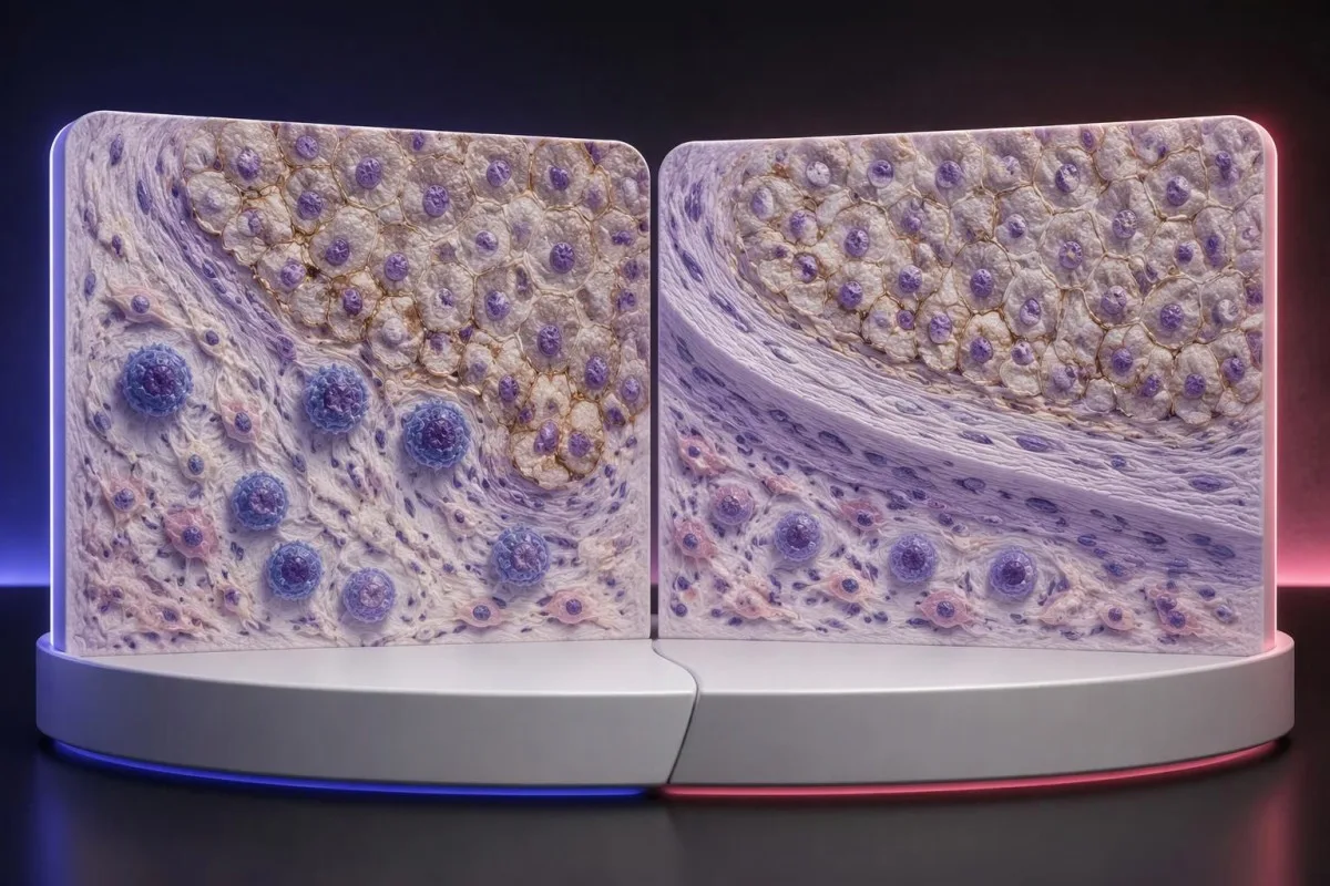
The 44.8% response rate in KEYNOTE-024 describes a trial population under defined conditions, not an individual's calculated chance of success (Reck et al., 2016). Objective response also means measurable tumor shrinkage by trial criteria; stable disease and survival benefit are different outcomes that should not be collapsed into that single percentage.
PD-L1 is only 1 part of a broader immune environment. A cancer can show substantial staining while lacking effective immune-cell access, recognizable targets, or intact antigen presentation; these mechanisms help explain why expression enriches for benefit in some settings without functioning as a deterministic on–off switch.
Higher does not always mean better across different cancers and combinations. A CPS 30 in one disease cannot be ranked against TPS 60% in another, and the same CPS threshold may have different predictive value when used for monotherapy versus a combination that already provides benefit through other mechanisms.
Some melanoma immunotherapy regimens do not require a positive PD-L1 test, so a low score should not be interpreted using lung-cancer monotherapy rules. Our melanoma warning-sign guide addresses recognition and assessment, while treatment benefit must be discussed using disease-specific trial evidence and the person's overall condition.
Чи можуть запропонувати імунотерапію, якщо результат PD-L1 негативний?
PD-L1-negative cancers can still have appropriate immunotherapy options. TPS <1% or CPS <1 may exclude a particular PD-L1-selected regimen, but some combinations, earlier-stage pathways, and treatments selected by other biomarkers do not require PD-L1 positivity.
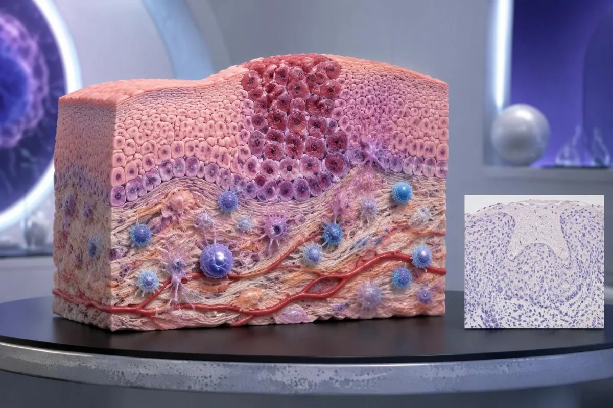
PD-L1-negative should first be translated into the laboratory's actual definition. TPS <1%, CPS <1, and an assay-specific IC-negative category are not identical measurements; an unreportable or inadequate sample is a fourth situation and should not be grouped with a technically valid negative result.
Ан MSI-high or mismatch-repair-deficient cancer may support particular immunotherapy pathways without PD-L1 selection, depending on the disease, stage, prior treatment, and local authorization. Tumor MMR testing and Генетичне тестування на синдром Лінча answer related but different questions: an abnormal tumor result does not automatically prove an inherited syndrome.
Other genetic findings may point toward targeted treatment or different planning rather than immunotherapy. A result discussed in our BRCA variant interpretation guide does not turn CPS 0 into CPS 10; clinicians combine distinct biomarkers without pretending they measure the same biological feature.
No PD-L1 requirement is not the same as no selection criteria. A regimen can still require a particular histology, stage, treatment history, organ function, or absence of an actionable driver alteration; ask whether 1 negative score changes only the proposed option or the broader treatment plan.
Коли забір зразка або граничний показник можуть бути підставою для перегляду?
Sampling variation can change PD-L1 classification, especially near a treatment threshold. A CPS of 9 versus 10, or TPS 0.8% versus 1%, may prompt pathology review when the distinction would change an otherwise suitable treatment—but repeating every result is not automatically useful.
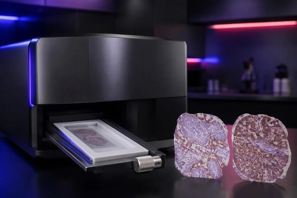
PD-L1 staining can be heterogeneous within 1 tissue block and between primary and metastatic sites. A small tissue examination may miss a highly expressing area or overrepresent it; the pathologist assesses the available viable tissue rather than assuming a single selected field describes the whole cancer.
Pretreatment and post-treatment specimens are not always equivalent. Chemotherapy, radiotherapy, and changes in the immune environment can alter expression, while decalcification of some bone specimens can impair antigen detection; ask whether a lower result reflects biology, processing limitations, or simply a different sampled site.
Borderline results require the actual assay rules, not homemade averaging. If 2 reports show CPS 8 and CPS 12, calculating CPS 10 does not create a newly validated result; the laboratory and oncology team should decide which specimen and assessment are appropriate for the treatment indication.
Kantesti cannot verify viable-cell counts, staining controls, or tissue suitability from a score alone. Our клінічна валідація та нагляд should be read in that limited context; when another tissue procedure is considered, its risks and likely impact on treatment need to be weighed against the uncertainty in the existing result.
Які інші біомаркери слід розглядати разом із PD-L1?
Other biomarkers can take priority over PD-L1, depending on the cancer. In advanced non-small-cell lung cancer, molecular alterations such as EGFR mutations and ALK rearrangements affect treatment planning; in selected other cancers, HER2, MSI/MMR, or additional molecular findings may change the preferred pathway.

A high TPS does not cancel an actionable molecular finding. The pivotal KEYNOTE-024 і KEYNOTE-189 populations excluded relevant EGFR or ALK alterations, so their results should not be applied unchanged to those groups; treatment sequence also matters because some targeted therapies and checkpoint inhibitors can interact through toxicity.
Tumor mutational burden, often measured in mutations per megabase, is a different biomarker from PD-L1. A threshold such as 10 mutations/Mb has been used in a specific tissue-agnostic treatment context, but assay differences, cancer type, authorization, and clinical setting prevent it from becoming a universal immunotherapy rule.
A liquid molecular test may find circulating tumor DNA, but it does not automatically replace tissue PD-L1 immunohistochemistry. The 2 tests measure different things—genetic material versus protein location within cells—and a negative liquid result may also reflect limited DNA shedding rather than absence of a tumor alteration.
Kantesti AI distinguishes the role of supporting laboratory markers from tissue-based treatment biomarkers in our довідник референсних значень біомаркерів крові. A blood-cell ratio, LDH level, or serum tumor-marker concentration should not be used to manufacture TPS 50%, estimate CPS 10, or bypass an indicated molecular workup.
Які перевірки безпеки важливі, якщо розглядають імунотерапію?
PD-L1 scores predict neither treatment safety nor immune-related toxicity. A TPS of 90% does not make checkpoint treatment safer than TPS 1%; the team assesses medical history, organ function, medications, and the particular regimen separately from the tissue biomarker.
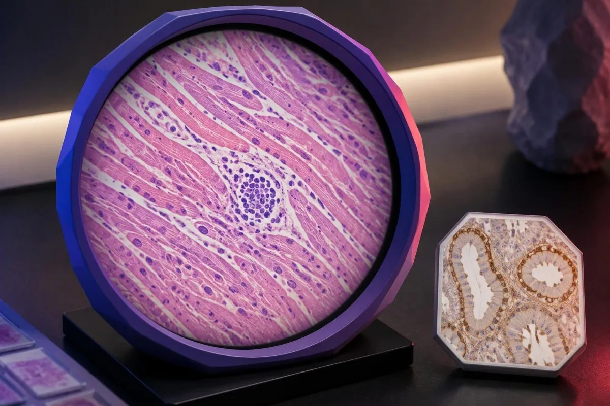
Checkpoint inhibitors can cause immune-related effects involving the thyroid, liver, lungs, bowel, kidneys, heart, and other organs. Baseline testing commonly includes liver chemistry, creatinine, thyroid measurements, and a complete blood count, with monitoring schedules varying by regimen; 1 normal baseline panel does not prevent later toxicity.
New chest pain, palpitations, fainting, or breathlessness during treatment requires prompt assessment because myocarditis, although uncommon, can be serious. Our myocarditis test interpretation guide explains why troponin supports evaluation but cannot determine the cause by itself; severe symptoms warrant emergency care rather than waiting for the next scheduled panel.
Kidney changes need interpretation against the person's own baseline and treatment exposures. A hypothetical creatinine rise from 0.8 to 1.6 mg/dL is a doubling even if the absolute value looks only modestly elevated; our kidney function testing guide explains supporting measurements, not an immunotherapy-specific treatment decision.
Pre-existing autoimmune disease, organ transplantation, ongoing immunosuppression, or previous severe immune-related toxicity can complicate treatment planning even when CPS is 100. Report new persistent diarrhea, worsening cough, jaundice, unusual weakness, or marked thirst promptly; neither a high PD-L1 score nor previously reassuring blood results justify delaying symptom assessment.
Про що запитати на прийомі в онколога?
Принесіть complete PD-L1 pathology report and ask which exact treatment decision it informs. Four useful starting questions are: Which score was used? Which cutoff applies? Is the specimen adequate? What options remain if the result is below the cutoff?

A hypothetical report reading 22C3, CPS 12, adequate tissue is still incomplete as a treatment answer until the cancer, stage, and regimen are specified. Ask whether CPS ≥10 is relevant to the proposed option, whether other biomarkers are pending, and whether a combination would be considered under different selection rules.
Request explanations of 3 outcomes separately: tumor shrinkage, duration of disease control, and survival. A trial's response rate is not the same as its survival benefit, while median survival is a group midpoint—not an individual deadline—and the balance between rapid control and treatment toxicity may change the preferred regimen.
Кантесті – це сервісі інтерпретації тестів AI for supporting laboratory understanding, not independent immunotherapy eligibility assessment. If blood monitoring is planned, our посібник із підготовки до базового тестування can help organize previous values, units, medication lists, and collection dates so that the oncology team can compare results meaningfully.
Keep tissue scores separate from serial blood monitoring in 2 clearly labeled records. Our посібник з тенденцій безпеки препаратів explains why changes over time need context; our принципи відповідального використання ШІ also make clear that automated explanations should not trigger treatment changes without clinical review.
Наукові публікації та обмеження доказів щодо PD-L1
PD-L1 interpretation should be grounded in cancer-specific trials and validated pathology methods. The 3 clinical references below address high-TPS lung-cancer monotherapy, lung-cancer combination treatment, and CPS-selected head-and-neck treatment; the 2 separate Zenodo publications concern other laboratory topics and do not establish PD-L1 thresholds.

KEYNOTE-024 supports a defined TPS ≥50% advanced lung-cancer population, whereas KEYNOTE-189 supports a particular combination across PD-L1 categories (Reck et al., 2016; Gandhi et al., 2018). Those different study designs explain why evidence for one regimen cannot be substituted for evidence supporting another simply because both drugs affect the same immune pathway.
KEYNOTE-048 assessed CPS-defined recurrent or metastatic head-and-neck cancer populations, including CPS ≥1 and CPS ≥20 groups (Burtness et al., 2019). When reading a trial, check the histology, stage, previous treatment, comparator, and endpoint; a subgroup survival finding does not automatically quantify an individual's chance of measurable shrinkage.
Kantesti's 2 Zenodo publications listed below are educational resources on complement/ANA testing and Nipah-virus diagnostics, not PD-L1 clinical trials or assay-validation studies. The орієнтовне значення комплементу та ANA concerns different laboratory questions, and a DOI identifies a deposited publication without independently establishing clinical validity.
Use the original pathology result and current local treatment information to resolve eligibility—not a generalized positive/negative table or an AI-generated score conversion. Our стандарти медичного контенту emphasize source relevance and uncertainty; ask your oncology team to document the 1 specific regimen, applicable threshold, and remaining alternatives.
Поширені запитання
Що означає позитивний результат на PD-L1?
PD-L1-позитивний результат означає, що результат дослідження тканини відповідає заданим критеріям аналізу та пороговому значенню оцінки, наприклад TPS ≥1% або CPS ≥1 у відповідних клінічних умовах. Порогове значення залежить від типу раку, схеми лікування та місцевих вимог до лікування. Позитивний результат не доводить, що імунотерапія буде ефективною, а у звіті слід зазначати числову оцінку, а не лише позначку про позитивний результат.
У чому різниця між CPS і TPS?
TPS вимірює відсоток життєздатних пухлинних клітин із відповідним забарвленням мембран PD-L1, тоді як CPS охоплює кількість відповідно забарвлених позитивних пухлинних клітин, лімфоцитів і макрофагів, поділену на кількість життєздатних пухлинних клітин і помножену на 100. TPS виражають у діапазоні від 0% до 100%, а значення CPS обмежене 100. Ці показники не можна перерахувати один в один, оскільки CPS враховує забарвлення імунних клітин, яких TPS не враховує.
Чи означає CPS 10 10%-ву ймовірність того, що імунотерапія подіє?
CPS 10 не означає 10% ймовірності відповіді на імунотерапію. CPS — це розрахунок за результатами забарвлення тканини, у знаменнику якого використовують життєздатні пухлинні клітини, а не ймовірність відповіді на лікування для окремого пацієнта. CPS ≥10 є підставою для вибору певних схем лікування, специфічних для конкретного виду раку, але для оцінки ймовірної користі також необхідно враховувати діагноз раку, його стадію, інші біомаркери та наявні докази ефективності запропонованого лікування.
Чи можу я отримувати імунотерапію, якщо PD-L1 негативний?
Негативний результат PD-L1 не виключає всіх варіантів імунотерапії. Для деяких комбінацій хіміотерапії з імунотерапією та інших терапевтичних підходів, що обираються за біомаркерами, позитивність PD-L1 не потрібна, навіть якщо TPS становить менше ніж 1%. Відповідність критеріям залежить від типу та стадії раку, молекулярних характеристик, історії лікування, придатності пацієнта до лікування та місцевих дозволів; неадекватний зразок тканини не слід вважати достовірним негативним результатом.
Чи вважається рівень TPS 50% високим рівнем експресії PD-L1?
TPS ≥50% зазвичай описують як високу експресію PD-L1 у певних клінічних ситуаціях при недрібноклітинному раку легень. Цей поріг використовували в дослідженні KEYNOTE-024, у якому вивчали певну групу пацієнтів із поширеним раком легень без сенсибілізувальних мутацій EGFR або перебудов ALK. TPS 50% не є еквівалентом CPS 50 і не є універсальним порогом для кожного виду раку, стадії чи схеми імунотерапії.
Чи є PD-L1 аналізом крові та чи потрібно його повторювати?
Клінічне оцінювання PD-L1 за CPS і TPS зазвичай проводять за збереженою тканиною пухлини, дослідженою методом імуногістохімії, а не за звичайним зразком крові. Повторне оцінювання може бути корисним, якщо тканини недостатньо, результат близький до порогового значення, що може змінити рішення, наприклад CPS 10, або краще підходить інший зразок. Онкологи та патологи мають вирішити, чи ймовірно, що перегляд результатів, додаткові зрізи або інший зразок достатньо змінять тактику лікування, щоб виправдати пов’язані з цим зусилля та процедурні ризики.
Дізнайтеся, як читати аналіз крові
Завантажте лабораторний звіт, і ШІ Kantesti за лічені секунди пояснить кожен біомаркер простою мовою. Ним користуються понад 2 мільйони людей у всьому світі.
Посилання
- Klein, T., Mitchell, S., & Weber, H. (2026). Посібник із аналізу крові на C3, C4 і титр ANA. Медичні дослідження ШІ Kantesti. DOI
- Klein, T., Mitchell, S., & Weber, H. (2026). Аналіз крові на вірус Ніпах: посібник із раннього виявлення та діагностики, 2026. Медичні дослідження ШІ Kantesti. DOI
- Reck M et al. (2016). Pembrolizumab versus Chemotherapy for PD-L1–Positive Non–Small-Cell Lung Cancer. Медичний журнал Нової Англії. DOI
- Gandhi L et al. (2018). Pembrolizumab plus Chemotherapy in Metastatic Non–Small-Cell Lung Cancer. Медичний журнал Нової Англії. DOI
- Burtness B et al. (2019). Pembrolizumab alone or with chemotherapy versus cetuximab with chemotherapy for recurrent or metastatic squamous cell carcinoma of the head and neck (KEYNOTE-048): a randomised, open-label, phase 3 study. The Lancet. DOI
- 2 млн+Проаналізовані тести
- 127+Країни
- 100+Мови
Про медичну команду


Сигнали довіри E-E-A-T
Досвід
Лікарський клінічний огляд робочих процесів інтерпретації лабораторних показників.
Експертиза
Лабораторна медицина з акцентом на те, як біомаркери поводяться в клінічному контексті.
Авторитетність
Медичну рецензію провів доктор Томас Кляйн за участю доктора Сари Мітчелл і професора, доктора Ганса Вебера.
Довірливість
Інтерпретація на основі доказів із чіткими шляхами подальших дій, щоб зменшити тривогу.






Застереження щодо медичної інформації
Ця стаття призначена лише для освітніх цілей і не становить медичної поради. Завжди консультуйтеся з кваліфікованим медичним працівником щодо рішень про діагностику та лікування.
Опубліковано · Зв’язатися з редакційною командою
Опубліковано компанією Kantesti Ltd, зареєстрованою в Англії та Уельсі під номером компанії. 17090423, Лондон, Велика Британія.
