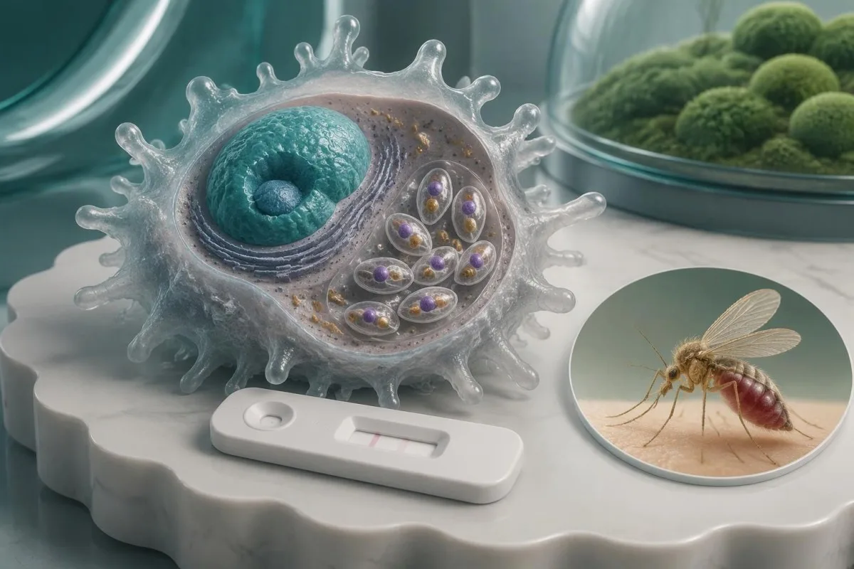
A positive antibody result can reflect past exposure rather than current illness. Symptoms, travel history, immune status, and the specimen tested determine what confirmation is needed.

Mambo muhimu ya kukumbuka
- Kingamwili chanya show an immune response to Leishmania antigens; they do not, by themselves, establish active leishmaniasis.
- Three main forms—visceral, cutaneous, and mucosal leishmaniasis—require different clinical assessments and specimen strategies.
- Visceral confirmation may combine compatible illness with serology, PCR, microscopy, or culture; some endemic programs accept positive rapid serology within a defined clinical algorithm.
- Cutaneous confirmation usually requires material from an affected skin site for PCR, microscopy, or culture; antibody testing performs poorly for localized disease.
- Regional test performance differs: a Cochrane review estimated rK39 sensitivity around 97% in the Indian subcontinent versus 85% in East Africa among people with suspected visceral disease.
- Immune suppression can weaken antibody responses and permit reactivation years after exposure; a negative antibody result does not safely exclude disease in a high-risk patient.
- Dalili za dharura za onyo include confusion, severe breathlessness, significant bleeding, or fever with an absolute neutrophil count below 0.5 × 10^9/L.
- Next appointment should include the complete laboratory report, travel locations and dates, previous leishmaniasis treatment, and a list of immune-suppressing medicines.
What does a positive leishmaniasis antibody actually mean?
A positive leishmaniasis antibody result means the assay detected antibodies reacting with Leishmania antigens—not necessarily active disease. Past infection, asymptomatic exposure, or cross-reactivity can produce positivity; active illness requires assessment of symptoms, exposure, immune status, and, when appropriate, parasite detection.
Leishmaniasis has 3 main clinical forms: visceral disease affecting internal organs, cutaneous disease affecting skin, and mucosal disease affecting structures such as the nose or mouth. An antibody result cannot reliably identify which form a person has, locate the affected tissue, or determine the responsible species; those distinctions change both the next test and the treatment discussion.
Antileishmanial antibodies may remain detectable for years after successful treatment, so a result obtained 2 years after recovery can still be positive without proving relapse. Conversely, someone with a newly developed persistent fever and an enlarged spleen needs assessment even if the antibody value is only slightly above the laboratory cutoff—clinical evidence matters more than the height of the flag.
Kantesti ni Mchambuzi wa mtihani wa damu wa AI that can help explain accompanying laboratory findings, but a positive antibody report does not supply the examination or tissue evidence needed for diagnosis. Our shirika na kusudi la kliniki distinguish interpretation support from medical care; bring the complete report, including at least the method, reference interval, and specimen type, to the clinician arranging follow-up.
Which leishmaniasis test did your laboratory perform?
The meaning of a positive leishmaniasis test depends on its method: rK39 rapid testing, ELISA, indirect fluorescent antibody testing, and direct agglutination testing detect antibodies, whereas PCR detects parasite DNA. Their numbers are not interchangeable, and no universal antibody cutoff establishes active disease.
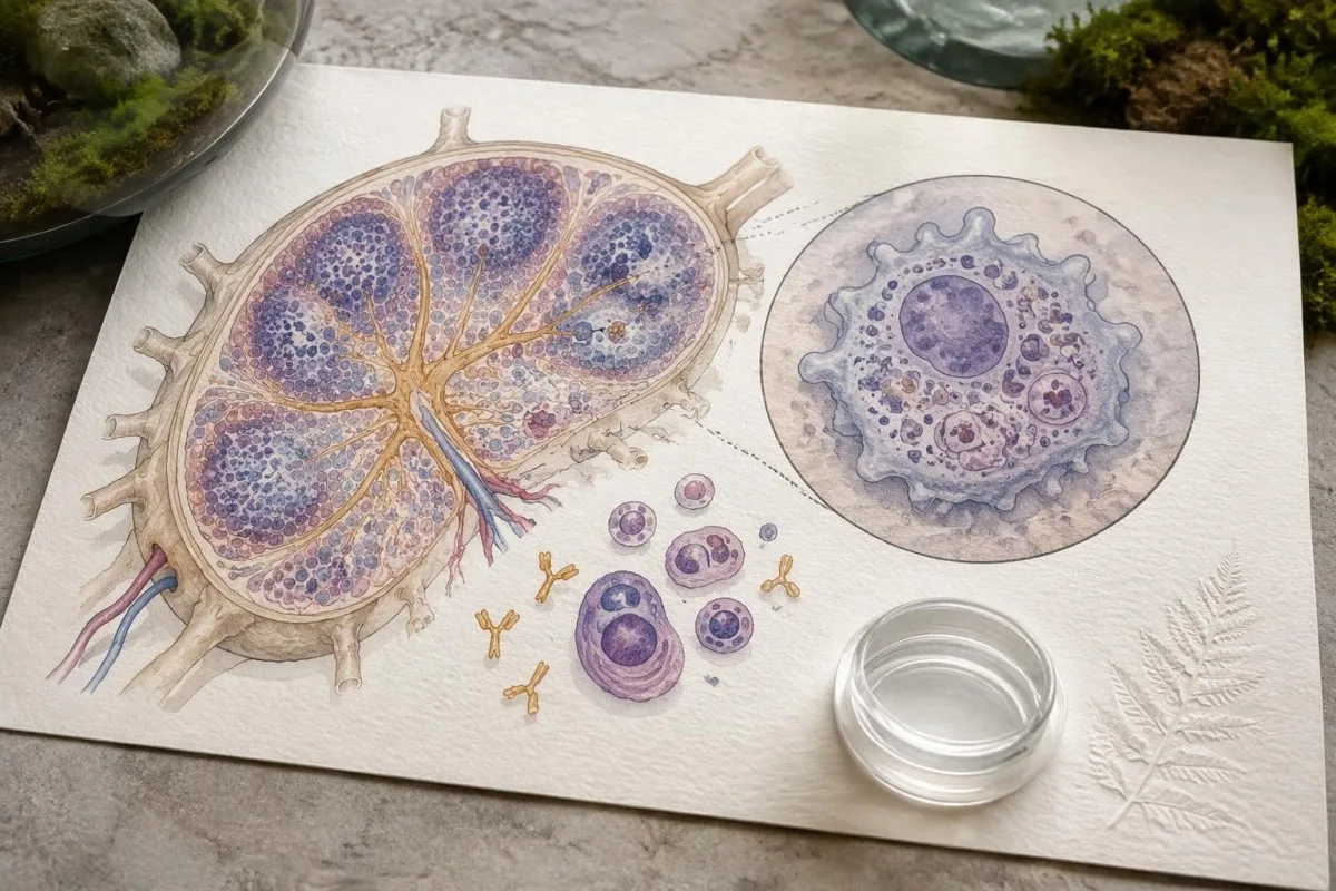
A report may show a qualitative result, an index value, or an antibody titer expressed as a dilution such as 1:160. In a clearly hypothetical assay with positivity beginning at an index of 1.1, a result of 1.4 means the signal exceeded that assay's threshold—not that illness is mild or that a particular parasite count is present; our mwongozo wa ubora dhidi ya kiasi explains this reporting distinction.
rK39 assays detect antibodies against a recombinant Leishmania antigen, but their sensitivity varies geographically and by product. Boelaert et al.'s 2014 Cochrane review estimated rK39 rapid-test sensitivity at approximately 97.0% in the Indian subcontinent and 85.3% in East Africa among people evaluated for suspected visceral leishmaniasis; these estimates should not be applied to asymptomatic screening or cutaneous disease.
Different laboratories may use different antigens, dilution series, and definitions of borderline results, even when both reports say 'Leishmania antibody.' Comparing a titer of 1:160 from one method with an index of 1.4 from another cannot establish a trend; ask whether a reference laboratory should review a discordant result before treating the apparent numerical change as biological deterioration.
Why can a positive antibody result be misleading?
A positive antibody result can mislead because past exposure, cross-reactivity, and low pretest probability reduce its ability to identify active illness. The same assay result carries different diagnostic weight in a symptomatic person from an endemic area and an asymptomatic person without plausible exposure.
Some Leishmania antibody assays cross-react with antibodies to Trypanosoma cruzi, the parasite responsible for Chagas disease, although the extent depends on the antigen and method. A person with exposure in Latin America and 2 reactive parasite antibody tests may need reference-laboratory clarification rather than an assumption of two active infections; our Chagas confirmation pathway explains why assay selection matters.
Consider a hypothetical group of 1,000 people in which 1% have active disease, using an imaginary test with 90% sensitivity and 95% specificity: approximately 9 true-positive and 50 false-positive results would occur, making only about 15% of positive results true positives. These are teaching assumptions, not performance figures for a particular leishmaniasis assay, but they show why testing people without exposure can generate confusing alarms.
Now keep the same hypothetical assay but increase disease prevalence to 30%: approximately 270 true positives and 35 false positives would occur, so about 89% of positives would identify disease. The individual estimate still depends on symptoms, past infection, and immune status; our kutafsiri matokeo chanya ya kingamwili helps separate evidence of immune recognition from evidence that treatment is needed.
Which findings make visceral leishmaniasis more likely?
Visceral leishmaniasis becomes more concerning when prolonged fever, weight loss, and an enlarged spleen accompany compatible exposure, particularly with low blood-cell counts. Antibodies strengthen that clinical pattern but do not explain every fever, anemia, or abnormal protein result.
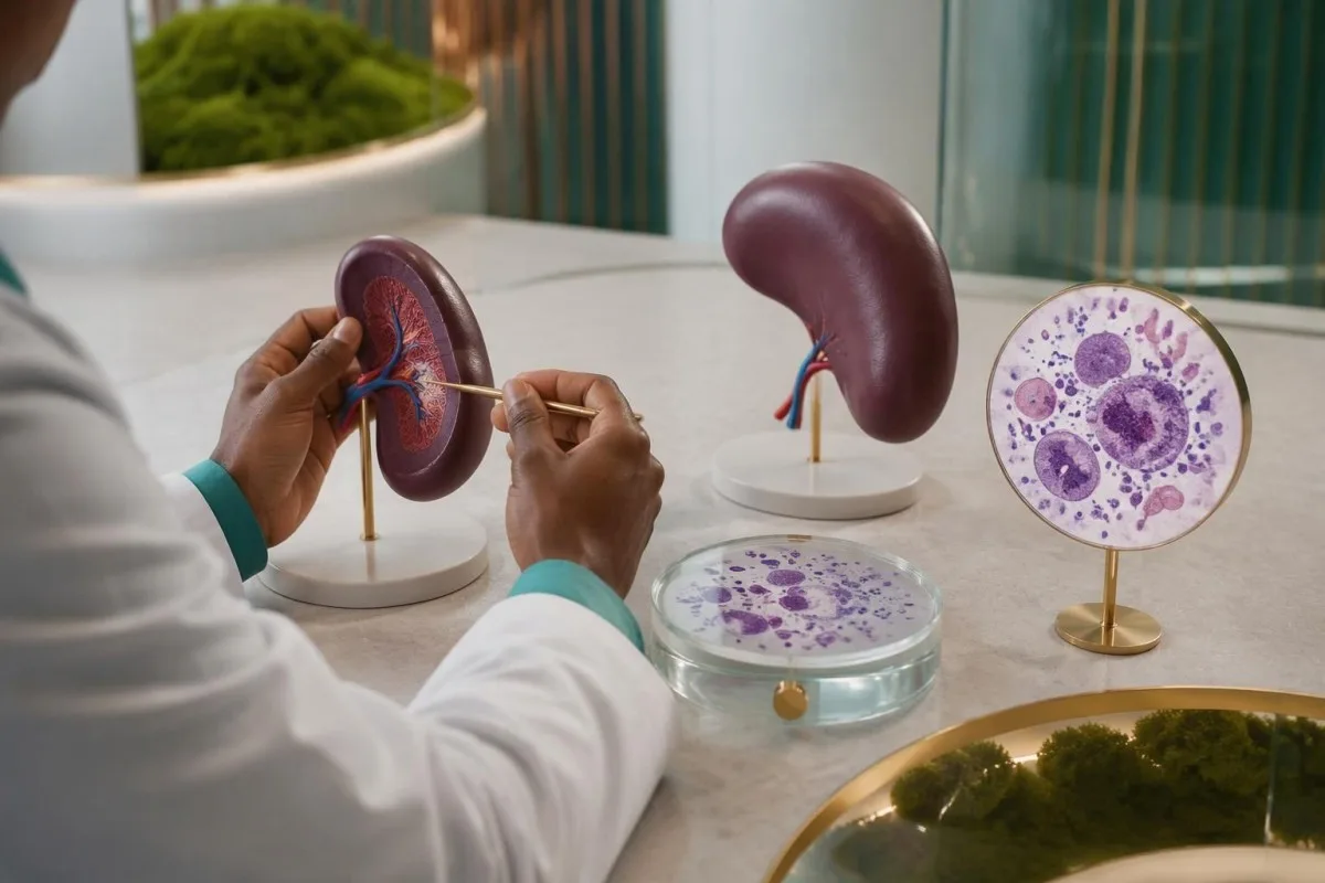
Visceral leishmaniasis can lower all 3 major blood-cell lines: red cells, white cells, and platelets, producing pancytopenia. Bone-marrow involvement, inflammatory effects, and trapping of cellular elements in an enlarged spleen can contribute together; our hemoglobini na hematokriti helps readers understand anemia without mistaking a single low value for a parasite diagnosis.
For example, a hypothetical traveler with 3 weeks of fever, hemoglobin 8.5 g/dL, white-cell count 2.2 × 10^9/L, platelets 70 × 10^9/L, and splenomegaly needs prompt investigation regardless of the antibody index. This combination supports a serious systemic process, but malaria, hematologic disease, disseminated fungal infection, and other conditions remain possible; diagnostic testing should address those alternatives rather than stop at serology.
Visceral disease may also produce polyclonal hypergammaglobulinemia and low albumin, so a hypothetical total protein of 9.0 g/dL with albumin 2.8 g/dL warrants contextual interpretation rather than a reflex assumption of a monoclonal disorder. Our mwongozo wa tafsiri ya protini ya damu explains the pattern; ultrasound can assess spleen size, but spleen enlargement itself cannot establish the cause.
How is visceral leishmaniasis diagnosis confirmed?
Visceral leishmaniasis diagnosis uses a compatible clinical syndrome plus laboratory evidence chosen for the setting. PCR, microscopy, and culture can detect the parasite; in some endemic treatment programs, positive rK39 serology with a defined clinical case pattern supports diagnosis without invasive sampling.
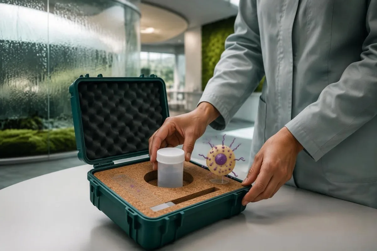
The 2016 IDSA/ASTMH guideline recommends combining diagnostic approaches and using molecular testing when available rather than relying on a single assay in every patient (Aronson et al., 2016). Peripheral blood or buffy-coat PCR may be useful, especially with immune suppression, but bone marrow or other tissue may still be needed when suspicion remains high; a negative result from 1 compartment does not guarantee absence elsewhere.
Aronson et al. report microscopy sensitivities of approximately 52–85% for bone marrow aspirates and 93–99% for splenic aspirates, with yield affected by expertise and parasite burden. Splenic aspiration has a potentially serious bleeding risk and belongs only in appropriately selected patients at experienced centers; the highest-yield specimen is not automatically the safest first choice, particularly when platelet counts or coagulation are abnormal.
A clinician may investigate other causes of the same syndrome while obtaining confirmatory samples, especially if antibody and PCR findings disagree. Our Histoplasma antigen follow-up guide describes another systemic illness that can overlap clinically; obtaining specimens before antiparasitic therapy is useful when feasible, but a critically ill person should not wait several days solely to complete an ideal diagnostic sequence.
Does a positive leishmaniasis PCR test prove active infection?
A positive leishmaniasis PCR test establishes detection of Leishmania DNA in the tested specimen, assuming a valid assay and appropriate contamination controls. It supports active disease strongly when the specimen and symptoms fit, but DNA detection alone does not prove parasite viability or explain every symptom.
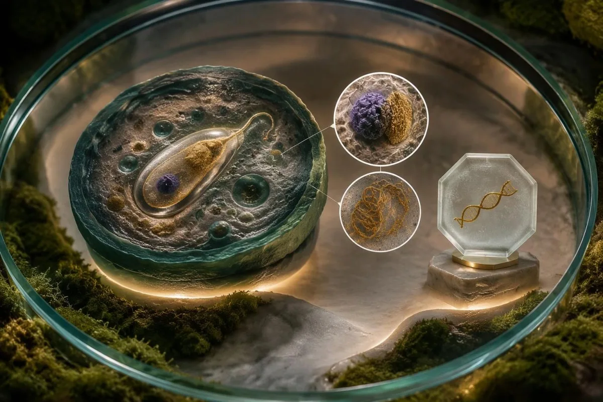
PCR can detect low-level or asymptomatic parasitemia in some exposed populations, and residual genetic material may complicate interpretation after treatment. A hypothetical weakly positive blood PCR in a well person is therefore different from a positive bone-marrow PCR in someone with fever and pancytopenia; clinicians consider at least 3 variables together—clinical syndrome, specimen source, and previous treatment—before interpreting detection as disease requiring therapy.
Cycle-threshold values are not a universal Leishmania parasite-count scale, and a Ct of 35 from 1 laboratory cannot be compared directly with Ct 35 from another assay. Extraction volume, genetic target, calibration, and detection limits differ; our Babesia antibody and infection guide illustrates a related diagnostic distinction, although the organisms and recommended testing pathways are not interchangeable.
Kantesti ni huduma ya kutafsiri vipimo vya maabara ya AI that can explain why antibody results, DNA detection, and accompanying CBC findings answer different questions, rather than collapsing them into a single diagnosis. Our mwongozo wa teknolojia ya tafsiri ya AI describes the interpretation approach; no AI analysis can recover the missing specimen source or assay validation details from a report that contains only 1 word: 'positive.'
Why does cutaneous leishmaniasis need a skin specimen?
Cutaneous leishmaniasis is usually confirmed by testing material from an affected skin site, not by measuring antibodies in blood. Localized disease may generate little detectable circulating antibody, while skin PCR, microscopy, or culture can directly investigate the area causing concern.
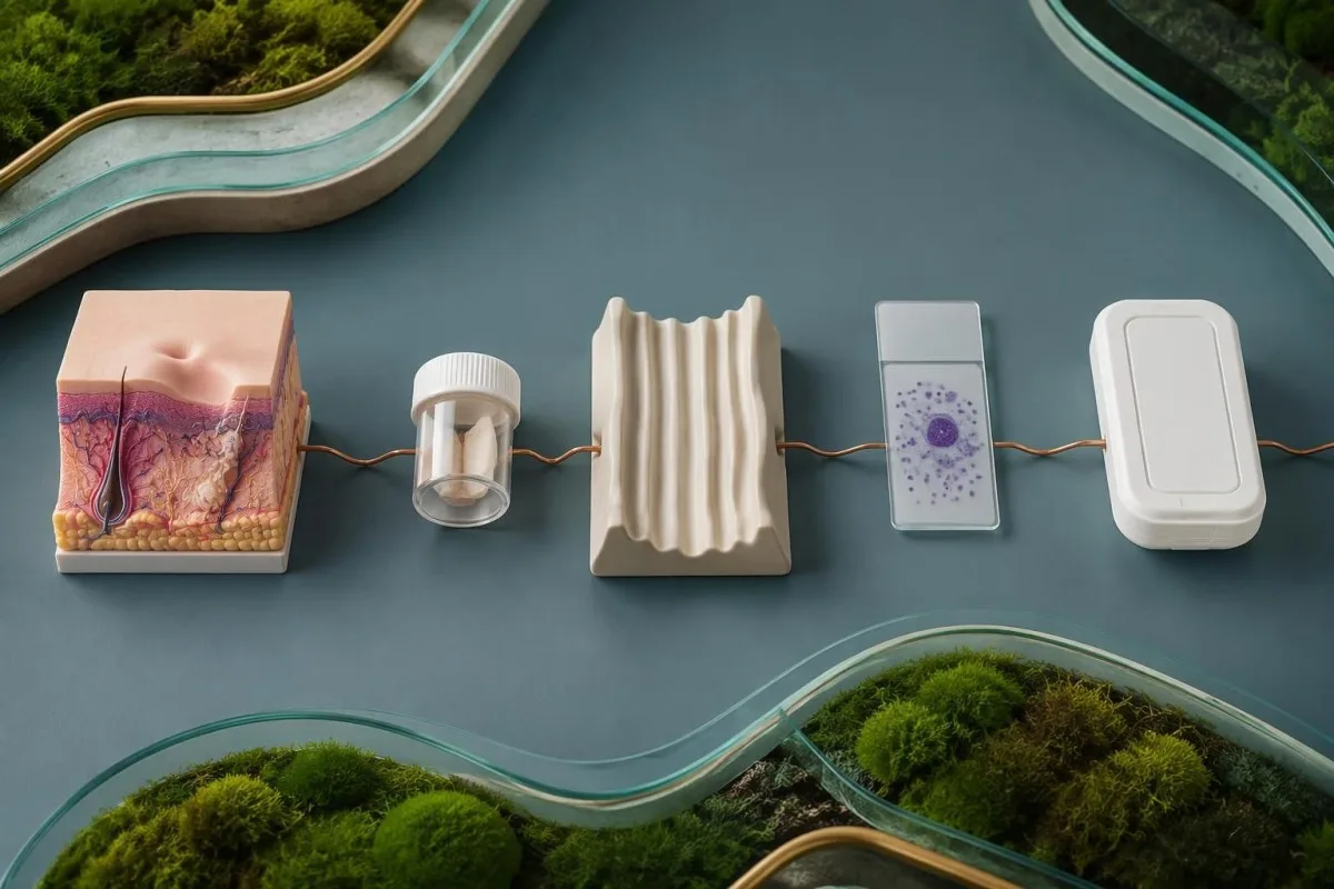
The active raised edge of a persistent affected area often provides useful material, but the optimal sampling method depends on its age, appearance, and the laboratory protocol. Clinicians may use scraping, aspiration, or a biopsy; requesting at least 2 diagnostic approaches, such as PCR and histology, can be useful because one assesses parasite material while the other also evaluates alternative explanations.
A hypothetical person with a single painless forearm sore persisting for 8 weeks after travel may need lesion-directed testing even if Leishmania antibodies are negative and the CBC is normal. Fungal disease, atypical mycobacteria, inflammatory disorders, and skin cancer can resemble cutaneous leishmaniasis; sending appropriate tissue for broader evaluation prevents a parasite-focused request from overlooking another treatable cause.
Species identification matters because some Leishmania species, particularly members of the Viannia subgenus acquired in the Americas, carry a greater concern for later mucosal involvement. Our skin-site PCR testing guide explains why material must come from the relevant affected site, although the exact collection requirements differ; ask before sampling whether the laboratory can identify species from the submitted specimen.
How does immune suppression change test interpretation?
Immune suppression can reduce antibody production, increase the risk of visceral disease, and permit reactivation after remote exposure. A negative antibody result is therefore less reassuring in someone with HIV, a transplant, or substantial immune-suppressing treatment; PCR and direct parasite detection become particularly valuable.
HIV-associated risk cannot be judged from viral load alone: Hesabu ya CD4 describes a different aspect of immune function, and counts below 200 cells/µL indicate advanced immunodeficiency. Our HIV viral-load interpretation guide explains the distinction; even a person with suppressed HIV RNA may need careful evaluation if immune recovery is incomplete and compatible symptoms or exposure are present.
Leishmania can persist after apparent recovery, so immune-suppressing treatment begun 5 years after travel can still make that older exposure clinically relevant. Corticosteroids, transplant medicines, some biologic therapies, and disorders affecting immune function may alter the diagnostic strategy, but risk varies by drug and intensity; patients should provide medication names and doses without stopping prescribed treatment on their own.
A low total IgG concentration offers 1 possible explanation for unreliable antibody-based screening, but normal immunoglobulin levels do not guarantee normal parasite-specific responses. Our immunoglobulin result interpretation separates these measurements; when symptoms remain concerning, repeating the same antibody assay without changing the diagnostic approach may add little compared with testing an appropriate specimen for parasite DNA or organisms.
Which travel details change the meaning of a positive result?
Historia ya safari changes both the probability of leishmaniasis and the most useful diagnostic pathway. Exact destinations, rural or outdoor exposure, dates, and previous residence matter more than a continent name because Leishmania species, clinical forms, and antibody-test performance vary within regions.

Leishmaniasis is usually transmitted through the bite of an infected female phlebotomine sand fly, and transmission can occur in parts of the Mediterranean as well as tropical regions. A traveler who visited southern Europe for 2 weeks may have a relevant exposure despite never visiting a destination they considered tropical; absence of a remembered bite does not exclude exposure because these insects are small.
Cutaneous disease often appears weeks to months after exposure, whereas visceral disease may emerge months later or reactivate years afterward, particularly with immune suppression. Record all relevant destinations over more than the last 12 months if your clinician requests them; our travel-associated parasite antibody guide illustrates why an exposure timeline can remain useful long after the journey has ended.
Kantesti AI can help organize the distinction between exposure evidence and routine laboratory abnormalities, but the travel history must come from the person or their records—not from a positive assay. For an appointment, write a 4-part timeline covering destination, dates, onset of symptoms, and immune-suppressing treatment; this can help the clinician choose a geographically appropriate assay and determine whether species identification would change management.
Why can the right test on the wrong specimen miss disease?
Specimen choice and handling affect sensitivity because Leishmania distribution differs between visceral and localized skin disease. Blood PCR may help investigate visceral disease, while an affected skin specimen is usually more informative for cutaneous disease; collection medium, storage, and transport must match the laboratory method.
Reference laboratories may request EDTA whole blood, buffy coat, fresh tissue, or another specific preparation for PCR, depending on the validated assay. Serum used for an antibody test is not automatically a substitute for every molecular protocol; our PCR sample timing guide explains the general principle of matching collection to the test, although disease-specific instructions must come from the receiving laboratory.
Formalin-fixed tissue supports histology and sometimes validated PCR, but it cannot be used for parasite culture because fixation destroys viability. Before a skin biopsy, the clinician may need to divide material into 2 separately handled portions, one for histopathology and another for fresh-tissue testing; requesting culture only after all material has entered formalin can require an avoidable second procedure.
A negative result obtained after 5 days of treatment may reflect reduced detectable parasite burden rather than an incorrect original diagnosis, although the size of that effect depends on method and specimen. Tell the laboratory and clinician the treatment start date, previous medicines, and sample collection date; if suspicion remains high, the next step may be a different compartment or method rather than repeating an inadequately matched sample.
Which symptoms or laboratory findings need urgent care?
Confusion, severe breathlessness, fainting, significant bleeding, or rapidly worsening illness require emergency evaluation, regardless of the antibody result. Persistent fever with weight loss or spleen enlargement needs prompt medical review; new nose, mouth, or throat symptoms after compatible exposure need assessment for mucosal disease.
Homa pamoja na absolute neutrophil count below 0.5 × 10^9/L is an emergency because serious infection can progress rapidly, even before its cause is known. Platelets below 20 × 10^9/L generally warrant urgent clinician-directed assessment, with greater urgency if bleeding occurs; abnormal coagulation and falling counts may need parallel investigation, as explained in our Mwongozo wa muundo wa vipimo vya maabara vya DIC.
Visceral leishmaniasis can trigger hemophagocytic lymphohistiocytosis, an excessive immune-activation syndrome that may resemble severe infection or hematologic disease. A ferritin value above 500 ng/mL is only one component of established HLH criteria and is far too nonspecific to diagnose the syndrome alone; our HScore interpretation guide explains why fever, cytopenias, organ findings, and additional measurements must be considered together.
Persistent nasal blockage, recurrent nosebleeds, mouth changes, or hoarseness can indicate mucosal involvement, sometimes years after a skin lesion has healed; breathing difficulty is an emergency. Do not wait 2 weeks for a routine antibody repeat if systemic or airway symptoms are progressing, and tell the assessing team about exposure; fever after travel to a malaria-endemic area also requires urgent evaluation for malaria rather than attribution to Leishmania antibodies.
How do clinicians reason through different positive results?
Clinicians interpret a positive leishmaniasis result by asking whether exposure, symptoms, immune status, and parasite evidence agree. The following 3 hypothetical examples show why identical antibody positivity can lead to reassurance with follow-up, lesion-directed testing, or urgent systemic investigation.
Example 1: a hypothetical 42-year-old completed treatment for visceral leishmaniasis 3 years ago, feels well, and has stable blood counts but a persistently positive antibody assay. Persistent antibodies are plausible, so the result alone does not establish relapse or justify retreatment; new fever, weight loss, or immune suppression would change the assessment and might justify direct parasite testing rather than simply checking another antibody titer.
Example 2: a hypothetical 29-year-old has an 8-week skin lesion after travel, normal routine blood tests, and a weakly positive antibody result. The affected skin still needs appropriate PCR, microscopy, or histology because serology neither confirms its cause nor identifies the species; our limits of fever antibody tests describes a parallel diagnostic pitfall, not a reason to apply typhoid testing rules to leishmaniasis.
Example 3: a hypothetical 58-year-old taking transplant medicines develops fever, spleen enlargement, platelets of 45 × 10^9/L, and a negative antibody test. At Kantesti, we explain why that negative result cannot cancel the clinical concern: immune suppression can impair antibody responses, and prompt clinician-led PCR or tissue evaluation may be necessary; these examples are teaching scenarios, not patient encounters or substitutes for individualized medical advice.
Does a positive result mean treatment or repeat testing?
A positive result does not automatically mean treatment, and successful treatment does not require antibodies to become negative. Confirmed visceral disease generally needs systemic therapy; cutaneous management depends on species, site, lesion burden, mucosal risk, immune status, and the likelihood of spontaneous healing.
Untreated visceral leishmaniasis can be life-threatening, so compatible illness with convincing diagnostic evidence needs specialist management rather than watchful waiting based on a low antibody index. By contrast, some uncomplicated cutaneous presentations may be observed or treated locally, while others need systemic treatment; a single 1-cm lesion on a cosmetically sensitive site can require a different approach from a similarly sized lesion elsewhere.
When systemic therapy is selected, safety monitoring may include at least 3 groups of measurements: kidney function, electrolytes, and liver tests, alongside the CBC when indicated by the medicine and illness. Liposomal amphotericin B, for example, can affect renal function and potassium or magnesium; our mwongozo wa mwenendo wa usalama wa dawa explains why changes from baseline can matter even before an isolated value crosses a reference limit.
Kantesti ni jukwaa la tafsiri ya vipimo vya damu la AI that can help explain these follow-up trends, but treatment success is assessed through clinical recovery and clinician-selected testing rather than AI confirmation of cure. The IDSA/ASTMH guideline recommends monitoring treated cutaneous lesions for miezi 6-12 for evidence of therapeutic failure (Aronson et al., 2016); visceral follow-up and relapse surveillance must be individualized, especially when immune suppression increases ongoing risk.
Machapisho ya utafiti na vikwazo vya tafsiri ya akili bandia (AI)
AI interpretation supports explanation; it does not confirm leishmaniasis or validate a parasite assay. Disease-specific diagnostic recommendations come from clinical guidelines and test-accuracy evidence, while technical research about an interpretation engine answers separate questions about structured output, evaluation methods, and oversight.
The Kantesti technical benchmark listed below evaluates the blood-test interpretation engine using visa 100,000 vya majaribio vya kubuni, as stated in its publication title. Synthetic-case performance must not be presented as prospective diagnostic accuracy for leishmaniasis, proof of PCR sensitivity, or evidence that treatment decisions are safe without a clinician; our mfumo wa uthibitishaji wa matibabu describes the evaluation and oversight context rather than replacing disease-specific evidence.
The Mfumo wa Uthibitisho wa Kimatibabu v2.0 publication describes a framework for assessment and clinical oversight; it is not, by its title or purpose, a clinical trial demonstrating leishmaniasis outcomes. Our viwango vya maudhui ya matibabu separate technical validation, clinical evidence, and educational writing, so readers can judge each claim against the type of evidence actually supporting it rather than treating a DOI as a guarantee of clinical utility.
For a positive leishmaniasis report, the most useful next step is a clinician-led review of mambo 4 muhimu: the assay, the specimen, the exposure history, and the current clinical picture. Kantesti can help explain routine laboratory patterns during that process, but an urgent symptom takes priority over any upload or repeat antibody test; the references below distinguish external leishmaniasis guidance from publications about our interpretation methodology.
Maswali ya mara kwa mara
Je, kipimo chanya cha kingamwili dhidi ya leishmaniasis kinamaanisha nina leishmaniasis hai?
Matokeo chanya ya kipimo cha kingamwili za leishmaniasis pekee hayathibitishi uwepo wa leishmaniasis iliyo hai, kwa sababu kingamwili zinaweza kubaki baada ya maambukizi ya awali au kuwepo kutokana na kuathiriwa na vimelea bila dalili au mwitikio mtambuka. Kwa hiyo, matokeo yanayoendelea kuwa chanya miaka 2 baada ya matibabu yenye mafanikio yanaweza kuashiria kumbukumbu ya kinga badala ya kurudia kwa ugonjwa. Madaktari hutathmini dalili, historia ya safari, hali ya kinga na aina mahususi ya kipimo, kisha hutumia PCR, uchunguzi wa hadubini, utamaduni wa vimelea au uchunguzi wa tishu inapofaa. Homa inayoendelea, kupungua uzito au kuongezeka kwa ukubwa wa wengu kunahitaji uchunguzi wa haraka wa kitabibu.
Kipimo cha rK39 cha leishmaniasis kina usahihi kiasi gani?
Usahihi wa kipimo cha haraka cha rK39 hutofautiana kulingana na eneo la kijiografia, bidhaa na kundi la wagonjwa. Tathmini ya Cochrane ya mwaka 2014 ilikadiria unyeti kuwa takriban 97.0% katika bara dogo la India na 85.3% Afrika Mashariki miongoni mwa watu waliochunguzwa kwa tuhuma za leishmaniasis ya viungo vya ndani. Takwimu hizi hazielezi utendaji wa kipimo katika uchunguzi wa watu wasio na dalili, ugonjwa wa ngozi uliolokalika, au kila kundi la watu wenye mfumo wa kinga uliodhoofika. Kipimo hasi hakipaswi kusitisha uchunguzi wakati tuhuma za kitabibu bado ni kubwa.
Je, kipimo cha PCR cha leishmaniasis kinaweza kuwa chanya bila dalili?
Kipimo cha PCR cha leishmaniasis kinaweza kugundua DNA ya vimelea kwa baadhi ya watu wasio na dalili, hivyo matokeo chanya hayathibitishi moja kwa moja kuwa mtu ana ugonjwa wenye dalili au anahitaji matibabu. Tafsiri hutegemea angalau mambo 3: chanzo cha sampuli, matokeo ya sasa ya uchunguzi wa kliniki, na matibabu au kukabiliwa na vimelea hapo awali. PCR hugundua DNA badala ya kuthibitisha moja kwa moja kwamba vimelea bado vina uwezo wa kuishi. Madaktari wanaweza kutafuta ushahidi wa ziada pale ugunduzi unapokuwa dhaifu, usiotarajiwa au usiolingana na hali ya kliniki.
Je, kipimo cha damu kinaweza kutambua leishmaniasis ya ngozi?
Kipimo cha kingamwili katika damu kwa kawaida hakitoshi kuthibitisha leishmaniasi ya ngozi, kwa sababu ugonjwa uliolokalika kwenye ngozi unaweza kusababisha kingamwili chache zinazozunguka kwenye damu. Kidonda cha ngozi kinachoendelea kwa muda mrefu—kwa mfano, kinachodumu kwa wiki 8 baada ya safari husika—kwa kawaida huhitaji kupimwa kwa sampuli kutoka kwenye eneo lililoathirika. PCR, uchunguzi wa hadubini, utamaduni wa vijidudu au histolojia vinaweza kutoa ushahidi wa moja kwa moja zaidi na kutathmini visababishi vingine vinavyowezekana. Utambuzi wa spishi unaweza pia kuathiri matibabu na tathmini ya hatari ya baadaye ya kuathirika kwa utando wa kamasi.
Je, kingamwili za leishmaniasis zinaweza kuendelea kuwa chanya baada ya matibabu?
Kingamwili za leishmaniasis zinaweza kuendelea kuwa chanya kwa miaka kadhaa baada ya matibabu kufanikiwa, kwa hiyo kutoweka kwa kingamwili si kigezo cha kuaminika cha kuthibitisha kupona. Matokeo chanya miaka 3 baada ya matibabu hayawezi kuthibitisha kurudia kwa ugonjwa bila dalili zinazoendana na ugonjwa huo au ushahidi wa ziada. Kupona kiafya na vipimo vya maabara au vya vimelea vinavyochaguliwa na daktari ndivyo vinavyoongoza ufuatiliaji. Watu ambao baadaye kinga yao hudhoofika wanaweza kuhitaji tathmini tofauti ya kurudia kwa ugonjwa, kwa sababu maambukizi ya awali yanaweza kuendelea kuwa na umuhimu.
Ni lini matokeo chanya ya leishmaniasis yanapaswa kuhitaji matibabu ya haraka?
Matokeo chanya ya leishmaniasis yanahitaji tathmini ya haraka ya kitabibu yanapoambatana na homa inayoendelea, kupungua kwa uzito, kuongezeka kwa ukubwa wa wengu, au kupungua kwa kiasi kikubwa kwa hesabu za seli za damu. Kuchanganyikiwa, upungufu mkubwa wa pumzi, kuzimia, kutokwa na damu kwa kiasi kikubwa, au matatizo ya njia ya hewa kunahitaji tathmini ya dharura bila kujali matokeo ya kipimo. Homa pamoja na hesabu kamili ya neutrofili iliyo chini ya 0.5 × 10^9/L ni dharura, na kiwango cha chembe sahani kilicho chini ya 20 × 10^9/L kwa kawaida kinahitaji tathmini ya haraka inayoongozwa na daktari. Dalili mpya zinazoendelea za pua, mdomo au koo baada ya kukabiliwa na hali inayoweza kusababisha maambukizi pia zinahitaji tathmini ya ugonjwa wa utando wa ute.
Pata maelezo ya jinsi ya kusoma matokeo ya vipimo vya damu
Pakia ripoti yako ya maabara na AI ya Kantesti itaeleza kila kiashiria cha kibayolojia kwa lugha rahisi ndani ya sekunde chache. Inatumiwa na zaidi ya watu milioni 2 duniani kote.
Marejeleo
- Klein, T., Mitchell, S., & Weber, H. (2026). Kielelezo cha Kiufundi cha Kiotomatiki kilichosajiliwa awali, chenye Rubriki, cha injini ya Ufafanuzi wa Vipimo vya Damu ya Kantesti kwenye visa 100,000 vya majaribio vya kubuni. Utafiti wa Kimatibabu wa AI ya Kantesti. DOI
- Klein, T., Mitchell, S., & Weber, H. (2026). Mfumo wa Uthibitisho wa Kitabibu v2.0 (Ukurasa wa Uthibitisho wa Kitabibu). Utafiti wa Kimatibabu wa AI ya Kantesti. DOI
- Aronson N et al. (2016). Diagnosis and Treatment of Leishmaniasis: Clinical Practice Guidelines by the Infectious Diseases Society of America (IDSA) and the American Society of Tropical Medicine and Hygiene (ASTMH). Magonjwa ya Kuambukiza ya Kliniki. DOI
- Boelaert M et al. (2014). Rapid tests for the diagnosis of visceral leishmaniasis in patients with suspected disease. Hifadhidata ya Cochrane ya Mapitio ya Kitaratibu. DOI
- 2M+Vipimo vilivyochanganuliwa
- 127+Nchi
- 100+Lugha
Kuhusu timu ya matibabu


E-E-A-T Trust Signals
Uzoefu
Mapitio ya kimatibabu inayoongozwa na daktari ya mifumo ya tafsiri ya maabara.
Utaalamu
Kuzingatia dawa za maabara kuhusu jinsi viashiria (biomarkers) vinavyobadilika katika muktadha wa kliniki.
Mamlaka
Imekaguliwa kimatibabu na Dkt. Thomas Klein, kwa michango kutoka kwa Dkt. Sarah Mitchell na Prof. Dkt. Hans Weber.
Uaminifu
Tafsiri inayotegemea ushahidi yenye njia zilizo wazi za ufuatiliaji ili kupunguza tahadhari za hofu.





Kanusho la kimatibabu
Makala haya ni kwa madhumuni ya elimu tu na si ushauri wa kimatibabu. Wasiliana na mtoa huduma aliyehitimu kila wakati kwa maamuzi ya utambuzi na matibabu.
Imechapishwa · Wasiliana na timu ya uhariri
Imechapishwa na Kantesti Ltd, imesajiliwa nchini Uingereza na Wales, nambari ya kampuni. 17090423, London, Uingereza.
