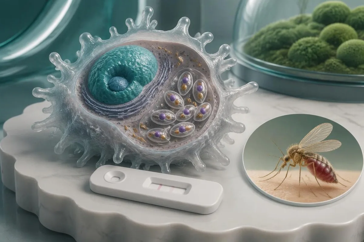
Müsbət anticisim nəticəsi hazırkı xəstəlikdən çox, əvvəllər yoluxmaya məruz qalmanı göstərə bilər. Hansı təsdiqləyici müayinənin lazım olduğunu simptomlar, səyahət tarixçəsi, immun status və yoxlanılan nümunə müəyyən edir.

Əsas nəticələr
- Müsbət antitelər Leyşmaniya antigenlərinə qarşı immun cavabı göstərir; təkbaşına aktiv leyşmaniozun olduğunu təsdiqləmir.
- Üç əsas forma—visseral, dəri və selikli qişa leyşmaniozu—fərqli klinik qiymətləndirmələr və nümunə götürmə strategiyaları tələb edir.
- Visseral leyşmaniozun təsdiqi uyğun gələn klinik mənzərənin serologiya, PCR, mikroskopiya və ya kultura ilə birlikdə qiymətləndirilməsini əhatə edə bilər; bəzi endemik proqramlarda müəyyən edilmiş klinik alqoritm çərçivəsində müsbət sürətli seroloji test kifayət sayılır.
- Dəri leyşmaniozunun təsdiqi adətən PCR, mikroskopiya və ya kultura üçün zədələnmiş dəri nahiyəsindən material götürülməsini tələb edir; anticisim testi lokalizə olunmuş xəstəlikdə zəif nəticə verir.
- Regional test performansı fərqlənir: Cochrane icmalında visseral xəstəlik şübhəsi olan şəxslər arasında rK39-un həssaslığı Hindistan yarımqitəsində təxminən 97%, Şərqi Afrikada isə 85% qiymətləndirilib.
- İmmunosupressiya anticisim reaksiyalarını zəiflədə və təmasdan illər sonra yenidən aktivləşməyə imkan verə bilər; mənfi anticisim nəticəsi yüksək riskli pasiyentdə xəstəliyi təhlükəsiz şəkildə istisna etmir.
- Təcili xəbərdarlıq əlamətləri çaşqınlıq, ağır təngnəfəslik, əhəmiyyətli qanaxma və ya mütləq neytrofil sayı 0.5 × 10^9/L-dən aşağı olduqda qızdırma daxildir.
- Növbəti qəbul tam laboratoriya hesabatını, səyahət edilən yerləri və tarixləri, leişmaniozun əvvəlki müalicəsini və immuniteti zəiflədən dərmanların siyahısını əhatə etməlidir.
Müsbət leyşmanioz anticisimi əslində nə deməkdir?
A leişmanioz anticisimləri müsbət nəticə o deməkdir ki, analiz Leishmania antigenləri ilə reaksiyaya girən anticisimləri aşkar edib — bu, mütləq aktiv xəstəlik demək deyil. Keçmiş infeksiya, simptomsuz təmas və ya çarpaz reaktivlik müsbət nəticəyə səbəb ola bilər; aktiv xəstəlik simptomların, təmasın, immun statusun və uyğun hallarda parazitin aşkar edilməsinin qiymətləndirilməsini tələb edir.
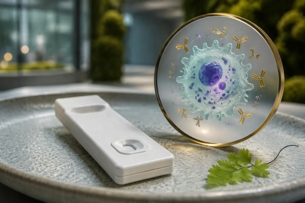
Leişmaniozun 3 əsas klinik forması var: daxili orqanları zədələyən visseral xəstəlik, dərini zədələyən dəri forması və burun və ya ağız kimi strukturları zədələyən selikli qişa forması. Anticisim nəticəsi insanın hansı formaya malik olduğunu, zədələnmiş toxumanın yerini və ya hansı növün səbəb olduğunu etibarlı şəkildə müəyyən edə bilməz; bu fərqlər həm növbəti testi, həm də müalicə müzakirəsini dəyişir.
Leyşmaniyaya qarşı anticisimlər uğurlu müalicədən sonra illərlə aşkarlana bilər; buna görə sağaldıqdan 2 il sonra alınan nəticə residivi sübut etmədən də müsbət ola bilər. Əksinə, yeni yaranmış davamlı qızdırması və böyümüş dalağı olan şəxs, anticisim göstəricisi laboratoriyanın kəsim həddindən cüzi yüksək olsa belə, qiymətləndirilməlidir — klinik sübutlar göstəricinin nə qədər yüksək olmasından daha vacibdir.
Kantesti bir AI qan testi analizatoru yanaşı laboratoriya nəticələrini izah etməyə kömək edə bilər, lakin müsbət anticisim hesabatı diaqnoz üçün lazım olan müayinəni və toxuma sübutlarını təmin etmir. Bizim təşkilat və kliniki məqsədimiz şərhin dəstəklənməsini tibbi yardımdan fərqləndirin; təqibi təşkil edən klinisyə ən azı metod, referens intervalı və nümunə növü daxil olmaqla tam hesabatı təqdim edin.
Laboratoriyanız hansı leyşmanioz testini aparıb?
Leyşmanioz testinin müsbət nəticəsinin mənası onun metodundanasılıdır: rK39 sürətli testləri, ELISA, dolayı flüoressent anticisim testi və birbaşa aqqlütinasiya testi anticisimləri, PCR isə parazit DNT-sini aşkar edir. Onların rəqəmləri bir-biri ilə əvəz edilə bilməz və aktiv xəstəliyi təsdiqləyən universal anticisim kəsim həddi yoxdur.
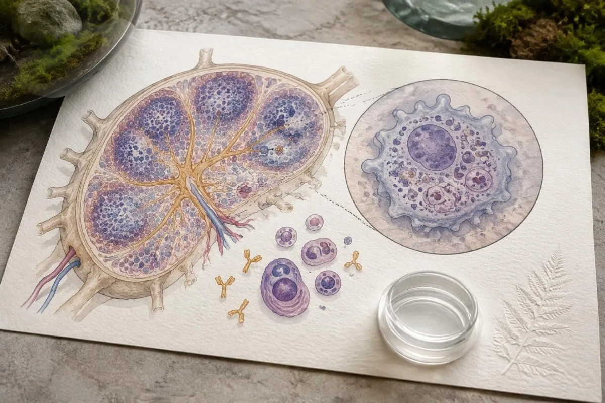
Hesabatda keyfiyyət nəticəsi, indeks göstəricisi və ya 1:160 kimi durulaşdırma şəklində ifadə olunan anticisim titri göstərilə bilər. Müsbətlik indeksin 1.1-dən başladığı, sırf hipotetik analizdə 1.4 nəticəsi siqnalın həmin analizin həddini keçdiyini bildirir — xəstəliyin yüngül olduğunu və ya müəyyən sayda parazitin mövcudluğunu yox; bizim keyfiyyətə qarşı kəmiyyət təlimatımız bu hesabat fərqini izah edir.
rK39 analizləri rekombinant Leishmania antigeninə qarşı anticisimləri aşkar edir, lakin həssaslığı coğrafi bölgəyə və məhsula görə dəyişir. Boelaert və həmmüəlliflərinin 2014-cü il Cochrane icmalında rK39 sürətli testlərinin həssaslığı təxminən belə qiymətləndirilmişdir: Hindistan yarımadasında 97.0%, Şərqi Afrikada isə 85.3% visseral leişmaniozdan şübhələnilən şəxslər arasında; bu göstəricilər simptomsuz skrininqə və ya dəri formasına şamil edilməməlidir.
Hər iki hesabatda 'Leishmania anticismi' yazılsa belə, müxtəlif laboratoriyalar fərqli antigenlərdən, durulaşdırma seriyalarından və sərhəd nəticələrin fərqli təriflərindən istifadə edə bilər. Bir metodla alınmış 1:160 titrini digər metodun 1.4 indeks göstəricisi ilə müqayisə etmək tendensiyanı müəyyən etmir; rəqəmlərdəki görünən dəyişikliyi bioloji pisləşmə kimi qəbul etməzdən əvvəl uyğunsuz nəticəni referens laboratoriyanın nəzərdən keçirməli olub-olmadığını soruşun.
Müsbət anticisim nəticəsi niyə yanıltıcı ola bilər?
Müsbət anticisim nəticəsi yanılda bilər, çünki əvvəlki təmas, çarpaz reaktivlik və aşağı ilkin test ehtimalı reduce its ability to identify active illness. The same assay result carries different diagnostic weight in a symptomatic person from an endemic area and an asymptomatic person without plausible exposure.
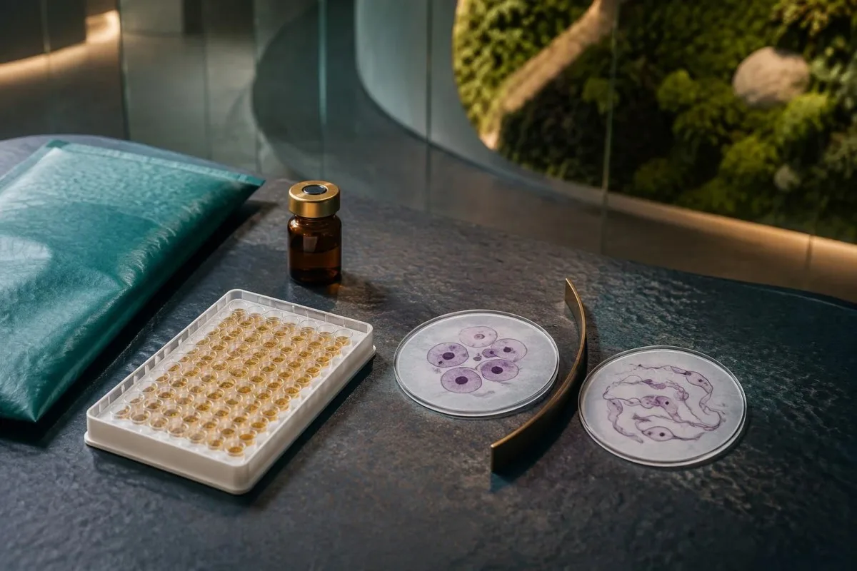
Some Leishmania antibody assays cross-react with antibodies to Trypanosoma cruzi, the parasite responsible for Chagas disease, although the extent depends on the antigen and method. A person with exposure in Latin America and 2 reactive parasite antibody tests may need reference-laboratory clarification rather than an assumption of two active infections; our Chagas confirmation pathway explains why assay selection matters.
Consider a hypothetical group of 1,000 people in which 1% have active disease, using an imaginary test with 90% sensitivity and 95% specificity: approximately 9 true-positive and 50 false-positive results would occur, making only about 15% of positive results true positives. These are teaching assumptions, not performance figures for a particular leishmaniasis assay, but they show why testing people without exposure can generate confusing alarms.
Now keep the same hypothetical assay but increase disease prevalence to 30%: approximately 270 true positives and 35 false positives would occur, so about 89% of positives would identify disease. The individual estimate still depends on symptoms, past infection, and immune status; our müsbət anticisim nəticələrinin şərhinə dair bələdçimiz helps separate evidence of immune recognition from evidence that treatment is needed.
Hansı tapıntılar visseral leyşmanioz ehtimalını artırır?
Visceral leishmaniasis becomes more concerning when prolonged fever, weight loss, and an enlarged spleen accompany compatible exposure, particularly with low blood-cell counts. Antibodies strengthen that clinical pattern but do not explain every fever, anemia, or abnormal protein result.
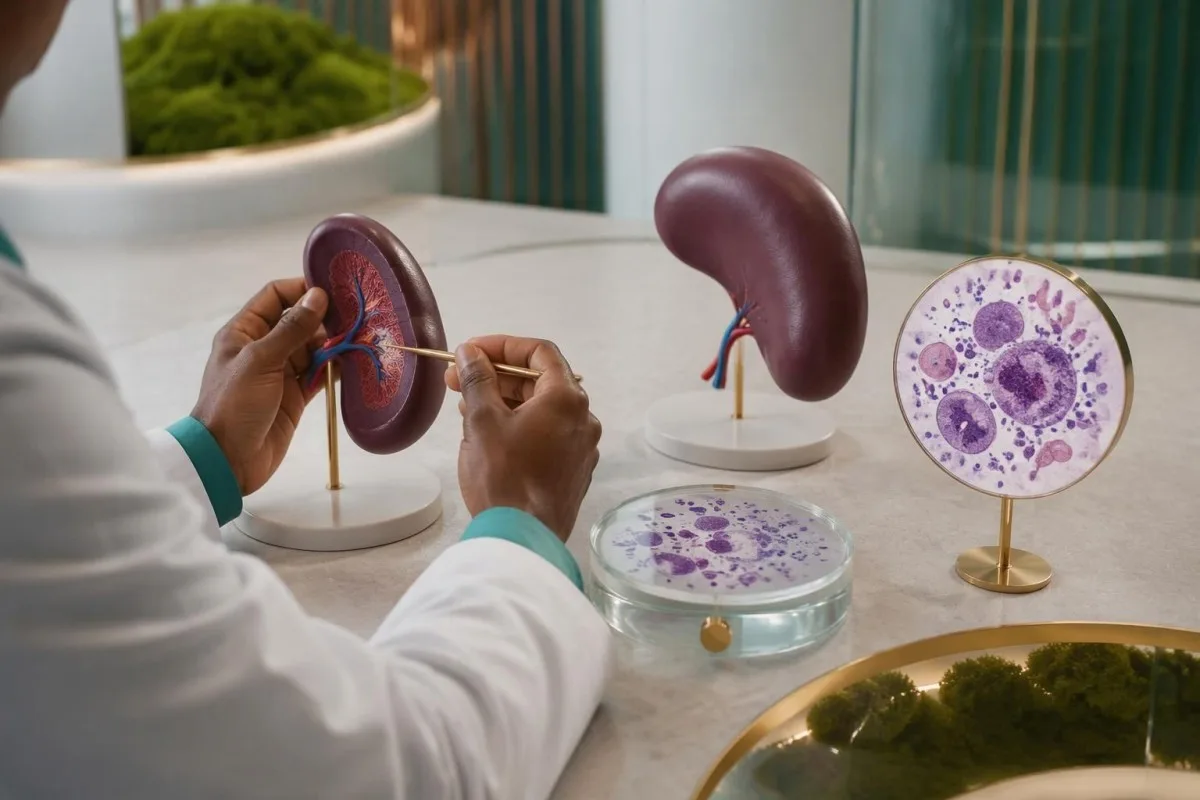
Visceral leishmaniasis can lower all 3 major blood-cell lines: red cells, white cells, and platelets, producing pancytopenia. Bone-marrow involvement, inflammatory effects, and trapping of cellular elements in an enlarged spleen can contribute together; our hemoglobin və hematokrit izahımız helps readers understand anemia without mistaking a single low value for a parasite diagnosis.
For example, a hypothetical traveler with 3 weeks of fever, hemoglobin 8.5 g/dL, white-cell count 2.2 × 10^9/L, platelets 70 × 10^9/L, and splenomegaly needs prompt investigation regardless of the antibody index. This combination supports a serious systemic process, but malaria, hematologic disease, disseminated fungal infection, and other conditions remain possible; diagnostic testing should address those alternatives rather than stop at serology.
Visceral disease may also produce polyclonal hypergammaglobulinemia and low albumin, so a hypothetical total protein of 9.0 g/dL with albumin 2.8 g/dL warrants contextual interpretation rather than a reflex assumption of a monoclonal disorder. Our serum zülallarının interpretasiya bələdçisi explains the pattern; ultrasound can assess spleen size, but spleen enlargement itself cannot establish the cause.
Visseral leyşmanioz diaqnozu necə təsdiqlənir?
Visceral leishmaniasis diagnosis uses a compatible clinical syndrome plus laboratory evidence chosen for the setting. PCR, microscopy, and culture can detect the parasite; in some endemic treatment programs, positive rK39 serology with a defined clinical case pattern supports diagnosis without invasive sampling.
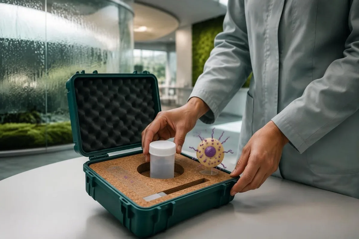
The 2016 IDSA/ASTMH guideline recommends combining diagnostic approaches and using molecular testing when available rather than relying on a single assay in every patient (Aronson et al., 2016). Peripheral blood or buffy-coat PCR may be useful, especially with immune suppression, but bone marrow or other tissue may still be needed when suspicion remains high; a negative result from 1 compartment does not guarantee absence elsewhere.
Aronson et al. report microscopy sensitivities of approximately 52–85% for bone marrow aspirates and 93–99% for splenic aspirates, with yield affected by expertise and parasite burden. Splenic aspiration has a potentially serious bleeding risk and belongs only in appropriately selected patients at experienced centers; the highest-yield specimen is not automatically the safest first choice, particularly when platelet counts or coagulation are abnormal.
A clinician may investigate other causes of the same syndrome while obtaining confirmatory samples, especially if antibody and PCR findings disagree. Our Histoplasma antigen follow-up guide describes another systemic illness that can overlap clinically; obtaining specimens before antiparasitic therapy is useful when feasible, but a critically ill person should not wait several days solely to complete an ideal diagnostic sequence.
Müsbət leyşmanioz PCR testi aktiv infeksiyanı sübut edirmi?
A positive leishmaniasis PCR test establishes detection of Leishmania DNA in the tested specimen, assuming a valid assay and appropriate contamination controls. It supports active disease strongly when the specimen and symptoms fit, but DNA detection alone does not prove parasite viability or explain every symptom.
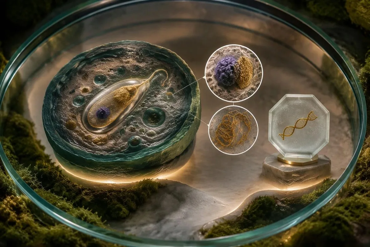
PCR can detect low-level or asymptomatic parasitemia in some exposed populations, and residual genetic material may complicate interpretation after treatment. A hypothetical weakly positive blood PCR in a well person is therefore different from a positive bone-marrow PCR in someone with fever and pancytopenia; clinicians consider at least 3 variables together—clinical syndrome, specimen source, and previous treatment—before interpreting detection as disease requiring therapy.
Cycle-threshold values are not a universal Leishmania parasite-count scale, and a Ct of 35 from 1 laboratory cannot be compared directly with Ct 35 from another assay. Extraction volume, genetic target, calibration, and detection limits differ; our Babesia antibody and infection guide illustrates a related diagnostic distinction, although the organisms and recommended testing pathways are not interchangeable.
Kantesti bir AI laboratoriya testi şərh xidməti that can explain why antibody results, DNA detection, and accompanying CBC findings answer different questions, rather than collapsing them into a single diagnosis. Our AI şərh texnologiyası bələdçisi describes the interpretation approach; no AI analysis can recover the missing specimen source or assay validation details from a report that contains only 1 word: 'positive.'
Dəri leyşmaniozu üçün niyə dəri nümunəsi lazımdır?
Cutaneous leishmaniasis is usually confirmed by testing material from an affected skin site, not by measuring antibodies in blood. Localized disease may generate little detectable circulating antibody, while skin PCR, microscopy, or culture can directly investigate the area causing concern.
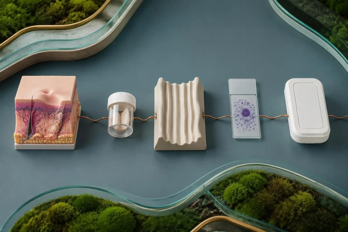
The active raised edge of a persistent affected area often provides useful material, but the optimal sampling method depends on its age, appearance, and the laboratory protocol. Clinicians may use scraping, aspiration, or a biopsy; requesting at least 2 diagnostic approaches, such as PCR and histology, can be useful because one assesses parasite material while the other also evaluates alternative explanations.
A hypothetical person with a single painless forearm sore persisting for 8 weeks after travel may need lesion-directed testing even if Leishmania antibodies are negative and the CBC is normal. Fungal disease, atypical mycobacteria, inflammatory disorders, and skin cancer can resemble cutaneous leishmaniasis; sending appropriate tissue for broader evaluation prevents a parasite-focused request from overlooking another treatable cause.
Species identification matters because some Leishmania species, particularly members of the Viannia subgenus acquired in the Americas, carry a greater concern for later mucosal involvement. Our skin-site PCR testing guide explains why material must come from the relevant affected site, although the exact collection requirements differ; ask before sampling whether the laboratory can identify species from the submitted specimen.
İmmunosupressiya test nəticələrinin şərhini necə dəyişir?
İmmunosupressiya can reduce antibody production, increase the risk of visceral disease, and permit reactivation after remote exposure. A negative antibody result is therefore less reassuring in someone with HIV, a transplant, or substantial immune-suppressing treatment; PCR and direct parasite detection become particularly valuable.

HIV-associated risk cannot be judged from viral load alone: CD4 sayı describes a different aspect of immune function, and counts below 200 cells/µL indicate advanced immunodeficiency. Our HIV viral-load interpretation guide explains the distinction; even a person with suppressed HIV RNA may need careful evaluation if immune recovery is incomplete and compatible symptoms or exposure are present.
Leishmania can persist after apparent recovery, so immune-suppressing treatment begun 5 years after travel can still make that older exposure clinically relevant. Corticosteroids, transplant medicines, some biologic therapies, and disorders affecting immune function may alter the diagnostic strategy, but risk varies by drug and intensity; patients should provide medication names and doses without stopping prescribed treatment on their own.
A low total IgG concentration offers 1 possible explanation for unreliable antibody-based screening, but normal immunoglobulin levels do not guarantee normal parasite-specific responses. Our immunoglobulin result interpretation separates these measurements; when symptoms remain concerning, repeating the same antibody assay without changing the diagnostic approach may add little compared with testing an appropriate specimen for parasite DNA or organisms.
Səyahətlə bağlı hansı təfərrüatlar müsbət nəticənin mənasını dəyişir?
Səyahət tarixi changes both the probability of leishmaniasis and the most useful diagnostic pathway. Exact destinations, rural or outdoor exposure, dates, and previous residence matter more than a continent name because Leishmania species, clinical forms, and antibody-test performance vary within regions.

Leishmaniasis is usually transmitted through the bite of an infected female phlebotomine sand fly, and transmission can occur in parts of the Mediterranean as well as tropical regions. A traveler who visited southern Europe for 2 weeks may have a relevant exposure despite never visiting a destination they considered tropical; absence of a remembered bite does not exclude exposure because these insects are small.
Cutaneous disease often appears weeks to months after exposure, whereas visceral disease may emerge months later or reactivate years afterward, particularly with immune suppression. Record all relevant destinations over more than the last 12 months if your clinician requests them; our travel-associated parasite antibody guide illustrates why an exposure timeline can remain useful long after the journey has ended.
Kantesti AI can help organize the distinction between exposure evidence and routine laboratory abnormalities, but the travel history must come from the person or their records—not from a positive assay. For an appointment, write a 4-part timeline covering destination, dates, onset of symptoms, and immune-suppressing treatment; this can help the clinician choose a geographically appropriate assay and determine whether species identification would change management.
Yanlış nümunədə düzgün test aparılması xəstəliyi niyə aşkarlamaya bilər?
Specimen choice and handling affect sensitivity because Leishmania distribution differs between visceral and localized skin disease. Blood PCR may help investigate visceral disease, while an affected skin specimen is usually more informative for cutaneous disease; collection medium, storage, and transport must match the laboratory method.
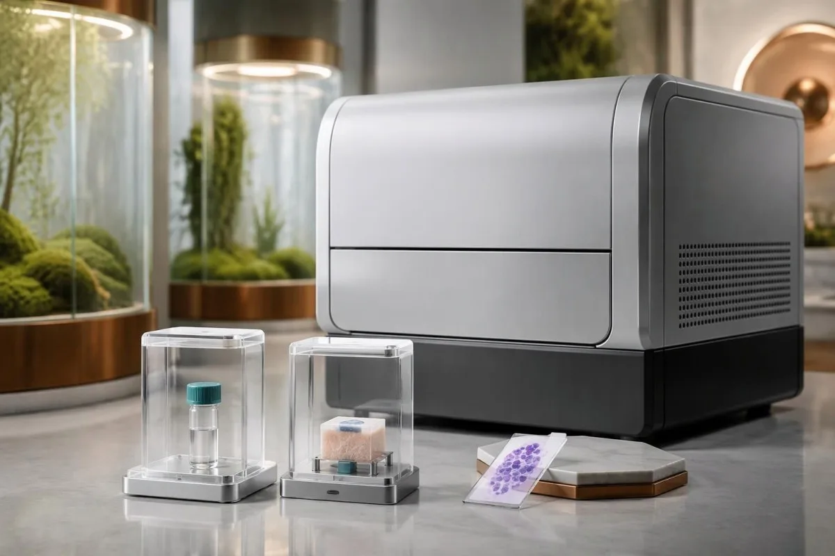
Reference laboratories may request EDTA whole blood, buffy coat, fresh tissue, or another specific preparation for PCR, depending on the validated assay. Serum used for an antibody test is not automatically a substitute for every molecular protocol; our PCR sample timing guide explains the general principle of matching collection to the test, although disease-specific instructions must come from the receiving laboratory.
Formalin-fixed tissue supports histology and sometimes validated PCR, but it cannot be used for parasite culture because fixation destroys viability. Before a skin biopsy, the clinician may need to divide material into 2 separately handled portions, one for histopathology and another for fresh-tissue testing; requesting culture only after all material has entered formalin can require an avoidable second procedure.
A negative result obtained after 5 days of treatment may reflect reduced detectable parasite burden rather than an incorrect original diagnosis, although the size of that effect depends on method and specimen. Tell the laboratory and clinician the treatment start date, previous medicines, and sample collection date; if suspicion remains high, the next step may be a different compartment or method rather than repeating an inadequately matched sample.
Hansı simptomlar və ya laboratoriya tapıntıları təcili tibbi yardım tələb edir?
Confusion, severe breathlessness, fainting, significant bleeding, or rapidly worsening illness require emergency evaluation, regardless of the antibody result. Persistent fever with weight loss or spleen enlargement needs prompt medical review; new nose, mouth, or throat symptoms after compatible exposure need assessment for mucosal disease.
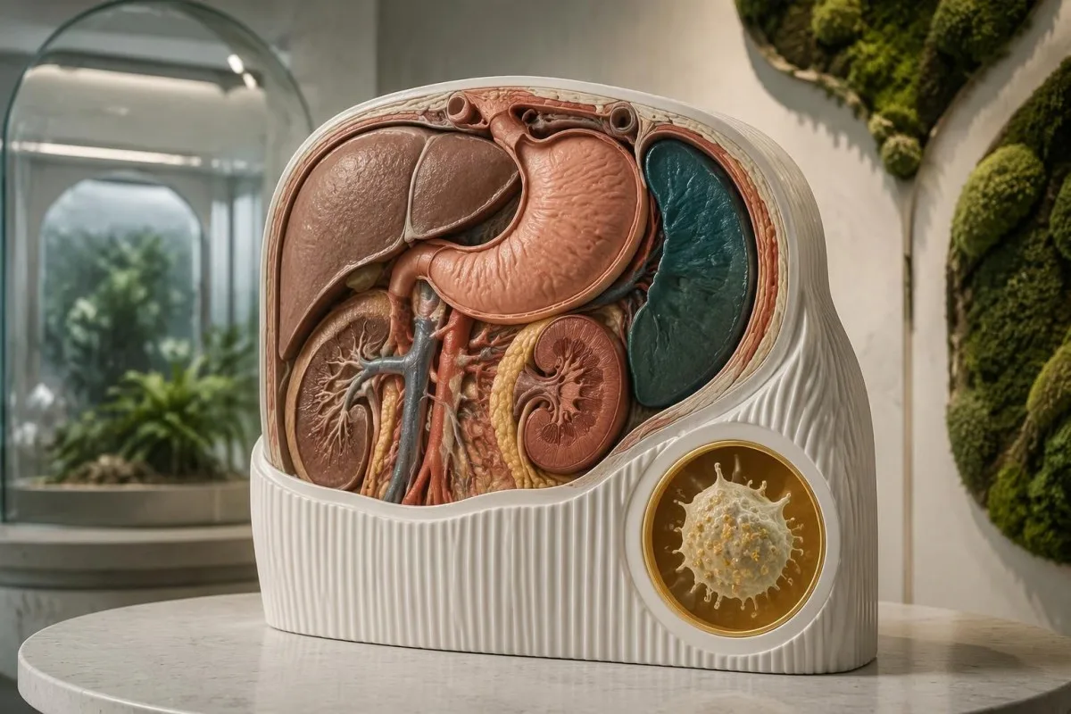
ilə müşayiət olunan qızdırma absolute neutrophil count below 0.5 × 10^9/L is an emergency because serious infection can progress rapidly, even before its cause is known. Platelets below 20 × 10^9/L generally warrant urgent clinician-directed assessment, with greater urgency if bleeding occurs; abnormal coagulation and falling counts may need parallel investigation, as explained in our DIC laborator göstəriciləri üzrə bələdçi.
Visceral leishmaniasis can trigger hemophagocytic lymphohistiocytosis, an excessive immune-activation syndrome that may resemble severe infection or hematologic disease. A ferritin value above 500 ng/mL is only one component of established HLH criteria and is far too nonspecific to diagnose the syndrome alone; our HScore interpretation guide explains why fever, cytopenias, organ findings, and additional measurements must be considered together.
Persistent nasal blockage, recurrent nosebleeds, mouth changes, or hoarseness can indicate mucosal involvement, sometimes years after a skin lesion has healed; breathing difficulty is an emergency. Do not wait 2 weeks for a routine antibody repeat if systemic or airway symptoms are progressing, and tell the assessing team about exposure; fever after travel to a malaria-endemic area also requires urgent evaluation for malaria rather than attribution to Leishmania antibodies.
Klinik mütəxəssislər fərqli müsbət nəticələri necə qiymətləndirirlər?
Clinicians interpret a positive leishmaniasis result by asking whether exposure, symptoms, immune status, and parasite evidence agree. The following 3 hypothetical examples show why identical antibody positivity can lead to reassurance with follow-up, lesion-directed testing, or urgent systemic investigation.
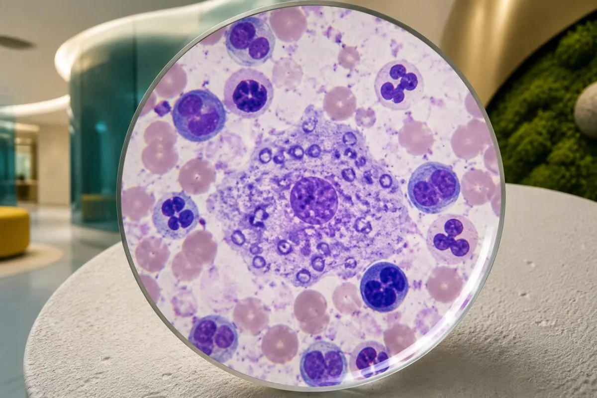
Example 1: a hypothetical 42-year-old completed treatment for visceral leishmaniasis 3 years ago, feels well, and has stable blood counts but a persistently positive antibody assay. Persistent antibodies are plausible, so the result alone does not establish relapse or justify retreatment; new fever, weight loss, or immune suppression would change the assessment and might justify direct parasite testing rather than simply checking another antibody titer.
Example 2: a hypothetical 29-year-old has an 8-week skin lesion after travel, normal routine blood tests, and a weakly positive antibody result. The affected skin still needs appropriate PCR, microscopy, or histology because serology neither confirms its cause nor identifies the species; our limits of fever antibody tests describes a parallel diagnostic pitfall, not a reason to apply typhoid testing rules to leishmaniasis.
Example 3: a hypothetical 58-year-old taking transplant medicines develops fever, spleen enlargement, platelets of 45 × 10^9/L, and a negative antibody test. At Kantesti, we explain why that negative result cannot cancel the clinical concern: immune suppression can impair antibody responses, and prompt clinician-led PCR or tissue evaluation may be necessary; these examples are teaching scenarios, not patient encounters or substitutes for individualized medical advice.
Müsbət nəticə müalicə və ya təkrar test tələb edirmi?
A positive result does not automatically mean treatment, and successful treatment does not require antibodies to become negative. Confirmed visceral disease generally needs systemic therapy; cutaneous management depends on species, site, lesion burden, mucosal risk, immune status, and the likelihood of spontaneous healing.

Untreated visceral leishmaniasis can be life-threatening, so compatible illness with convincing diagnostic evidence needs specialist management rather than watchful waiting based on a low antibody index. By contrast, some uncomplicated cutaneous presentations may be observed or treated locally, while others need systemic treatment; a single 1-cm lesion on a cosmetically sensitive site can require a different approach from a similarly sized lesion elsewhere.
When systemic therapy is selected, safety monitoring may include at least 3 groups of measurements: kidney function, electrolytes, and liver tests, alongside the CBC when indicated by the medicine and illness. Liposomal amphotericin B, for example, can affect renal function and potassium or magnesium; our dərman təhlükəsizliyi trend bələdçisi explains why changes from baseline can matter even before an isolated value crosses a reference limit.
Kantesti bir AI qan analizi nəticələrinin şərhi platforması that can help explain these follow-up trends, but treatment success is assessed through clinical recovery and clinician-selected testing rather than AI confirmation of cure. The IDSA/ASTMH guideline recommends monitoring treated cutaneous lesions for 6–12 ayda bir for evidence of therapeutic failure (Aronson et al., 2016); visceral follow-up and relapse surveillance must be individualized, especially when immune suppression increases ongoing risk.
Tədqiqat nəşrləri və süni intellekt təfsirinin məhdudiyyətləri
AI interpretation supports explanation; it does not confirm leishmaniasis or validate a parasite assay. Disease-specific diagnostic recommendations come from clinical guidelines and test-accuracy evidence, while technical research about an interpretation engine answers separate questions about structured output, evaluation methods, and oversight.
The Kantesti technical benchmark listed below evaluates the blood-test interpretation engine using 100 000 sintetik test nümunəsini, as stated in its publication title. Synthetic-case performance must not be presented as prospective diagnostic accuracy for leishmaniasis, proof of PCR sensitivity, or evidence that treatment decisions are safe without a clinician; our tibbi validasiya çərçivəmiz describes the evaluation and oversight context rather than replacing disease-specific evidence.
The Klinik Təsdiqləmə Çərçivəsi v2.0 publication describes a framework for assessment and clinical oversight; it is not, by its title or purpose, a clinical trial demonstrating leishmaniasis outcomes. Our tibbi məzmun standartları separate technical validation, clinical evidence, and educational writing, so readers can judge each claim against the type of evidence actually supporting it rather than treating a DOI as a guarantee of clinical utility.
For a positive leishmaniasis report, the most useful next step is a clinician-led review of 4 əsas məqamı: the assay, the specimen, the exposure history, and the current clinical picture. Kantesti can help explain routine laboratory patterns during that process, but an urgent symptom takes priority over any upload or repeat antibody test; the references below distinguish external leishmaniasis guidance from publications about our interpretation methodology.
Tez-tez verilən suallar
Leişmanioz anticisminin müsbət olması məndə aktiv leişmanioz olduğu deməkdirmi?
Leishmanioz anticisimləri üçün müsbət nəticə özlüyündə aktiv leishmaniozu təsdiqləmir, çünki anticisimlər əvvəlki infeksiyadan sonra qala və ya simptomsuz təmas zamanı, yaxud çarpaz reaktivlik səbəbindən aşkar oluna bilər. Buna görə də uğurlu müalicədən 2 il sonra nəticənin hələ də müsbət olması residivdən deyil, immun yaddaşdan qaynaqlana bilər. Həkimlər simptomları, səyahət tarixçəsini, immun statusunu və istifadə olunan konkret testi qiymətləndirir, sonra isə lazım gəldikdə PCR, mikroskopiya, kultivasiya və ya toxuma müayinəsindən istifadə edirlər. Davamlı qızdırma, çəki itkisi və ya dalağın böyüməsi dərhal tibbi müayinə tələb edir.
rK39 leyşmanioz testi nə dərəcədə dəqiqdir?
rK39 sürətli testinin dəqiqliyi coğrafi bölgədən, məhsuldan və pasiyent populyasiyasından asılı olaraq dəyişir. 2014-cü ildə aparılmış Cochrane icmalında visseral leyşmanioz şübhəsi ilə müayinə olunan şəxslər arasında həssaslığın Hindistan yarımadasında təxminən 97,0%, Şərqi Afrikada isə 85,3% olduğu təxmin edilmişdir. Bu göstəricilər simptomsuz skrininqdə, lokal dəri leyşmaniozunda və ya bütün immunosupressiv pasiyent populyasiyalarında testin göstəricilərini əks etdirmir. Klinik şübhə yüksək olaraq qalırsa, mənfi test nəticəsi müayinənin dayandırılmasına səbəb olmamalıdır.
Leyşmanioz üçün PCR testi simptomlar olmadan da müsbət ola bilərmi?
Leyşmanioz üzrə PCR testi bəzi simptomsuz şəxslərdə parazitin DNT-sini aşkar edə bilər, buna görə müsbət nəticə simptomatik xəstəliyin və ya müalicəyə ehtiyacın olduğunu avtomatik olaraq təsdiqləmir. Nəticənin şərhi ən azı 3 amildən asılıdır: nümunənin götürüldüyü mənbə, hazırkı klinik əlamətlər və əvvəlki müalicə və ya yoluxma. PCR parazitin canlı olduğunu birbaşa sübut etmək əvəzinə DNT-ni aşkar edir. Aşkarlanma zəif olduqda, gözlənilməz nəticə verdikdə və ya klinik mənzərəyə uyğun gəlmədikdə həkimlər əlavə sübutlar axtara bilərlər.
Qan analizi dəri leyşmaniozunu diaqnoz edə bilərmi?
Qanda anticisim testi dəri leyşmaniozunu təsdiqləmək üçün, ümumiyyətlə, kifayət etmir, çünki lokal dəri xəstəliyi qanda az miqdarda anticisim yaranmasına səbəb ola bilər. Davamlı dəri zədələnməsi — məsələn, müvafiq səyahətdən sonra 8 həftə davam edən zədələnmə — adətən zədələnmiş nahiyədən götürülmüş materialın müayinəsini tələb edir. PCR, mikroskopiya, kultura və ya histologiya daha birbaşa sübut verə və alternativ səbəbləri qiymətləndirməyə kömək edə bilər. Növün müəyyənləşdirilməsi müalicəyə və sonradan selikli qişaların zədələnməsi riskinin qiymətləndirilməsinə də təsir göstərə bilər.
Leyşmanioz anticisimləri müalicədən sonra da müsbət qala bilərmi?
Uğurlu müalicədən sonra leyşmanioz anticisimləri illərlə müsbət qala bilər, buna görə də sağalma üçün anticisimlərin yoxa çıxması etibarlı meyar deyil. Müalicədən 3 il sonra müsbət nəticə uyğun simptomlar və ya əlavə sübutlar olmadan residivin baş verdiyini təsdiqləyə bilməz. Sonrakı müşahidə klinik sağalma və həkimin seçdiyi laborator müayinələr və ya parazitin aşkarlanması testləri əsasında aparılır. Sonradan immun sistemi zəifləyən şəxslərdə residivin qiymətləndirilməsi fərqli aparılmalıdır, çünki əvvəlki yoluxma hələ də əhəmiyyət daşıya bilər.
Leişmanioz testinin müsbət nəticəsi nə zaman təcili tibbi yardıma müraciət etməyi tələb etməlidir?
Müsbət leyşmanioz nəticəsi davamlı qızdırma, çəki itkisi, dalağın böyüməsi və ya qan hüceyrələrinin sayının əhəmiyyətli dərəcədə azalması ilə müşayiət olunarsa, təcili tibbi qiymətləndirmə tələb edir. Analiz nəticəsindən asılı olmayaraq, çaşqınlıq, ciddi nəfəs darlığı, huşunu itirmə, əhəmiyyətli qanaxma və ya tənəffüs yollarında çətinlik təcili müayinə tələb edir. Mütləq neytrofil sayı 0.5 × 10^9/L-dən aşağı olduqda qızdırma təcili hal sayılır; trombositlərin sayı 20 × 10^9/L-dən aşağı olduqda isə, ümumiyyətlə, həkimin göstərişi ilə təcili qiymətləndirmə tələb olunur. Uyğun təmasdan sonra burun, ağız və ya boğazda yaranan yeni və davamlı simptomlar selikli qişa leyşmaniozunun qiymətləndirilməsini də tələb edir.
Qan analizinin nəticələrini necə oxumaq
Laboratoriya hesabatınızı yükləyin, Kantesti-nin AI-si isə saniyələr ərzində hər biomarkeri sadə dildə izah etsin. Dünyada 2 milyondan çox insan istifadə edir.
İstinadlar
- Klein, T., Mitchell, S., & Weber, H. (2026). Kantesti Qan Testi Şərh Mühərrikinin 100,000 Sintetik Test Halında Pre-Registered, Rubrik-Əsaslı Avtomatlaşdırılmış Texniki Benchmarku. Kantesti AI tibbi araşdırmaları. DOI
- Klein, T., Mitchell, S., & Weber, H. (2026). Klinik Təsdiqləmə Çərçivəsi v2.0 (Tibbi Təsdiqləmə Səhifəsi). Kantesti AI tibbi araşdırmaları. DOI
- Aronson N et al. (2016). Diagnosis and Treatment of Leishmaniasis: Clinical Practice Guidelines by the Infectious Diseases Society of America (IDSA) and the American Society of Tropical Medicine and Hygiene (ASTMH). Clinical Infectious Diseases. DOI
- Boelaert M et al. (2014). Rapid tests for the diagnosis of visceral leishmaniasis in patients with suspected disease. Cochrane Sistematik İcmallar Bazası. DOI
- 2M+Təhlil edilmiş testlər
- 127+ölkələr
- 100+Dillər
Tibbi komanda haqqında


E-E-A-T Etibar Siqnalları
Təcrübə
Həkim rəhbərliyi ilə laborator şərh iş axınlarının klinik icmalı.
Ekspertiza
Biomarkerlərin klinik kontekstdə necə davrandığına dair laboratoriya təbabəti diqqəti.
Səlahiyyətlilik
Dr. Thomas Klein tərəfindən tibbi baxımdan nəzərdən keçirilib; Dr. Sarah Mitchell və Prof. Dr. Hans Weber töhfə veriblər.
Etibarlılıq
Aydın izləmə yolları ilə həyəcanı azaltmağa yönəlmiş sübuta əsaslanan şərh.




Tibbi məsuliyyətdən imtina bildirişi
Bu məqalə yalnız təhsil məqsədlidir və tibbi məsləhət təşkil etmir. Diaqnoz və müalicə qərarları üçün həmişə ixtisaslı səhiyyə işçisi ilə məsləhətləşin.
Dərc edilmişdir · Redaksiya komandası ilə əlaqə saxlayın
İngiltərə və Uelsdə qeydiyyatdan keçmiş Kantesti Ltd tərəfindən nəşr olunub, şirkət №. 17090423, London, Birləşmiş Krallıq.
