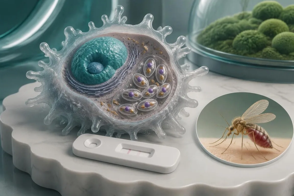
Pozitiven rezultat protiteles lahko odraža preteklo izpostavljenost in ne trenutne bolezni. Simptomi, potovalna anamneza, imunski status in testirani vzorec določajo, katera potrditev je potrebna.

Ključne ugotovitve
- Pozitivna protitelesa kažejo imunski odziv na antigene Leishmania; sami po sebi ne potrjujejo aktivne leishmanioze.
- Tri glavne oblike—visceralna, kožna in mukozna leishmanioza—zahtevajo različne klinične ocene in strategije odvzema vzorcev.
- Potrditev visceralne leishmanioze lahko temelji na kombinaciji ustrezne klinične slike s serologijo, PCR, mikroskopijo ali kulturo; nekateri programi na endemičnih območjih sprejemajo pozitiven hitri serološki test v okviru določenega kliničnega algoritma.
- Potrditev kožne leishmanioze običajno zahteva vzorec z prizadetega predela kože za PCR, mikroskopijo ali kulturo; testiranje protiteles se pri lokalizirani bolezni slabo obnese.
- Uspešnost testov po regijah se razlikuje: pregled Cochrane je pri ljudeh s sumom na visceralno bolezen ocenil občutljivost testa rK39 na približno 97 % na Indijski podcelini v primerjavi z 85% v Vzhodni Afriki.
- Imunosupresija lahko oslabijo odziv protiteles in omogočijo ponovno aktivacijo več let po izpostavitvi; negativen rezultat protiteles pri bolniku z visokim tveganjem varno ne izključuje bolezni.
- Opozorilni znaki za nujno pomoč vključujejo zmedenost, hudo težko dihanje, pomembno krvavitev ali zvišano telesno temperaturo ob absolutnem številu nevtrofilcev pod 0.5 × 10^9/L.
- Naslednji pregled naj vključujejo celotno laboratorijsko poročilo, kraje in datume potovanj, predhodno zdravljenje lišmanioze ter seznam zdravil, ki zavirajo imunski sistem.
Kaj dejansko pomeni pozitiven rezultat testa na protitelesa proti leishmaniozi?
A pozitivna protitelesa proti lišmaniji rezultat pomeni, da je test zaznal protitelesa, ki reagirajo z antigeni Leishmania – ne pa nujno aktivne bolezni. Pozitiven rezultat lahko povzročijo pretekla okužba, asimptomatska izpostavljenost ali navzkrižna reaktivnost; za oceno aktivne bolezni je treba upoštevati simptome, izpostavljenost, imunski status in po potrebi dokazati prisotnost parazita.
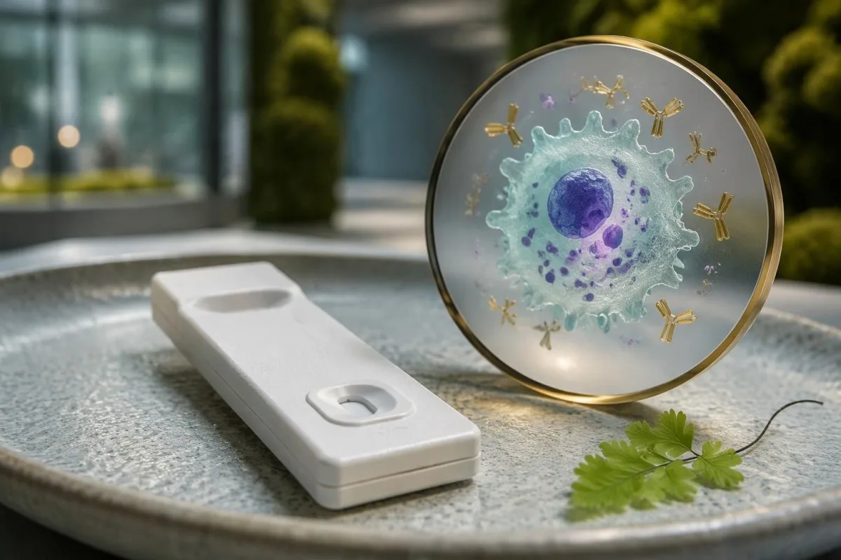
Lišmanioza ima 3 glavne klinične oblike: visceralno obliko, ki prizadene notranje organe, kožno obliko, ki prizadene kožo, in mukozno obliko, ki prizadene strukture, kot sta nos ali usta. Na podlagi rezultata protiteles ni mogoče zanesljivo ugotoviti, katero obliko ima oseba, kje v telesu je prizadeto tkivo ali katera vrsta parazita je povzročitelj; te razlike vplivajo tako na izbiro naslednjega testa kot na pogovor o zdravljenju.
Protitelesa proti lišmaniji so lahko zaznavna še več let po uspešnem zdravljenju, zato je lahko rezultat, pridobljen 2 leti po ozdravitvi, še vedno pozitiven, ne da bi to dokazovalo ponovitev bolezni. Po drugi strani pa je treba osebo z novo vztrajno vročino in povečano vranico oceniti tudi, če je vrednost protiteles le malo nad laboratorijsko mejno vrednostjo – klinični znaki so pomembnejši od višine oznake.
Kantesti je AI analizator krvi ki lahko pomagajo pojasniti spremljajoče laboratorijske izvide, vendar pozitivno poročilo o protitelesih ne zagotavlja izvida pregleda ali dokazov iz tkiva, potrebnih za diagnozo. Naša organizacija in klinični namen razlikuje podporo pri razlagi izvidov od zdravstvene oskrbe; zdravniku, ki organizira nadaljnjo obravnavo, prinesite celotno poročilo, vključno vsaj z metodo, referenčnim intervalom in vrsto vzorca.
Kateri test za leishmaniozo je opravil vaš laboratorij?
Pomen pozitivnega testa za lišmaniozo je odvisen od njegove metode: hitri testi rK39, ELISA, testiranje protiteles z indirektno imunofluorescenco in testiranje z direktno aglutinacijo zaznavajo protitelesa, medtem ko PCR zaznava DNK parazita. Njihove vrednosti niso medsebojno primerljive in nobena univerzalna mejna vrednost protiteles ne potrjuje aktivne bolezni.
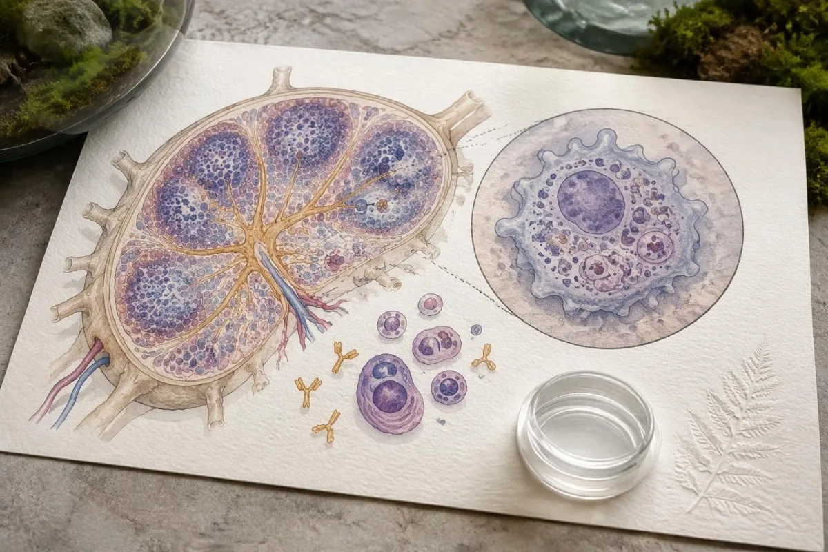
Poročilo lahko prikazuje kvalitativni rezultat, vrednost indeksa ali titer protiteles, izražen kot razredčitev, na primer 1:160. Pri povsem hipotetičnem testu, pri katerem se pozitivnost začne pri indeksu 1.1, rezultat 1.4 pomeni, da je signal presegel mejno vrednost tega testa – ne pa, da je bolezen blaga ali da je prisotno določeno število parazitov; naša vodnik po kvalitativnih v primerjavi s kvantitativnimi metodami pojasnjuje to razliko pri poročanju.
Testi rK39 zaznavajo protitelesa proti rekombinantnemu antigenu Leishmania, vendar se njihova občutljivost razlikuje glede na geografsko območje in uporabljeni izdelek. Cochraneov pregled Boelaerta in sodelavcev iz leta 2014 je ocenil občutljivost hitrih testov rK39 na približno 97.0% na indijski podcelini in 85.3% v vzhodni Afriki pri osebah, pregledanih zaradi suma na visceralno lišmaniozo; teh ocen ne smemo uporabljati za asimptomatsko presejanje ali kožno obliko bolezni.
Različni laboratoriji lahko uporabljajo različne antigene, serije razredčitev in opredelitve mejnih rezultatov, tudi če je v obeh poročilih navedeno 'protitelesa proti Leishmania'. Primerjava titra 1:160, pridobljenega z eno metodo, z indeksom 1.4, pridobljenim z drugo, ne more pokazati trenda; vprašajte, ali naj neskladen rezultat pregleda referenčni laboratorij, preden navidezno številčno spremembo obravnavate kot biološko poslabšanje.
Zakaj je lahko pozitiven rezultat protiteles zavajajoč?
Pozitiven rezultat protiteles je lahko zavajajoč, ker pretekla izpostavljenost, navzkrižna reaktivnost in nizka predtestna verjetnost reduce its ability to identify active illness. The same assay result carries different diagnostic weight in a symptomatic person from an endemic area and an asymptomatic person without plausible exposure.
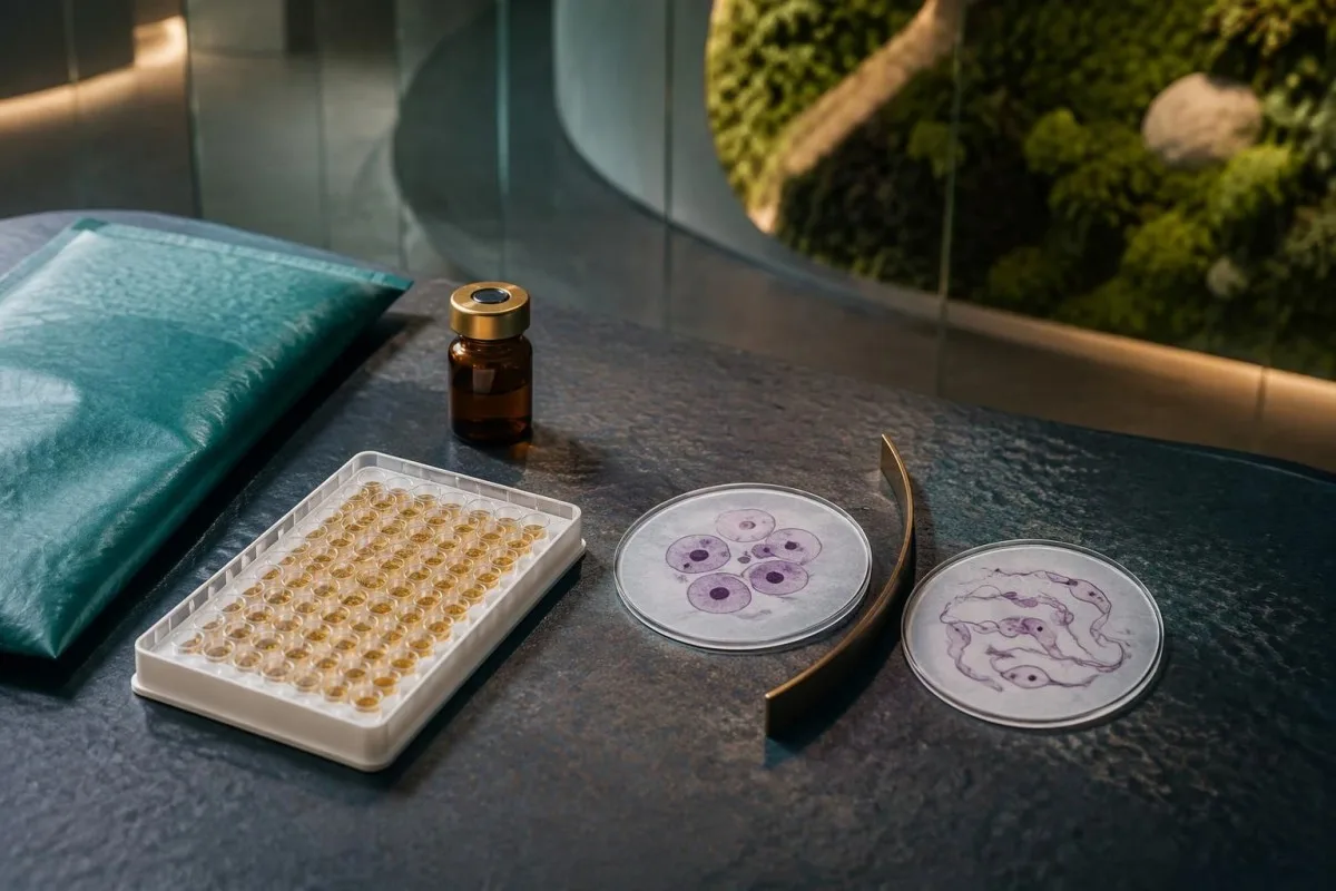
Some Leishmania antibody assays cross-react with antibodies to Trypanosoma cruzi, the parasite responsible for Chagas disease, although the extent depends on the antigen and method. A person with exposure in Latin America and 2 reactive parasite antibody tests may need reference-laboratory clarification rather than an assumption of two active infections; our Chagas confirmation pathway explains why assay selection matters.
Consider a hypothetical group of 1,000 people in which 1% have active disease, using an imaginary test with 90% sensitivity and 95% specificity: approximately 9 true-positive and 50 false-positive results would occur, making only about 15% of positive results true positives. These are teaching assumptions, not performance figures for a particular leishmaniasis assay, but they show why testing people without exposure can generate confusing alarms.
Now keep the same hypothetical assay but increase disease prevalence to 30%: approximately 270 true positives and 35 false positives would occur, so about 89% of positives would identify disease. The individual estimate still depends on symptoms, past infection, and immune status; our vodnik za razlago pozitivnih protiteles helps separate evidence of immune recognition from evidence that treatment is needed.
Kateri izvidi povečujejo verjetnost visceralne leishmanioze?
Visceral leishmaniasis becomes more concerning when prolonged fever, weight loss, and an enlarged spleen accompany compatible exposure, particularly with low blood-cell counts. Antibodies strengthen that clinical pattern but do not explain every fever, anemia, or abnormal protein result.
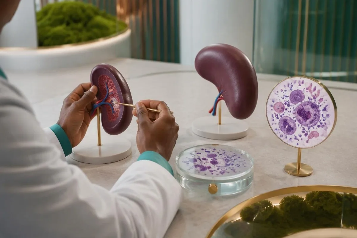
Visceral leishmaniasis can lower all 3 major blood-cell lines: red cells, white cells, and platelets, producing pancytopenia. Bone-marrow involvement, inflammatory effects, and trapping of cellular elements in an enlarged spleen can contribute together; our razlaga hemoglobina in hematokrita helps readers understand anemia without mistaking a single low value for a parasite diagnosis.
For example, a hypothetical traveler with 3 weeks of fever, hemoglobin 8.5 g/dL, white-cell count 2.2 × 10^9/L, platelets 70 × 10^9/L, and splenomegaly needs prompt investigation regardless of the antibody index. This combination supports a serious systemic process, but malaria, hematologic disease, disseminated fungal infection, and other conditions remain possible; diagnostic testing should address those alternatives rather than stop at serology.
Visceral disease may also produce polyclonal hypergammaglobulinemia and low albumin, so a hypothetical total protein of 9.0 g/dL with albumin 2.8 g/dL warrants contextual interpretation rather than a reflex assumption of a monoclonal disorder. Our vodnik za interpretacijo serumskih proteinov explains the pattern; ultrasound can assess spleen size, but spleen enlargement itself cannot establish the cause.
Kako potrdimo diagnozo visceralne leishmanioze?
Visceral leishmaniasis diagnosis uses a compatible clinical syndrome plus laboratory evidence chosen for the setting. PCR, microscopy, and culture can detect the parasite; in some endemic treatment programs, positive rK39 serology with a defined clinical case pattern supports diagnosis without invasive sampling.
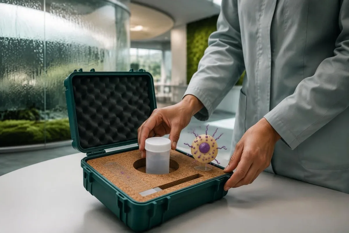
The 2016 IDSA/ASTMH guideline recommends combining diagnostic approaches and using molecular testing when available rather than relying on a single assay in every patient (Aronson et al., 2016). Peripheral blood or buffy-coat PCR may be useful, especially with immune suppression, but bone marrow or other tissue may still be needed when suspicion remains high; a negative result from 1 compartment does not guarantee absence elsewhere.
Aronson et al. report microscopy sensitivities of approximately 52–85% for bone marrow aspirates and 93–99% for splenic aspirates, with yield affected by expertise and parasite burden. Splenic aspiration has a potentially serious bleeding risk and belongs only in appropriately selected patients at experienced centers; the highest-yield specimen is not automatically the safest first choice, particularly when platelet counts or coagulation are abnormal.
A clinician may investigate other causes of the same syndrome while obtaining confirmatory samples, especially if antibody and PCR findings disagree. Our Histoplasma antigen follow-up guide describes another systemic illness that can overlap clinically; obtaining specimens before antiparasitic therapy is useful when feasible, but a critically ill person should not wait several days solely to complete an ideal diagnostic sequence.
Ali pozitiven rezultat PCR-testa za leishmaniozo dokazuje aktivno okužbo?
A positive leishmaniasis PCR test establishes detection of Leishmania DNA in the tested specimen, assuming a valid assay and appropriate contamination controls. It supports active disease strongly when the specimen and symptoms fit, but DNA detection alone does not prove parasite viability or explain every symptom.

PCR can detect low-level or asymptomatic parasitemia in some exposed populations, and residual genetic material may complicate interpretation after treatment. A hypothetical weakly positive blood PCR in a well person is therefore different from a positive bone-marrow PCR in someone with fever and pancytopenia; clinicians consider at least 3 variables together—clinical syndrome, specimen source, and previous treatment—before interpreting detection as disease requiring therapy.
Cycle-threshold values are not a universal Leishmania parasite-count scale, and a Ct of 35 from 1 laboratory cannot be compared directly with Ct 35 from another assay. Extraction volume, genetic target, calibration, and detection limits differ; our Babesia antibody and infection guide illustrates a related diagnostic distinction, although the organisms and recommended testing pathways are not interchangeable.
Kantesti je storitvi za interpretacijo laboratorijskih testov AI that can explain why antibody results, DNA detection, and accompanying CBC findings answer different questions, rather than collapsing them into a single diagnosis. Our pojasnjuje, kako Kantesti-jev nevronski sistem uteži sosednje biomarkerje, smer trenda in notranjo skladnost. Po mojih izkušnjah je to najpomembnejše za mejne vrednosti BUN med describes the interpretation approach; no AI analysis can recover the missing specimen source or assay validation details from a report that contains only 1 word: 'positive.'
Zakaj je pri kožni leishmaniozi potreben vzorec kože?
Cutaneous leishmaniasis is usually confirmed by testing material from an affected skin site, not by measuring antibodies in blood. Localized disease may generate little detectable circulating antibody, while skin PCR, microscopy, or culture can directly investigate the area causing concern.
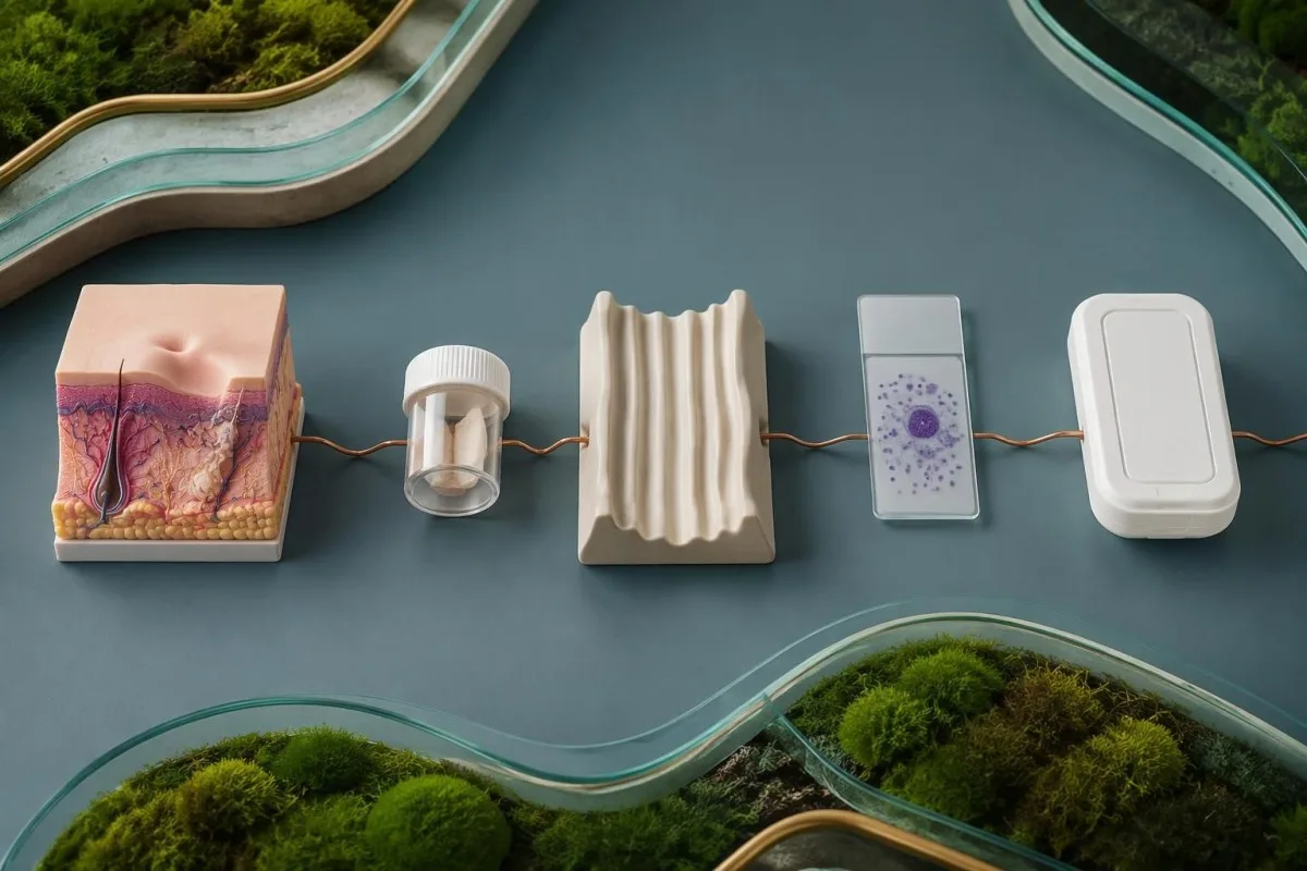
The active raised edge of a persistent affected area often provides useful material, but the optimal sampling method depends on its age, appearance, and the laboratory protocol. Clinicians may use scraping, aspiration, or a biopsy; requesting at least 2 diagnostic approaches, such as PCR and histology, can be useful because one assesses parasite material while the other also evaluates alternative explanations.
A hypothetical person with a single painless forearm sore persisting for 8 weeks after travel may need lesion-directed testing even if Leishmania antibodies are negative and the CBC is normal. Fungal disease, atypical mycobacteria, inflammatory disorders, and skin cancer can resemble cutaneous leishmaniasis; sending appropriate tissue for broader evaluation prevents a parasite-focused request from overlooking another treatable cause.
Species identification matters because some Leishmania species, particularly members of the Viannia subgenus acquired in the Americas, carry a greater concern for later mucosal involvement. Our skin-site PCR testing guide explains why material must come from the relevant affected site, although the exact collection requirements differ; ask before sampling whether the laboratory can identify species from the submitted specimen.
Kako imunosupresija vpliva na razlago rezultatov testov?
Imunosupresija can reduce antibody production, increase the risk of visceral disease, and permit reactivation after remote exposure. A negative antibody result is therefore less reassuring in someone with HIV, a transplant, or substantial immune-suppressing treatment; PCR and direct parasite detection become particularly valuable.

HIV-associated risk cannot be judged from viral load alone: Število CD4 describes a different aspect of immune function, and counts below 200 cells/µL indicate advanced immunodeficiency. Our HIV viral-load interpretation guide explains the distinction; even a person with suppressed HIV RNA may need careful evaluation if immune recovery is incomplete and compatible symptoms or exposure are present.
Leishmania can persist after apparent recovery, so immune-suppressing treatment begun 5 years after travel can still make that older exposure clinically relevant. Corticosteroids, transplant medicines, some biologic therapies, and disorders affecting immune function may alter the diagnostic strategy, but risk varies by drug and intensity; patients should provide medication names and doses without stopping prescribed treatment on their own.
A low total IgG concentration offers 1 possible explanation for unreliable antibody-based screening, but normal immunoglobulin levels do not guarantee normal parasite-specific responses. Our immunoglobulin result interpretation separates these measurements; when symptoms remain concerning, repeating the same antibody assay without changing the diagnostic approach may add little compared with testing an appropriate specimen for parasite DNA or organisms.
Kateri podatki o potovanju spremenijo pomen pozitivnega rezultata?
Potovalna anamneza changes both the probability of leishmaniasis and the most useful diagnostic pathway. Exact destinations, rural or outdoor exposure, dates, and previous residence matter more than a continent name because Leishmania species, clinical forms, and antibody-test performance vary within regions.

Leishmaniasis is usually transmitted through the bite of an infected female phlebotomine sand fly, and transmission can occur in parts of the Mediterranean as well as tropical regions. A traveler who visited southern Europe for 2 weeks may have a relevant exposure despite never visiting a destination they considered tropical; absence of a remembered bite does not exclude exposure because these insects are small.
Cutaneous disease often appears weeks to months after exposure, whereas visceral disease may emerge months later or reactivate years afterward, particularly with immune suppression. Record all relevant destinations over more than the last 12 months if your clinician requests them; our travel-associated parasite antibody guide illustrates why an exposure timeline can remain useful long after the journey has ended.
Kantesti AI can help organize the distinction between exposure evidence and routine laboratory abnormalities, but the travel history must come from the person or their records—not from a positive assay. For an appointment, write a 4-part timeline covering destination, dates, onset of symptoms, and immune-suppressing treatment; this can help the clinician choose a geographically appropriate assay and determine whether species identification would change management.
Zakaj lahko pravi test na napačnem vzorcu spregleda bolezen?
Specimen choice and handling affect sensitivity because Leishmania distribution differs between visceral and localized skin disease. Blood PCR may help investigate visceral disease, while an affected skin specimen is usually more informative for cutaneous disease; collection medium, storage, and transport must match the laboratory method.
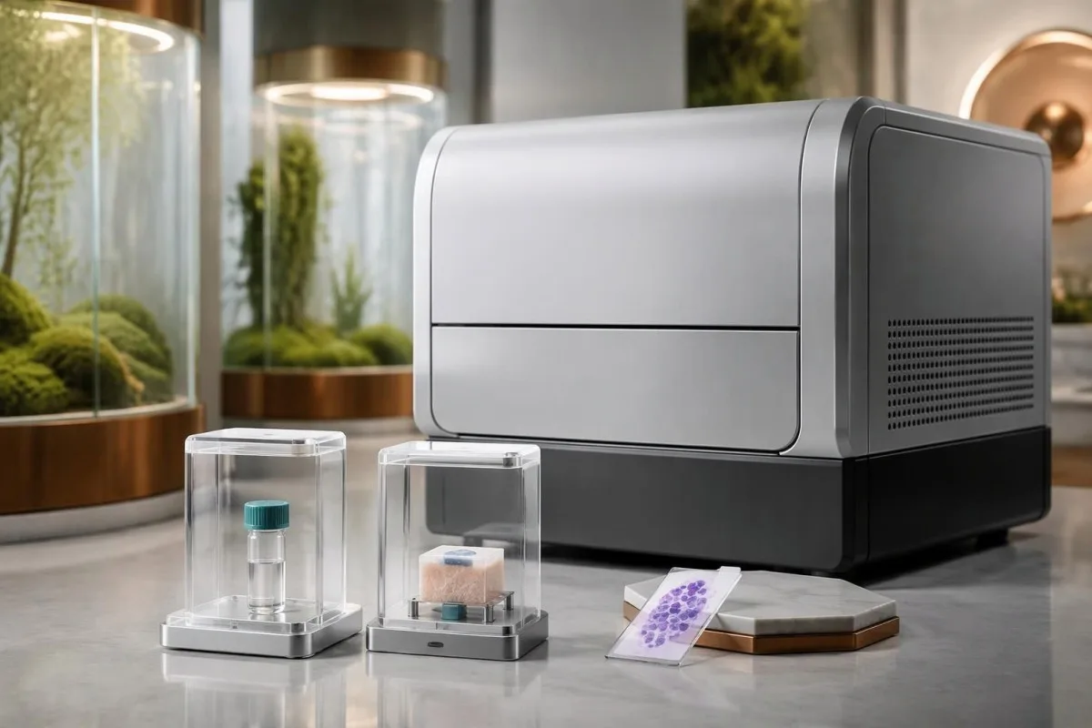
Reference laboratories may request EDTA whole blood, buffy coat, fresh tissue, or another specific preparation for PCR, depending on the validated assay. Serum used for an antibody test is not automatically a substitute for every molecular protocol; our PCR sample timing guide explains the general principle of matching collection to the test, although disease-specific instructions must come from the receiving laboratory.
Formalin-fixed tissue supports histology and sometimes validated PCR, but it cannot be used for parasite culture because fixation destroys viability. Before a skin biopsy, the clinician may need to divide material into 2 separately handled portions, one for histopathology and another for fresh-tissue testing; requesting culture only after all material has entered formalin can require an avoidable second procedure.
A negative result obtained after 5 days of treatment may reflect reduced detectable parasite burden rather than an incorrect original diagnosis, although the size of that effect depends on method and specimen. Tell the laboratory and clinician the treatment start date, previous medicines, and sample collection date; if suspicion remains high, the next step may be a different compartment or method rather than repeating an inadequately matched sample.
Kateri simptomi ali laboratorijski izvidi zahtevajo nujno oskrbo?
Confusion, severe breathlessness, fainting, significant bleeding, or rapidly worsening illness require emergency evaluation, regardless of the antibody result. Persistent fever with weight loss or spleen enlargement needs prompt medical review; new nose, mouth, or throat symptoms after compatible exposure need assessment for mucosal disease.

Vročina ob absolute neutrophil count below 0.5 × 10^9/L is an emergency because serious infection can progress rapidly, even before its cause is known. Platelets below 20 × 10^9/L generally warrant urgent clinician-directed assessment, with greater urgency if bleeding occurs; abnormal coagulation and falling counts may need parallel investigation, as explained in our Vodnik po laboratorijskem vzorcu DIC.
Visceral leishmaniasis can trigger hemophagocytic lymphohistiocytosis, an excessive immune-activation syndrome that may resemble severe infection or hematologic disease. A ferritin value above 500 ng/mL is only one component of established HLH criteria and is far too nonspecific to diagnose the syndrome alone; our HScore interpretation guide explains why fever, cytopenias, organ findings, and additional measurements must be considered together.
Persistent nasal blockage, recurrent nosebleeds, mouth changes, or hoarseness can indicate mucosal involvement, sometimes years after a skin lesion has healed; breathing difficulty is an emergency. Do not wait 2 weeks for a routine antibody repeat if systemic or airway symptoms are progressing, and tell the assessing team about exposure; fever after travel to a malaria-endemic area also requires urgent evaluation for malaria rather than attribution to Leishmania antibodies.
Kako zdravniki sklepajo na podlagi različnih pozitivnih rezultatov?
Clinicians interpret a positive leishmaniasis result by asking whether exposure, symptoms, immune status, and parasite evidence agree. The following 3 hypothetical examples show why identical antibody positivity can lead to reassurance with follow-up, lesion-directed testing, or urgent systemic investigation.
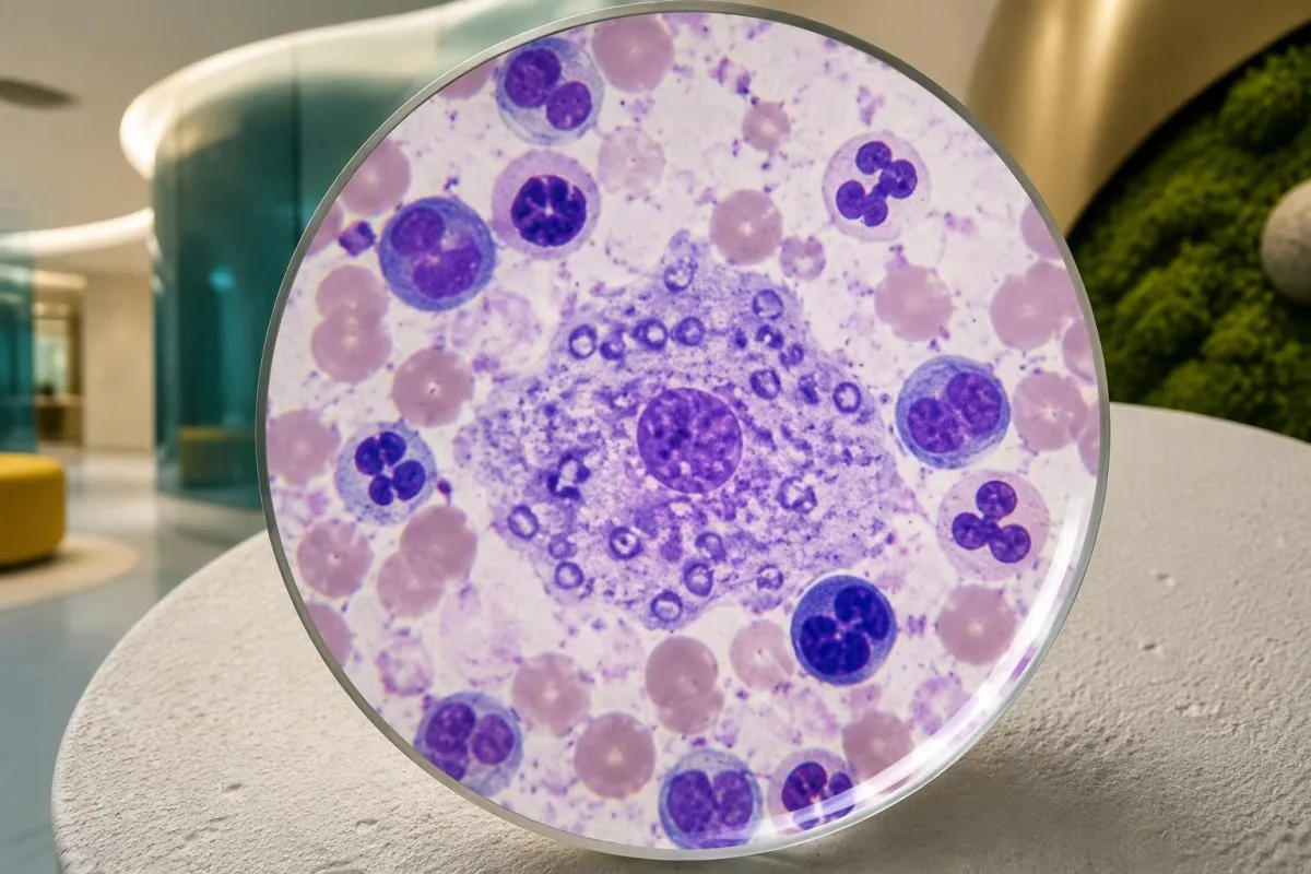
Example 1: a hypothetical 42-year-old completed treatment for visceral leishmaniasis 3 years ago, feels well, and has stable blood counts but a persistently positive antibody assay. Persistent antibodies are plausible, so the result alone does not establish relapse or justify retreatment; new fever, weight loss, or immune suppression would change the assessment and might justify direct parasite testing rather than simply checking another antibody titer.
Example 2: a hypothetical 29-year-old has an 8-week skin lesion after travel, normal routine blood tests, and a weakly positive antibody result. The affected skin still needs appropriate PCR, microscopy, or histology because serology neither confirms its cause nor identifies the species; our limits of fever antibody tests describes a parallel diagnostic pitfall, not a reason to apply typhoid testing rules to leishmaniasis.
Example 3: a hypothetical 58-year-old taking transplant medicines develops fever, spleen enlargement, platelets of 45 × 10^9/L, and a negative antibody test. At Kantesti, we explain why that negative result cannot cancel the clinical concern: immune suppression can impair antibody responses, and prompt clinician-led PCR or tissue evaluation may be necessary; these examples are teaching scenarios, not patient encounters or substitutes for individualized medical advice.
Ali pozitiven rezultat pomeni zdravljenje ali ponovitev testiranja?
A positive result does not automatically mean treatment, and successful treatment does not require antibodies to become negative. Confirmed visceral disease generally needs systemic therapy; cutaneous management depends on species, site, lesion burden, mucosal risk, immune status, and the likelihood of spontaneous healing.

Untreated visceral leishmaniasis can be life-threatening, so compatible illness with convincing diagnostic evidence needs specialist management rather than watchful waiting based on a low antibody index. By contrast, some uncomplicated cutaneous presentations may be observed or treated locally, while others need systemic treatment; a single 1-cm lesion on a cosmetically sensitive site can require a different approach from a similarly sized lesion elsewhere.
When systemic therapy is selected, safety monitoring may include at least 3 groups of measurements: kidney function, electrolytes, and liver tests, alongside the CBC when indicated by the medicine and illness. Liposomal amphotericin B, for example, can affect renal function and potassium or magnesium; our vodnik po trendih varnosti zdravil explains why changes from baseline can matter even before an isolated value crosses a reference limit.
Kantesti je platforma za razlaga krvne slike z AI that can help explain these follow-up trends, but treatment success is assessed through clinical recovery and clinician-selected testing rather than AI confirmation of cure. The IDSA/ASTMH guideline recommends monitoring treated cutaneous lesions for 6–12 mesecev for evidence of therapeutic failure (Aronson et al., 2016); visceral follow-up and relapse surveillance must be individualized, especially when immune suppression increases ongoing risk.
Raziskovalne publikacije in omejitve AI interpretacije
AI interpretation supports explanation; it does not confirm leishmaniasis or validate a parasite assay. Disease-specific diagnostic recommendations come from clinical guidelines and test-accuracy evidence, while technical research about an interpretation engine answers separate questions about structured output, evaluation methods, and oversight.
The Kantesti technical benchmark listed below evaluates the blood-test interpretation engine using 100.000 sintetičnih testnih primerov, as stated in its publication title. Synthetic-case performance must not be presented as prospective diagnostic accuracy for leishmaniasis, proof of PCR sensitivity, or evidence that treatment decisions are safe without a clinician; our medicinski validacijski okvir describes the evaluation and oversight context rather than replacing disease-specific evidence.
The Okvir klinične validacije v2.0 publication describes a framework for assessment and clinical oversight; it is not, by its title or purpose, a clinical trial demonstrating leishmaniasis outcomes. Our standardi za medicinske vsebine separate technical validation, clinical evidence, and educational writing, so readers can judge each claim against the type of evidence actually supporting it rather than treating a DOI as a guarantee of clinical utility.
For a positive leishmaniasis report, the most useful next step is a clinician-led review of 4 bistvene elemente: the assay, the specimen, the exposure history, and the current clinical picture. Kantesti can help explain routine laboratory patterns during that process, but an urgent symptom takes priority over any upload or repeat antibody test; the references below distinguish external leishmaniasis guidance from publications about our interpretation methodology.
Pogosta vprašanja
Ali pozitiven test na protitelesa proti lišmaniozi pomeni, da imam aktivno lišmaniozo?
Pozitiven rezultat testa protiteles proti lišmaniozi sam po sebi še ne potrjuje aktivne lišmanioze, saj lahko protitelesa vztrajajo po pretekli okužbi ali se pojavijo ob asimptomatski izpostavljenosti oziroma navzkrižni reaktivnosti. Rezultat, ki ostane pozitiven 2 leti po uspešnem zdravljenju, lahko zato odraža imunski spomin in ne ponovitve bolezni. Zdravniki ocenijo simptome, potovalno anamnezo, imunski status in natančno uporabljeno metodo testiranja, nato pa po potrebi uporabijo PCR, mikroskopijo, kulturo ali preiskavo tkiva. Vztrajna vročina, izguba telesne mase ali povečana vranica zahtevajo hiter zdravniški pregled.
Kako natančen je test rK39 za lišmaniozo?
Natančnost hitrega testa rK39 se razlikuje glede na geografsko območje, izdelek in populacijo bolnikov. Cochraneov pregled iz leta 2014 je ocenil občutljivost na približno 97,0 % na indijski podcelini in 85,3 % v vzhodni Afriki pri osebah, pri katerih so preiskovali sum na visceralno lišmaniozo. Te številke ne opisujejo uspešnosti pri asimptomatskem presejanju, lokalizirani kožni bolezni ali vseh populacijah z oslabljenim imunskim sistemom. Negativen rezultat testa ne bi smel zaključiti preiskav, če klinični sum ostaja velik.
Ali je lahko test PCR za lišmaniozo pozitiven tudi brez simptomov?
Test PCR za lišmaniozo lahko pri nekaterih ljudeh brez simptomov zazna DNK parazita, zato pozitiven rezultat sam po sebi ne potrjuje simptomatske bolezni ali potrebe po zdravljenju. Razlaga je odvisna od vsaj 3 dejavnikov: izvora vzorca, trenutnih kliničnih izvidov ter predhodnega zdravljenja ali izpostavljenosti. PCR zazna DNK, ne dokazuje pa neposredno, da je parazit živ. Zdravniki lahko poiščejo dodatne dokaze, kadar je zaznava šibka, nepričakovana ali neskladna s klinično sliko.
Ali je mogoče kožno lišmaniozo diagnosticirati s krvnim testom?
Krvni test na protitelesa običajno ne zadostuje za potrditev kožne lišmanioze, saj lahko lokalizirana kožna bolezen povzroči le malo protiteles v krvi. Pri vztrajni kožni spremembi, na primer takšni, ki traja 8 tednov po relevantnem potovanju, je običajno treba testirati vzorec z prizadetega mesta. PCR, mikroskopija, kultura ali histologija lahko zagotovijo neposrednejše dokaze in pomagajo oceniti druge možne vzroke. Določitev vrste lahko vpliva tudi na zdravljenje in oceno poznejšega tveganja za prizadetost sluznic.
Ali lahko protitelesa proti lišmaniozi ostanejo pozitivna po zdravljenju?
Protitelesa proti lišmaniozi so lahko pozitivna še leta po uspešnem zdravljenju, zato izginotje protiteles ni zanesljiv pogoj za ozdravitev. Pozitiven rezultat 3 leta po zdravljenju ne more potrditi ponovitve bolezni brez ustreznih simptomov ali dodatnih dokazov. Klinično okrevanje ter laboratorijske preiskave ali testiranje na parazite, ki jih izbere zdravnik, usmerjajo nadaljnje spremljanje. Pri osebah, ki pozneje postanejo imunsko oslabljene, je morda potrebna drugačna ocena ponovitve bolezni, saj lahko predhodna izpostavljenost še vedno vpliva na presojo.
Kdaj je treba ob pozitivnem izvidu na lišmaniozo nemudoma poiskati zdravniško pomoč?
Pozitiven rezultat testa na lišmaniozo zahteva nujno zdravniško oceno, če ga spremljajo vztrajna vročina, izguba telesne teže, povečana vranica ali znaten upad števila krvnih celic. Zmedenost, huda zadihanost, omedlevica, pomembna krvavitev ali težave z dihalnimi potmi zahtevajo nujno obravnavo ne glede na rezultat testa. Vročina ob absolutnem številu nevtrofilcev pod 0.5 × 10^9/L je nujno stanje, število trombocitov pod 20 × 10^9/L pa praviloma zahteva nujno oceno po navodilih zdravnika. Novi vztrajni simptomi v nosu, ustih ali žrelu po ustrezni izpostavljenosti zahtevajo tudi oceno glede bolezni sluznic.
Poskrbite, da vam razložijo rezultate krvnih preiskav
Naložite svoj laboratorijski izvid in AI Kantesti bo v nekaj sekundah preprosto razložil vsak biomarker. Uporablja ga več kot 2 milijona ljudi po vsem svetu.
Reference
- Klein, T., Mitchell, S., & Weber, H. (2026). Predregistrirana, na rubrikah zasnovana avtomatizirana tehnična primerjava (benchmark) interpretacijskega motorja Kantesti za analizo krvnih testov na 100.000 sintetičnih testnih primerih. Kantesti AI Medical Research. DOI
- Klein, T., Mitchell, S., & Weber, H. (2026). Klinični okvir validacije v2.0 (stran za medicinsko validacijo). Kantesti AI Medical Research. DOI
- Aronson N et al. (2016). Diagnosis and Treatment of Leishmaniasis: Clinical Practice Guidelines by the Infectious Diseases Society of America (IDSA) and the American Society of Tropical Medicine and Hygiene (ASTMH). Clinical Infectious Diseases. DOI
- Boelaert M et al. (2014). Rapid tests for the diagnosis of visceral leishmaniasis in patients with suspected disease. Cochrane Database of Systematic Reviews. DOI
- 2 milijona+Analizirani testi
- 127+Države
- 100+Jeziki
O zdravstveni ekipi


E-E-A-T zaupanja vredni signali
Izkušnje
Zdravniški klinični pregled delovnih postopkov za interpretacijo laboratorijskih izvidov.
Strokovno znanje
Laboratorijska medicina s poudarkom na tem, kako se biomarkerji obnašajo v kliničnem kontekstu.
Avtoritativnost
Zdravstveno pregledal dr. Thomas Klein, s prispevki dr. Sarah Mitchell in prof. dr. Hansa Webra.
Zanesljivost
Interpretacija na podlagi dokazov z jasnimi nadaljnjimi potmi za zmanjšanje alarmiranja.





Zdravstvena omejitev odgovornosti
Ta članek je namenjen izobraževalnim namenom in ne predstavlja medicinskega nasveta. Za odločitve o diagnozi in zdravljenju se vedno posvetujte z usposobljenim izvajalcem zdravstvenih storitev.
Objavljeno · Obrnite se na uredniško ekipo
Izdala družba Kantesti Ltd, registrirana v Angliji in Walesu, matična št. 17090423, London, Združeno kraljestvo.
