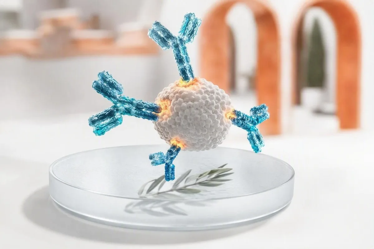
Een positief antilichaamresultaat betekent dat uw immuunsysteem een bepaald doelwit herkende; het bewijst op zichzelf geen actieve infectie of ziekte. De antilichaamklasse, testmethode, timing, symptomen en aanvullende tests bepalen of u geruststelling, een herhalingstest, bevestiging of snelle klinische zorg nodig heeft.


- Positief antilichaamresultaat toont meestal immuunherkenning, niet noodzakelijkerwijs huidige ziekte of besmettelijkheid.
- IgM versus IgG is een tijdelijke aanwijzing, geen klok: IgM kan maanden aanhouden en IgG kan vroeg verschijnen.
- Vaccinantilichamen kunnen immuniteitstests positief maken zonder dat dit duidt op een natuurlijke infectie.
- Allergeenspecifieke IgE boven 0,35 kUA/L duidt op sensibilisatie, maar symptomen na blootstelling vestigen een klinische allergie.
- ANA-positiviteit komt voor bij maximaal 1 op de 4 gezonde volwassenen met lage titers, vooral met de leeftijd; symptomen en specifieke antistoffen zijn belangrijker.
- HIV-screening reactiviteit vereist altijd het laboratoriumbevestigingsalgoritme voordat een diagnose wordt gesteld.
- Anti-tTG IgA is alleen betrouwbaar voor de beoordeling van coeliakie als de persoon gluten eet en adequate totale IgA heeft.
- Spoedsymptomen zoals ademhalingsmoeilijkheden, verwardheid, geelzucht, pijn op de borst of een wijdverspreide blaarvormige huiduitslag wegen zwaarder dan een routinematig antistofresultaat.
Wat een positief antilichaamresultaat u werkelijk vertelt
A positieve antistoftest betekent dat de test immuunproteïnen heeft gedetecteerd die een bepaald doelwit binden; het betekent niet niet automatisch dat u een actieve infectie, een auto-immuunziekte of een allergie heeft die behandeling nodig heeft. De volgende stap hangt af van het antistoftype, de sterkte van het resultaat, de symptomen, de blootstellingstijd en eventuele bijbehorende antigeen-, PCR- of orgaanfunctie-tests.
Antistoffen zijn B-celproteïnen die worden aangemaakt na vaccinatie, infectie, blootstelling aan allergenen, auto-immuniteit, of af en toe zonder klinisch schadelijke reden. Een positief resultaat beantwoordt één nauwe analytische vraag — “werd binding gedetecteerd boven deze laboratoriumafsnijding?” — in plaats van de bredere klinische vraag over wat er vandaag in uw lichaam gebeurt.
In mijn 15 jaar klinische praktijk is de meest voorkomende fout geweest om “positief” als een diagnose te lezen. De praktische regel van Dr. Thomas Klein is om eerst het doelwit—een virus, vaccinantigeen, voedseleiwit, schildklierenzym, nucleair antigeen of geneesmiddel— te identificeren en vervolgens te vragen of de test een eerdere blootstelling, een huidig organisme, sensibilisatie of immuunmisleiding detecteert.
Kantesti AI is een AI-bloedtestanalysator die antistofvlaggen naast datums, immunoglobulineklasse en gerelateerde resultaten leest in plaats van één enkele positieve lijn als een oordeel te behandelen. Die context is vooral nuttig wanneer patiënten rapporten van verschillende laboratoria vergelijken; kwalitatieve en kwantitatieve resultaten zijn niet onderling uitwisselbaar, zelfs als beide positief zeggen.
IgG versus IgM positief: waarom timing minder eenvoudig is dan het klinkt
IgM positief kan wijzen op een recente immuunrespons, terwijl IgG positief vaak wijst op eerdere blootstelling of vaccinrespons; geen van beide patronen dateert een ziekte op zichzelf betrouwbaar. Sommige infecties produceren IgG binnen 7-14 dagen, en IgM kan 3-12 maanden of langer detecteerbaar blijven.
IgM is een grote antistof met vijf eenheden die vaak vroeg detecteerbaar is, maar zijn grootte en brede binding maken het vatbaarder voor kruisreacties. Epstein-Barr-virus, parvovirus B19, reumafactor en sommige auto-immuunantistoffen kunnen misleidende IgM-reacties veroorzaken; een geïsoleerd laag IgM-resultaat verdient meer scepsis dan paniek.
IgG blijft meestal bestaan nadat het immuungeheugen zich heeft ontwikkeld, soms tientallen jaren. Een positief IgG-resultaat kan duiden op een kinderinfectie, een recente vaccinatie, passieve antistoffen van intraveneuze immunoglobuline of maternale overdracht naar een zuigeling; het kan geen bescherming bewijzen, tenzij die specifieke test een gevalideerde beschermende drempel heeft.
Gekoppelde monsters kunnen nuttiger zijn dan een enkele bloedafname: een viervoudige stijging van de antistoftiter tussen monsters die ongeveer 2-4 weken na elkaar zijn afgenomen, ondersteunt een recente infectie bij geselecteerde ziekten. Als het rapport totale IgG, IgA en IgM vermeldt, bekijk dan het patroon in plaats van één getal met onze richtlijn voor interpretatie van immunoglobulinen.
Wanneer infectieantilichamen actieve ziekte betekenen en wanneer niet
Antistoffen tegen infecties tonen meestal blootstelling aan, terwijl PCR, antigeentests, kweek of een oplopende titer betere middelen zijn om veel actieve infecties te detecteren. Een positief antistofresultaat zonder symptomen is vaak historisch, maar de uitzondering hangt af van het organisme en uw immuunstatus.
Voor hepatitis C betekent een reactieve antistof blootstelling op enig moment, maar alleen detecteerbaar HCV-RNA vestigt huidige viraemie. Voor hepatitis B is de combinatie van belang: oppervlakteantigeen, oppervlakteantistof en kernantistof scheiden actieve infectie, genezen infectie en vaccinimmuniteit veel beter dan één positieve marker.
HIV is een andere situatie waar de formulering enorm belangrijk is. Een reactieve vierde-generatie screening is geen diagnose; het laboratorium volgt dit op met een HIV-1/HIV-2 differentiatie immunoassay en, indien nodig, nucleïnezuurtesten. Een persoon die post-expositie profylaxe neemt, heeft tijdsafhankelijke testen nodig, zoals uitgelegd in onze HIV-test na PEP richtlijn.
Ik heb een gezonde patiënt gezien die bang was voor een positieve treponemale syfilisantistof na behandeling 18 jaar eerder. Treponemale testen blijven vaak levenslang positief, terwijl een RPR- of VDRL-titer helpt bij het beoordelen van de huidige activiteit en reactie; syfilistestpatronen maken dit onderscheid duidelijker.
Hoe vaccibrantilichamen verschillen van antilichamen na infectie
Vacciegerelateerde antistoffen tonen aan dat het immuunsysteem reageerde op een vaccinantigeen; ze betekenen niet dat het vaccin een actieve infectie veroorzaakte. Een positief resultaat kan nog steeds onder een bekende beschermende correlaat vallen, omdat bindingsantistoffen en functionele neutraliserende antistoffen niet identiek zijn.
Hepatitis B oppervlakte-antistofconcentraties van 10 mIU/mL of hoger gemeten 1-2 maanden na een gedocumenteerde vaccinreeks worden over het algemeen als beschermend beschouwd bij immunocompetente personen. Anti-HBc, de hepatitis B kernantistof, wordt niet geproduceerd door standaard hepatitis B vaccinatie; de aanwezigheid ervan wijst op natuurlijke blootstelling en moet leiden tot volledige paneelinterpretatie.
Rubella IgG wordt vaak gecontroleerd vóór de zwangerschap omdat het duidt op eerdere immuniteit, niet op een actieve rubella-ziekte. Laboratoria stellen assay-specifieke drempels in, en grensresultaten worden meestal behandeld als niet-immuun voor preconceptionele planning in plaats van geïnterpreteerd als recente infectie; de timing is gedetailleerd in onze rubellatest richtlijn.
Kantesti AI is een AI-bloedonderzoek uitslagplatform die vaccinpatroonantistoffen scheidt van infectiepatroonantistoffen wanneer het rapport de relevante begeleidende markers bevat. De praktische vraag is vaak niet “ben ik positief?”, maar “welk antigeen is gemeten, wanneer ben ik gevaccineerd, en verandert dit resultaat mijn huidige beschermingsplan?”
Waarom een positieve allergie-antilichaamtest mogelijk geen allergie betekent
Een positieve allergeen-specifieke IgE test geeft sensibilisatie aan, wat betekent dat IgE een allergeen herkent; het diagnosticeert geen klinische allergie zonder een compatibele reactiegeschiedenis. Een waarde boven 0,35 kUA/L is een gebruikelijke analytische positiviteitsdrempel, geen universele drempel voor het vermijden van een voedsel of medicijn.
Het verhaal vóór de test is belangrijker dan veel mensen beseffen. Als iemand wekelijks pinda's eet zonder netelroos, piepende ademhaling, braken of collaps, mag een lage positieve pinda IgE-waarde alleen niet leiden tot vermijding; onnodige voedselbeperking kan voedingskundige en sociale schade veroorzaken.
De door NIAID gesponsorde richtlijn voor voedselallergie stelt dat huidpriktests en specifieke IgE-tests mogelijke triggerfoods identificeren, maar zelf geen voedselallergie kunnen diagnosticeren (Sampson et al., 2010). In geselecteerde gevallen gebruikt een allergoloog component-resolved testen of een medisch begeleide orale voedselprovocatie, wat de referentiestandaard blijft voor het oplossen van onzekerheid.
Totale IgE wordt gemeten in IU/mL en kan hoog zijn bij eczeem, parasitaire blootstelling, roken en verschillende immuuncondities; het kan geen boosdoener-allergeen identificeren. Componenttesten kunnen een stabiele opslagproteïne onderscheiden van een cross-reactieve pollen-gerelateerde proteïne, een verschil dat wordt onderzocht in onze moleculaire allergietestartikel.
Waarom auto-immuunantilichamen symptomen en orgaanklachten vereisen
Auto-antistoffen kunnen voorafgaan aan de ziekte, de ziekte begeleiden of voorkomen bij gezonde mensen; een positief resultaat alleen stelt zelden een auto-immuun diagnose. Clinici handelen wanneer het antistofpatroon overeenkomt met symptomen, bevindingen bij lichamelijk onderzoek, ontstekingsmarkers of objectieve orgaanveranderingen.
Een antinucleaire antistof, of ANA, is positief bij lage titers bij ongeveer 13% van de mensen bij 1:80 en ongeveer 20% bij 1:40 in populatiestudies, met hogere percentages bij vrouwen en oudere volwassenen. Een homogeen of gespikkeld patroon stelt geen lupus vast; ANA wordt het best gebruikt als een toegangstest wanneer het klinische beeld een bindweefselziekte plausibel maakt.
Het classificatiekader voor lupus van EULAR/ACR uit 2019 gebruikt ANA bij een titer van ten minste 1:80 als criterium voor insluiting, waarna aanvullende gewogen klinische en immunologische kenmerken vereist zijn (Aringer et al., 2019). Nieuw eiwit in urine, lage C3/C4, cytopenieën, inflammatoire gewrichtszwelling of een overtuigende fotosensitieve uitslag veranderen de urgentie veel meer dan een zwakke ANA alleen.
Anti-dsDNA wordt betekenisvoller wanneer gecombineerd met laag complement en nierbevindingen, hoewel de testmethoden aanzienlijk verschillen. Onze anti-dsDNA-opvolggids En overzicht complementtesten verklaren waarom een specialist de testen op een ander platform kan herhalen.
Orgaanspecifieke antilichamen: voorbeelden van schildklier, coeliakie en maag
Orgaanspecifieke antistoffen identificeren immuun gerichtheid op een weefsel, maar ze tonen niet altijd aan of het orgaan momenteel beschadigd is of behandeling nodig heeft. Schildklierperoxidase-antistoffen, weefseltransglutaminase-antistoffen en intrinsieke-factor-antistoffen vereisen elk verschillende begeleidende testen.
TPO-antistoffen komen vaak voor bij Hashimoto-thyroïditis en kunnen jaren aanwezig zijn terwijl TSH en vrije T4 normaal blijven. Bij een volwassene die niet zwanger is met een normale schildklierfunctie, ondersteunt het resultaat meestal periodieke TSH-monitoring in plaats van schildklierhormoonbehandeling; zwangerschap, vruchtbaarheidsplannen en symptomen kunnen die aanpak veranderen.
Een positieve tTG-IgA suggereert coeliakie alleen als de persoon nog steeds gluten consumeert en het totale IgA adequaat is. Selectieve IgA-deficiëntie kan tTG-IgA vals-negatief maken, dus clinici gebruiken IgG-gebaseerde gedeamidineerde gliadinepeptiden of tTG-testen wanneer het totale IgA laag is; zie onze uitleg van valkuilen bij laag IgA.
Intrinsieke-factor-antistof is relatief specifiek, maar niet erg gevoelig voor pernicieuze anemie. Wanneer deze positief is naast laag B12, verhoogd methylmalonzuur en macrocytose, is het patroon overtuigend; zonder dat patroon vermijd ik het behandelen van een laboratoriumlabel in plaats van de patiënt. Pernicieuze anemie testen toont de gebruikelijke bevestigingsreeks.
Wat veroorzaakt een vals-positief antilichaamresultaat
Valse-positieve antistofresultaten ontstaan wanneer de test onbedoelde binding detecteert, niet wanneer het laboratorium het simpelweg “verkeerd heeft”. Kruisreactieve antistoffen, reumafactor, recente immuunstimulatie, passief immunoglobuline en lage pre-test waarschijnlijkheid zijn veelvoorkomende redenen.
Specificiteit is geen perfectie. Een test met 99% specificiteit produceert ongeveer 10 vals-positieve resultaten voor elke 1.000 werkelijk negatieve geteste personen; wanneer de aandoening zeldzaam is, kunnen die resultaten de ware positieven overtreffen. Daarom creëert brede screening bij iemand zonder symptomen vaak ambiguïteit in plaats van geruststelling.
Biotine is berucht om het interfereren met sommige hormoon- en cardiale testen, terwijl heterofiele antistoffen en reumafactor kunnen interfereren met bepaalde immunoassays. Het laboratorium kan soms het monster verdunnen, blokkerende reagentia gebruiken, het resultaat herhalen op een andere testarchitectuur, of een specifiekere bevestigende methode uitvoeren.
Een positief resultaat dat in tegenspraak is met een volledig onopmerkelijk klinisch beeld is geen reden om te negeren; het is een reden om te verifiëren. Kantesti AI vergelijkt discordante markers en testdatums, en onze checklist voor nauwkeurigheid van labuitslagen kan u helpen supplementen, recente infusies en ziekten vast te leggen voordat u met de aanvragende arts spreekt.
Antilichaamtest positief maar geen symptomen: een praktisch triageplan
Wanneer een antistoftest positief is, maar u zich goed voelt, hebben de meeste mensen opheldering nodig in plaats van spoedeisende zorg. De veiligste eerste stappen zijn het verifiëren van de exacte test, het controleren van blootstellings- en vaccinatie datums, en het zoeken naar de bijbehorende test die huidige ziekte of orgaanbetrokkenheid bevestigt.
Begin met het rapport zelf: noteer de naam van het analiet, de antistofklasse, de numerieke index of titer, het referentiebereik, de laboratoriummethode en de afnamedatum. “Reactief” kan een signaal-tot-afkapratio betekenen die nauwelijks boven 1,0 ligt, of een veel sterker resultaat; laboratoria gebruiken niet identieke drempels, en de omvang volgt slechts soms de klinische waarschijnlijkheid.
Vraag vervolgens of er een test voor actieve ziekte bestaat. Voorbeelden zijn PCR voor hepatitis C, seriële RPR voor syfilis, ontlasting- of ademtesten voor actieve Helicobacter pylori, TSH/vrije T4 voor schildklierantistoffen, en urinetesten plus creatinine voor lupus-gerelateerde nierproblemen.
Vanaf 8 september 2026 zou ik niet aanraden om op eigen initiatief antibiotica, antivirale middelen, glutenvrije diëten, schildkliermedicatie of voedselvermijding te starten op basis van een geïsoleerd antistofresultaat. Bewaar de originele PDF, noteer uw baseline met longitudinale labtracking, en regel routinematige controle tenzij er rode-vlag symptomen aanwezig zijn.
Bevestigende tests die de volgende stap veranderen
De beste bevestigingstest hangt af van wat het antistofdoelwit is: moleculaire detectie bevestigt veel infecties, functionele assays verhelderen allergieën, en orgaanfunctie-testen beoordelen auto-immune impact. Het herhalen van dezelfde antistoftest is alleen nuttig wanneer de verwachte verandering in de tijd klinische betekenis heeft.
Voor een reactieve HIV-screening is het reflex-bevestigingspad een differentiatie-assay gevolgd door HIV-nucleïnezuurtesten als de resultaten onbepaald zijn. Voor een hepatitis C-antistof is HCV RNA de beslissende follow-up; voor een positieve hepatitis B-kernantistof verhelderen het oppervlakteantigeen, de oppervlakteantistof en soms HBV DNA het patroon.
Voor vermoedelijke coeliakie moeten biopten worden genomen terwijl de glutenblootstelling voldoende is voor testen. Voor mogelijke voedselallergie overweegt een allergoloog de blootstellingsgeschiedenis, het specifieke IgE-niveau, het componentprofiel en het uitdagingrisico — niet een reeks van niet-gerelateerde lage positieven die zonder klinische vraag zijn besteld.
Kantesti AI is een AI-aangedreven tool voor analyse van bloedtesten die deze gekoppelde markers kan organiseren in een voor artsen begrijpelijke vragenlijst, maar het vervangt geen bevestigend testen of diagnose. Als u niet zeker weet hoeveel u een afwijkend resultaat kunt vertrouwen, onze medische validatiestandaarden beschrijft waarom verificatie en klinisch toezicht belangrijk zijn.
Symptomen die een positief antilichaamresultaat urgenter maken
Een positief antistofresultaat vereist dringend onderzoek wanneer het gepaard gaat met ademhalingsmoeilijkheden, zwelling van het gezicht of de keel, flauwvallen, verwardheid, pijn op de borst, geelzucht, snel uitbreidende huiduitslag, ernstige uitdroging of nieuwe neurologische zwakte. Symptomen — niet alleen het antistofgetal — bepalen de urgentie.
Mogelijke anafylaxie is een noodsituatie: plotselinge wijdverspreide netelroos met piepende ademhaling, keelvernauwing, aanhoudend braken, duizeligheid of lage bloeddruk na blootstelling vereist spoedeisende hulp en voorgeschreven adrenaline indien beschikbaar. Een positieve IgE voor voedsel of gif van maanden geleden voorspelt dit directe risico noch sluit het uit.
Nieuwe geelzucht met donkere urine, hevige buikpijn, koorts of verwardheid moeten aanleiding geven tot een beoordeling op dezelfde dag, ongeacht of virale hepatitis antistoffen positief zijn of niet. Leverenzymen, bilirubine, INR en directe pathogentests helpen bij het onderscheiden van de immuungeschiedenis van actieve hepatobiliaire ziekte; leverpanelcomponenten zijn nuttige context.
Auto-immune problemen worden tijdgevoelig met hoesten van bloed, kortademigheid, snel stijgend creatinine, bloederige diarree, gezichtsverandering, convulsies of een nieuwe eenzijdige zwakte. Ik heb geleerd iemand niet gerust te stellen op basis van een borderline antistoftiter wanneer hun symptoomtraject duidelijk verslechtert.
Betekent een hoger antilichaamgetal een ernstigere ziekte?
Een hogere antistofindex of titer vergroot soms de waarschijnlijkheid dat een resultaat geldig is, maar meet meestal de ziekteseveriteit niet. Assaywaarden zijn methode-specifiek, en een resultaat van 80 AU/mL kan niet direct worden vergeleken met 80 U/mL van een ander laboratorium.
Bij auto-immune ziekten kunnen anti-dsDNA trends soms de lupusactiviteit volgen, vooral wanneer ze consistent met dezelfde methode worden gemeten en geïnterpreteerd met complement en urine-eiwit. ANA-titer is echter een slechte maat voor de ernst van lupus en mag bij een stabiele persoon niet herhaaldelijk worden opgevolgd.
Bij infecties kan een sterk antistofsineaal eerdere blootstelling ondersteunen, maar duidt zelden op infectieusheid. Neutraliserende antistoftest zijn gespecialiseerd en hun beschermende afkapwaarden kunnen variëren per pathogeen, leeftijd, vaccinproduct en immuunsuppressie; een eenvoudige commerciële IgG-assay is geen persoonlijke immuniteitspaspoort.
Bij het vergelijken van rapporten, houd het laboratorium, de assay en de eenheden zichtbaar. Een werkelijke biologische verandering moet de gewone analytische en dagelijkse variatie overschrijden, daarom richt onze vergelijkgids voor bloedonderzoek zich op datums en methoden in plaats van alleen kleurgecodeerde vlaggen.
Zwangerschap, kinderen en immuunsuppressie veranderen de interpretatie
Zwangerschap, zuigelingenleeftijd en immuunonderdrukkende behandeling kunnen zowel de antistofproductie als de veiligheid van wachten op bevestiging beïnvloeden. Bij deze groepen kan een positief resultaat maternale antistoffen, verminderde vaccinrespons, reactiviteitsrisico of een behoefte aan vroegtijdig specialistisch advies weerspiegelen.
Maternaal IgG gaat over de placenta en kan nog maanden aantoonbaar zijn bij een zuigeling, terwijl maternaal IgM over het algemeen niet passeert. Daarom weerspiegelt geïsoleerde IgG-positiviteit bij een zuigeling vaak maternale immuniteit, terwijl organismspecifieke IgM of directe detectie meer gerichte evaluatie kan verdienen, afhankelijk van de infectie en leeftijd.
Tijdens de zwangerschap worden rubella- of parvovirus-antilichaamresultaten geïnterpreteerd met blootstellingsdata, aviditeitstests en herhaalde monsters in plaats van een eenmalige IgM alleen. Valse-positieve IgM is een bekend probleem en vermijdbare alarmen kunnen leiden tot onnodige invasieve onderzoeken; verloskundige of infectieziekte-inbreng is verstandig wanneer blootstelling geloofwaardig is.
Mensen die rituximab, hoge doses corticosteroïden, chemotherapie of transplantatiemedicatie ontvangen, kunnen zwakke of afwezige antilichaamreacties hebben ondanks infectie of vaccinatie. Voor gezinsbeslissingen, de interpretatielimieten voor kinder-AI en de medische adviesraad van Kantesti versterken de noodzaak van door clinici geleide beoordeling.
Hoe u zich voorbereidt op een nuttige beoordeling door een arts van een positief resultaat
Breng het volledige laboratoriumrapport mee, de reden waarom de test is aangevraagd, data van symptomen of blootstelling, vaccinatiegeschiedenis, medicijnen en eerdere resultaten. Deze informatie kan een schijnbaar vaag positief antilichaamresultaat omzetten in een duidelijke volgende actie binnen één afspraak.
Schrijf vijf feiten op vóór het bezoek: de eerste dag van de symptomen, de laatste plausibele blootstelling, vaccinatie-data, immuunsuppressieve medicijnen of immunoglobuline-infusies, en of de symptomen verbeteren of verergeren. Koorts die 3 dagen voor bemonstering begon, wordt heel anders geïnterpreteerd dan vermoeidheid die 6 maanden geleden begon.
Stel drie gerichte vragen: “Wat detecteert deze test?”, “Welke test stelt actieve ziekte of orgaanschade vast?” en “Welk resultaat zou de behandeling veranderen?” Dr. Thomas Klein vindt dat deze vragen onnodige herhaalde panels verminderen en patiënten helpen vertrekken met een gedefinieerd tijdsbestek - vandaag, 1-2 weken, of routinematige monitoring.
Kantesti AI ondersteunt veilige rapportbeoordeling in meer dan 75 talen en kan ontbrekende bijbehorende tests uit een gefotografeerde of geüploade PDF in ongeveer 60 seconden naar voren halen. Onze AI-technologiegids legt de context-matching benadering uit, terwijl ons klinisch team de medische supervisie achter de patiëntgerichte interpretatie verzorgt.
Veelgestelde vragen
Wat betekent een positief antistofresultaat als ik geen symptomen heb?
Een positief antilichaamresultaat zonder symptomen duidt meestal op eerdere blootstelling, vaccinatie, sensibilisatie of een resultaat dat bevestiging behoeft in plaats van een actieve ziekte. De volgende stap is afhankelijk van het doelwit: hepatitis C-antilichamen behoeven HCV RNA, een reactieve HIV-screening behoeft het laboratoriumbevestigingsalgoritme en schildklierantilichamen behoeven TSH en vrij T4. Een geïsoleerd laag-positief resultaat is minder overtuigend wanneer er geen compatibele blootstelling of symptoomgeschiedenis is. Zoek onmiddellijk zorg als er nieuwe geelzucht, ademhalingsmoeilijkheden, verwarring, pijn op de borst of een snel verergerende huiduitslag optreedt.
Betekent IgM positief en IgG negatief altijd een nieuwe infectie?
IgM positief met IgG negatief kan vroeg in een infectie optreden, maar bewijst niet altijd een nieuwe infectie. IgM kan kruisreactief zijn met niet-gerelateerde immuunproteïnen, en sommige tests produceren vaker valspositieve IgM-resultaten dan IgG-resultaten. Clinici kunnen serologie herhalen na 1–2 weken, PCR of antigeentesten aanvragen, of een specifiekere bevestigende test gebruiken. De juiste interpretatie hangt ook af van de tijdslijn van de ziekte en het specifieke geteste organisme.
Kan een vaccin een antistoftest positief maken?
Ja, vaccins maken antilichaamtests vaak positief voor het antigeen dat in het vaccin zit. Voor hepatitis B geeft een anti-HBs-concentratie van ten minste 10 mIU/mL, gemeten 1–2 maanden na een voltooide serie, over het algemeen een beschermende respons op het vaccin aan bij immunocompetente personen. Standaard hepatitis B-vaccinatie produceert geen anti-HBc, dus positief zijn voor kernantilichamen suggereert natuurlijke blootstelling in plaats van alleen vaccinatie. Vaccingerelateerde positiviteit betekent niet dat u een actieve infectie heeft.
Kan je een positieve auto-immuunantilichaamtest hebben en toch gezond zijn?
Ja, gezonde mensen kunnen positieve auto-antilichamen hebben, vooral ANA-resultaten met een lage titer. ANA is detecteerbaar bij ongeveer 13% van de mensen bij een titer van 1:80 en ongeveer 20% bij 1:40, dus het is geen op zichzelf staande lupustest. Symptomen zoals inflammatoire gewrichtszwelling, mondzweren, fotosensitieve uitslag, laag complement, eiwit in urine of abnormale bloedtellingen bepalen of specialistische beoordeling nodig is. Een positief resultaat moet worden geïnterpreteerd in de context van de reden waarom de test is aangevraagd.
Betekent een positieve IgE-bloedtest dat ik dat voedsel moet vermijden?
Nee, een positief resultaat voor specifieke IgE-antistoffen tegen voedsel betekent sensibilisatie en bewijst niet dat het eten van het voedsel een allergische reactie veroorzaakt. Veel laboratoria noemen resultaten boven 0,35 kUA/L positief, maar die afkapwaarde is bedoeld voor detectie in plaats van een universele vermijdingsbeslissing. Een persoon die het voedsel regelmatig eet zonder netelroos, piepende ademhaling, herhaaldelijk braken of flauwvallen, moet het niet automatisch uit het dieet verwijderen. Een allergoloog kan de reactiegeschiedenis, componententesten en soms een gecontroleerde orale voedselprovocatie gebruiken om het risico te verduidelijken.
Hoe lang blijven antistoffen positief na een infectie?
Antilichaamduur varieert sterk per infectie en antilichaamklasse. IgM kan binnen enkele weken verdwijnen, maar kan 3-12 maanden of langer aanhouden, terwijl IgG jarenlang of levenslang meetbaar kan blijven. Treponemale syfilisantilichamen blijven vaak levenslang positief na succesvolle behandeling, terwijl RPR- of VDRL-titers worden gebruikt om de activiteit te volgen. Een positief antilichaamresultaat kan u daarom zonder andere klinische en laboratoriuminformatie niet betrouwbaar vertellen wanneer een infectie heeft plaatsgevonden.
Ontvang vandaag nog AI-aangedreven bloedtestanalyse
Sluit je aan bij meer dan 2 miljoen gebruikers wereldwijd die Kantesti vertrouwen voor directe, nauwkeurige analyse van labtests. Upload je bloedwaarden resultaten en ontvang binnen enkele seconden een uitgebreide interpretatie van 15,000+-biomarkers.
📚 Geraadpleegde wetenschappelijke publicaties
Klein, T., Mitchell, S., & Weber, H. (2026). Meertalige AI-ondersteunde klinische beslissingsondersteuning voor vroege Hantavirus triage: Ontwerp, technische validatie en implementatie in de praktijk op basis van 50.000 geïnterpreteerde bloedtestrapporten. Figshare. https://www.researchgate.net/. Kantesti AI medisch onderzoek.
Klein, T., Mitchell, S., & Weber, H. (2026). Een vooraf geregistreerde, op beoordelingscriteria gebaseerde geautomatiseerde technische benchmark van de Kantesti bloedtestinterpretatiemotor op 100.000 synthetische testgevallen. Figshare. https://www.academia.edu/. Kantesti AI medisch onderzoek.
📖 Externe medische referenties
⚕️ Medische disclaimer
Dit artikel is uitsluitend bedoeld voor educatieve doeleinden en vormt geen medisch advies. Raadpleeg altijd een gekwalificeerde zorgverlener voor beslissingen over diagnose en behandeling.
E-E-A-T Vertrouwenssignalen
Ervaring
Klinische beoordeling door artsen van lab-interpretatieworkflows.
Expertise
Laboratoriumgeneeskunde met focus op hoe biomarkers zich gedragen in een klinische context.
Gezag
Geschreven door Dr. Thomas Klein, met review door Dr. Sarah Mitchell en Prof. Dr. Hans Weber.
Betrouwbaarheid
Evidence-based interpretatie met duidelijke vervolgstappen om onrust te verminderen.