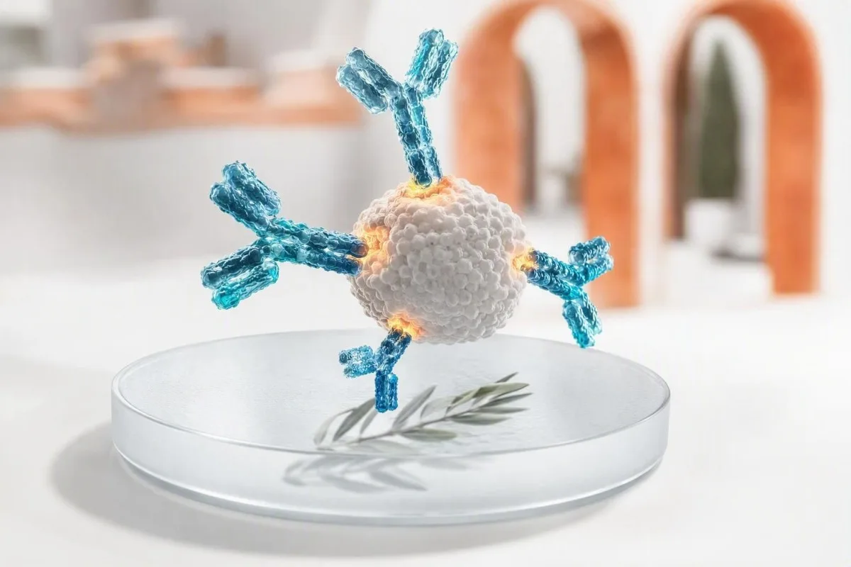
ئەنجامی ئەرێنیی دژەتەن مانای ئەوەیە کە سیستمی بەرگریی جەستەت ئامانجێکی دیاریکراوی ناسیوەتەوە؛ بە خۆی، ئەمە سەلمێنەری هەوکردنێکی چالاک یان نەخۆشی نییە. پۆلینی دژەتەن، شێوازی پشکنین، کات، نیشانەکان و پشکنینە هاوڕێکان دیاری دەکەن کە ئایا پێویستت بە دڵنیایی، دووبارەکردنەوەی پشکنین، پشتڕاستکردنەوە یان چاودێریی خێرای کلینیکی هەیە.


- ئەنجامی ئەرێنیی دژەتەن بە شێوەیەکی گشتی نیشانەی ناسینەوەی بەرگرییە، نەک هەمیشە نەخۆشیی ئێستا یان گوێزانەوە.
- IgM یان IgG ئاماژەیەکی کاتییە، نەک کاتژمێر: IgM لەوانەیە بۆ مانگێک بمێنێتەوە و IgG دەتوانێت زوو دەربکەوێت.
- دژەتەنەکانی کوتان دەتوانن پشکنینەکانی بەرگری ئەرێنی بکەن بێ ئەوەی هەوکردنی سروشتی نیشان بدەن.
- IgE ی تایبەت بە ماددە ئەلەرجیکەرەکان سەروو ٠.٣٥ kUA/L مانای هەستیارییە، بەڵام نیشانەکان دوای بەرکەوتن ئەلەرجییە کلینیکییەکان پشتڕاست دەکەنەوە.
- پۆزەتیڤی ANA لە %TP44T کەسی ساغدا ڕوودەدات بە تیتری نزم، بەتایبەتی لەگەڵ بەرزبوونەوەی تەمەن؛ نیشانەکان و دژەتەنە دیاریکراوەکان گرنگترن.
- چالاکی پشکنینی HIV هەمیشە پێویستی بە ڕێگەی پشتڕاستکردنەوەی تاقیگە هەیە پێش ئەوەی هیچ دەستنیشانکردنێک بکرێت.
- دژە tTG IgA تەنها کاتێک کەسەکە گلوتەم دەخوات و IgA ی تەواوی گونجاوی هەیە، بۆ هەڵسەنگاندنی سیلیاک دروستە.
- نەخۆشی/ئەلامەتە فوریتەکان وەک کێشەی هەناسەدان، تێکچوونی هۆش، زەردویی، ئازاری سنگ یان پێستی پڕ لە زیپکەی فراوان سەرووی ئەنجامی دژەتەنی ئاساییە.
ئەنجامی ئەرێنیی دژەتەن چی بە ڕاستی پێ دەڵێت
A پشکنینی دژەتەنی ئەرێنی مانای وایە تاقیکردنەوەکە پرۆتینی بەرگریی دۆزیوەتەوە کە بە ئامانجێک دەبەسترێتەوە؛ ne بە شێوەیەکی خۆکار مانای ئەوە نییە کە هەوکردنێکی چالاکت هەیە، یان نەخۆشییەکی ئۆتۆ-ئیمون، یان ئالێرژیت هەیە کە پێویستی بە چارەسەر بێت. هەنگاوی دواتر پشت بە جۆری دژەتەن، هێزی ئەنجامەکە، نیشانەکان، کاتی تێکەوتن و هەر ئانتیجینی هاوتا، PCR یان پشکنینەکانی ئەرکی ئەندامەکان دەبەستێت.
دژەتەنەکان پرۆتینی خانەکانی Bن کە دوای ڤاکسین، هەوکردن، تێکەوتن لەگەڵ ئالێرژین، ئۆتۆ-ئیمون، یان جاروبار بەهۆی هۆکارێکی قێزەونی کلینیکی دروست دەبن. ئەنجامێکی ئەرێنی وەڵامێکە بۆ پرسیارێکی تەسکی شیکاری—“ئایا بەستنەوە لەم سنوورەی تاقیگەوە دیاریکراوە؟”—لەبری پرسیارە کلینیکییە فراوانەکەی کە ئەمڕۆ لە جەستەتدا چی ڕوودەدات.
لە ١٥ ساڵ کارکردن لە بواری کلینیکیدا، هەڵەی باو بریتی بووە لە خوێندنەوەی “ئەرێنی” وەک دەستنیشانکردن. ڕێنمایی پراکتیکی دکتۆر تاماس کلاین ئەوەیە کە سەرەتا ئامانجەکە—ڤایرۆسێک، ئانتیجینی ڤاکسین، پرۆتینی خۆراک، ئەنزیمی دەرەکی، ئانتیجینی ناوەکی یان دەرمانێک—پاشان بپرسە ئایا پشکنینەکە تێکەوتنی پێشوو، ئۆرگانیزمی ئێستا، هەستیاری، یان تێکچوونی بەرگری دەستنیشان دەکات.
Kantesti AI یەک Analyzerê testa xwînê ya AI کە ئاڵای دژەتەنەکان لە تەنیشت ڕێکەوتەکان، پۆلی ئیمونوگلوبولین و ئەنجامە پەیوەندیدارەکانەوە دەخوێنێتەوە نەک مامەڵەکردن لەگەڵ یەک هێڵی ئەرێنی وەک بڕیارێک. ئەو چوارچێوەیە بەتایبەتی بەسوودە کاتێک نەخۆشەکان ڕاپۆرتەکان لە تاقیگەی جیاوازەوە بەراورد دەکەن؛; ئەنجامە کواڵتیڤ و کواینتیڤەکان تەنانەت کاتێک هەردووکیان ئەرێنی دەڵێن، ناکۆکن.
ئەرێنیی IgG یان IgM: بۆچی کات سادەتر نییە لەوەی دەردەکەوێت
IgM ئەرێنی دەتوانێت وەڵامدانەوەیەکی نوێی بەرگری نیشان بدات، لە کاتێکدا IgG بەدڵە زۆرجار نیشانەی تێکەوتنی پێشوو یان وەڵامدانەوەی ڤاکسینە؛ هیچ یەک لەم شێوازانە بە شێوەیەکی جێگیر نەخۆشییەک بە تەنها دیاری ناکەن. هەندێک هەوکردن IgG لە ماوەی ٧-١٤ ڕۆژدا بەرهەم دەهێنن، و IgM لەوانەیە بۆ ماوەی ٣-١٢ مانگ یان زیاتر ئاشکرا بمێنێتەوە.
IgM دژەتەنێکی گەورەی پێنج-یەکە، کە زۆرجار لە سەرەتاوە ئاشکرایە، بەڵام قەبارە و بەستنەوە فراوانەکەی زیاتر وای لێدەکات کە بەکتریاکان بخاتە ناو یەکتر؛ ڤایرۆسی ئێپشتاین-بار، پارڤۆڤایرۆسی B19، فاکتەری ڕێوماتیدی و هەندێک دژەتەنی ئۆتۆ-ئیمون دەتوانن هۆکاری وەڵامدانەوەی هەڵەی IgM بن؛ ئەنجامێکی کەم-ئاستی IgM ی تاکەکان شایستەی گومانکردنە زیاتر لە ترسان.
IgG بە شێوەیەکی گشتی دوای گەشەی بیرەوەری بەرگری بەردەوام دەبێت، هەندێک جار بۆ دەیان ساڵ. ئەنجامێکی ئەرێنی IgG دەتوانێت ڕەنگدانەوەی هەوکردنی منداڵی، ڤاکسینێکی نوێ، دژەتەنی ناچالاک لە ئیمونوگلوبولینی وریچکەی، یان گواستنەوەی دایک بۆ شیرپێداو بێت؛ ناتوانێت پارێزبەندی بسەلمێنێت مەگەر ئەو تاقیکردنەوەیە سنوورێکی پشتڕاستکراوی پارێزبەندی هەبێت.
نموونەی هاوتەریب دەتوانن زیاتر لە یەک کێشان سودیان هەبێت: چوار قات بەرزبوونەوەی تیتری دژەتەن لە نێوان نموونەکاندا کە نزیکەی ٢-٤ هەفتە لێک دوورن، پشتگیری لە هەوکردنی نوێ دەکات لە نەخۆشییە دیاریکراوەکاندا. ئەگەر ڕاپۆرتەکە IgG, IgA و IgM ی تەواو نیشان بدات، شێوازەکە بخوێنەوە نەک یەک ژمارە لەگەڵ ڕێبەری لێکدانەوەی ئینزیلۆبیولین.
کەی دژەتەنەکانی هەوکردن مانای نەخۆشیی چالاک دەدەن و کەی نادەن
دژەتەنەکانی نەخۆشی بە گشتی نیشانەی بەرکەوتنن، لە کاتێکدا PCR، پشکنینی ئه نتین، کەلتوور، یان زیادبوونی تیتر ئامرازی باشترن بۆ زانینی زۆرێک لە نەخۆشییە چالاکەکان. ئەنجامی ئەرێنیی دژەتەن بێ نیشانە زۆرجار مێژووییە، بەڵام ئیستیسنایەکەی بە هۆی جۆرە بەکتریایەکە و باری گشتی سیستمی بەرگری لەشەوە دەگۆڕێت.
بۆ هەوکردنی جگەر جۆری C، دژەتەنی ئەرێنی مانای بەرکەوتنە لە کاتێکدا، بەڵام تەنها RNAی HCV کە دەستنیشان دەکرێت، ڤایرۆسی ئێستا پشتڕاست دەکاتەوە. بۆ هەوکردنی جگەر جۆری B، کۆمەڵەکەی گرنگە: ئەنتاینی ڕووکراو، دژەتەنی ڕووکراو و دژەتەنی ناوەکییەکان نەخۆشی چالاک، نەخۆشی چاکبووەوە و بەرگری ڤاکسین باشتر جیا دەکەنەوە لە یەک نیشانەی ئەرێنی.
HIV نموونەیەکی ترە کە وشەکان زۆر گرنگن. شاشەی نەوەی چوارەمی ئەرێنی مانای دەستنیشانکردن نییە؛ تاقیگە بە پشکنینی جیاوازی HIV-1/HIV-2 و، کاتێک پێویست بێت، پشکنینی ترشی ناوکیی دێتە دوای. کەسێک کە چارەسەری پارێزگاری دوای بەرکەوتن وەردەگرێت پێویستی بە پشکنینی کاتی هەیە، وەک لە ڕێبەری پشکنینی HIV دوای PEP.
من نەخۆشێکی تەندروستم بینی کە ترسا بوو لە دژەتەنی تڕیپۆنێمی سیفلیس ئەرێنی دوای چارەسەرکردن ١٨ ساڵ پێشتر. پشکنینە تڕیپۆنێمییەکان بە گشتی بۆ ژیان ئەرێنی دەمێنێتەوە، لە کاتێکدا تیترەکانی RPR یان VDRL یارمەتی دەدەن بۆ قەیمکردنی چالاکی ئێستا و وەڵامدانەوە؛; نموونەکانی پشکنینی سیفلیس ئەم جیاوازییە روونتر دەکەنەوە.
چۆن دژەتەنەکانی کوتان جیاوازن لە دژەتەنەکانی دوای هەوکردن
دژەتەنەکانی پەیوەست بە ڤاکسین نیشان دەدەن کە سیستمی بەرگری وەڵامی ڤاکسینی داوەتەوە؛ ئەوانە مانای ئەوە نین کە ڤاکسینەکە بووەتە هۆی نەخۆشییەکی چالاک. ئەنجامی ئەرێنی هێشتا لە خوار سنوورێکی پارێزگاری ناسراو دەکەوێت، چونکە دژەتەنە پەیوەستکەرەکان و دژەتەنی بێ زیان کە رۆڵی چالاکیان هەیە وەک یەکتر نین.
ڕێژەکانی دژەتەنی ڕووکراوی هەوکردنی جگەر B 10 mIU/mL یان بەرزتر کە ١-٢ مانگ دوای زنجیرەیەکی ڤاکسینی نووسراو پشکنین کرابێت بە گشتی لە کەسانی بەرگریبەهێزدا پارێزەر دادەنرێت. دژەتەنی ناوکیی هەوکردنی جگەر B، دژەتەنی ناوکی هەوکردنی جگەر، بە ڤاکسینەکانی ئاسایی هەوکردنی جگەر B دروست ناکرێت؛ بوونی ئەمانە ئاماژەیە بۆ بەرکەوتنی سروشتی و دەبێت ببێتە هۆی لێکدانەوەیەکی تەواو.
IgGی کوکوولە بە گشتی پێش دووگیانی پشکنین دەکرێت چونکە ئاماژەیە بۆ بەرگری پێشوو، نە نەخۆشی کوکوولەی چالاک. تاقیگەکان سنوور دیاری دەکەن، و ئەنجامە سنووردارەکان بە گشتی وەک ناپارێزەر بۆ پلاندانانی پێش دووگیانی مامەڵەیان لەگەڵدا دەکرێت لەبری لێکدانەوەی نەخۆشی نوێ؛ کاتی نەخۆشی لە ڕێبەری ڕێبەری پشکنینی کوکوولە.
Kantesti AI یەک پلاتفۆرمی تێکست/وەشاندنی تاقیکردنی خوێنی AI کە جیاوازی دژەتەنی نموونەی ڤاکسین و دژەتەنی نموونەی نەخۆشی دەکات ئەگەر ڕاپۆرتەکە نیشانەکانی هاوڕێی پەیوەندیداری تێدابێت. پرسیارە کردارییەکە زۆرجار “ئایا من ئەرێنییم؟” نییە، بەڵکو “کام ئەنتاین پشکنین کرا، کەی ڤاکسین کرام، و ئایا ئەم ئەنجامە پلانی پارێزگاری ئێستای من دەگۆڕێت؟”
بۆچی ئەنجامی ئەرێنیی پشکنینی دژەتەنی ئەلەرجی مانای ئەلەرجی نەبێت
دژەتەنی تایبەتی هەستیاری IgE پشکنین نیشانەی هەستیارییە، واتە IgE هەستیارە بۆ مادەیەکی هەستیار؛ بەبێ مێژوویەکی هاوتا لە وەڵامدانەوەدا نەخۆشی ڕاستەقینەی هەستیاری دیاری ناکات. بەهایەک لە سەرووی 0.35 kUA/L سنوورێکی ئاسایی ئەرێنییە، سنوورێکی جیهانی نییە بۆ خۆلادان لە خۆراک یان دەرمان.
چیرۆکی پێش پشکنین گرنگترە لەوەی زۆر کەس پێی وایە. ئەگەر کەسێک هەفتانە گوێز بخوات بێ تووشبوون بە تا، پژمین، ڕشانەوە یان بێهۆشبوون، تەنها IgEی گوێزی کەم ئەرێنی نابێتە هۆی خۆلادان؛ سنووردارکردنی خۆراکی بێ سوود دەتوانێت زیانی خۆراکی و کۆمەڵایەتی دروست بکات.
ڕێنمایی خۆراکی NIAID-ی پاڵپشتی نیشانی داوە کە پشکنینی پێستی و IgE تایبەت خۆراکە هاندەرەکان دەستنیشان دەکەن بەڵام ناتوانن بە تەنیا نەخۆشی هەستیاری خۆراک دیاری بکەن (سامپسن و هاوکارانی، 2010). لە حاڵەتی دیاریکراودا، پزیشکی هەستیارییەکان پشکنینی چارەسەرکراوی پێکهاتە یان تاقیکردنەوەی دەمی خۆراکیی بە چاودێری پزیشکی، کە هێشتا ستانداردە بۆ چارەسەرکردنی نادڵنیایی.
IgE گشتی بە IU/mL پشکنین دەکرێت و دەکرێت لە eczema، بەرکەوتنی پارازیتی، جگەرەکێشان و چەندین حاڵەتی بەرگری لەشدا بەرز بێت؛ ناتوانێت هۆکاری هەستیاری دیاری بکات. پشکنینی پێکهاتە دەتوانێت جیاوازی نێوان پڕۆتینی نیشتەجێی جێگیر لەگەڵ پڕۆتینی پەیوەست بە پۆلێنی تێکەڵاو بکات، جیاوازییەک کە لە ڕێبەری ئێمەدا لێکۆڵینەوەی بۆ کراوە وتارێکی پشکنینی مۆلەکولیی ئەلێرژی.
بۆچی دژەتەنە خۆبەرگرییەکان پێویستیان بە نیشانە و ئاماژەی ئەندامەکانە
ئۆتۆمیون هاری هاری نەخۆشی بن. پۆزەتیف بە تەنها دەگمەنە یەکێک لە نەخۆشییە ئۆتۆمیونەکان بسەلمێنێت. پزیشکان کاتێک کاردەکەن کە شێوازی هاری لەگەڵ نیشانەکان، دەرئەنجامەکانی پشکنین، نیشانەکانی هەوکردن یان گۆڕانکارییەki شیاوی ئەندام یەک بگرێتەوە.
هاری دژە ناوکی، یان ANA, ، لە ڕێژەی کەمدا پۆزەتیڤە لە نزیکەی 13% خەڵک لە 1:80 و نزیکەی 20% لە 1:40 لە لێکۆڵینەوەی دانیشتوان، لەگەڵ ڕێژەی بەرزتر لە ژنان و بەساڵاچوان. شێوازێکی یەکگرتوو یان خاڵ خاڵ شێوازی لووس نادات؛ ANA باشترین بەکارهاتنی وەک پشکنینێکی دەروازەیە کاتێک وێنەی کلینیکی نەخۆشییە توێکڵدارەکان ڕاستە.
چوارچێوەی پۆلێنکردنی لووسی EULAR/ACRی 2019 ANA بە تیترێکی لانیکەم 1:80 وەک مەرجی سەرەکی بەکاردەهێنێت، پاشان پێویستی بە تایبەتمەندییە کلینیکی و ئیمونۆلۆجییە زیادکراوەکان هەیە (Aringer et al., 2019). پڕۆتینی نوێ لە میزدا، C3/C4ی کەم، سیتۆپینیا، هەوکردنی جومگەی قوڕقوشم یان تێکچوونی ڕەنگی پێست کە هەستیارە بەرامبەر ڕووناکی، زۆر زیاتر لە ANAیەکی لاواز ئاڵۆزییەکە دەگۆڕن.
Anti-dsDNA مانادارتر دەبێت کاتێک لەگەڵ کۆمپلێمێنت و دەرئەنجامەکانی گورچیلەدا یەک دەگرێتەوە، هەرچەندە میتۆدەکانی شیکردنەوە جیاوازن. ئێمە ڕێنمایی چاودێریکردنی anti-dsDNA û ڕوانینێکی گشتی پشکنینی کۆمپلێمێنت ڕوون بکەنەوە بۆچی پزیشکی پسپۆڕ لەوانەیە پشکنین دووبارە بکاتەوە لەسەر پلاتفۆرمێکی جیاواز.
دژەتەنە تایبەتەکانی ئەندامەکان: نموونەی ڕژێنی تووڵەڕژێن، سیلاک و گەدە
هاریی ئەندام-تایبەت ئاماژە بە هێرشی ئیمنی بۆسەر خانە دەدەن، بەڵام ئەوان هەمیشە نیشان نادەن کە ئایا ئەندامەکە ئێستا زیانی پێگەیشتووە یان پێویستی بە چارەسەرە. هاریی پەرۆکسیدازی تایرۆید، هاریی ترانسگلوتامینازی توێکڵ و هاریی فاکتەری ناوەکی هەر یەکەیان پێویستیان بە پشکنینی هاوڕێی جیاواز هەیە.
هاریی TPO لە تایرۆدایتی هاشیوموتۆدا باوە و دەتوانێت بۆ ساڵانێک بەردەوام بێت کاتێک TSH و free T4 ئاسایی دەمێنێتەوە. لە کەسێکی پێگەیشتووی نا دووگیان کە functionی تایرۆیدی ئاسایی هەیە، ئەنجامەکە بە شێوەیەکی گشتی پشتگیری لە چاودێریکردنی TSHی دورەیی دەکات نەک چارەسەری هۆرمۆنی تایرۆید؛ دووگیانی، پلانی زاوزێ و نیشانەکان دەتوانن ئەو ڕێگەیە بگۆڕن.
ئەزمونێکی tTG-IgA تەنها ئەگەر کەسەکە هێشتا گلوتینی بەکارهێناوە و IgAی گشتی بەسە، ئاماژە بە نەخۆشی سیلیاک دەکات. کەمیی IgAی هەڵبژێردراو دەتوانێت tTG-IgA بە هەڵە نەرێنی بکات، بۆیە پزیشکان دەمرەکانی گلوتامی دیئامیدی basi IgAی یان پشکنینی tTG بەکاردەهێنن کاتێک IgAی گشتی کەمە؛ سەیری ڕوونکردنەوەمان بکە لەسەر کەمبوونی IgA.
هاریی فاکتەری ناوەکی بە ڕێژەیەکی دیاریکراو بێهێزە بەڵام بە هێزی بەرز نییە بۆ کەمخوێنیی پرینیشیۆس. کاتێک لەگەڵ B12ی کەم، مێتیلمالۆنیک ئەسیدی بەرزبووەوە و ماکرۆسایتۆسیدا پۆزەتیڤە، شێوازەکە ڕازیکەرە؛ بێ ئەو شێوازە، من لەبری نەخۆش، لەبری نەخۆش، لەبری ناونیشانی تاقیگە چارەسەر دەکەم. پشکنینی کەمخوێنیی پرینیشیۆس ڕێبازی پشتڕاستکردنەوەی ئاسایی نیشان دەدات.
چی هۆکاری ئەنجامی هەڵەی ئەرێنیی دژەتەنە
ئەنجامی هاریی هەڵەیی پۆزەتیڤ کاتێک دروست دەبێت کە شیکارەکە پەیوەستبوونی نەویستراو دەستنیشان دەکات، نەک کاتێک تاقیگە سادە “هەڵەی کردووە”. هاریی کاریگەر، فاکتەری ڕێوماتۆیدی، هاندانی نوێی ئیمنی، پلاسما ئیمونوگڵوبولین و کەمبوونی پێش-پشکنینی هۆکارەکانی باون.
تایبەتمەندی کەمەڵی نییە. پشکنینێک کە 99% تایبەتمەندی هەیە نزیکەی 10 ئەنجامی هەڵەیی پۆزەتیڤ بۆ هەزار کەسی ڕاستەقینە نەرێنی تاقیکراو دروست دەکات؛ کاتێک حاڵەتەکە دەگمەنە، ئەو ئەنجامانە دەتوانن ژمارەی پۆزەتیڤە ڕاستەقینەکان تێپەڕێنن. ئەمەش هۆکارێکە بۆ ئەوەی پشکنینی بەرفراوان لە کەسێکدا بێ نیشانە زۆرجار بەشێوەیەکی زیاتر لێکتێنەگەیشتن دروست دەکات نەک دڵنیایی.
بایوتین بەناوبانگە بۆ تێکدانی هەندێک لە پشکنینەکانی هۆرمۆن و دڵ، لەکاتێکدا هاریی هیترۆفایل و فاکتەری ڕێوماتۆیدی دەتوانن تێکبەری هەندێک لە ئیمونۆئەسایکان بن. تاقیگە دەتوانێت جاروبارە نموونەکە بچووک بکاتەوە، ڕێگر بەکاربهێنێت، ئەنجامەکە لەسەر شیکارێکی تر دووبارە بکاتەوە، یان میتۆدێکی پشتڕاستکەرەوەی وردتر ئەنجام بدات.
ئەنجامێکی پۆزەتیڤ کە لەگەڵ وێنەیەکی تەواو بێ نیشانەی کلینیکی ناکۆکە شتێک نییە پشتگوێ بخرێت؛ هۆکارێکە بۆ پشتڕاستکردنەوە. Kantesti AI بەراورد دەکات لە نێوان نیشانە ناکۆکەکان و ڕێکەوتەکانی پشکنین، و ئێمە چکلیست دقتِ نتیجهٔ آزمایشگاهی دەتوانێت یارمەتیت بدات بۆ تۆمارکردنی زیادکراوەکان، ئینفیوژنەکانی دوایی و نەخۆشییەکان پێش قسەکردن لەگەڵ پزیشکی داواکار.
ئەنجامی پشکنینی دژەتەن ئەرێنییە بەڵام بێ نیشانەیە: پلانێکی پراکتیکی بۆ تریج
کاتێک پشکنینی هاری پۆزەتیڤە بەڵام تۆ باش هەست دەکەیت، زۆربەی خەڵک پێویستیان بە ڕوونکردنەوە هەیە نەک چارەسەری بەپەلە. هەنگاوە سەلامەتەکان بریتیین لە پشتڕاستکردنەوەی شیکارە وردەکە، پشکنینی ڕێکەوتەکانی تیشکدان و کوتان، و گەڕان بەدوای پشکنینی هاوڕێدا کە نەخۆشی یان تووشبوونی ئەندامی ئێستا پشتڕاست دەکاتەوە.
دەستپێ بکە لەگەڵ خۆی ڕاپۆرتەکە: ناو، پۆلی ئەنتی بادی، ژمارە یان تایتەر، بازنەی ڕێنوێنی، میتۆدی تاقیگە و ڕێکەوتی کۆکردنەوە تۆمار بکە. “ڕیئاکتیڤ” دەتوانێت مانای ڕێژەی سیگناڵ بۆ سنوور کە بە زەحمەت سەرووی 1.0 بێت یان ئەنجامێکی زۆر بەهێزتر بێت؛ تاقیگەکان سنووری یەکسان بەکار ناهێنن، و قەبارەکە تەنها جاروبارە ڕێگە بە گومانە کلینیکیەکان دەدات.
پاشان، بپرسە ئایا تاقیکردنەوەی نەخۆشی چالاک بوونی هەیە. نموونەکان بریتیین لە PCR بۆ هەوکردنی جگەر C، RPR ی دووپات بۆ نەخۆشیە سێفیلیس، پشکنینی پیسایی یان هەناسە بۆ H.pylori ی چالاک، TSH/free T4 بۆ ئەنتی بادی غوددە، و پشکنینی میز پلس کریاتینین بۆ نیگەرانی گورچیلەی پەیوەست بە لووس.
لە 2026/9/8 ەوە، من دانادانانم وەرنەگرت بۆ دەستپێکردنی خۆبەخۆی دەرمانەکانی دژە بەکتریایی، دژە ڤایرۆس، خۆراکە کەم گلووتینەکان، دەرمانی غوددە یان خۆجنکاندن تەنها لە ئەنجامێکی ئەنتی بادی یەکپارچە. PDF ەکە ڕەسەنەکە بپارێزە، بنەمای خۆت لەگەڵ پەسەندکردنی لابراتۆری بە شێوەی درێژخایەن, تۆمار بکە، و پێداچوونەوەی ئاسایی ڕێک بخە ئەگەر نیشانە سوورەکان بوونیان هەبێت.
پشکنینە پشتڕاستکەرەوەکان کە هەنگاوی داهاتوو دەگۆڕن
باشترین تاقیکردنەوەی پشتڕاستکردنەوە پشت دەبەستێت بەوەی کە ئەنتی بادیەکە چی ئامانج دەگرێت: دۆزینەوەی مۆلیکولی زۆرێک لە هەوکردنەکان پشتڕاست دەکاتەوە، تاقیکردنەوەی فانکشنیی ئەلەرژی ڕوون دەکاتەوە، و تاقیکردنەوەی توانای ئەندام הערساندن کاریگەری ئۆتۆئیمونیش دیاری دەکات. دووبارەکردنەوەی هەمان تاقیکردنەوەی ئەنتی بادی تەنها ئەوکاتە بەسوودە کە گۆڕانی چاوەڕوانکراو بە تێپەڕبوونی کات مانای کلینیکی هەبێت.
بۆ تاقیکردنەوەی HIV ی ئەرێنی، ڕێگای پشتڕاستکردنەوەی ڕیلێکس بریتییە لە تاقیکردنەوەی جیاوازی دواتر تاقیکردنەوەی ترشی ناوکی HIV ئەگەر ئەنجامەکان ناسەقامگیر بن. بۆ ئەنتی بادی C ی هەوکردنی جگەر، HCV RNA بەدواداچوونی یەکلاکەرەوەیە؛ بۆ ئەنتی بادی B ی ناوەکی ئەرێنی، پۆشی کاردانەوە، ئەنتی بادی پۆش و جاروبارە HBV DNA ڕەنگەکە ڕوون دەکەنەوە.
بۆ گومانی نەخۆشی زەردەویی، بڕیاری بایۆپسی دەبێت لە کاتێکدا کە ڕووبەڕووبوونەوەی گلووتین بەس بێت بۆ تاقیکردنەوە. بۆ گومانی ئەلەرژی خۆراک، پزیشکی ئەلەرژی مێژووی ڕووبەڕووبوونەوە، ئاستی IgE ی تایبەت، پێکهاتەی ڕەنگ و مەترسی هەڵمەتەکە لەبەرچاو دەگرێت — نا پانێلێک لە ئەنجامە نەرێنیە کەمەکان کە بەبێ پرسیارێکی کلینیکی داوا کرابن.
Kantesti AI یەک ئامێری توێژینەوەی تاقیکردنەوەی خوێن بە پشتبەستن بە AI کە دەتوانێت ئەم نیشانە پەیوەستەکان ڕێک بخات بۆ لیستێکی پرسیار کە ئامادەبێت بۆ پزیشک، بەڵام جێگای تاقیکردنەوەی پشتڕاستکردنەوە یان دەستنیشانکردن ناگرێتەوە. ئەگەر گومان لەوە دەکەیت کە چەندە پشت بە ئەنجامێکی دەرەوە بدەیت، ئۆستانداردەکانی ڕەسەنکردنی پزیشکی باس لەوە دەکەن کە بۆچی پشتڕاستکردنەوە و چاودێری کلینیکی گرنگن.
نیشانەکانی کە ئەنجامی ئەرێنیی دژەتەن گرنگتر دەکەن
ئەنجامێکی ئەنتی بادی ئەرێنی نیشانەی گرنگی دەدات کاتێک لەگەڵ کێشەی هەناسەدان، هەڵئاوسانی دەم و قوڕگ، بێهۆشبوون، شڵەژان، ئازاری سنگ، زەردی، پشێویەک بە خێرایی بڵاوبووەوە، یاخود لاوازی نوێی دەماری. نیشانەکان — ژمارەی ئەنتی بادیەکە بە تەنها — گرنگیەکە دیاری دەکەن.
گومانی ئەنەفیلێکسیا حاڵەتێکی لەناکاوە: خێزانی تووشبووی زەردەویی لەناکاو لەگەڵ دەنگی ناڕوون، تەنگی قوڕگ، ڕشانەوەی بەردەوام، بێهۆشی یان نزمی پەستانی خوێن دوای ڕووبەڕووبوونەوە پێویستی بە خزمەتگوزاری فریاکەوتن هەیە و ئەگەر بوونی هەبێت ئەدرینالین پێویستە. ئەنجامێکی ئەرێنی IgE ی خۆراک یان ژەهر کە مانگێک لەمەوبەر بووە نە پێشبینی دەکات و نە ڕەتیدەکاتەوە ئەو مەترسییە immediate ە.
زەردییەکی نوێ لەگەڵ میزێکی تاریک، ئازاری زگ، تا یان شڵەژان دەبێت هەمان ڕۆژ הערساندن بکرێت چ ئەنتی بادیەکانی هەوکردنی جگەری ڤایرۆسی ئەرێنی بن یان نا. ئەنزیمی جگەر، بیلی روبین، INR و تاقیکردنەوەی pathogens ی ڕاستەوخۆ یارمەتی جیاکردنەوەی مێژووی بەرگری دەدەن لە نەخۆشی چالاکی جگەر و زراو؛; اجزای پنل کبدی context ێکی بەسوودن.
نیگەرانی ئۆتۆئیمون کات-هەستیار دەبێت لەگەڵ خوێن هاتن لە قوڕگ، کورتەهەناسە، بەرزبوونی خێرای کریاتینین، سکچوونی خوێناوی، گۆڕانی بینین، لەرز یان لاوازی نوێی یەک لایەنە. من فێربووم کە پشت بە دڵنیایی دان بە کەسێک لەسەر بنەمای تایتەرێکی ئەنتی بادی سنووردار کاتێک ڕەوتی نیشانەکانی بە ڕوونی تێک دەچێت.
ئایا ژمارەیەکی زیاتری دژەتەن مانای نەخۆشیی سەختترە؟
ئیندێکس یان تایتەرێکی ئەنتی بادی بەرزتر جاروبارە گومان زیاتر دەکات کە ئەنجامەکە ڕاست بێت، بەڵام بەزۆری قەبارەی نەخۆشیەکە ناپێوێت. نرخی تاقیکردنەوەکان تایبەت بە میتۆدن، و ئەنجامێکی 80 AU/mL ناکرێت بە ڕاستەوخۆ بە 80 U/mL ەوە لە تاقیگەیەکی ترەوە بەراورد بکرێت.
لە نەخۆشی ئۆتۆئیمون، گۆڕانکاری anti-dsDNA دەکرێت جاروبارە دوای چالاکی لووس بکەوێت، بەتایبەتی کاتێک بەردەوام بە هەمان میتۆد ئەنجام دەدرێت و لەگەڵ کۆمێنت و پرۆتینی میزدا لێک دەدرێتەوە. بەڵام، تایتەری ANA، پێوانەیەکی خراپە بۆ قەبارەی لووس و نابێت بەدوایدا بگەڕێت بەدووبارە لە کەسێکی جێگیردا.
بۆ هەوکردنەکان، سیگناڵێکی بەهێزی ئەنتی بادی دەکرێت پشتگیری لە ڕووبەڕووبوونەوەی پێشوو بکات بەڵام بە زەحمەت نەخۆشی دەگەیەنێت. تاقیکردنەوەی ئەنتی بادی بێ کاریگەر تایبەتین و سنوورەکانی پارێزەرییان دەکرێت جیاواز بن بەپێی پەتایەکە، تەمەن، بەرهەمی ڤاکسین و بێ بەرگری؛ تاقیکردنەوەیەکی سادەی بازرگانی IgG پاسپۆرتی بەرگری تاکەکەسی نییە.
کاتێک ڕاپۆرتەکان بەراورد دەکەیت، تاقیگە، تاقیکردنەوە و یەکەکان ببینە. گۆڕانکاریەکی ڕاستەقینەی بایۆلۆجی دەبێت تێپەڕبکات بەسەر جیاوازی ئاسایی وردبین و ڕۆژانە، ئەوەش هۆکارێکە بۆ ئەوەی کە ڕێبەری بەراوردکردنی پشکنینی خوێن ئێمە سەرنج دەخەینە سەر ڕێکەوتەکان و میتۆدەکان نەک تەنها ئاماژە ڕەنگینەکان.
دووگیانی، منداڵان و کەمبوونەوەی بەرگری، شیکارکردنەوەی ئەنجامەکە دەگۆڕێت
دووگیانی، منداڵبوون و چارەسەری کەمبەرگری دەکرێت هەردوو بەرھەمھێنانی ئەنتی بادی و سەلامەتی چاوەڕوانی بۆ پشتڕاستکردنەوە بگۆڕێت. لەم گرووپانەدا، ئەنجامێکی ئەرێنی دەکرێت ئەنتی بادی دایکی، کەمبوونەوەی وەڵامدانەوەی ڤاکسین، مەترسی دووبارە چالاکبوون یان پێویستی بە ڕاوێژی زووتری پسپۆڕ نیشان بدات.
IgG ی دایک دەگوازرێتەوە بۆ منداڵ و بۆ ماوەی چەند مانگێک دەمێنێتەوە، لەکاتێکدا IgM ی دایک بەگشتی ناگوازرێتەوە. لەبەر ئەوە، پۆزەتیڤی تەنها IgG ی منداڵ زۆرجار وەڵامی بەرگری دایکی نیشان دەدات، لە کاتێکدا IgM ی تایبەت بەو میکرۆب یان دەرکەوتنی ڕاستەوخۆ شایانی هەڵسەنگاندنی وردترە بەپێی جۆری هەوکردن و تەمەن.
لە کاتی دووگیانیدا، ئەنجامی پۆزەتیڤی دژەتەنی ڕوبێلا یان پارڤۆ ڤایرۆس بە بەرواری تووشبوون، تاقیکردنەوەی توندی و نموونەی دووبارە هەڵدەسنگنرێت نەک تەنها IgM. IgM ی هەڵەی پۆزەتیڤ کێشەیەکی ناسراوە، و هۆشداری ناحەق دەکرێت ببێتە هۆی لێکۆڵینەوەی ناپێویست؛ بچوونە ژوورەوەی پسپۆڕی نەخۆشییەکانی ژنان یان نەخۆشییە گوازراوەکان هەنگاوێکی ژیرانەیە کاتێک تووشبوونەکە جێی متمانەیە.
کەسانی وەرگرتنی ڕیتوکسیماپ، ستیرۆیدی بەرز، کیمیاوی یان دەرمانی گواستنەوەی ئەندام دەکرێت وەڵامی دژەتەنی لاواز یان نەبوو هەبێت سەرەڕای هەوکردن یان کوتان. بۆ بڕیاری خێزان،, سنوورەکانی لێکدانەوەی AI ی منداڵان û دەستەی ڕاوێژکاری پزیشکی Kantesti پێویستی بە وردبینی لەلایەن پزیشکەوە بەهێز دەکات.
چۆن ئامادەبیت بۆ پێداچوونەوەیەکی سوودبەخشی کلینیککار بۆ ئەنجامێکی ئەرێنی
ڕاپۆرتی تەواوی تاقیگاکە، هۆکاری داواکردنی تاقیکردنەوەکە، بەرواری نیشانەکان یان تووشبوون، مێژووی کوتان، دەرمانەکان و ئەنجامە پێشووەکان بهێنە. ئەم زانیارییانە دەتوانن ئەنجامێکی پۆزەتیڤی دژەتەنی بەدیاریکراوی نادیار بگۆڕن بۆ هەنگاوێکی ڕوون لەماوەی یەک سەردان.
پێنج ڕاستی بنووسە پێش سەردانەکە: یەکەم ڕۆژی نیشانەکان، دوایین جار تووشبوون، بەرواری کوتان، دەرمانی کەمکەرەوەی بەرگری یان وەرگرتنی گلۆبوڵینی ئیمون، و چاوخشاندنەوەی نیشانەکان باشتر دەبن یان خراپتر. تاوەکێک کە سێ ڕۆژ پێش وەرگرتنی نموونە دەستیپێکردووە زۆر جیاوازتر لە ماندوویەتییەک کە شەش مانگ لەمەوبەر دەستیپێکردووە هەڵدەسنگنرێت.
سێ پرسیاری چڕ بنووسە: “ئەم تاقیکردنەوەیە چی دەستنیشان دەکات؟”، “کام تاقیکردنەوە نەخۆشی چالاک یان کاریگەری ئەندام دیاری دەکات؟”، و “کام ئەنجام چارەسەرەکە دەگۆڕێت؟” دکتۆر تۆماس کلاین پێی وایە ئەم پرسیارانە پەنێڵی دووبارە بێسوود کەم دەکەنەوە و یارمەتی نەخۆشەکان دەدات بە چوارچێوەیەکی کاتی دیاریکراو ڕۆیشتن - ئەمڕۆ، ١-٢ هەفتە، یان چاودێری ئاسایی.
Kantesti AI پشتگیری لە وردبینی ڕاپۆرتی پارێزراو دەکات بە زیاتر لە 75 زمان و دەتوانێت تاقیکردنەوەی هاوشانەی ونبوو لە PDF ێکی وێنەگیراو یان بارکراو لە ماوەی 60 چرکەدا پیشان بدات. ئێمە ڕێنمایی تەکنەلۆژیای AI ڕێگەی گونجاندنی چوارچێوەکە ڕووندەکاتەوە، لە کاتێکدا تیمی کلینیکی ئێمە چاودێری پزیشکی پشت ئەنجامدان دەکات بۆ لێکدانەوەی ڕووبەڕووی نەخۆش.
Pirsên Pir tên Pirsîn
واتهمان چییە ئەنجامێکی پۆزەتیڤی دژەتەن (antibody) هەبێت و هیچ نیشانەیەکی نەخۆشیم نەبێت؟
ئەنجامێکی ئەرێنیی دژەتەن لە کاتێکدا کە هیچ نیشانەیەک بوونی نییە، زۆربەی جار ئاماژەیە بۆ بەرکەوتنێکی پێشوو، کوتان، هەستیاری یان ئەنجامێک کە پێویستی بە پشتڕاستکردنەوە هەیە نەک نەخۆشییەکی چالاک. هەنگاوی دواتر پشت دەبەستێت بە ئامانجەکە: دژەتەنی هەوکردنی جگەر (hepatitis C) پێویستی بە RNAی HCV هەیە، شاشەیەکی کاراکتەری HIV پێویستی بە ئەلگۆریتمی پشتڕاستکردنەوەی تاقیگەیە، و دژەتەنەکانی غوددەی خۆرەپەپکە (thyroid antibodies) پێویستیان بە TSH و T4ی ئازاد هەیە. ئەنجامێکی تاک و کەم-ئەرێنی بڕواپێنەرترە کاتێک هیچ بەرکەوتنێکی گونجاو یان مێژووی نیشانەیەک بوونی نییە. لە حاڵەتی نوێی زەردویی، سەختی هەناسەدان، تێکچوونی هۆش، ئازاری سنگ یان پەرەسەندنی خێرای پێستدا، خێرا سەردانی پزیشک بکە.
ئایا پۆزەتیڤی IgM و نەرێنیی IgG هەمیشە مانای تووشبوونێکی نوێیە؟
ئای جی ئێم (IgM) ئەرێنی و ئای جی جی (IgG) نەرێنی لەوانەیە لە سەرەتای تووشبوونێکدا ڕووبدات، بەڵام هەمیشە سەلمێنەری تووشبوونی نوێ نییە. ئای جی ئێم (IgM) لەوانەیە لەگەڵ پرۆتینە بێ پەیوەندەکانی دیکەی بەرگریدا کاردانەوەی هاوبەش بکات، هەروەها هەندێک پشکنین ئەنجامی نەرێنی درۆ بۆ ئای جی ئێم (IgM) زیاتر لە ئای جی جی (IgG) دەردەخەن. پزیشکان لەوانەیە پشکنینی خوێن دووپات بکەنەوە دوای 1-2 هەفتە، داوای پشکنینی PCR یان ئهنتيجين (antigen) بکەن، یان پشکنینێکی پشتڕاستکەرەوەی تایبەتتر بەکاربهێنن. لێکدانەوەی دروست هەروەها پشت بە ماوەی نەخۆشییەکە و جۆرە بەکتریا یان ڤایرۆسەکە دەبەستێت کە پشکنینەکەی بۆ کراوە.
ئایا کوتانێک دەتوانێت پشکنینی دژەتەن ئەرێنی بکات؟
بەڵێ، ڤاکسینەکان بە شێوەیەکی گشتی پشکنینی دژەتەنەکان (antibody tests) ئەرێنی دەکەن بۆ ئەو پارتۆجینەی کە لە ڤاکسینەکەدایە. بۆ هەوکردنی جگەر ب، ئاستێکی دژەتەنی HBs (anti-HBs)ی لانیکەم 10 mIU/mL کە 1-2 مانگ دوای تەواوکردنی زنجیرەی ڤاکسینەکە ئەنجام بدرێت بە گشتی نیشانەی وەڵامدانەوەی پارێزگاری ڤاکسینە لە کەسانی ئاساییدا. ڤاکسینکردنی ئاسایی هەوکردنی جگەر ب دژەتەنی HBc (anti-HBc) دروست ناکات، بۆیە ئەرێنیبوونی دژەتەنی ناوکی (core-antibody) ئاماژەیە بۆ بەرکەوتنی سروشتی نەک تەنها ڤاکسین. ئەرێنیبوونی پەیوەندیدار بە ڤاکسین مانای ئەوە نییە کە تۆ هەوکردنێکی چالاکت هەیە.
دەکرێت پشکنینی ئەنتیبۆدی ئۆتۆimmine ئەرێنی بێت و تەندروست بیت؟
بەڵێ، کەسانی تەندروست دەتوانن ئەنتیبۆدی ئۆتۆimmuned positiveیان هەبێت، بە تایبەت ئەنجامی نزم ALT. ALT لە نزیکەی 13% خەڵکدا بە تایتڵی 1:80 و لە دەوروبەری 20%دا بە 1:40 دەستنیشان دەکرێت، بۆیە تاقیکردنەوەیەکی سەربەخۆی لووس نییە. نیشانەکانی وەک هەوکردنی جومگە، برینی دەم، پەڵەی شێرپەنجەیی، کەمبوونی تەواوکەر، پرۆتین لە میزدا یان ژمارەی نائاسایی خوێن دیاری دەکات کە ئایا پێویستە هەڵسەنگاندنی پسپۆڕانە بکرێت. ئەنجامێکی ئەرێنی دەبێت لە چوارچێوەی هۆکاری داواکردنی تاقیکردنەوەکەدا لێکبدرێتەوە.
مانای پۆزەتیڤ بوونی پشکنینی خوێن بۆ IgE ئەوەیە کە دەبێت خۆم لەو خۆراکە بپارێزم؟
نا، ئەرێتی ئی جێی تایبەت بە خۆراک مانای هەستیارییە و سەلمێنەری ئەوە نییە کە خواردنی خۆراکەکە هۆکاری کاردانەوەی ئەلێرژیک بێت. زۆرێک لە تاقیگەکان ئەنجامەکانی سەرووی 0.35 kUA/L بە ئەرێنی ناودەبەن، بەڵام ئەو سنوورە بۆ دۆزینەوە دیزاین کراوە نەک بڕیاردانی گشتگیر بۆ خۆلێکردنەوە. کەسێک کە بەردەوام خۆراکەکە دەخوات بەبێ گرژبوونی پێست، پژمە، ڕشانەوەی دووبارە یان بێهێزی، نابێت بە شێوەیەکی خۆکارانە لە ژەهری بکات. پسپۆڕی نەخۆشییە ئەلێرژییەکان دەتوانێت مێژووی کاردانەوە، تاقیکردنەوەی پێکهاتەکان و هەندێک جار تاقیکردنەوەیەکی چاودێریکراوی خواردنی زارەکی بەکاربهێنێت بۆ ڕوون کردنەوەی مەترسی.
چەند ماوەیەک ئیستیجابات (Antibodies) ئەرێنی دەمێننەوە دوای تووشبوون؟
ماوەی دژەتەنەکان بە شێوەیەکی زۆر جیاوازە بەپێی جۆری هەوکردن و پۆلینی دژەتەنەکان. IgM لە ماوەی چەند هەفتەیەکدا لەناو دەچێت بەڵام دەتوانێت بۆ ماوەی ٣–١٢ مانگ یان زیاتر بەردەوام بێت، لە کاتێکدا IgG دەتوانێت بۆ ساڵان یان هەتاهەتایی پێوانە بکرێت. دژەتەنەکانی بریاسی (syphilis) ترپۆنێمی زۆرجار دوای چارەسەرێکی سەرکەوتوو بۆ ماوەی ژیان ئەرێنی دەمێنێتەوە، لە کاتێکدا تیتری RPR یان VDRL بۆ چاودێرکردنی چالاکی بەکاردەهێنرێت. بۆیە ئەنجامێکی ئەرێنی دژەتەن ناتوانێت بە شێوەیەکی جێی متمانە پێتان بڵێت کەی تووشبوونێک ڕوویداوە بێ زانیاری دیکەی کلینیکی و تاقیگەیی.
ئەمڕۆ AI-پاوەرد لەسەر تاقیکردنەوەی خوێن بەدەست بهێنە
بە یارمەتی زیاتر لە 2 ملیۆن بەکارهێنەر لە هەموو جیهاندا کە Kantesti دەستپێدەکەن بۆ تاقیکردنەوەی لابراتۆری ڕاست و بەهێز لە کاتێکی کەم. ڕەخنەی تاقیکردنەوەی خوێنت بنێرە و تفسیرێکی تەواو لە 15,000+ نیشانەی زیستی (biomarkers) لە ماوەی چرکەکاندا وەرگرە.
📚 توێژینەوە سەرچاوە پەیوەندیدارەکان
کلاین، ت.، میچێڵ، س.، & وێبەر، ه. (2026). AI ێکی فرەزمان یارمەتی دەدات بۆ بڕیاردانی خێرا لەسەر triageی سەرەتایی Hantavirus: دیزاین، پشتڕاستکردنەوەی ئەندازەیی، و جێبەجێکردنی لە واقیعدا بۆ زیاتر لە 50,000 ڕاپۆرتی خوێن کە لێکدراونەتەوە. Figshare. https://www.researchgate.net/. Kantesti توێژینەوەی پزیشکی AI.
کلاین، ت.، میچێڵ، س.، & وێبەر، ه. (2026). تاقیکردنەوەیەکی بازبەند-بنەمای ئۆتۆماتیکی تەکنیکی پێش-تۆمارکراو لەسەر AI ی لێکدانەوەی تاقیکردنەوەی خوێن Kantesti لەسەر 100,000 حاڵەتی تاقیکردنەوەی سینتتیک. Figshare. https://www.academia.edu/. Kantesti توێژینەوەی پزیشکی AI.
📖 سەرچاوەی پزیشکی دەرەکی
⚕️ Daxuyaniya Bijîşkî
ئەم مادەیە تەنها بۆ. I think I must continue but user expects all items.
سەنگەری باوەڕپێکردن E-E-A-T
Tecribe
ڕەوی پشکنینی کلینیکی لەلایەن پزیشکەوە بۆ ڕێکخستنی ڕووداوەکانی لێکدانەوەی ئازمایش.
Pisporî
گرێدانی لابراتۆرییەی پزیشکی بەوەی چۆن بایۆمارکەرەکان لە کۆنتێکستی کلینیکی دەگۆڕن.
Desthilatdarî
نووسراوە لەلایەن د. توماس کلاین بە پشکنینی لەلایەن د. سارا میچێڵ و پڕۆف. د. هانس وێبەر.
Bawerî
لێکدانەوە بە بنەمای دڵنیابوون (Evidence-based) بە ڕێڕەوی دوایینەوەی ڕوون بۆ کەمکردنەوەی هەست بە ترس/هەڵوەشاندن.