Η νόσος Gaucher διαγιγνώσκεται με την απόδειξη μειωμένης δραστηριότητας της γλυκοκερεβροσιδάσης, συνήθως στα λευκοκύτταρα, ενώ η ανάλυση του GBA1 παρέχει γενετική επιβεβαίωση. Ένα χαμηλό αποτέλεσμα προσυμπτωματικού ελέγχου σε αποξηραμένη σταγόνα αίματος χρειάζεται διερεύνηση· η κατάσταση φορέα και ένα αυξημένο αποτέλεσμα lyso-Gb1 δεν ισοδυναμούν με διάγνωση.

Βασικά συμπεράσματα
- Προσυμπτωματικός έλεγχος σε αποξηραμένη σταγόνα αίματος εντοπίζει πιθανή ενζυμική ανεπάρκεια· 1 παθολογικό αποτέλεσμα προσυμπτωματικού ελέγχου δεν τεκμηριώνει τη νόσο Gaucher.
- Επιβεβαιωτικός ενζυμικός έλεγχος συνήθως μετρά τη γλυκοκερεβροσιδάση στα λευκοκύτταρα· δραστηριότητα κάτω από περίπου 10–15% της μέσης φυσιολογικής δραστηριότητας είναι χαρακτηριστική, με την επιφύλαξη της επικυρωμένης μεθόδου του εργαστηρίου.
- Επιβεβαίωση μέσω GBA1 εντοπίζει παθογόνες παραλλαγές και στα δύο αντίγραφα του γονιδίου· 2 παραλλαγές πρέπει να βρίσκονται σε διαφορετικά αντίγραφα για να τεκμηριωθεί αλληλόμορφος γονότυπος.
- Κατάσταση φορέα συνήθως σημαίνει 1 παθογόνο παραλλαγή του GBA1, όχι νόσο Gaucher· η ενζυμική δραστηριότητα αλληλεπικαλύπτεται μεταξύ φορέων και μη φορέων.
- Lyso-Gb1 υποστηρίζει τη διάγνωση και την παρακολούθηση, αλλά το δημοσιευμένο ερευνητικό όριο των 12 ng/mL δεν αποτελεί καθολικό όριο για όλα τα εργαστήρια.
- Αβέβαιες παραλλαγές απαιτούν εξειδικευμένη ερμηνεία· 2 παραλλαγές αβέβαιης κλινικής σημασίας δεν αρκούν από μόνες τους για να τεκμηριώσουν νόσο Gaucher.
- Οικογενειακός προγραμματισμός περιλαμβάνει πιθανότητα 25% για παιδί με τη νόσο σε κάθε κύηση, όταν και οι δύο γονείς φέρουν μια παθογόνο παραλλαγή στο ίδιο υπολειπόμενο γονίδιο.
- Η βαρύτητα της νόσου δεν μπορεί να υπολογιστεί από ένα μόνο ποσοστό ενζυμικής δραστηριότητας, αποτέλεσμα GBA1 ή μέτρηση lyso-Gb1.
Τι επιβεβαιώνει τη νόσο Gaucher μετά από ένα αποτέλεσμα προσυμπτωματικού ελέγχου;
Η νόσος Gaucher επιβεβαιώνεται με μειωμένη δραστηριότητα της γλυκοκερεβροσιδάσης σε κατάλληλο διαγνωστικό δείγμα, με υποστήριξη από τον έλεγχο GBA1. Ένα παθολογικό αποτέλεσμα σε αποξηραμένη σταγόνα αίματος συνήθως οδηγεί σε έλεγχο ενζύμου σε λευκοκύτταρα και ανάλυση και των δύο αντιγράφων του GBA1· τα συμπτώματα βοηθούν στην αξιολόγηση της προσβολής από τη νόσο, αλλά δεν είναι απαραίτητα για έγκαιρη διάγνωση.
Η διαγνωστική πορεία περιλαμβάνει 3 διακριτά ερωτήματα: εντόπισε ο προσυμπτωματικός έλεγχος πιθανή ανεπάρκεια, την επιβεβαιώνει αξιόπιστη κυτταρική δοκιμασία και ποιες γενετικές αλλαγές την εξηγούν; Η αντιμετώπιση αυτών των ερωτημάτων ξεχωριστά αποτρέπει ένα εύρημα προσυμπτωματικού ελέγχου, ένα εύρημα φορείας ή μια αβέβαιη παραλλαγή από το να οδηγήσει σε ισόβια διάγνωση χωρίς επαρκή τεκμηρίωση.
Το Kantesti είναι ένας αναλυτής εξετάσεων αίματος με ΤΝ που βοηθά στην επεξήγηση της ορολογίας μιας αναφοράς, χωρίς να αντικαθιστά την εξειδικευμένη επιβεβαίωση κληρονομικών ενζυμικών ανεπαρκειών. Η δική μας εταιρικό υπόβαθρο περιγράφει τον οργανισμό που βρίσκεται πίσω από αυτές τις επεξηγήσεις· για αυτή την πάθηση, διακρίνουμε 2 εργασίες—την κατανόηση μιας αναφοράς και την τεκμηρίωση της διάγνωσης.
Ένα μοριακό αποτέλεσμα που εντοπίζει 2 παθογόνες ή πιθανώς παθογόνες παραλλαγές του GBA1 σε αντίθετα αντίγραφα του γονιδίου μπορεί να τεκμηριώσει γενετική διάγνωση, ακόμη και πριν εμφανιστούν συμπτώματα. Ωστόσο, ο συνδυασμός του γενετικού αποτελέσματος με επικυρωμένη ενζυμική δοκιμασία είναι ιδιαίτερα χρήσιμος μετά τον προσυμπτωματικό έλεγχο, επειδή μια ασυμφωνία μπορεί να αποκαλύψει πρόβλημα με το δείγμα, ελλιπή έλεγχο ή εσφαλμένη ερμηνεία παραλλαγής.
Οι Dardis et al. (2022), στις εργαστηριακές κατευθυντήριες οδηγίες της Διεθνούς Ομάδας Εργασίας για τη Νόσο Gaucher, διακρίνουν τον προσυμπτωματικό έλεγχο από τη διαγνωστική μέτρηση ενζύμου και τη μοριακή ανάλυση. Ένα πρακτικό πρώτο βήμα είναι να λάβετε την πλήρη αναφορά—όχι μόνο τη λέξη 'θετικό'—συμπεριλαμβανομένου του τύπου δείγματος, της δραστηριότητας, του διαστήματος αναφοράς και του κατά πόσον πραγματοποιήθηκε εξέταση δεύτερου επιπέδου.
Γιατί μπορεί ένας προσυμπτωματικός έλεγχος σε αποξηραμένη σταγόνα αίματος να δείξει χαμηλή δραστηριότητα;
Η χαμηλή δραστηριότητα της γλυκοκερεβροσιδάσης σε αποξηραμένη σταγόνα αίματος μπορεί να υποδηλώνει νόσο Gaucher ή πρόβλημα συλλογής, μεταφοράς ή ανάλυσης. Το αποτέλεσμα εντοπίζει κάποιον που χρειάζεται περαιτέρω έλεγχο· δεν διακρίνει αξιόπιστα το πάσχον άτομο από τον φορέα με μία μόνο μέτρηση προσυμπτωματικού ελέγχου.

Μια αποξηραμένη σταγόνα αίματος περιέχει ένα μείγμα κυτταρικών στοιχείων και όχι ένα τυποποιημένο παρασκεύασμα απομονωμένων λευκοκυττάρων. Επειδή το σήμα του προσυμπτωματικού ελέγχου εξαρτάται εν μέρει από τη σύσταση του δείγματος, ο αιματοκρίτης και η αφθονία των λευκοκυττάρων μπορούν να επηρεάσουν τη δραστηριότητα· συνεπώς, το διαγνωστικό εργαστήριο ερμηνεύει 1 χαμηλό αποτέλεσμα λαμβάνοντας υπόψη την ποιότητα της συλλογής και τον δικό του αλγόριθμο προσυμπτωματικού ελέγχου.
Η θερμότητα, η υγρασία και η ανεπαρκής ξήρανση μπορούν να μειώσουν την ενζυμική δραστηριότητα πριν φτάσει το δείγμα στο εργαστήριο. Αν σε πολλές λυσοσωμικές ενζυμικές εξετάσεις της ίδιας κάρτας παρατηρούνται απροσδόκητα χαμηλές τιμές, η αλλοίωση του δείγματος γίνεται πιο πιθανή από την ύπαρξη αρκετών διαφορετικών διαταραχών· επομένως, η σύγκριση 2 ή περισσότερων σημάτων από εξετάσεις μπορεί να προσφέρει χρήσιμες πληροφορίες ποιοτικού ελέγχου χωρίς να αποδεικνύει την αιτία.
Για παράδειγμα, φανταστείτε μια κάρτα προσυμπτωματικού ελέγχου που εκτέθηκε σε παρατεταμένη μεταφορά υπό συνθήκες υγρασίας και έδειξε χαμηλή δραστηριότητα, ενώ στη συνέχεια ο έλεγχος λευκοκυττάρων από σωστά διατηρημένο δείγμα έδωσε φυσιολογικό αποτέλεσμα. Αυτός ο υποθετικός συνδυασμός αποτελεσμάτων υποστηρίζει την ύπαρξη τεχνικού artefact στον προσυμπτωματικό έλεγχο και όχι αποκατάσταση της ενζυμικής ανεπάρκειας· η δική μας οδηγός ποιότητας δείγματος εξηγεί τη γενικότερη αρχή ότι τα σφάλματα χειρισμού επηρεάζουν διαφορετικά τις εξετάσεις.
Τα προγράμματα νεογνικού προσυμπτωματικού ελέγχου δεν περιλαμβάνουν όλα τη νόσο Gaucher και τα πρωτόκολλα εξετάσεων δεύτερου επιπέδου διαφέρουν μεταξύ των δικαιοδοσιών. Θέστε 3 ερωτήματα: επρόκειτο για πληθυσμιακό προσυμπτωματικό έλεγχο ή για εξέταση λόγω συμπτωμάτων, μετρήθηκε το lyso-Gb1 και είχε ήδη πραγματοποιηθεί ανάλυση GBA1; Οι απαντήσεις καθορίζουν αν το επόμενο ραντεβού θα πρέπει να επικεντρωθεί στην επιβεβαίωση, στη γενετική ή και στα δύο.
Πώς γίνεται ο επιβεβαιωτικός έλεγχος της γλυκοκερεβροσιδάσης;
Confirmatory Gaucher disease enzyme testing usually measures acid beta-glucosidase, also called glucocerebrosidase, in leukocytes. Cultured skin fibroblasts provide another diagnostic specimen when needed; the laboratory uses an assay designed to measure the relevant lysosomal enzyme rather than interpreting a routine chemistry panel.
Glucocerebrosidase normally helps break down glucosylceramide inside lysosomes. Diagnostic assays measure the conversion of a laboratory substrate under controlled conditions; laboratories may report activity in nmol/hour/mg protein or another validated unit, so 2 reports with different denominators cannot be compared by their printed numbers alone.
Leukocyte separation and specimen stability matter because the diagnostic assay depends on viable enzyme activity, not merely the presence of DNA. Ask the receiving laboratory which collection tube, shipping conditions and arrival window it requires; a rule used for 1 hospital's routine tests may be inappropriate for a specialist metabolic laboratory.
Fasting is generally unnecessary for isolated enzyme and DNA testing, although additional tests ordered at the same visit may have separate requirements. Our G6PD enzyme testing explanation illustrates why cell type and assay context matter, but G6PD is a different enzyme with different inheritance and retesting considerations.
A diagnostic laboratory may use a control enzyme or other checks to determine whether the specimen is suitable. When the report says 'insufficient,' 'degraded' or 'repeat required,' the correct interpretation is that 1 test has not answered the question—not that the patient has a borderline form of Gaucher disease.
Πόσο χαμηλή πρέπει να είναι η δραστηριότητα της γλυκοκερεβροσιδάσης;
Activity below approximately 10–15% of mean normal glucocerebrosidase activity is characteristic of Gaucher disease in diagnostic cellular assays. The laboratory's affected range and assay validation govern interpretation; there is no single activity value in nmol/hour/mg that applies to every method.
The 10–15% description refers to a proportion of mean normal activity, not necessarily a proportion of the lower reference limit. That distinction matters: dividing a patient's result by the wrong reference number can create a misleading percentage, especially when the laboratory reports a broad interval instead of a control mean.
For a hypothetical assay with mean normal activity of 20 nmol/hour/mg protein, a result of 2 nmol/hour/mg represents 10% of that mean. This arithmetic explains the percentage; it does not establish a transferable cutoff, because another laboratory may use a different substrate, preparation, calibration or normalization method.
Carrier enzyme activity overlaps with activity in people without a disease-causing variant, so an intermediate value cannot reliably establish or exclude carrier status. The distinction between ποιοτικά και ποσοτικά αποτελέσματα is useful here: 'low' summarizes a measurement but does not explain its diagnostic reliability.
Residual activity is not a disease-severity calculator. A person with 5% activity is not necessarily more clinically affected than someone with 8%; substrate-based assays do not reproduce every aspect of enzyme function in tissues, and genotype, age, organ involvement and treatment history all contribute to the clinical picture.
| Επίπεδο | Εύρος | Τι σημαίνει |
|---|---|---|
| Within the laboratory's normal interval | Method-specific activity units | Usually argues against classic GBA1-related enzyme deficiency when specimen quality is satisfactory. |
| Intermediate or borderline activity | Below normal but outside a clear affected range | Needs laboratory review; cannot reliably classify carrier status. |
| Markedly deficient diagnostic activity | Often below approximately 10–15% of mean normal | Supports Gaucher disease in a validated cellular assay; correlate with GBA1 findings. |
| Unsuitable specimen | No interpretable diagnostic threshold | Requires repeat collection; specimen failure is not evidence of severe disease. |
Ποιος είναι ο ρόλος της lyso-Gb1 στη διάγνωση της νόσου Gaucher;
Lyso-Gb1, also called glucosylsphingosine, is a biomarker that supports Gaucher diagnosis and helps monitor disease activity and treatment response. It complements enzyme and GBA1 testing; a single concentration does not independently identify the genotype, clinical subtype or need for treatment.

Rolfs et al. (2013), in PLOS ONE, reported excellent diagnostic discrimination using a 12 ng/mL cutoff in a selected non-Jewish Caucasian cohort. That finding supports the biomarker's usefulness, but the cohort, assay and study design matter; it should not be converted into a universal screening threshold for every newborn or laboratory.
Plasma and dried blood spot lyso-Gb1 results are not automatically interchangeable, and laboratories may report ng/mL or nmol/L. A hypothetical plasma result of 60 ng/mL against that laboratory's upper limit of 6 ng/mL is 10 times the upper limit; the comparison should use the same specimen type and method.
Lyso-Gb1 is not lyso-Gb3: the latter is associated with Fabry disease and reflects a different lipid pathway. Kantesti's educational explanations keep these names separate because confusing the 2 can send follow-up in the wrong direction; our Fabry testing pathway explains the corresponding enzyme and gene tests.
A substantial lyso-Gb1 elevation strengthens concern when diagnostic enzyme activity is deficient, whereas a discordant biomarker result prompts review rather than automatic dismissal. For follow-up, compare at least 2 measurements using a consistent method and interpret the trend with platelet count, organ assessment, skeletal findings and treatment timing.
Τι τεκμηριώνουν στην πραγματικότητα τα αποτελέσματα γενετικού ελέγχου του GBA1;
GBA1 genetic test results establish which variants were detected and whether they support a disease-causing genotype. Gaucher disease is autosomal recessive: 2 pathogenic or likely pathogenic variants on opposite gene copies support a genetic diagnosis, while one detected variant usually indicates carrier status or incomplete testing.

The report should state the exact variant, its classification, zygosity and the test's technical limitations. A variant of uncertain significance, or VUS, is not a confirmed disease-causing finding; detecting 2 VUS does not establish Gaucher disease without additional evidence, even when the report uses alarming language such as 'potentially relevant.'
Kantesti is an AI blood test interpretation platform that can help explain the difference between 1 detected variant and a confirmed biallelic result. Our οδηγός ορολογίας βιοδεικτών supports report reading, but AI explanation does not substitute for a clinical laboratory's variant classification or a genetic counselor's assessment.
Two variants can sit together on the same gene copy, called 'in cis,' leaving the other copy unaffected. Variants on opposite copies are 'in trans'; parental testing can help establish this phase, so the phrase '2 variants found' must not be treated as synonymous with 'both copies affected.'
A negative limited mutation panel is different from negative comprehensive analysis. If a panel examines only selected variants, its 1 negative result leaves untested regions and structural changes unresolved; ask whether sequencing, deletion/duplication analysis and methods capable of detecting recombinant alleles were included before interpreting the result as exclusion.
Γιατί ένα αποτέλεσμα φορείας του GBA1 δεν σημαίνει νόσο Gaucher;
A GBA1 carrier usually has one disease-causing variant and does not have Gaucher disease. Carrier status mainly changes reproductive counseling, while certain heterozygous GBA1 variants also increase Parkinson disease risk; neither issue turns 1 carrier finding into a diagnosis of a lysosomal storage disorder.
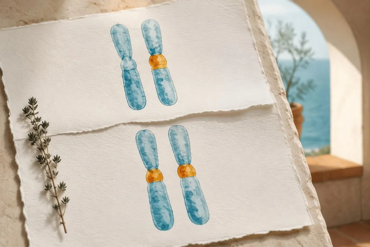
When both parents carry a disease-causing variant in GBA1, each pregnancy has a 25% probability of an affected child, a 50% probability of a carrier child and a 25% probability of a child who inherits neither familial variant. These probabilities reset with every pregnancy; previous outcomes do not change the next calculation.
Carrier interpretation depends on the gene's inheritance and the test's limitations. Our SMA carrier result guide shows how hidden risk can depend on gene-copy arrangement, while Fragile X carrier interpretation uses a different mechanism; neither algorithm should be imported into GBA1 counseling.
Sidransky et al. (2009), in the New England Journal of Medicine, found an overall odds ratio of 5.43 for the association between GBA mutations and Parkinson disease in a multicenter case-control study. An odds ratio is not a lifetime probability: most carriers do not inevitably develop Parkinson disease, and individual risk varies by variant and other factors.
Symptoms still deserve assessment in someone labeled a carrier. If a hypothetical person has 1 detected pathogenic variant plus clearly deficient leukocyte activity, clinicians should look for an undetected second allele or another explanation rather than assuming that carrier status alone accounts for the findings.
Γιατί ο έλεγχος του GBA1 πρέπει να γίνεται σε εξειδικευμένο εργαστήριο;
GBA1 testing is technically challenging because the nearby GBAP1 pseudogene has approximately 96% sequence similarity to GBA1. A suitable assay must distinguish the functional gene from the pseudogene and address recombinant or structural alleles that a limited test may miss or misassign.
Short DNA reads can map ambiguously when 2 regions are highly similar, and gene–pseudogene recombination can produce complex alleles. This is why 'sequencing performed' is not enough information: the laboratory should explain whether its method is validated for the GBA1 locus and how it handles difficult regions.
Variant names also changed with the protein numbering convention. The older N370S name corresponds to p.Asn409Ser, and L444P corresponds to p.Leu483Pro; the 39-amino-acid difference can make the same variant appear to be 2 different findings when comparing an older family report with a newer result.
Not every GBA1-associated Parkinson risk variant causes Gaucher disease on its own. For example, p.Glu365Lys, historically E326K, requires careful classification rather than automatic counting as 1 Gaucher-causing allele; our genetic result interpretation guide explains why a variant's significance must be matched to the condition under investigation.
Consumer genotyping and unvalidated raw-data interpretation should not confirm or exclude Gaucher disease. If a report lists only 1 common variant, request clinical confirmation and ask about the second allele; if it lists a complex allele, ask whether the changes are in cis and whether parental testing could clarify them.
Τι γίνεται αν τα αποτελέσματα ενζύμου, lyso-Gb1 και DNA διαφωνούν;
Discordant Gaucher results require specimen review, method review and specialist interpretation before a final diagnosis. Low enzyme activity with 1 detected variant, or elevated lyso-Gb1 with apparently normal activity, can reflect incomplete genetic testing, analytical limitations or a less common biological explanation.

A hypothetical combination of deficient leukocyte activity, elevated lyso-Gb1 and 1 pathogenic GBA1 variant strongly justifies a search for the missing allele. The next investigation may involve deletion/duplication assessment, recombinant-allele analysis or another validated method; repeating the same limited panel does not necessarily add useful information.
Rare saposin C deficiency, related to PSAP, can produce a Gaucher-like biochemical and clinical picture despite relatively preserved activity in some artificial-substrate assays. This is a specialist differential diagnosis, not a reason to distrust every normal enzyme test; it becomes relevant when 2 or more independent findings remain unexplained.
Allogeneic stem-cell transplantation can alter leukocyte enzyme activity and the source of DNA in circulating cells, so the laboratory needs that history. Recent transfusions should also be disclosed, although their impact depends on product and assay; η σύγκριση αποτελεσμάτων μεταξύ επισκέψεων requires more than assuming every numerical change reflects disease.
Kantesti AI cannot resolve gene phase, laboratory contamination or donor-cell chimerism from a report alone. Our AI explanation safety guide separates readable explanations from diagnostic verification; carry all 3 reports—enzyme, genetics and biomarker—to the same specialist review instead of interpreting each in isolation.
Ποιες αιματολογικές τιμές και συμπτώματα εγείρουν υποψίες;
Unexplained spleen enlargement, thrombocytopenia, anemia and skeletal symptoms can prompt Gaucher testing, but none confirms the disease. The combination matters more than 1 isolated abnormality, and clinicians must consider hematological, hepatic and other causes alongside a lysosomal storage disorder.
For example, a hypothetical adult with platelets of 85 × 10^9/L, hemoglobin of 10.4 g/dL and an enlarged spleen deserves an explanation for the combined pattern. Those numbers do not diagnose Gaucher disease; they justify assessment for sequestration, marrow disorders, liver disease and other causes rather than attributing everything to iron deficiency.
A low automated platelet count should first be checked for clumping when the clinical context suggests an artifact. Our επεξήγηση συσσωμάτωσης αιμοπεταλίων describes this common alternative; confirming 1 truly low count prevents a false laboratory abnormality from driving an unnecessary rare-disease workup.
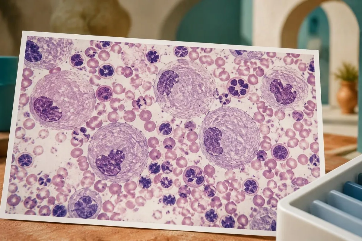
Bone marrow examination is not routinely required to diagnose Gaucher disease when enzyme and genetic testing are available. Gaucher-like or 'pseudo-Gaucher' cells can occur in other conditions, including disorders with high cell turnover; our marrow diagnosis guide explains why cellular appearance and disease-specific confirmation answer different questions.
A normal CBC does not exclude early or mild Gaucher disease, particularly when 2 familial pathogenic variants are known. Conversely, a marrow report mentioning storage-like cells should lead to biochemical confirmation rather than immediate labeling; ask which alternative causes were considered and whether the enzyme specimen was collected before any treatment.
Μπορούν αυτές οι εξετάσεις να προσδιορίσουν τον τύπο και τη βαρύτητα της νόσου Gaucher;
Enzyme and GBA1 tests establish the biochemical and genetic basis of Gaucher disease, but clinical assessment determines subtype and organ involvement. The conventional classification uses types 1, 2 and 3, with primary neurological involvement distinguishing neuronopathic forms; these categories do not replace an individualized assessment.
Type 1 lacks the primary neuronopathic pattern of types 2 and 3, although people with type 1 or heterozygous GBA1 variants can still have other neurological conditions. The distinction is clinically specific; a future Parkinson risk association is not equivalent to classifying someone as having neuronopathic Gaucher disease.
The p.Asn409Ser variant is strongly associated with non-neuronopathic disease when present in a Gaucher genotype, but genotype does not precisely predict skeletal burden or treatment timing. A hypothetical pair of siblings with the same 2 variants can still differ in spleen size, platelet count, bone involvement and age at recognition.
Skeletal assessment may require imaging because routine calcium or alkaline phosphatase results can miss Gaucher-related bone involvement. Our bone health laboratory guide explains what general bone tests can and cannot show; a normal result on 1 chemistry panel is not a substitute for indicated imaging.
Neurological assessment focuses on symptoms, examination and age-appropriate specialist evaluation, including eye movement abnormalities when relevant. Our neurofilament interpretation guide explains why a nonspecific neuronal-injury marker cannot determine Gaucher subtype; neither that marker nor 1 lyso-Gb1 concentration provides a stand-alone classification.
Ποιοι πρέπει να υποβληθούν σε οικογενειακό έλεγχο μετά την επιβεβαίωση;
After Gaucher disease is confirmed, genetic counseling can identify relatives who need targeted testing and explain reproductive probabilities. Testing for the family's 2 established variants is often more informative than repeating a broad screening panel, particularly when the original report clearly documents their phase and classification.
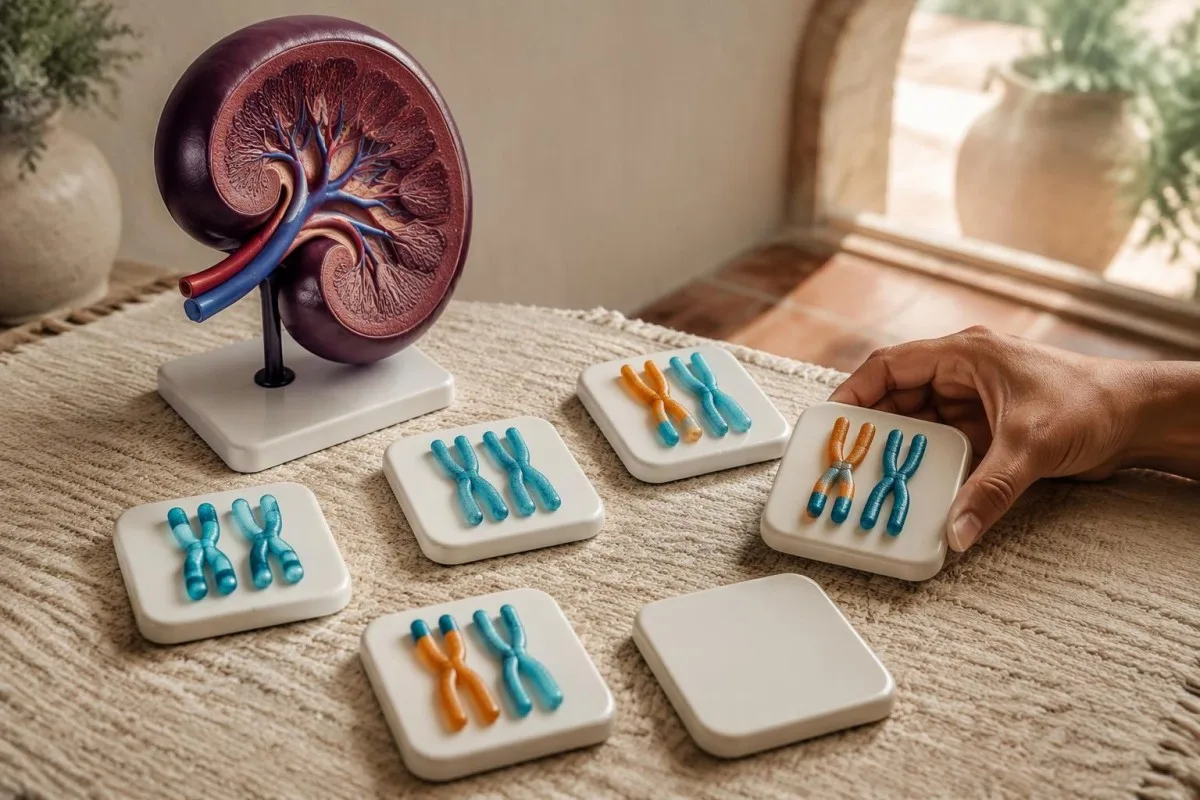
Full siblings of an affected person generally have the familiar 25% affected, 50% carrier and 25% noncarrier probabilities when both parents are carriers. These are pretest probabilities, not predictions about a particular sibling; an apparently well relative may still need testing because symptom severity and age at presentation vary.
Partner testing helps estimate reproductive risk, but the scope of the assay matters. A negative panel testing only several common variants leaves more residual uncertainty than a validated comprehensive assay; ask the counselor to explain both the detected result and the remaining risk rather than treating 1 negative screen as a guarantee.
Testing minors raises 2 different questions: could identifying disease change their childhood care, and is the request solely to establish future reproductive carrier status? Diagnostic testing for a potentially affected child can have immediate value, while carrier-only testing may warrant a different consent discussion based on local practice and family circumstances.
Share the original clinical laboratory report, including exact variant names and specimen details, rather than a rewritten family summary. Our secure result sharing guide explains practical consent considerations; each adult relative should control access to their own results, even when 1 family member coordinates appointments.
Τι ακολουθεί μετά την επιβεβαίωση της νόσου Gaucher;
A confirmed diagnosis leads to specialist assessment, baseline measurements and a decision about treatment or surveillance. Enzyme replacement or substrate reduction therapy is considered according to clinical involvement, not simply because 1 enzyme result is low or lyso-Gb1 exceeds a reference limit.

Baseline assessment commonly includes CBC, liver-related tests, spleen and liver measurements, skeletal evaluation and a disease-specific biomarker. Establishing these findings before treatment makes later changes interpretable; 2 lyso-Gb1 results without information about method, treatment timing and organ status provide a much weaker follow-up picture.
Chitotriosidase and CCL18 may support monitoring, but they are not substitutes for diagnostic enzyme testing. A common 24-base-pair CHIT1 duplication can eliminate chitotriosidase activity when present on both copies, so an undetectable chitotriosidase result can reflect inherited marker deficiency rather than an absence of Gaucher disease.
Kantesti's trend tools can help organize routine results, but they do not establish whether specialized Gaucher testing is analytically comparable or whether treatment should change. Genetic information deserves particular care; our οδηγίες για το απόρρητο και τα δεδομένα explains data handling, and patients should avoid including relatives' identifiable reports without consent.
Children need age-specific interpretation, growth assessment and pediatric metabolic oversight rather than adult reference ranges applied mechanically. Our καθοδήγησή μας για την ασφάλεια της παιδιατρικής ΤΝ describes that boundary; seek urgent care for uncontrolled bleeding, severe new abdominal pain or acute neurological change instead of waiting for a second routine monitoring result.
Ποια στοιχεία υποστηρίζουν τη διαγνωστική προσέγγιση και τι μπορεί να εξηγήσει η τεχνητή νοημοσύνη;
The diagnostic pathway is supported by specialist laboratory guidance, biomarker research and clinical genetic evidence—not by AI output alone. The 3 directly relevant medical references below address laboratory confirmation, lyso-Gb1 and GBA-associated Parkinson risk; our separate publications provide background on routine laboratory interpretation.

Kantesti is an AI-powered blood test analysis tool that helps translate report language into questions for a clinician, rather than verifying a rare-disease diagnosis. Our πληροφορίες κλινικής μεθοδολογίας explains broader oversight; performance on routine panels does not establish diagnostic accuracy for GBA1 phase, rare alleles or specialized enzyme assays.
Dardis et al. (2022) provide the central laboratory framework, while Rolfs et al. (2013) provide evidence for lyso-Gb1 as a diagnostic and follow-up biomarker. Their findings answer 2 different questions: how to establish enzyme deficiency and how to measure an associated biochemical signal; neither supports interpreting a carrier screen as affected status.
Our publication on coagulation test interpretation is relevant background when a confirmed patient is being assessed for bleeding, but it is not a Gaucher diagnostic study. The associated Zenodo DOI below documents that educational publication, not independent validation of a Gaucher test or a treatment threshold.
Το συνοδευτικό οδηγός ερμηνείας πρωτεϊνών ορού explains routine protein measurements that may accompany broader assessment; albumin and globulins do not confirm this disorder. Both of the 2 Zenodo references are included transparently as related educational publications, separate from the directly relevant medical evidence.
Συχνές ερωτήσεις
Πώς διαγιγνώσκεται η νόσος Gaucher μετά από παθολογικό αποτέλεσμα σε αποξηραμένη σταγόνα αίματος;
An abnormal dried blood spot is usually followed by confirmatory glucocerebrosidase activity testing in leukocytes and GBA1 analysis. Diagnostic cellular activity below approximately 10–15% of mean normal is characteristic, although interpretation depends on the laboratory's method and specimen quality. Two pathogenic or likely pathogenic GBA1 variants on opposite gene copies support genetic confirmation. Lyso-Gb1 adds biochemical evidence but does not replace those tests.
Σημαίνει η χαμηλή δραστηριότητα της γλυκοκερεβροσιδάσης ότι έχω νόσο Gaucher;
Η χαμηλή δραστηριότητα της γλυκοκερεβροσιδάσης δεν σημαίνει πάντα νόσο Gaucher, ιδίως όταν το αποτέλεσμα προέρχεται από έλεγχο σε αποξηραμένη σταγόνα αίματος. Η ποιότητα της συλλογής, οι συνθήκες μεταφοράς και η σύσταση του δείγματος μπορούν να επηρεάσουν τη δραστηριότητα που μετράται κατά τον έλεγχο. Ένα αποτέλεσμα με έντονα μειωμένη δραστηριότητα σε επικυρωμένη δοκιμασία σε λευκοκύτταρα ή ινοβλάστες αποτελεί πολύ ισχυρότερη διαγνωστική ένδειξη από 1 χαμηλή μέτρηση ελέγχου. Τα οριακά ή ασύμφωνα αποτελέσματα χρειάζονται εργαστηριακή αξιολόγηση και συσχέτιση με τον έλεγχο του GBA1.
Μπορεί ένας φορέας του GBA1 να έχει νόσο Gaucher;
Ένα άτομο με μόνο 1 παθογόνο παραλλαγή του GBA1 είναι συνήθως φορέας και όχι άτομο με νόσο Gaucher. Εάν τα συμπτώματα και οι επιβεβαιωτικές ενζυμικές εξετάσεις υποδηλώνουν έντονα νόσο, οι κλινικοί ιατροί θα πρέπει να διερευνήσουν εάν δεν εντοπίστηκε δεύτερη παραλλαγή ή εάν υπάρχει άλλη εξήγηση. Η ενζυμική δραστηριότητα από μόνη της δεν μπορεί να εντοπίσει αξιόπιστα τους φορείς, επειδή τα εύρη τιμών των φορέων και των μη φορέων επικαλύπτονται. Ορισμένες ετερόζυγες παραλλαγές του GBA1 αυξάνουν επίσης τον κίνδυνο νόσου του Parkinson, αλλά αυτή η συσχέτιση δεν αποτελεί διάγνωση ούτε της νόσου του Parkinson ούτε της νόσου Gaucher.
Ποιο επίπεδο lyso-Gb1 επιβεβαιώνει τη νόσο Gaucher;
No single lyso-Gb1 concentration universally confirms Gaucher disease across all laboratories and specimen types. Rolfs et al. (2013) reported excellent discrimination with a 12 ng/mL cutoff in a selected research cohort, but that threshold is assay- and population-dependent. Interpret the result using the reporting laboratory's interval and whether the specimen was plasma or a dried blood spot. Confirmation relies on diagnostic enzyme testing and appropriate genetic evidence.
Αποδεικνύουν δύο παραλλαγές του GBA1 τη νόσο Gaucher;
Η ανίχνευση δύο παραλλαγών του GBA1 δεν αποδεικνύει αυτομάτως νόσο Gaucher. Και οι δύο πρέπει να είναι παθογόνες ή πιθανώς παθογόνες και να επηρεάζουν διαφορετικά αντίγραφα του γονιδίου, αντί να βρίσκονται μαζί στο ίδιο αντίγραφο. Δύο παραλλαγές αβέβαιης κλινικής σημασίας δεν αρκούν από μόνες τους για να τεκμηριώσουν τη διάγνωση. Ο έλεγχος των γονέων, η επανεξέταση της εξέτασης και η επιβεβαίωση της ενζυμικής δραστηριότητας μπορούν να βοηθήσουν στην αποσαφήνιση αβέβαιων αποτελεσμάτων.
Χρειάζεται βιοψία μυελού των οστών για τη διάγνωση της νόσου Gaucher;
Η βιοψία μυελού των οστών δεν είναι συνήθως απαραίτητη όταν είναι διαθέσιμες διαγνωστικές εξετάσεις ενζύμων και GBA1. Κύτταρα τύπου Gaucher μπορεί να εμφανιστούν και σε άλλες διαταραχές, επομένως η εμφάνισή τους δεν είναι επαρκώς ειδική ώστε να επιβεβαιώσει από μόνη της τη διάγνωση. Οι κλινικοί ιατροί μπορεί και πάλι να συστήσουν εξέταση του μυελού για διαφορετικό αιματολογικό ερώτημα, όπως ανεξήγητες ανωμαλίες που αφορούν 2 ή περισσότερες κυτταρικές σειρές. Εάν αναφερθούν κύτταρα τύπου αποθήκευσης, εξακολουθούν να απαιτούνται βιοχημικές εξετάσεις ειδικές για τη νόσο.
Πρέπει να υποβάλλονται σε εξετάσεις οι συγγενείς εάν επιβεβαιωθεί η νόσος Gaucher;
Relatives may benefit from targeted testing after the family's disease-causing GBA1 variants are established. When both parents are carriers, each full sibling has a pretest probability of 25% affected, 50% carrier and 25% inheriting neither familial variant. Genetic counseling explains which relatives need testing, what a negative result leaves unresolved and how results affect reproductive planning. Testing an apparently well sibling can still be useful because Gaucher disease severity and age at recognition vary.
Δείτε πώς να διαβάσετε εξετάσεις αίματος
Ανεβάστε την εργαστηριακή σας αναφορά και η τεχνητή νοημοσύνη της Kantesti εξηγεί κάθε βιοδείκτη με απλή γλώσσα μέσα σε λίγα δευτερόλεπτα. Την έχουν χρησιμοποιήσει περισσότεροι από 2 εκατομμύρια άνθρωποι παγκοσμίως.
Αναφορές
- Klein, T., Mitchell, S., & Weber, H. (2026). Φυσιολογικό εύρος aPTT: Οδηγός για D-Dimer, πρωτεΐνη C και πήξη του αίματος. Ιατρική έρευνα της τεχνητής νοημοσύνης Kantesti. DOI
- Klein, T., Mitchell, S., & Weber, H. (2026). Οδηγός πρωτεϊνών ορού: Σφαιρίνες, αλβουμίνη και εξέταση αίματος για τον λόγο A/G. Ιατρική έρευνα της τεχνητής νοημοσύνης Kantesti. DOI
- Dardis A et al. (2022). Patient centered guidelines for the laboratory diagnosis of Gaucher disease type 1. Orphanet Journal of Rare Diseases.
- Rolfs A et al. (2013). Glucosylsphingosine is a highly sensitive and specific biomarker for primary diagnostic and follow-up monitoring in Gaucher disease in a non-Jewish, Caucasian cohort of Gaucher disease patients. PLOS ONE. DOI
- Sidransky E et al. (2009). Multicenter analysis of glucocerebrosidase mutations in Parkinson's disease. New England Journal of Medicine. DOI PubMed
- 2+ εκατομμύριαΕξετάσεις που αναλύθηκαν
- 127+χωρών
- 100+Γλώσσες
Σχετικά με την ιατρική ομάδα


Σήματα εμπιστοσύνης E-E-A-T
Εμπειρία
Κλινική ανασκόπηση από ιατρό για τις ροές εργασίας ερμηνείας εργαστηριακών αποτελεσμάτων.
Πραγματογνωμοσύνη
Εστίαση στην εργαστηριακή ιατρική στο πώς συμπεριφέρονται οι βιοδείκτες στο κλινικό πλαίσιο.
Αυθεντικότητα
Ιατρική αξιολόγηση από τον Δρ. Thomas Klein, με συνεισφορές από τη Δρ. Sarah Mitchell και τον Καθ. Δρ. Hans Weber.
Αξιοπιστία
Ερμηνεία βασισμένη σε τεκμηριωμένα δεδομένα με σαφείς οδούς παρακολούθησης για τη μείωση του συναγερμού.




Ιατρική αποποίηση ευθύνης
Αυτό το άρθρο προορίζεται μόνο για εκπαιδευτικούς σκοπούς και δεν αποτελεί ιατρική συμβουλή. Συμβουλεύεστε πάντα έναν κατάλληλο επαγγελματία υγείας για αποφάσεις σχετικά με τη διάγνωση και τη θεραπεία.
Δημοσιεύθηκε · Επικοινωνήστε με τη συντακτική ομάδα
Εκδίδεται από την Kantesti Ltd, εγγεγραμμένη στην Αγγλία και την Ουαλία, αριθμός εταιρείας. 17090423, Λονδίνο, Ηνωμένο Βασίλειο.
