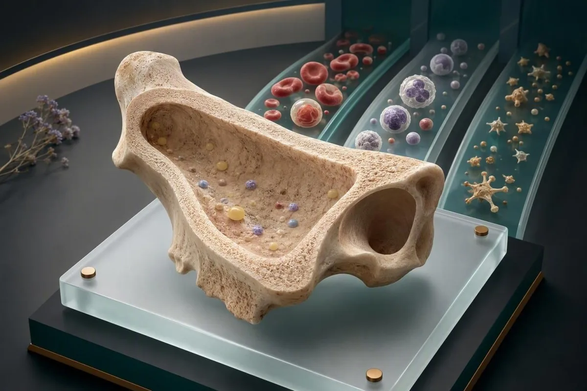
Anemia aplastyczna zazwyczaj daje charakterystyczny trójliniowy obraz morfologii krwi: niedokrwistość, mała liczba białych krwinek i mała liczba płytek krwi, z nieproporcjonalnie niską liczbą retikulocytów. Wymaga to pilnego potwierdzenia klinicznego, ponieważ kilka odwracalnych i poważnych stanów może początkowo wyglądać podobnie.


- Pancytoopenia oznacza niski poziom czerwonych krwinek, białych krwinek i płytek krwi; jest to obraz, a nie diagnoza.
- Bezwzględna liczba retikulocytów poniżej 20 × 10⁹/L sugeruje znacząco zmniejszoną produkcję szpiku przy obecności niedokrwistości.
- Ciężka anemia aplastyczna jest definiowana przez hipocelularny szpik kostny plus co najmniej dwa z następujących kryteriów: ANC poniżej 0,5 × 10⁹/L, płytki krwi poniżej 20 × 10⁹/L lub retikulocyty poniżej 20 × 10⁹/L.
- ANC poniżej 0,5 × 10⁹/L z gorączką 38,0°C lub wyższą wymaga pilnej oceny tego samego dnia.
- Płytki krwi poniżej 10 × 10⁹/L wiążą się ze znacznym ryzykiem spontanicznych krwawień i zazwyczaj wymagają pilnej opieki szpitalnej.
- Makrocytoza może wystąpić w anemii aplastycznej, ale MCV powyżej 110 fL powinno skłonić klinicystów do aktywnego wykluczenia niedoboru witaminy B12 lub kwasu foliowego.
- Blasty, znaczna dysplazja, komórki łezkowate lub schizocyty w rozmazie sugerują inne schorzenia niż niepowikłana niedokrwistość aplastyczna i zmieniają podejście diagnostyczne.
- Nie leczyć się samodzielnie trójliniowo niskie wyniki CBC z samym żelazem; żelazo nie przywraca produkcji szpiku i może opóźnić pilną ocenę.
Obraz morfologii krwi budzący obawy dotyczące anemii aplastycznej
Jakiś badanie krwi w kierunku niedokrwistości aplastycznej zwykle wykazuje pancytopenię: obniżoną hemoglobinę, niską liczbę neutrofilów i niską liczbę płytek krwi występujące jednocześnie, podczas gdy odpowiedź retikulocytarna pozostaje niska, a nie kompensacyjna. Taka kombinacja powinna skłonić do pilnej konsultacji lekarskiej, zwłaszcza jeśli liczba komórek spadła w więcej niż jednym poprzednim badaniu CBC.
U dorosłych typowy zakres referencyjny CBC wynosi około 120–155 g/L hemoglobiny dla wielu kobiet i 135–175 g/L dla wielu mężczyzn, 4,0–11,0 × 10⁹/L białych krwinek i 150–400 × 10⁹/L płytek krwi; laboratoria ustalają własne limity. Niedokrwistość aplastyczna obniża wszystkie trzy linie, ponieważ szpik nie produkuje wystarczającej ilości nowych elementów komórkowych. Podstawowe informacje można znaleźć w naszym przewodniku po tym, co obejmuje CBC.
Wzór jest bardziej informacyjny niż jakikolwiek pojedynczy niski wynik. Hemoglobina 92 g/L po obfitej utracie krwi miesiączkowej może oznaczać niedobór żelaza, podczas gdy hemoglobina 92 g/L plus ANC 0,7 × 10⁹/L i płytki 34 × 10⁹/L to problem z produkcją szpiku, dopóki nie zostanie udowodnione inaczej. W mojej praktyce klinicznej niebezpieczeństwo nie polega na samej anemii – to ryzyko infekcji i krwawienia pojawiające się cicho obok niej.
Kantesti jest Analizator do badań krwi AI która odczytuje składniki CBC jako powiązany wzorzec, a nie jako trzy niepowiązane sygnały ostrzegawcze. Nasz system może pomóc w organizacji raportu i poprzednich wyników, ale nie może zdiagnozować niewydolności szpiku ani zastąpić pilnej oceny hematologicznej. Od 26 sierpnia 2026 r. każde nowe trójliniowe cytopenie wymagają oceny przez lekarza w ciągu kilku dni, a nie rutynowej corocznej kontroli.
Prawidłowo wyglądające MCV nie sprawia, że pancytopenia jest łagodna. Wczesna niedokrwistość aplastyczna może być normocytarna, łagodnie makrocytarna lub sporadycznie mikrocytarna, jeśli współistnieje niedobór żelaza; odpowiedź szpiku i rozmaz obwodowy często dają więcej wskazówek niż samo MCV. Praktyczna zasada dr Thomasa Kleina jest prosta: zapytaj, czy szpik odpowiada, zanim założysz żywieniowe wyjaśnienie.
Dlaczego wzorzec jest często pomijany
Badanie CBC może oznaczyć każdy wynik osobno, więc pacjenci czasami skupiają się na najniższej liczbie i pomijają grupę. Spadek liczby płytek krwi z 260 do 92 × 10⁹/L, nawet jeśli technicznie jest powyżej progu alarmowego, jest klinicznie istotny, gdy hemoglobina i neutrofile spadają równolegle. Trendy w badaniach krwi często ujawniają trajektorię wcześniej niż pojedynczy raport.
Niedokrwistość plus niska liczba retikulocytów: wskazówka dotycząca wydolności szpiku
A niska liczba retikulocytów u osoby z anemią oznacza, że szpik nie zastępuje odpowiednio czerwonych krwinek, i jest to jedna z najbardziej pomocnych wskazówek wspierających niedokrwistość aplastyczną. Bezwzględna liczba retikulocytów ma większe znaczenie niż procent retikulocytów, ponieważ procent może wyglądać zwodniczo normalnie, gdy całkowita liczba czerwonych krwinek jest niska.
Większość laboratoriów podaje zakres referencyjny dla bezwzględnej liczby retikulocytów u dorosłych wynoszący około 25–100 × 10⁹/L, chociaż metody się różnią. W kryteriach ciężkiej niedokrwistości aplastycznej, bezwzględna liczba retikulocytów poniżej 20 × 10⁹/L jest jednym z wyznaczających niski wskaźnik produkcji środków, gdy komórkowość szpiku jest znacznie zmniejszona. Szczegółowy przewodnik po liczbie retikulocytów wyjaśnia, dlaczego wyniki oparte tylko na procentach mogą wprowadzać w błąd.
Natomiast ostra utrata krwi lub hemoliza powinny zwykle zwiększać liczbę retikulocytów po kilku dniach, często powyżej 100 × 10⁹/L, jeśli zapas żelaza i funkcja szpiku są prawidłowe; niska haptoglobina, wzrost stężenia bilirubiny pośredniej i podwyższone LDH wspierają hemolizę; to inna fizjologiczna historia niż aplazja. Rozróżnienie ma znaczenie, ponieważ leczenie “niedokrwistości” bez identyfikacji produkcji vs niszczenia prowadzi do opóźnień.
U pacjentki, którą widziałem z hemoglobiną 76 g/L, procent retikulocytów wynosił 1,2%, co wydawało się niepozorne, dopóki nie obliczono bezwzględnej liczby na 18 × 10⁹/L. To nie była odpowiedź regeneracyjna. Gdy niedokrwistość jest znacząca, lekarze mogą używać wskaźnika produkcji retikulocytów, ale bezwzględne retikulocyty są zazwyczaj mniej podatne na błędy w codziennej ocenie.
Retikulocyty mogą być przejściowo niskie podczas ciężkiej infekcji, po chemioterapii, przy toksyczności alkoholu lub w zaawansowanej chorobie nerek, więc ten wynik nie jest specyficzny dla niedokrwistości aplastycznej. Przegląd Younga z 2018 r. w New England Journal of Medicine podkreśla, że diagnoza wymaga oceny szpiku i wykluczenia dziedzicznych, immunologicznych, toksycznych i klonalnych alternatyw – a nie pojedynczego wyniku retikulocytów (Young, 2018).
Neutropenia: liczba w morfologii krwi, która wpływa na pilność postępowania w przypadku infekcji
Ten bezwzględną liczbę neutrofili (ANC) lepiej niż całkowita liczba białych krwinek przewiduje ryzyko infekcji bakteryjnych i grzybiczych. ANC poniżej 0,5 × 10⁹/L to ciężka neutropenia, a gorączka wynosząca lub przekraczająca 38,0°C jednorazowo lub utrzymująca się przez godzinę przy 38,0°C wymaga pilnej oceny.
ANC oblicza się jako liczbę białych krwinek pomnożoną przez procent neutrofilów plus procent pałeczkowatych. Na przykład, WBC 1,4 × 10⁹/L z 40% neutrofilami daje ANC 0,56 × 10⁹/L – znacznie bardziej użyteczne niż sama liczba całkowita. Nasza przewodnikiem progów ANC wyjaśnia zmienność związaną z wiekiem i ciążą, ale próg 0,5 × 10⁹/L pozostaje kluczową linią zagrożenia dla dorosłych.
Ciężka anaplastic anemia często powoduje neutropenię bez ziarnistości toksycznej lub przesunięcia w lewo, które mogą towarzyszyć infekcji bakteryjnej. Niemniej jednak pacjent może mieć zarówno niewydolność szpiku, jak i infekcję; brak podwyższonej liczby białych krwinek nigdy nie wyklucza sepsy. Gorączka, dreszcze, nowe splątanie, duszność lub szybko nasilający się ból gardła nie powinny czekać na powtórzenie pozaustrojowego CBC.
Bardzo ciężka anaplastic anemia stosuje jeszcze niższy próg ANC: poniżej 0,2 × 10⁹/L. Powodem oddzielenia 0,2 od 0,5 jest praktyczny aspekt – przy tych liczbach zwykłe drobnoustroje jamy ustnej i skóry mogą stać się niebezpieczne przy niewielkim zaczerwienieniu lub ropieniu miejscowym. W szpitalu lekarze mogą pobrać posiewy i rozpocząć empiryczne leczenie przeciwbakteryjne, zanim przyczyna zostanie w pełni ustalona.
Niska liczba limfocytów może współistnieć, ale nie zastępuje ANC w decyzjach dotyczących ostrego ryzyka. Jeśli brakuje rozmazu, poproś laboratorium lub lekarza o jego dostarczenie, zamiast zgadywać na podstawie całkowitej liczby WBC. Objawy niskiego poziomu limfocytów wymagają dalszej obserwacji, jednak głębokość neutropenii decyduje o decyzji podejmowanej tego samego dnia.
Kiedy gorączka jest ważniejsza niż wydruk CBC
Temperatura 38,0°C u osoby z ANC 0,4 × 10⁹/L jest stanem pilnym, nawet jeśli pacjent czuje się “w większości dobrze”, a gorączka spada po podaniu paracetamolu. Leki przeciwgorączkowe mogą zmniejszyć widoczny sygnał bez leczenia podstawowego problemu. Zabierz CBC i listę leków do pilnej opieki; nie przyjmuj pozostałości antybiotyków przed wykonaniem posiewów, chyba że lekarz tak zaleci.
Małopłytkowość: progi krwawienia przy podejrzeniu niewydolności szpiku
Płytki krwi poniżej 50 × 10⁹/L zwiększają ryzyko siniaków i krwawienia związanego z procedurami, podczas gdy liczby poniżej 10 × 10⁹/L mogą powodować spontaniczne krwawienie. W anaplastic anemia małopłytkowość zwykle występuje wraz z anemią i neutropenią, a nie jako izolowany problem z płytkami krwi.
Wybroczyny, krwawienia z nosa trwające ponad 20 minut, krwawienia z dziąseł, obfite krwawienia miesiączkowe, krew w moczu, smoliste stolce lub nagły silny ból głowy wymagają pilnego kontaktu z lekarzem, gdy liczba płytek jest niska. Liczba płytek 18 × 10⁹/L nie jest automatycznie katastrofalna, ale nie jest to liczba, którą należy zarządzać za pomocą porad internetowych. Zobacz naszą praktyczną dyskusję na temat progów płytek przed zabiegami stomatologicznymi.
Rozmaz pomaga odróżnić zmniejszoną produkcję od obwodowego niszczenia. W immunologicznej małopłytkowości płytki krwi mogą być jedynym niskim parametrem, a często widoczne są duże młode płytki; anaplastic anemia częściej ma niski lub prawidłowy odsetek niedojrzałych płytek, ponieważ sama produkcja jest zmniejszona. Są to tendencje, nie zasady, a preparat musi być odczytany przez wyszkolonego morfologa.
Unikaj aspiryny, ibuprofenu, naproksenu i nieprzepisanych megadawek oleju rybiego, dopóki lekarz nie oceni znaczącej małopłytkowości, ponieważ mogą one upośledzać czynność płytek krwi, nawet jeśli liczba liczbowa się nie zmieniła. Alkohol może nasilać supresję szpiku i czynność płytek krwi. Acetaminofen/paracetamol również nie jest uniwersalnie odpowiedni – dawka i stan wątroby nadal mają znaczenie.
Liczba płytek krwi może być fałszywie niska, gdy płytki krwi zlepiają się w próbce EDTA. Laboratorium może zgłosić “obecne zlepki płytek” i powtórzyć liczbę w probówce cytrynianowej; nazywa się to pseudomałopłytkowością. Wyjaśnia to izolowaną niską liczbę płytek, a nie przekonującą pancytopenię z niską liczbą retikulocytów.
Jak klinicyści klasyfikują ciężką i bardzo ciężką anemię aplastyczną
Ciężka anaplastic anemia wymaga hipocelularnego szpiku kostnego – zazwyczaj poniżej 25% komórkowości – plus co najmniej dwa kryteria obwodowe: ANC poniżej 0,5 × 10⁹/L, płytki poniżej 20 × 10⁹/L lub bezwzględne retikulocyty poniżej 20 × 10⁹/L. Bardzo ciężka choroba spełnia te kryteria z ANC poniżej 0,2 × 10⁹/L.
Te progi pochodzą z długo istniejących ram Camitty, ale są narzędziami do wstępnej kwalifikacji i planowania leczenia, a nie diagnozą z arkusza kalkulacyjnego. Szpik może być plamisty, a niedawne przetoczenie krwi może zmienić obraz anemii. Lekarze uwzględniają również objawy, historię infekcji, zapotrzebowanie na przetoczenia, wiek, opcje dawców i dowody na zaburzenia klonalne.
Umiarkowana niedokrwistość aplastyczna nie jest tak precyzyjnie zdefiniowana. Osoba z liczbą płytek krwi 58 × 10⁹/L, ANC 0,9 × 10⁹/L i hemoglobiną 98 g/L może nie spełniać kryteriów ciężkich, ale nadal może się pogorszyć lub wymagać leczenia. Ta niejednoznaczność jest właśnie powodem, dla którego seryjne CBC i kontrola hematologiczna są ważniejsze niż próba przypisania uspokajającej etykiety w domu.
Wytyczne British Society for Haematology opisują badanie szpiku, cytogenetykę, testy przepływowe dla klonów PNH i ocenę dziedzicznych niedomóg szpiku jako części ścieżki diagnostycznej (Killick et al., 2016). Prawidłowy wynik cytogenetyczny obniża obawy o niektóre zespoły mielodysplastyczne, ale sam w sobie nie potwierdza immunologicznej niedokrwistości aplastycznej.
Kantesti AI podkreśla kombinacje, które przekraczają pilne progi, w tym ANC poniżej 0,5 × 10⁹/L i płytki krwi poniżej 20 × 10⁹/L, dzięki czemu użytkownicy mogą zobaczyć klastra przed wizytą. Nasza analiza jest wskazówką bezpieczeństwa, a nie oceną ciężkości; podstawowe kryteria wymagają badania klinicznego i wyników badania szpiku. W kontekście indywidualnych flag raportu przeczytaj tego, co oznacza „poza zakresem”.
MCV, RDW i obraz rozmazu krwi, które doprecyzowują obraz morfologii krwi
Niedokrwistość aplastyczna często ma prawidłową lub lekko podwyższoną MCV, ale sama MCV nie może odróżnić jej od niedoboru B12, niedoboru kwasu foliowego, skutków alkoholu, choroby wątroby lub mielodysplazji. Ręczny rozmaz obwodowy jest niezbędny, gdy pancytopenia jest nowa, postępująca lub niewyjaśniona.
Zakres referencyjny MCV wynosi zazwyczaj 80–100 fL. Lekka makrocytoza w okolicy 101–105 fL może wystąpić w niedokrwistości aplastycznej, ponieważ stresująca produkcja szpiku wpływa na dojrzewanie krwinek czerwonych, ale MCV powyżej 110 fL powinno skłonić do świadomego przeglądu B12, kwasu foliowego, leków, alkoholu, tarczycy i wątroby. Porównaj wskaźniki w naszym Przewodnik MCV vs MCH.
RDW mierzy zmienność wielkości krwinek czerwonych i często wynosi 11,5–14,5%, w zależności od analizatora. Prawidłowy RDW nie wyklucza niewydolności szpiku; w rzeczywistości dość jednorodna populacja zmniejszonych krwinek czerwonych może wystąpić, gdy produkcja po prostu spada. Ostro podwyższony RDW może zamiast tego sugerować mieszane niedobory, niedawne przetoczenie, powrót do zdrowia po leczeniu lub dysplazję.
Blasty, promielocyty, zaznaczone hipogranulacje, neutrofile typu pseudo-Pelgera lub znaczące formy dysplastyczne przesuwają obawy w kierunku ostrej białaczki lub zespołu mielodysplastycznego i powinny przyspieszyć ocenę specjalistyczną. Schistocyty wskazują inny kierunek – fragmentację krwinek czerwonych spowodowaną mikroangiopatią – i mogą być pilne w połączeniu z niską liczbą płytek krwi, uszkodzeniem nerek lub objawami neurologicznymi. Nasz przewodnik po schistocytach obejmuje ten odrębny wzorzec nagły.
Kantesti jest Narzędzie do analizy wyników badań krwi oparte na AI który może porównywać MCV, RDW, retikulocyty i historyczne wyniki, ale nie zastępuje przeanalizowanego rozmazu. Z mojego doświadczenia wynika, że właśnie w rozmazie pozornie proste “niskie CBC” staje się ostro ukierunkowanym pytaniem diagnostycznym. przewodnik technologiczny wyjaśnia, jak nasza AI zachowuje oryginalne jednostki i zakresy referencyjne laboratorium.
Niedobór witaminy B12, kwasu foliowego i miedzi może naśladować niewydolność szpiku
Niedobór witaminy B12, kwasu foliowego i miedzi może powodować pancytopenię i musi zostać wykluczony przed zaklasyfikowaniem wzorca CBC jako niedokrwistości aplastycznej. Te niedobory są szczególnie prawdopodobne w przypadku diet restrykcyjnych, zaburzeń wchłaniania, operacji bariatrycznych, intensywnego stosowania cynku lub niektórych leków.
Niedobór B12 często powoduje makrocytozę, hipersegmentację neutrofilów i podwyższony poziom kwasu metylomalonowego, ale u niektórych pacjentów MCV jest poniżej 100 fL, gdy współistnieje niedobór żelaza. Serum B12 poniżej 148 pmol/L, czyli około 200 pg/mL, jest często uważane za niedobór, podczas gdy 148–221 pmol/L może wymagać potwierdzenia kwasem metylomalonowym lub homocysteine. Nasz porównanie B12 i kwasu foliowego wyjaśnia, dlaczego sam kwas foliowy może maskować objawy hematologiczne, podczas gdy uszkodzenia neurologiczne postępują.
Niedobór kwasu foliowego może obniżyć wszystkie trzy linie w zaawansowanych przypadkach, szczególnie przy słabym spożyciu, spożyciu alkoholu, zaburzeniach wchłaniania, metotreksacie, trimetoprimie lub niektórych lekach przeciwdrgawkowych. Kwas foliowy w krwinkach czerwonych może lepiej odzwierciedlać zapasy tkankowe niż pojedynczy poziom w surowicy w niektórych sytuacjach, ale dostępność i interpretacja oznaczeń różnią się. Nie należy rozpoczynać podawania dużych dawek kwasu foliowego przed oceną B12, chyba że lekarz ma ku temu szczególny powód.
Niedobór miedzi jest rzadziej sprawdzany, ale może powodować niedokrwistość i neutropenię oraz przypominać mielodysplazję w badaniu szpiku. Nadmiar suplementowanego cynku jest klasyczną odwracalną przyczyną, ponieważ cynk indukuje jelitową metalotioneinę, która preferencyjnie wiąże miedź. badanie pod kątem niskiego poziomu miedzi jest szczególnie istotne w przypadku narażenia na kremy do protez, operacji bariatrycznych lub dawek cynku powyżej 40 mg dziennie.
Odżywianie może być częścią rozwiązania, ale nie powinno być powodem do odkładania oceny szpiku w przypadku ciężkiej pancytopenii. Suplementacja żelazem, B12, kwasem foliowym i miedzią jest dostosowana do potwierdzonych niedoborów i kontekstu medycznego; nieograniczona suplementacja może zaciemnić wyniki użytecznych badań. Jest to jeden z tych obszarów, gdzie kontekst ma większe znaczenie niż pojedyncza, graniczna liczba.
Przyczyny niedoboru wszystkich rodzajów komórek krwi (pancytopenii) związane z lekami, toksynami i wirusami, które należy wykluczyć w pierwszej kolejności
Leki, toksyny i choroby wirusowe mogą hamować produkcję szpiku i dawać obraz morfologii krwi obwodowej (CBC) podobny do anemii aplastycznej, czasem odwracalny. Pełny wywiad dotyczący ekspozycji powinien obejmować leki na receptę, ostatnio stosowane antybiotyki, leki przeciwpadaczkowe, immunoterapie, produkty ziołowe, rozpuszczalniki, alkohol i substancje rekreacyjne.
Chemioterapia jest oczywistą przyczyną, ale mniej oczywiste czynniki obejmują chloramfenikol w regionach, gdzie jest on nadal stosowany, leki przeciwtarczycowe, linezolid, niektóre leki przeciwpadaczkowe i terapie immunomodulujące. Czas ma znaczenie: bezpośrednia toksyczność leku dla szpiku często pojawia się tygodnie lub miesiące po ekspozycji, podczas gdy reakcje immunologiczne na leki mogą być mniej przewidywalne. Nigdy nie przerywaj przepisanych leków nagle bez konsultacji z lekarzem prowadzącym cytopenię.
Niedokrwistość aplastyczna związana z zapaleniem wątroby może wystąpić tygodnie lub miesiące po ostrym epizodzie zapalenia wątroby, często u młodszych osób, a rutynowe badania na obecność wirusów zapalenia wątroby typu A, B, C, E, HIV, EBV, CMV i parwowirusów mogą być zlecone na podstawie wywiadu. Negatywny wynik badania przesiewowego w kierunku wirusów nie wyklucza immunologicznej niedokrwistości aplastycznej. Po prostu eliminuje ważne konkurencyjne wyjaśnienia i kieruje środkami ostrożności dotyczącymi infekcji.
Ekspozycja na benzen pozostaje istotnym ryzykiem zawodowym i domowym, w tym praca w słabo wentylowanych zakładach przemysłowych, obsługa paliw i niektóre rozpuszczalniki. Lekarze pytają o zadania w pracy, a nie o stanowiska, ponieważ “kierownik biura u dostawcy farb” może nadal wiązać się z okresową ekspozycją w magazynie. Jeśli dostępne, przynieś nazwy produktów lub karty charakterystyki; niejasne wspomnienia są powszechne i nikt nie powinien czuć się winny.
Morfologia krwi obwodowej (CBC) wykonana krótko po znaczącej chorobie wirusowej może wymagać powtórzenia w ciągu dni lub tygodni, jeśli stan pacjenta jest stabilny, ale ciężkich wyników nie należy obserwować pasywnie. Zmiany CBC po chorobie mogą pomóc określić czas, podczas gdy gorączka z ciężką neutropenią pozostaje stanem nagłym, niezależnie od podejrzewanego czynnika wywołującego.
Przyczyny pancytopenii, które wyglądają inaczej niż anemia aplastyczna
Pancytopenia ma wiele przyczyn, a anemia aplastyczna charakteryzuje się hipocelularnym szpikiem bez nacieku złośliwego lub znacznej dysplazji. Główne alternatywy obejmują ostrą białaczkę, zespół mielodysplastyczny, naciek szpiku, hipersplenizm, ciężkie stany niedoboru, zespół hemofagocytarny, infekcję i skutki działania leków.
Ostra białaczka może powodować niskie wartości liczbowe, ponieważ blasty wypierają normalny szpik, ale krążące blasty nie zawsze są widoczne. Zespół mielodysplastyczny staje się bardziej prawdopodobny przy dysplazji, charakterystycznych zmianach cytogenetycznych lub utrzymującej się makrocytozie u starszych dorosłych, chociaż sam wiek niczego nie dowodzi. Ścieżka w naszym przewodniku po badaniach krwi pod kątem chorób nowotworowych pokazuje, dlaczego rozmaz, cytometria przepływowa i badania szpiku są uzupełniające.
Hipersplenizm obniża liczbę krążących komórek poprzez sekwestrację, zwykle z splenomegalią, nadciśnieniem wrotnym, chorobą wątroby lub niektórymi zaburzeniami hematologicznymi. Retikulocyty mogą być normalne lub podwyższone zamiast głęboko zahamowane, a komórkowość szpiku jest zazwyczaj zachowana lub zwiększona. USG może być pomocne, ale nie eliminuje potrzeby badania niepokojącego rozmazu lub ciężkiej neutropenii.
Napadowa hemoglobinuria nocna (PNH) może nakładać się biologicznie z anemią aplastyczną, dlatego lekarze często zlecają wysokoczułą cytometrię przepływową w celu wykrycia granulocytów i monocytów z niedoborem kotwicy GPI. Ciemny mocz nie jest ani wymagany, ani specyficzny. Wykrycie małego klonu PNH może potwierdzić kontekst immunologicznego niewydolności szpiku, jednak leczenie zależy od wielkości klonu, hemolizy, ryzyka zakrzepicy i ogólnego obrazu szpiku.
Kantesti jest usłudze interpretacji testów laboratoryjnych AI które mogą ujawnić wzorce pancytopenii i potencjalne alternatywne wskazówki, w tym bilirubinę, LDH, wskaźniki wątrobowe i historyczny spadek liczby płytek krwi. Nie można określić, czy szpik jest hipocelularny, nacieczony czy dysplastyczny – są to pytania dla hematologii i patologii. Lista kontrolna drugiej opinii może pomóc pacjentom w przygotowaniu ukierunkowanych pytań.
Pilne sygnały ostrzegawcze: kiedy niskie wyniki morfologii krwi wymagają pilnej interwencji
Opieka nagła jest wskazana przy gorączce 38,0°C lub wyższej z ANC poniżej 0,5 × 10⁹/L, niekontrolowanym krwawieniu, smolistych stolcach, kaszlu z krwią, silnym nowym bólu głowy, dezorientacji, omdleniach, bólu w klatce piersiowej lub duszności w spoczynku. Nie czekaj na interpretację online, jeśli wystąpi którykolwiek z tych objawów.
Głęboka anemia może upośledzać dostarczanie tlenu nawet bez widocznego krwawienia. Hemoglobina poniżej 70 g/L jest często stosowanym progiem rozważenia transfuzji u stabilnych pacjentów szpitalnych, ale objawy, aktywne krwawienie, choroby sercowo-naczyniowe, ciąża i przewlekłość wpływają na decyzje. Osoba z hemoglobiną 82 g/L, która omdlewa lub ma duszności w spoczynku, wymaga oceny pilniej niż osoba zdrowa z tą samą liczbą.
Płytki krwi poniżej 10 × 10⁹/L często są leczone pilnymi decyzjami specjalistycznymi, ponieważ zwiększa się prawdopodobieństwo spontanicznego krwawienia z błon śluzowych lub krwawienia wewnętrznego. Unikaj sportów kontaktowych, wstrzyknięć domięśniowych, termometrów doodbytniczych i zabiegów stomatologicznych, dopóki zespół leczący nie zaleci inaczej. W przypadku urazu głowy przy jakiejkolwiek niskiej liczbie płytek krwi należy pilnie ocenić stan, nawet jeśli objawy wydają się niewielkie.
Normalna temperatura nie gwarantuje bezpieczeństwa w ciężkiej neutropenii; osoby starsze, osoby przyjmujące steroidy i bardzo chorzy pacjenci mogą nie gorączkować. Nowe owrzodzenia jamy ustnej, bolesność podczas połykania, ból okolicy odbytu lub szybko pogarszające się zmęczenie mogą być wczesnymi wskazówkami infekcji. Laboratoryjne wzorce sepsy dostarczają kontekstu, ale objawy i parametry życiowe decydują o natychmiastowym działaniu.
Dr Thomas Klein zaleca pacjentom fotografowanie lub pobieranie oryginalnego raportu, zapisywanie daty badania oraz przynoszenie wszystkich pojemników z lekami i suplementami do pilnego przeglądu. Ten niewielki krok często pozwala zidentyfikować czasowo zależne narażenie na leki lub wyjaśnić, czy “nowa” niska liczba już występowała. Nie prowadź pojazdu, jeśli odczuwasz zawroty głowy, omdlenia lub aktywne krwawienie.
Jakie badania potwierdzają lub wykluczają anemię aplastyczną po wykonaniu morfologii krwi
Morfologia krwi (CBC) nie może potwierdzić aplastycznej anemii; potwierdzenie zwykle wymaga aspiratu szpiku kostnego i biopsji trepanowej wykazującej hipocelularyzację, a także badań wykluczających przyczyny klonalne, dziedziczne, zakaźne, żywieniowe i związane z narażeniem. Tempo badań zależy od ciężkości liczby i objawów.
Próbka szpiku ocenia celularyzację, architekturę, blasty, zwłóknienie, zapasy żelaza i dysplazję. U dorosłych oczekiwana celularyzacja wynosi około 100 minus wiek jako przybliżone oszacowanie dydaktyczne, ale jest to niedokładne i jakość próbki ma znaczenie. 30-latek z celularyzacją szpiku wynoszącą 10% jest bardzo różny od 80-latka z 10%, a jednak obaj potrzebują fachowej interpretacji w kontekście.
Cytogenetyka, mikromacierze chromosomowe lub panele sekwencjonujące mogą pomóc odróżnić hipocelularny zespół mielodysplastyczny od nabytej aplastycznej anemii. Badania klonów PNH, zaburzeń telomerów, anemii Fanconiego i innych dziedzicznych stanów niewydolności szpiku są wybierane w zależności od wieku, historii rodzinnej, wyników badania fizykalnego i planowania przeszczepu. Badania genetyczne nie są zarezerwowane tylko dla dzieci; coraz częściej rozpoznawane są dziedziczne choroby o początku u dorosłych.
Podstawowe badania często obejmują B12, kwas foliowy, miedź, profil wątrobowy i nerkowy, badania tarczycy, gdy są wskazane, markery hemolizy, serologię wirusową, testy ciążowe, gdy są istotne, oraz badania autoimmunologiczne dostosowane do historii choroby. Celem nie jest zlecanie każdego badania. Chodzi o uniknięcie przeoczenia odwracalnych przyczyn, jednocześnie dążąc do ostatecznej oceny.
Kantesti AI może zorganizować raport w tych kategoriach i zidentyfikować brakujące podstawy, ale zarządzanie kliniczne ma znaczenie, gdy wyniki są obarczone wysokim ryzykiem. Nasz standardy walidacji medycznej opisuje granicę między interpretacją wspomaganą przez sztuczną inteligencję a diagnozą medyczną. Zapytaj, kto będzie analizował próbkę szpiku i czy będzie ona oceniana przez hematopatologa.
Kiedy powtórzyć morfologię krwi przed uznaniem wyniku za wiarygodny
Powtórzenie morfologii krwi (CBC) jest wskazane, gdy krzepnięcie próbki, agregacja płytek, rozcieńczenie płynami dożylnymi lub flagi analizatora mogą zniekształcić wynik, ale powtórne badanie nigdy nie powinno opóźniać nagłej pomocy medycznej w przypadku ciężkich cytopenii lub niepokojących objawów. Laboratoria często mogą zweryfikować wynik z oryginalnej probówki w ciągu kilku godzin.
Agregacja płytek w probówce EDTA może spowodować fałszywie niską liczbę płytek, podczas gdy częściowo zakrzepnięta probówka może nieprzewidywalnie obniżyć liczbę płytek i białych krwinek. Ocena rozmazu pod mikroskopem i oznaczenie liczby płytek w probówce z cytrynianem mogą rozwiązać problem. powtórne badanie po hemolizie przewodnikiem wyjaśnia, dlaczego uwagi dotyczące jakości próbki są klinicznie użyteczne, a nie tylko rutynowe czynności laboratoryjne.
Płyny dożylne mogą rozcieńczać hemoglobinę i płytki krwi, zwłaszcza po podaniu kilku litrów, ale rzadko wyjaśniają głęboko obniżoną liczbę ANC lub retikulocytów. Z drugiej strony, odwodnienie może tymczasowo zagęścić wartości i ukryć głębokość anemii. Porównaj czas pobrania próbki, ostatnie podane płyny, przetoczenia, ostre choroby i poprzednie wartości podstawowe, zanim stwierdzisz, że zmiana liczbowa odzwierciedla biologię szpiku.
Transfuzja komplikuje interpretację, ponieważ czerwone krwinki dawcy mogą poprawić hemoglobinę bez korygowania neutropenii, liczby płytek lub retikulocytów. Wzrost hemoglobiny po transfuzji nie powinien być mylony z regeneracją szpiku. Lekarze śledzą zatem daty transfuzji wraz z trendami liczby retikulocytów i odstępem do kolejnej wymaganej transfuzji.
“Kontrola różnicowa” laboratoryjna może oznaczyć gwałtowne odejście od poprzednich wartości, ale rzeczywista niewydolność szpiku może rozwinąć się między rutynowymi badaniami. Jeśli raport wygląda na nieprawdopodobny, zadzwoń niezwłocznie do zlecającego lekarza lub laboratorium; nie powtarzaj go po prostu w odległym punkcie pobrań i nie trać ciągłości. Oryginalne laboratorium może porównać flagi analizatora i mikroskopię.
Czego nie należy robić, czekając na ocenę hematologiczną
Nie rozpoczynaj podawania żelaza, dużych dawek kwasu foliowego, ziołowych “wzmacniaczy krwi”, aspiryny ani pozostałych antybiotyków tylko dlatego, że morfologia krwi jest niska. W podejrzeniu aplastycznej anemii najbezpieczniejsze pierwsze kroki to pilny przegląd medyczny, środki ostrożności dotyczące infekcji, środki ostrożności dotyczące krwawienia oraz pełna historia leków i suplementów.
Niedobór żelaza jest powszechny, ale żelazo nie wyjaśnia niskiej liczby neutrofili i płytek u większości dorosłych. Przyjmowanie 45–65 mg żelaza elementarnego każdego dnia może powodować nudności, zaparcia i mylące zmiany w badaniach żelaza bez rozwiązania problemu niewydolności szpiku. Sprawdź poziom ferrytyny, wysycenie transferyny, CRP i ogólny wzorzec morfologii krwi, zanim założysz, że zmęczenie równa się niedoborowi żelaza; nasz zasób dotyczący badań żelaza pomaga w rozróżnieniu tych stanów.
Unikaj zatłoczonych, zamkniętych przestrzeni i bliskiego kontaktu z osobami z objawami ze strony układu oddechowego, jeśli ANC jest bardzo niski, ale nie izoluj się od niezbędnej opieki medycznej. Higiena rąk, natychmiastowe pomiary temperatury przy złym samopoczuciu i bezpieczeństwo żywności są rozsądne. Ekstremalne “diety neutropeniczne” mają ograniczone dowody i mogą pogorszyć odżywianie, dlatego zamiast przestrzegać restrykcyjnych list z mediów społecznościowych, poproś zespół leczący o lokalnie odpowiednie wskazówki.
Prowadź jednostronicowy harmonogram: pierwsza nieprawidłowa morfologia krwi, poprzednia prawidłowa morfologia krwi, infekcje w ciągu ostatnich 6 miesięcy, rozpoczęte lub zakończone leki, podróże, narażenie w miejscu pracy, przetoczenia, rodzinne choroby krwi i objawy krwawienia. Te informacje mogą zaoszczędzić całą wizytę w poradni. narzędzie do śledzenia wyników laboratoryjnych oferuje praktyczną strukturę do zapisywania dat i jednostek.
Kantesti’s platforma do interpretacji biomarkerów przez AI może pomóc użytkownikom w porównywaniu seryjnych wyników morfologii krwi (CBC) i przygotowywaniu pytań w ich własnym języku w 75+ językach. Nie jest to usługa ratunkowa i nie może oceniać objawów, badać pacjenta ani organizować transfuzji. Używaj go do zrozumienia wyniku, a następnie postępuj zgodnie z już rozpoczętą ścieżką kliniczną.
Pytania, które sprawią, że wizyta u hematologa będzie bardziej owocna
Najbardziej przydatne pytania hematologiczne dotyczą ciężkości, prawdopodobnego mechanizmu, progów infekcji i krwawienia oraz planu diagnostycznego – a nie tego, czy pojedyncza liczba jest “zła”. Przynieś oryginalne wyniki, ponieważ trendy w ANC, płytkach krwi, hemoglobinie, MCV i retikulocytach kierują decyzjami.
Zapytaj: “Czy spełniam kryteria ciężkiej niedokrwistości aplastycznej i które kryteria spełniam?” Odpowiedź powinna obejmować komórkowość szpiku, ANC, liczbę płytek krwi i bezwzględną liczbę retikulocytów. Jeśli diagnoza pozostaje niepewna, zapytaj, jakie wyniki wskazują na hipoplastyczny zespół mielodysplastyczny, ostrą białaczkę, niedobór żywieniowy lub supresję polekową oraz jakie badanie je rozróżni.
Poproś o jasne progi bezpieczeństwa: przy jakiej temperaturze należy się pilnie zgłosić, jakie objawy krwawienia niwelują konieczność oczekiwania, jakich leków przeciwbólowych należy unikać i czy mogę podróżować? Te instrukcje różnią się w zależności od ANC i głębokości płytek krwi. Osoba z ANC 1,1 × 10⁹/L i płytkami krwi 95 × 10⁹/L otrzymuje inne porady niż osoba z ANC 0,18 × 10⁹/L i płytkami krwi 8 × 10⁹/L.
Jeśli potwierdzono niedokrwistość aplastyczną, dyskusja może obejmować terapię immunosupresyjną, eltrombopag w wybranych protokołach i przeszczepienie komórek macierzystych krwiotwórczych w zależności od wieku, dostępności dawcy, ciężkości choroby i lokalnej wiedzy specjalistycznej. Żadnego wyboru leczenia nie należy wywnioskować z internetowego wyjaśnienia CBC. Przewidywany przebieg i ryzyko są indywidualne, czasami naprawdę niepewne i najlepiej decyduje o nich zespół ds. niewydolności szpiku.
Nasz Rada doradcza ds. medycznych pomaga określić zasady przeglądu klinicznego leżące u podstaw treści Kantesti, w tym zasady, że wzorce CBC wysokiego ryzyka wymagają eskalacji profesjonalnej. Weź ze sobą osobę towarzyszącą, jeśli to możliwe; te wizyty zawierają wiele nieznanych terminów, a drugi słuchacz poprawia zapamiętywanie.
Bezpieczny plan dalszych działań po nieprawidłowej morfologii krwi z pancytopenią
Nową pancytopenię z niską liczbą retikulocytów należy traktować jako potencjalną chorobę produkcyjną szpiku, dopóki lekarz nie zidentyfikuje innej przyczyny. Opieka tego samego dnia jest potrzebna w przypadku gorączki z ciężką neutropenią, aktywnego krwawienia, objawów neurologicznych, bólu w klatce piersiowej, omdleń lub duszności w spoczynku.
Po pierwsze, zweryfikuj dokładne wartości i jednostki: hemoglobina, WBC z ANC, płytki krwi, MCV, RDW i bezwzględne retikulocyty. Po drugie, porównaj je z ostatnim dostępnym CBC, nawet tym sprzed 2 lat. Stabilny łagodny niski wynik i szybki spadek wszystkich trzech linii krwi nie są tym samym problemem klinicznym; porównanie CBC obok siebie może sprawić, że ta różnica będzie widoczna.
Po trzecie, poszukaj poziomu opieki, którego wymagają Twoje objawy i progi. ANC poniżej 0,5 × 10⁹/L z gorączką, płytki poniżej 10 × 10⁹/L lub znaczące krwawienie nie powinny czekać na standardowe skierowanie. W przypadku stabilnej, ale niewyjaśnionej pancytopenii, umów się na pilną konsultację lekarską i zapytaj, czy potrzebny jest przegląd rozmazu, powtórne CBC, badania żywieniowe, badania wirusologiczne i skierowanie do hematologa.
Po czwarte, nie interpretuj pojedynczego “normalnego” badania towarzyszącego jako uspokojenia. Prawidłowe ferrytyny nie wykluczają niedoboru B12, prawidłowe LDH nie wykluczają niewydolności szpiku, a prawidłowe MCV nie niweluje trzech linii komórkowych. Cały wzorzec, jego czas i badanie fizykalne określają ryzyko.
Kantesti AI obsługuje ustrukturyzowany przegląd wyników i analizę trendów historycznych, podczas gdy diagnoza i leczenie pozostają w gestii Twojego zespołu klinicznego. Ten artykuł został przejrzany medycznie zgodnie ze standardami opisanymi przez proces nadzoru klinicznego Kantesti. Praktycznym celem nie jest wzbudzenie strachu przed każdym niskim oznaczeniem – jest nim rozpoznanie kombinacji, których nigdy nie należy ignorować.
Często zadawane pytania
Jak wygląda niedokrwistość aplastyczna w badaniu morfologii krwi obwodowej (CBC)?
Niedokrwistość aplastyczna zazwyczaj objawia się pancytopenią: niskim stężeniem hemoglobiny, niską liczbą białych krwinek – zwłaszcza neutrofili – oraz niską liczbą płytek krwi w tym samym obrazie CBC. Potwierdzającym wskaźnikiem produkcji jest nieproporcjonalnie niski bezwzględny odsetek retikulocytów, często poniżej 20 × 10⁹/L w ciężkiej postaci choroby. MCV może być prawidłowe lub nieznacznie podwyższone, więc prawidłowe MCV nie wyklucza tego schorzenia. Rozpoznanie wymaga potwierdzenia hipocelularności szpiku kostnego i wykluczenia innych przyczyn pancytopenii.
Czy anemia aplastyczna może początkowo mieć prawidłowe białe krwinki?
Tak, wczesna postać anemii aplastycznej może początkowo dotyczyć jednej lub dwóch linii komórek krwi, zanim rozwinie się jawna pancytopenia. Spadająca liczba płytek krwi lub postępująca niedokrwistość z bezwzględną liczbą retikulocytów poniżej około 25 × 10⁹/L może poprzedzać ciężką neutropenię. Lekarze porównują więc wcześniejsze wyniki CBC, zamiast polegać na pojedynczym prawidłowym wyniku WBC. Nowy trend spadkowy w wielu liniach komórkowych powinien skłonić do terminowej konsultacji lekarskiej.
Jaki wynik liczby retikulocytów sugeruje anemię aplastyczną?
Bezwzględna liczba retikulocytów poniżej 20 × 10⁹/L przemawia za ciężką niedostateczną produkcją szpiku przy obecności anemii i jest częścią kryteriów ciężkiej anemi aplastycznej. Wiele laboratoriów stosuje dorosły przedział referencyjny bliski 25–100 × 10⁹/L, chociaż dokładny zakres różni się w zależności od metody. Procentowy udział retikulocytów może być zwodniczo prawidłowy w ciężkiej anemii, dlatego preferowana jest liczba bezwzględna. Niskie stężenie retikulocytów nie jest specyficzne dla anemii aplastycznej i występuje również w niedoborach składników odżywczych, chorobach nerek, infekcjach i supresji polekowej.
Czy liczba płytek krwi 20 jest niebezpieczna w niedokrwistości aplastycznej?
Liczba płytek krwi wynosząca 20 × 10⁹/L jest bardzo niska i wymaga pilnej oceny przez hematologa, szczególnie w przypadku krwawienia z nosa, krwawienia z dziąseł, obfitego krwawienia miesiączkowego, czarnych stolców, krwi w moczu lub bólu głowy. Liczba poniżej 10 × 10⁹/L niesie ze sobą wyższe ryzyko samoistnego krwawienia i często jest leczona w szpitalu w zależności od objawów i przyczyny. Unikać aspiryny i NLPZ, chyba że lekarz prowadzący wyraźnie je zaleci. Pilność sytuacji wzrasta, gdy obecna jest również niedokrwistość i neutropenia.
Kiedy niski poziom neutrofili jest stanem nagłym?
ANC poniżej 0,5 × 10⁹/L oznacza ciężką neutropenię, a temperatura 38,0°C lub wyższa wymaga pilnej oceny tego samego dnia. Bardzo ciężka niedokrwistość aplastyczna charakteryzuje się ANC poniżej 0,2 × 10⁹/L, gdzie poważne infekcje mogą wystąpić z niewielkimi objawami miejscowymi. Dreszcze, nowe splątanie, duszność, bolesność podczas połykania lub ból okolicy odbytu również wymagają pilnej pomocy, nawet bez udokumentowanej gorączki. Całkowita liczba białych krwinek jest mniej przydatna niż ANC do podejmowania tej decyzji.
Czy niedobór witaminy B12 można pomylić z anemią aplastyczną?
Niedobór witaminy B12 może powodować anemię, małą liczbę białych krwinek, małą liczbę płytek krwi, makrocytozę i niską liczbę retikulocytów, dlatego początkowo może przypominać anemię aplastyczną. Stężenie B12 w surowicy poniżej 148 pmol/L, około 200 pg/mL, jest powszechnie uważane za niedobór, podczas gdy wyniki graniczne mogą wymagać badania kwasu metylomalonowego. Hipersegmentowane neutrofile i MCV powyżej 110 fL wskazują na niedobór B12 lub kwasu foliowego, ale mieszany niedobór żelaza może maskować makrocytozę. Badanie szpiku kostnego i ukierunkowane badania krwi pozwalają odróżnić te stany, gdy CBC jest ciężkie lub niejasne.
Uzyskaj analizę wyników badań krwi zasilaną przez AI już dziś
Dołącz do ponad 2 milionów użytkowników na całym świecie, którzy ufają Kantesti w zakresie natychmiastowej, dokładnej analizy badań laboratoryjnych. Prześlij swoje wyniki badań krwi i otrzymaj kompleksową interpretację biomarkerów 15,000+ w kilka sekund.
📚 Publikacje badawcze z odniesieniami
Klein, T., Mitchell, S., & Weber, H. (2026). Przewodnik: grupa krwi B Rh-, badanie LDH i liczba retikulocytów.Kantesti AI Medical Research.
Klein, T., Mitchell, S., & Weber, H. (2026). Biegunka po poście, czarne plamki w stolcu i przewodnik po układzie pokarmowym 2026.Kantesti AI Medical Research.
📖 Zewnętrzne medyczne źródła odniesienia
⚕️ Zastrzeżenie medyczne
Ten artykuł ma wyłącznie charakter edukacyjny i nie stanowi porady medycznej. Zawsze konsultuj decyzje dotyczące diagnozy i leczenia z wykwalifikowanym pracownikiem ochrony zdrowia.
Sygnały zaufania E-E-A-T
Doświadczenie
Kliniczna weryfikacja procesów interpretacji przez lekarza.
Ekspertyza
Medycyna laboratoryjna skupiona na tym, jak zachowują się biomarkery w kontekście klinicznym.
Autorytatywność
Napisane przez dr. Thomasa Kleina, z recenzją dr Sarah Mitchell i prof. dr. Hansa Webera.
Solidność
Interpretacja oparta na dowodach, z jasnymi ścieżkami dalszego postępowania, aby ograniczyć alarm.