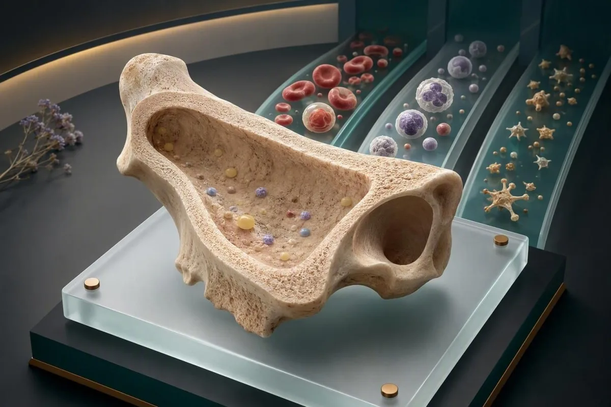
Aplasiaanemia produserer vanligvis et markant tre-linjers CBC-mønster: anemi, lave hvite blodlegemer og lave blodplater med en upassende lav retikulocytt-telling. Mønsteret krever rask klinisk bekreftelse fordi flere reversible og alvorlige tilstander kan se like ut ved første øyekast.


- Панкytапенія betyr lave røde blodlegemer, hvite blodlegemer og blodplater; det er et mønster, ikke en diagnose.
- Абсалютны падлік ретыкулоцитов under 20 × 10⁹/L støtter markert redusert beinmargsproduksjon når anemi er til stede.
- Alvorlig aplastisk anemi er definert av hypocellulær beinmarg pluss minst to av: ANC under 0,5 × 10⁹/L, blodplater under 20 × 10⁹/L, eller retikulocytter under 20 × 10⁹/L.
- ANC under 0,5 × 10⁹/L med feber på 38,0 °C eller høyere krever akuttvurdering samme dag.
- Blodplater under 10 × 10⁹/L medfører en betydelig risiko for spontan blødning og krever vanligvis akutt sykehusrettet behandling.
- Макрацытоз kan forekomme ved aplastisk anemi, men en MCV over 110 fL bør få klinikere til aktivt å utelukke B12- eller folatmangel.
- Blasts, marcada displasia, células em lágrima ou esquizócitos em um esfregaço apontam para longe da anemia aplásica não complicada e alteram a investigação.
- Не займацца самалячэннем um hemograma completo tri-linear baixo com ferro isoladamente; o ferro não restaura a produção da medula e pode atrasar a avaliação urgente.
CBC-mønsteret som vekker bekymring for aplastisk anemi
Ан exame de sangue para anemia aplásica geralmente mostra pancitopenia: hemoglobina baixa, neutrófilos baixos e plaquetas baixas ocorrendo juntos, enquanto a resposta de reticulócitos permanece baixa em vez de compensatória. Essa combinação deve desencadear revisão clínica urgente, especialmente se as contagens caíram em mais de um hemograma anterior.
Em adultos, um intervalo de referência típico do hemograma completo é hemoglobina de aproximadamente 120–155 g/L para muitas mulheres e 135–175 g/L para muitos homens, leucócitos 4,0–11,0 × 10⁹/L e plaquetas 150–400 × 10⁹/L; os laboratórios definem seus próprios limites. A anemia aplásica diminui todas as três linhagens porque a medula não está produzindo elementos celulares novos suficientes. Para uma orientação básica, consulte nosso guia para таму, што ўключае CBC.
O padrão é mais informativo do que qualquer resultado isolado baixo. Uma hemoglobina de 92 g/L após perda menstrual intensa pode ser deficiência de ferro, enquanto hemoglobina de 92 g/L mais ANC de 0,7 × 10⁹/L e plaquetas de 34 × 10⁹/L é um problema de produção da medula até prova em contrário. Na minha experiência clínica, o perigo não é a anemia isolada – é o risco de infecção e sangramento que chegam silenciosamente junto com ela.
Кантэсці — гэта Аналізатар крыві са штучным інтэлектам que lê os componentes do hemograma completo como um padrão interligado, não como três bandeiras vermelhas não relacionadas. Nosso sistema pode ajudar a organizar o relatório e os resultados anteriores, mas não pode diagnosticar falência medular ou substituir uma avaliação hematológica urgente. A partir de 26 de agosto de 2026, qualquer nova citopenia tri-linear merece a revisão de um clínico real em poucos dias, não um acompanhamento anual de rotina.
Um VCM de aparência normal não torna a pancitopenia benigna. A anemia aplásica precoce pode ser normocítica, levemente macrocítica ou ocasionalmente microcítica se a deficiência de ferro coexistir; a resposta da medula e o esfregaço periférico geralmente fornecem mais direcionamento do que o VCM isoladamente. A regra prática do Dr. Thomas Klein é simples: pergunte se a medula está respondendo antes de assumir uma explicação nutricional.
Por que o padrão é frequentemente perdido
Um hemograma completo pode sinalizar cada resultado separadamente, de modo que os pacientes às vezes se concentram no menor número e negligenciam o aglomerado. Uma queda nas plaquetas de 260 para 92 × 10⁹/L, mesmo que tecnicamente acima de um limiar de emergência, é clinicamente significativa quando a hemoglobina e os neutrófilos estão caindo em paralelo. Tendências de exames de sangue muitas vezes revelam essa trajetória mais cedo do que um único relatório.
Anemi pluss en lav retikulocytt-telling: ledetråden om beinmargsutgang
A нізкая колькасць ретыкулоцитов em uma pessoa anêmica significa que a medula não está substituindo os glóbulos vermelhos adequadamente, e é uma das pistas mais úteis que apoiam a anemia aplásica. A contagem absoluta de reticulócitos é mais importante do que a porcentagem de reticulócitos, porque uma porcentagem pode parecer enganosamente normal quando as células vermelhas totais são baixas.
A maioria dos laboratórios relata um intervalo de referência absoluto de reticulócitos em adultos em torno de 25–100 × 10⁹/L, embora os métodos variem. Nos critérios de anemia aplásica grave, uma contagem absoluta de reticulócitos abaixo de 20 × 10⁹/L é uma das medidas definidoras de baixa produção quando a celularidade da medula está marcadamente reduzida. Um detalhado кіраўніцтва па колькасці ретыкулоцитов explica por que resultados apenas em porcentagem podem enganar.
Em contraste, perda sanguínea aguda ou hemólise geralmente devem aumentar os reticulócitos após vários dias, muitas vezes acima de 100 × 10⁹/L se o suprimento de ferro e a função da medula estiverem intactos; haptoglobina baixa, bilirrubina indireta elevada e LDH elevado apoiam hemólise; essas são uma história fisiológica diferente da aplasia. A distinção é importante porque o tratamento da “anemia” sem identificar produção versus destruição é o que causa atrasos.
Uma paciente que vi com hemoglobina de 76 g/L tinha uma porcentagem de reticulócitos de 1,2%, que parecia normal até que a contagem absoluta foi calculada em 18 × 10⁹/L. Essa não foi uma resposta de recuperação. Quando a anemia é substancial, os clínicos podem usar um índice de produção de reticulócitos, mas os reticulócitos absolutos são geralmente menos propensos a erros no triagem do dia a dia.
Os reticulócitos podem estar baixos transitoriamente durante infecção grave, após quimioterapia, com toxicidade alcoólica ou em doença renal avançada, portanto este resultado não é específico para anemia aplásica. A revisão de Young de 2018 no New England Journal of Medicine enfatiza que o diagnóstico requer avaliação da medula e exclusão de alternativas herdadas, imunes, tóxicas e clonais – não um resultado de reticulócitos isoladamente (Young, 2018).
Lave nøytrofiler: CBC-tallet som endrer infeksjonsurgens
Гэты абсалютную колькасць нейтрофілаў (ANC) лепш прагназуе рызыку бактэрыяльных і грыбковых інфекцый, чым агульны падлік лейкацытаў. ANC ніжэй за 0,5 × 10⁹/л з'яўляецца цяжкай нейтропеніяй, а ліхаманка ад 38,0°C адзін раз або 38,0°C, якая захоўваецца на працягу гадзіны, патрабуе экстранай ацэнкі.
ANC разлічваецца як лейкацыты, памножаныя на адсотак нейтрофілов плюс адсотак палачак. Напрыклад, WBC 1,4 × 10⁹/л з 40% нейтрофіламі дае ANC 0,56 × 10⁹/л — значна больш паказальна, чым проста агульная колькасць. Наш арыенцірам для ANC тлумачыць варыяцыі ўзросту і цяжарнасці, але парог 0,5 × 10⁹/л застаецца ключавой небяспечнай мяжой для дарослых.
Цяжкая апластычная анемія часта выклікае нейтропенію без таксічнай грануляцыі або зсуву налева, якія могуць суправаджаць бактэрыяльную інфекцыю. Тым не менш, у пацыента можа быць як згасанне касцявога мозгу, так і інфекцыя; адсутнасць высокага падліку лейкацытаў ніколі не выключае сепсіс. Ліхаманка, дрыжыкі, новая спутанность свядомасці, дыхавіца або хуткае пагаршэнне болю ў горле не павінны чакаць паўторнага амбулаторнага CBC.
Вельмі цяжкая апластычная анемія выкарыстоўвае яшчэ больш нізкі парог ANC: ніжэй за 0,2 × 10⁹/л. Прычына аддзялення 0,2 ад 0,5 з'яўляецца практычнай — пры такім падліку звычайныя аральныя і скурныя арганізмы могуць стаць небяспечнымі пры невялікім мясцовым пачырваненні або гнойным выдзяленні. У бальніцы лекары могуць атрымаць пасевы і пачаць эмпірычнае антымікробнае лячэнне да таго, як прычына будзе цалкам устаноўлена.
Нізкі падлік лімфацытаў можа сустракацца, але не замяняе ANC пры прыняцці рашэнняў аб вострым рызыцы. Калі няма разгорнутай формы, запытайце яе ў лабараторыі або ў лекара, замест таго, каб здагадвацца па агульным WBC. Сімптомы нізкіх лімфацытаў патрабуюць далейшага назірання, але глыбіня нейтрофілов вызначае рашэнне ў той жа дзень.
Калі ліхаманка важная больш, чым раздрукоўка CBC
Тэмпература 38,0°C у чалавека з ANC 0,4 × 10⁹/л з'яўляецца тэрміновай, нават калі ён адчувае сябе “больш-менш нармальна”, а ліхаманка зніжаецца пасля парацэтамолу. Антыпірэтыкі могуць знізіць бачны сігнал, не лечачы асноўную праблему. Вазьміце CBC і спіс лекаў у экстраны кабінет; не прымайце рэшткі антыбіётыкаў перад пасевамі, калі гэта не будзе рэкамендавана лекарам.
Lave blodplater: blødningsgrenser ved mistenkt beinmargssvikt
Трамбацыты ніжэй за 50 × 10⁹/л павялічваюць сінякі і рызыку крывацёкаў, звязаных з працэдурамі, у той час як колькасць ніжэй за 10 × 10⁹/л можа выклікаць спантанныя крывацёкі. Пры апластычнай анеміі трамбацытапенія звычайна сустракаецца разам з анеміяй і нейтропеніяй, а не як ізаляваная праблема трамбацытаў.
Пецехіі, насавыя крывацёкі, якія доўжацца больш за 20 хвілін, крывацёк з дзёсен, моцныя менструальныя крывацёкі, кроў у мачы, чорны крэсла або раптоўны моцны галаўны боль патрабуюць тэрміновага медыцынскага кантакту пры нізкім узроўні трамбацытаў. Колькасць трамбацытаў 18 × 10⁹/л не з'яўляецца аўтаматычна катастрафічнай, але гэта не той паказнік, які варта кіраваць парадамі з інтэрнэту. Глядзіце наша практычнае абмеркаванне парогаў трамбацытаў перад стаматалагічным лячэннем.
Мазок дапамагае адрозніць зніжэнне выпрацоўкі ад перыферычнага разбурэння. Пры імуннай трамбацытапеніі трамбацыты могуць быць адзіным нізкім паказчыкам, і часта бачныя больш буйныя маладыя трамбацыты; апластычная анемія часцей мае нізкую або нармальную фракцыю няспелых трамбацытаў, таму што зніжаецца сама выпрацоўка. Гэта тэндэнцыі, а не правілы, і чытаць мазок павінен падрыхтаваны марфолаг.
Пазбягайце аспірыну, ібупрафэну, напроксена і нерэцэптурных мегадаз алею рыбы, пакуль лекар не агледзіць значную трамбацытапенію, бо яны могуць парушыць функцыю трамбацытаў, нават калі колькасць не змянілася. Алкаголь можа пагоршыць падаўленне касцявога мозгу і функцыю трамбацытаў. Ацэтамінафен/парацэтамол таксама не заўсёды падыходзіць — дазоўка і стан печані па-ранейшаму важныя.
Колькасць трамбацытаў можа быць ілжыва нізкай, калі трамбацыты згортваюцца ў ўзоры EDTA. Лабараторыя можа паведаміць “згорткі трамбацытаў прысутнічаюць” і паўтарыць падлік у цытратным шпрыцы; гэта называецца псеўдатромбацытапенія. Гэта тлумачыць ізаляваныя нізкія трамбацыты, а не пераканаўчую панцитопению з нізкім падлікам ретыкулацытаў.
Hvordan klinikere graderer alvorlig og svært alvorlig aplastisk anemi
Цяжкая апластычная анемія патрабуе гіпаклетачнага касцявога мозгу — звычайна ніжэй за 25% клетачнасць — плюс па меншай меры два перыферыйных крытэрыі: ANC ніжэй за 0,5 × 10⁹/л, трамбацыты ніжэй за 20 × 10⁹/л або абсалютныя ретыкулацыты ніжэй за 20 × 10⁹/л. Вельмі цяжкае захворванне адпавядае гэтым крытэрыям пры ANC ніжэй за 0,2 × 10⁹/л.
Гэтыя парогі паходзяць з даўно існуючай сістэмы Camitta, але яны з'яўляюцца інструментамі сартавання і планавання лячэння, а не дыягназам, зробленым па электроннай табліцы. Касцяны мозг можа быць лакальным, а нядаўняя трансфузія можа змяніць выгляд анеміі. Клініцысты таксама ўлічваюць сімптомы, гісторыю інфекцый, патрэбу ў трансфузіі, узрост, варыянты донараў і прыкметы кланальнага расстройства.
Умераная апластычная анемія вызначаецца не так дакладна. Чалавек з трамбацытамі 58 × 10⁹/л, ANC 0,9 × 10⁹/л і гемаглабінам 98 г/л можа не адпавядаць крытэрыям цяжкай формы, але ўсё яшчэ можа пагоршыцца альбо патрабаваць лячэння. Менавіта гэтая неадназначнасць робіць серыйныя CBC і назіранне ў гематалогіі больш важнымі, чым спроба прысвоіць супакойлівую этыкетку дома.
Кіруючы прынцып Брытанскага таварыства гематалогіі апісвае даследаванне касцявога мозгу, цытагенетыку, тэсціраванне плыні на наяўнасць клонаў PNH і ацэнку спадчыннай недастатковасці касцявога мозгу як часткі дыягнастычнага шляху (Killick et al., 2016). Нармальны цытагенетычны вынік зніжае занепакоенасць некаторымі миелодиспластическими сіндромамі, але сам па сабе не даказвае імунную апластычную анемію.
Kantesti AI падкрэслівае камбінацыі, якія перасякаюць тэрміновыя парогі, уключаючы ANC ніжэй за 0,5 × 10⁹/л і трамбацыты ніжэй за 20 × 10⁹/л, так што карыстальнікі могуць убачыць кластар перад прызначэннем. Наш аналіз з'яўляецца падказкай бяспекі, а не ацэнкай цяжкасці; асноўныя крытэрыі патрабуюць клінічнага агляду і дадзеных абследавання касцявога мозгу. Для кантэксту адзінкавых паведамленняў, прачытайце таго, што азначае «па-за межамі».
MCV, RDW og blodutstryksfunn som avgrenser CBC-mønsteret
Апластычная анемія часта мае нармальны або злёгку павышаны MCV, але MCV сам па сабе не можа адрозніць яе ад дэфіцыту B12, дэфіцыту фалійнай кіслаты, уздзеяння алкаголю, захворвання печані або миелодисплазии. Ручны мазок перыферычнай крыві неабходны, калі панцитапенія новая, прагрэсіўная або невытлумачальная.
Дыяпазон спасылак MCV звычайна складае 80–100 фл. Слабая макроцитоз каля 101–105 фл можа адбывацца пры апластычнай анеміі, таму што стрэсавая выпрацоўка касцявога мозгу змяняе паспяванне эрытрацытаў, але MCV вышэй за 110 фл павінна выклікаць абдуманы агляд B12, фалійнай кіслаты, лекаў, алкаголю, шчытападобнай залозы і печані. Параўнайце паказчыкі ў нашым Кіраўніцтва па MCV супраць MCH.
RDW вымярае варыяцыю памеру эрытрацытаў і часта складае 11,5–14,5%, у залежнасці ад аналізатара. Нармальны RDW не выключае недастатковасць касцявога мозгу; насамрэч, даволі аднастайная папуляцыя зніжаных эрытрацытаў можа адбыцца, калі выпрацоўка проста змяншаецца. Рэзка высокі RDW можа замест гэтага паказваць на змешаныя дэфіцыты, нядаўнюю трансфузію, аднаўленне пасля лячэння або дысплазію.
Бласты, проміелацыты, выяўленая гіпагрануляцыя, псевдо-Пельгеровскія нейтрофілы або значныя дыспластычныя формы перамяшчаюць занепакоенасць у бок вострага лейкозу або миелодиспластического сіндрому і павінны паскорыць ацэнку спецыяліста. Шыстацыты паказваюць у іншым кірунку - фрагментацыя эрытрацытаў пры мікраангіяпатыі - і могуць быць тэрміновымі пры спалучэнні з нізкім утрыманнем трамбацытаў, пашкоджаннем нырак або неўралагічнымі сімптомамі. Наш кіраўніцтва па шыстацытах ахоплівае гэты асобны надзвычайны шаблон.
Кантэсці — гэта Інструмент аналізу вынікаў аналізу крыві з падтрымкай AI які можа параўноўваць MCV, RDW, рэтыкулацыты і гістарычныя падлікі, але ён не замяняе прагледжаны мазок. На мой вопыт, менавіта ў мазку відавочна простая “нізкая КБС” ператвараецца ў дакладна сфакусаванае дыягнастычнае пытанне. гід па тэхналогіі тлумачыць, як наш AI захоўвае арыгінальныя адзінкі і эталонныя інтэрвалы лабараторыі.
B12-, folat- og kobbermangel kan etterligne beinmargssvikt
Дэфіцыт вітаміна B12, фалійнай кіслаты і медзі можа выклікаць панцитапенію і павінен быць выключаны перад пазначэннем шаблону КБС як апластычная анемія. Гэтыя дэфіцыты асабліва верагодныя пры абмежавальных дыетах, парушэнні ўсмоктвання, барыятрычнай хірургіі, ужыванні вялікай колькасці цынку або некаторых лекаў.
Дэфіцыт B12 часта выклікае макроцитоз, гіперсегментаваныя нейтрофілы і павышэнне метилмалоновой кіслаты, але ў некаторых пацыентаў MCV ніжэй за 100 фл, калі сустракаецца дэфіцыт жалеза. Серумны B12 ніжэй за 148 пмоль/л, прыкладна 200 пг/мл, часта лічыцца дэфіцытам, у той час як 148–221 пмоль/л можа патрабаваць пацвярджэння метилмалоновой кіслаты або гомацыстэіна. Наша параўнанне B12 і фалійнай кіслаты тлумачыць, чаму толькі фалійная кіслата можа маскіраваць гематалагічныя прыкметы, пакуль неўралагічнае пашкоджанне працягваецца.
Дэфіцыт фалійнай кіслаты можа знізіць усе тры лініі ў запушчаных выпадках, асабліва пры дрэнным спажыванні, ужыванні алкаголю, парушэнні ўсмоктвання, метотрэксаце, триметоприме або некаторых супрацьсутаргавых прэпаратах. Фалійная кіслата ў эрытрацытах можа лепш адлюстроўваць запасы тканін, чым адзінкавы ўзровень у сыроватцы крыві ў некаторых сітуацыях, але даступнасць і інтэрпрэтацыя аналізаў вар'іруюцца. Не пачынайце прыём высокіх доз фалійнай кіслаты перад ацэнкай B12, калі ў лекара няма канкрэтнай прычыны.
Дэфіцыт медзі правяраецца радзей, але ён можа выклікаць анемію і нейтропенію і можа нагадваць миелодисплазію пры даследаванні касцявога мозгу. Лішак дабаўкі цынку з'яўляецца класічнай зваротнай прычынай, таму што цынк індукуе кішачны металатэінеін, які выбарча звязвае медзь. абследаванне на нізкі ўзровень медзі асабліва актуальна для ўздзеяння на крэмы для зубных пратэзаў, барыятрычнай хірургіі або доз цынку вышэй за 40 мг у дзень.
Харчаванне можа быць часткай адказу, але яно не павінна станавіцца прычынай адтэрміноўкі ацэнкі касцявога мозгу пры цяжкай панцитапеніі. Замяшчэнне жалеза, B12, фалійнай кіслаты і медзі адаптаваны да пацверджаных дэфіцытаў і медыцынскага кантэксту; неразборлівая дабаўка можа зацемніць карысныя вынікі тэстаў. Гэта адна з тых абласцей, дзе кантэкст мае больш важнае значэнне, чым адзіная мяжовая лічба.
Medikament-, toksin- og virusårsaker til pancytopeni som bør sjekkes først
Медыкаменты, таксіны і вірусныя захворванні могуць зніжаць выпрацоўку касцявога мозгу і прыводзіць да змены ў CBC, падобнай на апластычную анемію, часам зваротнай. Поўная гісторыя ўздзеяння павінна ўключаць прызначаныя лекі, нядаўнія антыбіётыкі, супрацьсутаргавыя прэпараты, імунатэрапію, раслінныя прадукты, растваральнікі, алкаголь і рэкрэацыйныя рэчывы.
Хіміятэрапія з'яўляецца відавочнай прычынай, але менш відавочныя фактары ўключаюць хларамфенікол у рэгіёнах, дзе ён усё яшчэ выкарыстоўваецца, прэпараты супраць шчытападобнай залозы, лінезалід, некаторыя супрацьсутаргавыя прэпараты і імунамадулюючыя тэрапіі. Час мае значэнне: прамая таксічнасць прэпарата для касцявога мозгу часта з'яўляецца праз тыдні ці месяцы пасля ўздзеяння, у той час як імунныя рэакцыі на прэпараты могуць быць менш прадказальнымі. Ніколі не спыняйце прызначаны прэпарат раптоўна без кансультацыі з лекарам, які кіруе цитопенией.
Апластычная анемія, звязаная з гепатытам, можа развівацца праз тыдні ці месяцы пасля вострага эпізоду гепатыту, часта ў маладых людзей, і стандартнае тэсціраванне на гепатыт A, B, C, E, ВІЧ, ВЭБ, ЦМВ і парвовирус можа быць выбрана на аснове анамнезу. Адмоўны вірусны скрынінг не выключае імунную апластычную анемію. Ён проста выключае важныя супастаўныя тлумачэнні і накіроўвае прафілактыку інфекцый.
Уздзеянне бензолу застаецца актуальным прафесійным і хатнім рызыкай, у тым ліку праца ў дрэнна вентыляваных прамысловых умовах, зварот з палівам і некаторымі растваральнікамі. Лекары пытаюцца пра задачы на працы, а не пра назвы пасад, таму што “офіс-менеджар у пастаўшчыка фарбы” усё яшчэ можа ўключаць перыядычнае ўздзеянне на склад. Прыносьце назвы прадуктаў або пашпарты бяспекі, калі яны ёсць; няясныя ўспаміны часта сустракаюцца, і ніхто не павінен адчуваць сябе вінаватым.
CBC, зроблены неўзабаве пасля значнага віруснага захворвання, можа запатрабаваць паўторнага правядзення на працягу некалькіх дзён ці тыдняў, калі пацыент стабільны, але цяжкія паказчыкі не варта пакідаць без увагі. Змены CBC пасля хваробы могуць дапамагчы вызначыць час, у той час як ліхаманка з цяжкай нейтропеніей застаецца надзвычайнай сітуацыяй незалежна ад падазронага трыгера.
Årsaker til pancytopeni som ser annerledes ut enn aplastisk anemi
Панцытапенія мае шмат прычын, і апластычная анемія адрозніваецца гіпоклетачным касцяным мозгам без злаякаснага прарастанне або значнай дысплазіі. Асноўныя альтэрнатывы ўключаюць востры лейкоз, миелодиспластический сіндром, прарастанне касцявога мозгу, гіперспленізм, цяжкія дэфіцытныя стану, гемафагацытарныя сіндромы, інфекцыі і ўплыў лекаў.
Востры лейкоз можа выклікаць нізкія паказчыкі, таму што бласты выцясняюць нармальны касцяны мозг, але цыркулюючыя бласты бачныя не заўсёды. Миелодиспластический сіндром становіцца больш верагодным пры дысплазіі, характэрных цитогенетических зменах або ўстойлівай макроцитозе у пажылых людзей, хоць узрост сам па сабе нічога не даказвае. Шлях у нашым кіраўніцтве па тэсціраванні на рак крыві паказвае, чаму мазкі, проточная цытаметрыя і даследаванні касцявога мозгу з'яўляюцца дадатковымі.
Гиперспленизм зніжае цыркулюючыя паказчыкі за кошт секвестрацыі, звычайна пры спленомегалии, портальной гіпертэнзіі, захворваннях печані або некаторых гематалагічных засмучэннях. Рэтыкулацыты могуць быць нармальнымі або павышанымі, а не значна зніжанымі, а клетачнасць касцявога мозгу звычайна захоўваецца або павялічваецца. Ультрагукавое даследаванне можа быць карысным, але яно не адмяняе неабходнасць даследавання падазронага мазка або цяжкай нейтропеніі.
Параксізмальная начная гемаглабінурыя (PNH) можа перакрывацца біялагічна з апластычнай анеміяй, таму лекары часта прызначаюць высокаадчувальную проточною цытаметрыю для гранулоцитов і моноцитов з дэфіцытам GPI-анкера. Цёмная мача не з'яўляецца неабходнай або спецыфічнай. Выяўленне невялікага клона PNH можа падтрымліваць кантэкст імуннай недастатковасці касцявога мозгу, але лячэнне залежыць ад памеру клона, гемолізу, рызыкі тромбозу і агульнай карціны касцявога мозгу.
Кантэсці — гэта сервісі інтерпретації тестів ШІ якія могуць выявіць шаблоны панцитопеніі і патэнцыйныя альтэрнатыўныя падказкі, уключаючы білірубін, ЛДГ, паказчыкі печані і гістарычнае зніжэнне трамбацытаў. Гэта не можа вызначыць, ці з'яўляецца касцяны мозг гіпоклетачным, інфільтраваным або дыспластычным — гэта пытанні для гематалогіі і паталагічнай анатоміі. Чэк-ліст для другой думкі можа дапамагчы пацыентам падрыхтаваць сфакусаваныя пытанні.
Akutte varselsignaler: når lave CBC-resultater krever akuttmedisinsk hjelp
Неадкладная дапамога неабходная пры ліхаманцы 38,0°C або вышэй з ANC ніжэй за 0,5 × 10⁹/л, некантралюемым крывацёкам, чорным кале, кашлем з крывёй, моцным новым галаўным болем, разгубленасцю, стратай прытомнасці, болем у грудзях або дыхавіцай у спакоі. Не чакайце онлайн-трактоўкі, калі нешта з гэтага адбываецца.
Цяжкая анемія можа парушыць дастаўку кіслароду нават без бачнага крывацёку. Гемаглабін ніжэй за 70 г/л з'яўляецца распаўсюджаным парогам для разгляду пералівання крыві ў стабільных шпітальных пацыентаў, але сімптомы, актыўнае крывацёк, сардэчна-сасудзістыя захворванні, цяжарнасць і хранічнасць змяняюць рашэнні. Чалавек з гемаглабінам 82 г/л, які губляе прытомнасць або дыхавіцу ў стане спакою, патрабуе ацэнкі больш тэрмінова, чым здаровы чалавек з такім жа паказчыкам.
Трамбацыты ніжэй за 10 × 10⁹/л часта лечацца неадкладнымі рашэннямі спецыяліста, таму што больш верагодным становіцца спантаннае слізіста-кішачнае або ўнутранае крывацёк. Пазбягайце кантактных відаў спорту, нутрацягліцавых ін'екцый, рэктальных тэрмометраў і стаматалагічных працэдур, пакуль лекары не параяць іншае. Калі ёсць траўма галавы пры любым нізкім узроўні трамбацытаў, неадкладна звярніцеся за ацэнкай, нават калі сімптомы здаюцца нязначнымі.
Нармальная тэмпература не гарантуе бяспекі пры цяжкай нейтропеніі; пажылыя людзі, людзі, якія прымаюць стэроіды, і вельмі хворыя пацыенты могуць не мець ліхаманкі. Новыя язвы ў роце, боль пры глытанні, перыянальная боль або хуткае пагаршэнне стомленасці могуць быць раннімі прыкметамі інфекцыі. Лабараторныя шаблоны сепсісу даюць кантэкст, але сімптомы і жыццёва важныя паказчыкі вызначаюць неадкладныя дзеянні.
Доктар Томас Кляйн раіць пацыентам фатаграфаваць або спампоўваць арыгінальны справаздачу, запісваць дату аналізу і прыносіць усе кантэйнеры з лекамі і дадаткамі для тэрміновага агляду. Гэты невялікі крок часта дазваляе выявіць своечасовае ўздзеянне наркотыкаў або высветліць, ці быў “новы” нізкі паказчык ужо ў тэндэнцыі. Не кіруйце аўтамабілем, калі вы адчуваеце галавакружэнне, непрытомнасць або актыўнае крывацёк.
Hvilke tester som bekrefter eller utelukker aplastisk anemi etter CBC
Агульны аналіз крыві (CBC) не можа пацвердзіць апластычную анемію; пацвярджэнне звычайна патрабуе пункцыі і біяпсіі касцявога мозгу, якія паказваюць гіпаклетачнасць, а таксама аналізы, якія выключаюць кланальныя, спадчынныя, інфекцыйныя, харчовыя і звязаныя з уздзеяннем прычыны. Тэмп тэсціравання залежыць ад цяжкасці паказчыкаў і сімптомаў.
Узор касцявога мозгу ацэньвае клетачнасць, архітэктуру, бласты, фіброз, запасы жалеза і дысплазію. У дарослых чаканая клетачнасць прыкладна роўная 100 мінус узрост як грубая ацэнка навучання, але гэта не дакладна, і якасць узору мае значэнне. 30-гадовы чалавек з клетачнасцю касцявога мозгу 10% вельмі адрозніваецца ад 80-гадовага чалавека з 10%, аднак абодва патрабуюць экспертнай інтэрпрэтацыі ў кантэксце.
Цытагенетыка, храмасомны мікрамасіў або секвенсораўныя панэлі могуць дапамагчы адрозніць гіпаклетачны миелодиспластический сіндром ад набытай апластычнай анеміі. Тэсціраванне на клоны PNH, захворванні тэламераў, анемію Фанконі і іншыя спадчынныя захворванні недастатковасці касцявога мозгу выбіраецца па ўзросце, сямейнай гісторыі, фізічных знаходках і планаванні трансплантацыі. Генетычнае тэсціраванне не зарэзервавана толькі для дзяцей; усё часцей прызнаецца спадчыннае захворванне, якое пачынаецца ў дарослым узросце.
Базавыя аналізы часта ўключаюць B12, фоліевую кіслату, медзь, профіль печані і нырак, даследаванні шчытападобнай залозы пры паказаннях, маркеры гемолізу, вірусную сералогію, тэставанне на цяжарнасць пры адпаведных абставінах і аўтаімунныя даследаванні, прызначаныя гісторыяй. Мэта не ў тым, каб прызначыць усе аналізы. Гэта пазбегнуць прапуску адваротлівых прычын, адначасова хутка пераходзячы да канчатковай ацэнкі.
Kantesti AI можа арганізаваць справаздачу па гэтых катэгорыях і вызначыць адсутныя асновы, але клінічнае кіраванне мае значэнне, калі вынікі маюць высокія стаўкі. Наш стандарты медыцынскай валідацыі апісвае мяжу паміж інтэрпрэтацыяй з дапамогай ІІ і медыцынскім дыягназам. Спытайце, хто будзе разглядаць узор касцявога мозгу і ці будзе ён ацэнены гематапатолагам.
Når man skal gjenta en CBC før man antar at resultatet er reelt
Паўторны агульны аналіз крыві (CBC) мэтазгодны, калі згортванне ўзору, агрэгацыя трамбацытаў, развядзенне інфузійнымі вадкасцямі або сцягі аналізатара могуць сказіць вынік, але паўторнае тэсціраванне ніколі не павінна затрымліваць экстраную дапамогу пры цяжкіх цитапеніях або трывожных сімптомах. Лабараторыі часта могуць пацвердзіць вынік з арыгінальнай прабіркі на працягу некалькіх гадзін.
Зліпанне трамбацытаў EDTA можа прывесці да ілжыва нізкага ліку трамбацытаў, у той час як часткова згорнутая прабірка можа непрадказальна знізіць колькасць трамбацытаў і лейкацытаў. Ручны агляд мазка і аналіз колькасці трамбацытаў у цытратнай прабірцы могуць вырашыць праблему. паўторнае тэсціраванне пасля гемолізу кіраўніцтва тлумачыць, чаму каментарыі да якасці ўзору з'яўляюцца клінічна карыснымі, а не простай лабараторнай падрыхтоўкай.
Інфузійная вадкасць можа разводзіць гемаглабін і трамбацыты, асабліва пасля некалькіх літраў, але яна рэдка тлумачыць глыбока нізкі ўзровень ANC або рэтыкулацытаў. Наадварот, абязводжванне можа часова канцэнтраваць значэнні і хаваць глыбіню анеміі. Параўнайце час адбору, нядаўнія вадкасці, пераліванні, вострае захворванне і папярэднюю базу перад тым, як зрабіць выснову, што лікавая змена адлюстроўвае біялогію касцявога мозгу.
Пераліванне ўскладняе інтэрпрэтацыю, таму што донарскія эрытрацыты могуць палепшыць гемаглабін без карэкцыі нейтропеніі, трамбацытаў або рэтыкулацытаў. Павышэнне гемаглабіну пасля пералівання не варта блытаць з аднаўленнем касцявога мозгу. Таму клініцысты адсочваюць даты пералівання разам з тэндэнцыямі рэтыкулацытаў і інтэрвалам да наступнага неабходнага пералівання.
Лабараторная “дэльта-праверка” можа адзначыць рэзкае адхіленне ад папярэдніх значэнняў, але сапраўднае збой касцявога мозгу можа развіцца паміж звычайнымі аналізамі. Калі справаздача выглядае непраўдападобнай, неадкладна патэлефануйце лекару, які прызначыў аналіз, або ў лабараторыю; не проста паўтарайце яе ў аддаленым пункце аналізу і губляйце сувязь. Арыгінальная лабараторыя можа параўнаць сцягі аналізатара і мікраскапію.
Hva man ikke skal gjøre mens man venter på hematologisk vurdering
Не пачынайце прыём жалеза, высокіх доз фоліевай кіслаты, травяных “кровездавальнікаў”, аспірыну або рэшткавых антыбіётыкаў толькі таму, што агульны аналіз крыві (CBC) нізкі. Пры падазрэнні на апластычную анемію найбяспечнейшымі першымі дзеяннямі з'яўляюцца тэрміновы медыцынскі агляд, прафілактыка інфекцый, прафілактыка крывацёкаў і поўная гісторыя прыёму лекаў і дадаткаў.
Дэфіцыт жалеза распаўсюджаны, але жалеза не тлумачыць нізкі ўзровень нейтрофілов і трамбацытаў у большасці дарослых. Прыём 45–65 мг элементарнага жалеза кожны дзень можа выклікаць млоснасць, завала і ўводзіць у зман змены ў аналізах жалеза без ліквідацыі збою касцявога мозгу. Праверце ферытын, насычэнне трансферыну, CRP і агульную карціну CBC перад тым, як меркаваць, што стомленасць роўная дэфіцыту жалеза; наш рэсурс па аналізах жалеза дапамагае з гэтымі адрозненнямі.
Узбягайце людных закрытых памяшканняў і блізкага кантакту з людзьмі з рэспіраторнымі сімптомамі, калі ANC моцна нізкі, але не ізалюйце сябе ад неабходнай медыцынскай дапамогі. Гігіена рук, своечасовая праверка тэмпературы пры захворванні і бяспека харчавання разумныя. Экстрэмальныя “нейтропенічныя дыеты” маюць абмежаваныя доказы і могуць пагоршыць харчаванне, таму запытайце ў лекара мясцовыя рэкамендацыі, а не прытрымлівайцеся абмежавальных спісаў у сацыяльных сетках.
Захоўвайце аднастаронкавую храналогію: першы ненармальны CBC, папярэдні нармальны CBC, інфекцыі за апошнія 6 месяцаў, пачатыя або спыненыя лекі, падарожжы, уздзеянне на працоўным месцы, пераліванні, сямейныя захворванні крыві і сімптомы крывацёку. Гэтая інфармацыя можа зэканоміць увесь візіт да лекара. Трэкер лабараторных вынікаў прапануе практычную структуру для запісу дат і адзінак.
Kantesti’s платформа інтэрпрэтацыі біямаркераў AI можа да помогне на потребителите да сравняват серийни CBC доклади и да подготвят въпроси на собствен език на 75+ езици. Това не е спешна услуга и не може да вижда симптоми, да преглежда пациент или да организира трансфузионна поддръжка. Използвайте го, за да разберете доклада, след което следвайте клиничния път, който вече е в ход.
Spørsmål som gjør en hematologisk avtale mer nyttig
Най-полезните въпроси по хематология са относно тежестта, вероятния механизъм, праговете за инфекция и кървене, и диагностичния план — а не дали една изолирана цифра е “лоша”. Носете оригинални доклади, защото тенденциите в ANC, тромбоцитите, хемоглобина, MCV и ретикулоцитите ръководят решенията.
Попитайте: “Отговарям ли на критериите за тежка апластична анемия и кои критерии отговарям?” Отговорът трябва да включва костномозъчна клетъчност, ANC, брой на тромбоцитите и абсолютен брой на ретикулоцитите. Ако диагнозата остане несигурна, попитайте кои находки подкрепят хипоцелуларен миелодиспластичен синдром, остра левкемия, хранителен дефицит или потискане, свързано с лекарства, и кой тест ще ги разграничи.
Поискайте изрични прагове за безопасност: при каква температура трябва да се явя спешно, кои симптоми на кървене надделяват над изчакването, кои болкоуспокояващи трябва да избягвам и мога ли да пътувам? Тези инструкции варират в зависимост от ANC и дълбочината на тромбоцитите. Човек с ANC 1,1 × 10⁹/л и тромбоцити 95 × 10⁹/л получава различен съвет от някой с ANC 0,18 × 10⁹/л и тромбоцити 8 × 10⁹/л.
Ако апластичната анемия е потвърдена, обсъждането може да включва имуносупресивна терапия, елтромбопаг по избрани протоколи и трансплантация на хемопоетични стволови клетки в зависимост от възрастта, наличността на донор, тежестта на заболяването и местния опит. Никакъв избор на лечение не трябва да се подразбира от онлайн обяснение на CBC. Очакваният ход и рисковете са лични, понякога наистина несигурни и най-добре се решават от екип за костномозъчна недостатъчност.
Наш Медыцынская кансультатыўная рада помага за определяне на принципите на клиничния преглед зад съдържанието на Kantesti, включително правилото, че моделите на CBC с висок риск изискват професионална ескалация. Вземете придружител, ако е възможно; тези срещи съдържат много непознати термини, а друг слушател подобрява запомнянето.
En trygg neste-trinnsplan etter en unormal pancytopeni CBC
Новата панцитопения с нисък брой ретикулоцити трябва да се третира като възможно нарушение на костномозъчното производство, докато клиницист не установи друга причина. Необходими са грижи в същия ден за температура с тежка неутропения, активно кървене, неврологични симптоми, болка в гърдите, припадък или задух в покой.
Първо, проверете точните стойности и единици: хемоглобин, WBC с ANC, тромбоцити, MCV, RDW и абсолютни ретикулоцити. Второ, сравнете ги с последния наличен CBC, дори такъв от преди 2 години. Стабилен лек нисък брой и бърз спад по трите линии не са един и същ клиничен проблем; CBC сравнение страница до страница може да направи тази разлика видима.
Трето, потърсете нивото на грижа, което вашите симптоми и прагове изискват. ANC под 0,5 × 10⁹/л с температура, тромбоцити под 10 × 10⁹/л или значително кървене не трябва да чакат стандартна препратка. За стабилна, но необяснима панцитопения, уговорете навременен преглед от клиницист и попитайте дали е необходим преглед на петно, повторен CBC, хранителни проучвания, вирусологични изследвания и хематологична препратка.
Четвърто, не интерпретирайте един “нормален” съпътстващ тест като успокоение. Нормалният феритин не изключва дефицит на B12, нормалното LDH не изключва костномозъчна недостатъчност, а нормалното MCV не отменя три ниски клетъчни линии. Целият модел, неговият времеви фактор и прегледът определят риска.
Kantesti AI поддържа структуриран преглед на резултатите и анализ на исторически тенденции, докато диагнозата и лечението остават с вашия клиничен екип. Тази статия е медицински прегледана съгласно стандартите, описани от процеса на клиничен надзор на Kantesti. Практическата цел не е да ви накара да се страхувате от всеки нисък флаг — тя е да разпознаете комбинациите, които никога не трябва да се пренебрегват.
Часта задаваныя пытанні
Jak wygląda niedokrwistość aplastyczna w badaniu morfologii krwi obwodowej (CBC)?
Anemia aplástica geralmente aparece como pancitopenia: hemoglobina baixa, leucócitos baixos — especialmente neutrófilos — e plaquetas baixas no mesmo CBC. A pista de suporte na produção é uma contagem absoluta de reticulócitos inapropriadamente baixa, frequentemente abaixo de 20 × 10⁹/L em doença grave. O MCV pode ser normal ou levemente elevado, portanto, um MCV normal não exclui a condição. O diagnóstico requer evidência de hipocelularidade na medula óssea e exclusão de outras causas de pancitopenia.
Aplasia pode ter glóbulos brancos normais no início?
Sim, a anemia aplásica inicial pode afetar inicialmente uma ou duas linhagens de células sanguíneas antes que a pancitopenia clara se desenvolva. Uma contagem decrescente de plaquetas ou anemia progressiva com reticulócitos absolutos abaixo de cerca de 25 × 10⁹/L pode preceder neutropenia grave. Os clínicos, portanto, comparam CBCs anteriores em vez de confiar em um único resultado normal de WBC. Uma nova tendência descendente em múltiplas linhagens deve levar a uma revisão médica oportuna.
Qual contagem de reticulócitos sugere anemia aplásica?
Un recuento absoluto de reticulocitos por debajo de 20 × 10⁹/L apoya la hipoproducción medular grave cuando existe anemia y es parte de los criterios de anemia aplásica grave. Muchos laboratorios utilizan un intervalo de referencia para adultos cercano a 25–100 × 10⁹/L, aunque el rango exacto varía según el método. El porcentaje de reticulocitos puede ser engañosamente normal en la anemia grave, por lo que se prefiere el recuento absoluto. Los reticulocitos bajos no son específicos de la anemia aplásica y también ocurren con deficiencia de nutrientes, enfermedad renal, infección y supresión relacionada con fármacos.
É perigoso um contagem de plaquetas de 20 na anemia aplásica?
Uma contagem de plaquetas de 20 × 10⁹/L é gravemente baixa e requer avaliação urgente dirigida pela hematologia, particularmente se houver sangramento nasal, sangramento gengival, sangramento menstrual intenso, fezes escuras, sangue na urina ou dor de cabeça. Contagens abaixo de 10 × 10⁹/L apresentam um risco maior de sangramento espontâneo e são frequentemente tratadas no hospital, de acordo com os sintomas e a causa da pessoa. Evite aspirina e AINEs, a menos que o médico assistente os aprove especificamente. A urgência aumenta ainda mais quando anemia e neutropenia também estão presentes.
När är lågt neutrofilvärde en nödsituation?
Un ANC inferior a 0,5 × 10⁹/L indica neutropenia grave e una temperatura di 38,0°C o superiore richiede una valutazione di emergenza in giornata. L'anemia aplastica molto grave utilizza un ANC inferiore a 0,2 × 10⁹/L, dove può verificarsi un'infezione grave con pochi sintomi locali. Brividi, nuova confusione, dispnea, deglutizione dolorosa o dolore perianale giustificano anche cure urgenti anche in assenza di febbre documentata. Il conteggio totale dei globuli bianchi da solo è meno utile dell'ANC per questa decisione.
A deficiência de vitamina B12 pode ser confundida com anemia aplásica?
A deficiência de vitamina B12 pode causar anemia, leucopenia, plaquetopenia, macrocitose e baixa contagem de reticulócitos, podendo assemelhar-se inicialmente à anemia aplásica. Níveis de B12 sérica abaixo de 148 pmol/L, aproximadamente 200 pg/mL, são comumente considerados deficientes, enquanto resultados limítrofes podem necessitar de teste de ácido metilmalônico. Neutrófilos hipersegmentados e um MCV acima de 110 fL favorecem a deficiência de B12 ou folato, mas a deficiência mista de ferro pode mascarar a macrocitose. O exame da medula óssea e testes sanguíneos direcionados separam essas condições quando o CBC é grave ou incerto.
Атрымаць аналіз крыві з AI сёння
Далучайцеся больш чым да 2 мільёнаў карыстальнікаў па ўсім свеце, якія давяраюць Kantesti для імгненнага, дакладнага аналізу лабараторных тэстаў. Загрузіце вынікі аналізу крыві і атрымайце поўную інтэрпрэтацыю біямаркераў 15,000+ за лічаныя секунды.
📚 Публікацыі даследаванняў са спасылкамі
Кляйн, Т., Мітчэл, С., і Вебер, Г. (2026). Кроў тыпу B (Rh-), даведнік па аналізе LDH і падліку ретыкулоцитов. Kantesti AI Medical Research.
Кляйн, Т., Мітчэл, С., і Вебер, Г. (2026). Дыярэя пасля галадання, чорныя плямкі ў кале і кіраўніцтва па ЖКТ 2026. Kantesti AI Medical Research.
📖 Знешнія медыцынскія спасылкі
⚕️ Медыцынская адмова ад адказнасці
Гэты артыкул прызначаны толькі для адукацыйных мэт і не з’яўляецца медычнай парадай. Заўсёды кансультуйцеся з кваліфікаваным спецыялістам аховы здароўя для прыняцця рашэнняў па дыягностыцы і лячэнні.
Сігналы даверу E-E-A-T
Вопыт
Клінічны агляд, які вядзе лекар, працэсаў інтэрпрэтацыі лабараторных паказчыкаў.
Экспертыза
Фокус лабараторнай медыцыны на тым, як біямаркеры паводзяць сябе ў клінічным кантэксце.
Аўтарытэтнасць
Напісана доктарам Томасам Кляйнам, з аглядам доктара Сара Мітчэл і праф. доктара Ганса Вебера.
Надзейнасць
Інтэрпрэтацыя на аснове доказаў з выразнымі шляхамі далейшых дзеянняў, каб паменшыць трывогу.