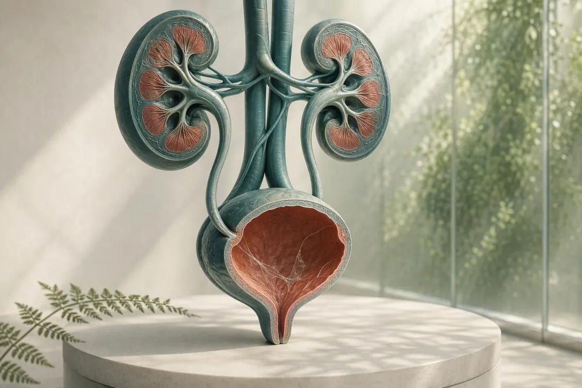
Slijm op een urine microscopisch verslag is meestal een probleem met de monsterafname, geen diagnose. De omliggende urinetest bevindingen, symptomen en de kwaliteit van het monster bepalen of verder onderzoek nodig is.


- Slijmdraden alleen zijn vaak onschadelijk, vooral in een enkel schoon-catch monster zonder urinewegsymptomen.
- Pyurie-drempel van meer dan 5 witte cellen per hoog-vermogen veld ondersteunt ontsteking van de urinewegen, maar bewijst geen bacteriële urineweginfectie.
- Kweektrigger is slijm plus branden, urgentie, koorts, flankpijn, nitrieten of significante witte cellen—niet alleen slijm.
- Plaveiselcellen van ongeveer meer dan 10 per hoog-vermogen veld geven vaak aan dat er sprake is van besmetting van de geslachtsdelen/huid en maken een urineuitslag minder betrouwbaar.
- Zichtbaar bloed met koliekachtige zijpijn vereist een snelle beoordeling op een steen, zelfs als er ook slijm wordt gerapporteerd.
- Zwangerschap verandert de drempel voor opvolging omdat asymptomatische bacteriurie meestal wordt gescreend met urinekweek, niet alleen met microscopie.
- Troebele urine kan kristallen, geconcentreerde urine, vaginale afscheiding, sperma of cellen weerspiegelen; het kan geen infectie diagnosticeren op uiterlijk.
- Herhaalmonster is zinvol wanneer slijm de enige ongebruikelijke bevinding is en de inzameling geen zorgvuldige midstream clean-catch was.
Wat slijmdraden bij urinetest meestal betekenen
Slijmdraden in urine zijn meestal strengen van normale beschermende afscheiding of genitale contaminatie, en een geïsoleerd resultaat wijst zelden op een nierziekte. De bevinding wordt zinvol wanneer deze verschijnt met urinewegsymptomen, witte bloedcellen, bacteriën, bloed of eiwit.
Laboratoria identificeren slijm als vezelige, doorschijnende strengen die onder de microscoop worden gezien; veel rapporten rangschikken het simpelweg als zeldzaam, weinig, matig of veel in plaats van een numeriek referentiebereik toe te kennen. Urine bevat van nature een kleine hoeveelheid glycoproteïne-rijk materiaal uit de urinewegen, en vaginale of urethrale afscheidingen kunnen tijdens de inzameling meer toevoegen.
De formulering van het rapport is minder alarmerend dan het klinkt. Vanaf 26 augustus 2026, is er geen gevalideerde slijmdraad telling die een urineweginfectie, steen, kanker of nierfalen diagnosticeert; het is een contextuele microscopische observatie. Voor een bredere kaart van de andere items op een dipstick en microscoop rapport, zie onze complete handleiding voor urineonderzoek.
In mijn klinische ervaring, een symptoomvrije volwassene met slijm gemarkeerd als “matig”, 0-2 witte bloedcellen per hoogvermogen veld, negatief nitriet, en geen bloed wordt meestal het beste geholpen door geruststelling of een correct verzamelde herhalingsmonster. Dr. Thomas Klein ziet vaak onnodige angst voor antibiotica ontstaan omdat een laboratoriumvlag wordt aangezien voor een ziekte-etiket.
Kantesti is een AI bloedonderzoek uitslag platform dat niergerelateerde bloedmarkers, zoals creatinine en eGFR, naast een urinesediment bevinding kan plaatsen—maar het kan een urineweginfectie niet alleen op basis van slijm diagnosticeren. Een urine microscopie resultaat heeft het klinische verhaal nodig dat erbij kwam.
Waarom een lab het markeert
Laboratoriuminformatiesystemen markeren slijm omdat het visueel aanwezig is, niet omdat het laboratorium een gevaarlijke afsnijding heeft vastgesteld. Een vlag betekent daarom “waargenomen” in plaats van “abnormale ziektemarker”, net zoals een geïsoleerde spoorbevinding geen behandeling kan vereisen.
Waar slijm in een urinemonster vandaan komt
Slijm in urine kan afkomstig zijn van de urethra, blaaswand, baarmoederhals of vagina, sperma, of de buitenste genitale huid tijdens de inzameling. De bron wordt vaak afgeleid uit de rest van het microscopische patroon in plaats van uit de strengen zelf.
De blaas en urethra hebben een beschermende oppervlakte laag die mucines bevat, waaronder uroplakin-geassocieerd materiaal, dat wrijving en microbieel aanhechting vermindert. Kleine hoeveelheden kunnen in urine afscheiden na uitdroging, recente seksuele activiteit, lichte irritatie, of simpelweg omdat een geconcentreerd eerste ochtend specimen strengen gemakkelijker zichtbaar maakt.
Voor mensen die menstrueren of vaginale afscheiding hebben, kunnen leukocyten en slijm in de cup komen zonder uit de blaas te komen. Meer dan 10 plaveiselcellen per hoogvermogen veld maakt contaminatie waarschijnlijker, hoewel laboratoria verschillende rapportagedrempels gebruiken. Onze gids voor epitheelcellen in urine legt uit waarom dit de betrouwbaarheid van een resultaat verandert.
Sperma kan ook draadachtig materiaal of troebelheid veroorzaken voor enkele uren na de ejaculatie. Dat maakt de urine niet onveilig, maar het kan microscopie bemoeilijken; als er een kweek wordt overwogen, adviseer ik over het algemeen om een nieuw monster te verzamelen ten minste 24 uur later indien mogelijk.
Waarom seks en anatomie de uitslag beïnvloeden
Een urinebeker isoleert de blaas niet van nabijgelegen weefsel. Daarom wordt “slijm aanwezig” vaker gerapporteerd in monsters met externe afscheidingen, terwijl een gekatheteriseerde urine soms wordt gebruikt wanneer clinici een schoner antwoord nodig hebben.
Wanneer slijmdraden wijzen op een urineweginfectie
Slijmdraden ondersteunen een mogelijke urineweginfectie (UTI) alleen wanneer ze gepaard gaan met symptomen en objectieve inflammatoire bevindingen, met name pyurie, nitrietpositiviteit, of een overtuigende kweek. Slijm op zich is geen test voor UTI.
Een symptomatische lagere UTI veroorzaakt gewoonlijk branden, aandrang, frequentie, suprapubische pijn en soms een nieuwe urinegeur. In een correct verzameld monster, meer dan 5 witte bloedcellen per hoog-vermogen veld wordt vaak pyurie genoemd; het ondersteunt urineweginflammatie, maar kan ook voorkomen bij stenen, seksueel overdraagbare aandoeningen en contaminatie.
Nitriet is zeer specifiek wanneer positief, maar mist infecties veroorzaakt door organismen die nitraat niet reduceren, en een negatief nitrietresultaat sluit een UTI niet uit. Leukocytenesterase detecteert witte-celenzymactiviteit en kan vals positief zijn wanneer vaginale afscheidingen het monster contamineren; onze uitleg van leukocytenesterase-uitslagen behandelt die valkuilen.
De richtlijn van de IDSA raadt af om asymptomatische bacteriurie te screenen of te behandelen bij gezonde niet-zwangere volwassenen, omdat antibiotica schade toebrengen zonder voordeel (Nicolle et al., 2019). Dat principe is vooral relevant voor slijm: geen branden, geen koorts en geen geplande urologische ingreep betekent meestal geen antibioticum, simpelweg omdat een microscopisch veld er rommelig uitzag.
Wanneer kweek de betere test is
Urinekweek is nuttiger dan herhaalde dipstick wanneer symptomen aanhouden, symptomen terugkeren binnen 4 weken, zwangerschap aanwezig is, pyelonefritis wordt vermoed, of eerdere antibiotica het resultaat hebben beïnvloed. Gemengde bacteriële groei duidt vaak op contaminatie van de verzameling in plaats van een enkele urinewegpathogeen; zie interpretatie van urinekweek.
Slijm, kristallen, stenen en mechanische irritatie
Slijm met ernstige golfachtige flankpijn of bloed in de urine kan voorkomen bij een urinesteen, maar slijm bevestigt of sluit een steen niet uit. Bloed, kristallen, pijnpatroon, beeldvorming en nierfunctie zijn belangrijker.
Een steen kan de urinewegen schrapen of blokkeren, wat rode bloedcellen, witte bloedcellen en extra slijm produceert. Het klassieke symptoom is abrupte koliekpijn die uitstraalt naar de lies, vaak met misselijkheid; zichtbaar rood of theekleurig urine vergroot de urgentie. Lees meer over rode vlaggen bij bloed in de urine in plaats van aan te nemen dat elke roze monster een simpele infectie is.
Calciumoxalaatkristallen kunnen voorkomen bij gezonde mensen, vooral in geconcentreerde zure urine, dus één kristaltype bewijst geen actieve steen. Bij een persoon met pijn, terugkerende stenen of aanhoudende hematurie kunnen clinici echografie of laag-dosis CT zonder contrast gebruiken in plaats van alleen af te gaan op sediment. Onze bron over calciumoxalaatkristallen beschrijft de beperkingen van kristalrapporten.
Een belangrijke uitzondering is obstructie met infectie: koorts van 38,0°C of hoger, flankpijn, braken en onvermogen om urine te laten lopen, rechtvaardigt een spoedbeoordeling. De zorg is niet het slijm; het is een geïnfecteerd, geblokkeerd urinewegen, wat snel kan verslechteren.
Irritatie zonder steen
Recent kathetergebruik, blaasprocedures, intensief fietsen en bekkenbestraling kunnen de lagere urinewegen irriteren en slijm of witte bloedcellen verhogen. Een arts moet deze resultaten interpreteren tegen de timing, omdat een monster dat binnen 48 uur na instrumentatie is afgenomen, niet gelijkwaardig is aan een routine screeningsmonster.
Betekenis van troebele urine: wat uiterlijk wel en niet kan vertellen
Troebele urine kan worden veroorzaakt door geconcentreerde zouten, kristallen, slijm, cellen, genitale afscheiding of bacteriën, dus uiterlijk alleen kan geen infectie diagnosticeren. Urine die troebel is, maar pijnloos en kortdurend, is meestal niet dringend.
Fosfaatkristallen kunnen alkalisch urine troebel maken nadat deze is afgekoeld, terwijl uraatkristallen zure geconcentreerde urine kunnen vertroebelen. Een monster dat langer dan 2 uur op kamertemperatuur blijft staan, kan steeds troebeler worden doordat cellen afbreken en bacteriën zich vermenigvuldigen, daarom is verse verwerking of koeling belangrijk.
Troebelheid plus dysurie, urgentie en pyurie verdient testen; troebelheid na inspanning of slechte vochtinname verbetert vaak met gewone hydratatie. Streef naar urine die bleekgeel is in plaats van overmatige waterinname te forceren - zeer heldere urine betekent niet dat de nieren een infectie “wegspoelen”. Ons gedetailleerde overzicht van oorzaken van troebele urine scheidt visuele aanwijzingen van betrouwbare tests.
Kantesti is een AI-bloedtestanalysator ontworpen om bloedmarkers in een klinische context te interpreteren; een hoge creatinine of verlaagde eGFR naast urinewegenafwijkingen kan medische beoordeling rechtvaardigen, terwijl troebele urine alleen geen verminderde nierfiltratie vaststelt. Een basis metaboolpaneel kan nuttige context toevoegen wanneer symptomen wijzen op uitdroging of obstructie.
Geur is geen kweek
Sterk ruikende urine weerspiegelt vaak concentratie, asperge-metabolieten, B-vitamines of een container die te lang heeft gestaan. Een nieuwe vieze geur met koorts of urinewegsymptomen is een reden om beoordeling te zoeken, maar geur is geen vervanging voor microscopie en kweek.
Vaginale afscheiding en seksueel overdraagbare aandoeningen
Urethrale of vaginale afscheiding kan lijken op slijm in urine, en nieuwe afscheiding met brandende urinewegen vereist seksuele gezondheidstesten naast een urineonderzoek. Routinematige urinekweek kan chlamydia en gonorroe missen.
Chlamydia en gonorroe kunnen dysurie en steriele pyurie veroorzaken - witte bloedcellen zonder routinematige bacteriële groei - met name na een nieuwe seksuele blootstelling. Nucleic acid amplification testing (NAAT), vaak met behulp van eerste-vangst urine in plaats van een midstream specimen, is de juiste test omdat standaardkweek gericht is op andere organismen.
Bij mensen met een vagina kunnen bacteriële vaginose, candidiasis en cervicale ontsteking afscheiding toevoegen aan een midstream cup; een vaginale swab of onderzoek kan informatiever zijn dan het herhalen van urine microscopie. Bij mensen met een penis mag zichtbare urethrale afscheiding niet worden afgedaan als “slijmdraden”. Het praktische onderscheid is afscheiding die buiten de urinewegen wordt opgemerkt versus strengen die alleen door het laboratorium worden gemeld.
De CDC STI-behandelrichtlijn van 2021 beveelt NAAT-gebaseerde testen aan op relevante anatomische locaties op basis van blootstellingsgeschiedenis, niet alleen op symptomen (Workowski et al., 2021). Als er bekkenpijn, testikelpijn, koorts, zwangerschap of mogelijke aanranding bij betrokken is, zoek dan dezelfde dag klinisch advies in plaats van zelfbehandeling met resterende antibiotica.
Waarom antibiotica het beeld kunnen vertroebelen
Het nemen van zelfs 1 of 2 doses antibiotica vóór een kweek kan de normale bacteriële groei onderdrukken terwijl urinewegsymptomen en witte bloedcellen aanhouden. Vertel de arts precies welk medicijn, welke dosis en hoe laat de laatste dosis is ingenomen; dat detail verandert hoe een negatieve kweek wordt geïnterpreteerd.
Hoe een urinemonster af te nemen dat de vraag beantwoordt
Een midstream clean-catch monster vermindert slijm, plaveiselcellen en gemengde bacteriële groei beter dan het opvangen van het eerste of laatste deel van de stroom. De techniek is eenvoudig, maar de eerste 2 seconden maken een onevenredig verschil.
Was de handen, scheid de genitale huid of trek de voorhuid terug indien comfortabel, reinig volgens de instructies van de kit, begin met urineren in het toilet, vang vervolgens het middelste deel op zonder dat de cup de huid raakt. Een monster van 20–30 ml is meestal voldoende; het vullen van een grote container tot de rand verbetert de test niet.
Lever de cup direct in, idealiter binnen 1 uur; indien vertraging onvermijdelijk is, koel deze dan volgens de instructies van het laboratorium en breng deze binnen 24 uur terug. Koeling vertraagt de bacteriële groei, maar herstelt een monster dat al gecontamineerd was niet. Dit is met name belangrijk wanneer de aanvraag een kweek bevat en niet alleen een dipstick.
Kantesti is een AI-aangedreven tool voor analyse van bloedtesten gebruikt in 127+ landen, en onze door artsen beoordeelde workflows beschouwen de kwaliteit van het specimen als onderdeel van de interpretatie in plaats van een voetnoot. Hetzelfde principe geldt voor elke laboratoriumwaarde: een technisch slecht monster kan een medisch overtuigend maar misleidend resultaat opleveren. Zie onze laboratorium nauwkeurigheid checklist voor vragen die u kunt stellen voordat u handelt op basis van een geïsoleerd signaal.
Verzamel niet tijdens deze situaties als u kunt wachten
Vermijd routinetesten tijdens hevige menstruatie, direct na geslachtsgemeenschap, of na het gebruik van vaginale crèmes, tenzij de arts dit specifiek vraagt. Wanneer testen niet kan wachten, vertel het laboratorium of de arts, omdat die context slijm, rode bloedcellen of externe cellen kan verklaren.
Hoe artsen slijm lezen met de rest van de urinetest
Het meest nuttige patroon voor urineanalyse combineert symptomen met witte bloedcellen, rode bloedcellen, nitriet, leukocyt-esterase, eiwit, glucose, specifiek gewicht en epitheelcellen. Slijm is een minder belangrijk ondersteunend kenmerk in dat patroon.
Een schoon monster met slijm, 0-2 witte cellen per hoog-vermogen veld, negatief nitriet en geen bloed heeft doorgaans geen behandeling nodig. Slijm met meer dan 5 witte cellen per hoog-vermogen veld en positieve leukocytenesterase verhoogt de waarschijnlijkheid van ontsteking; het toevoegen van nitriet of een enkele-organismenkweek vergroot het vertrouwen dat bacteriën verantwoordelijk zijn.
Eiwit heeft zijn eigen pad nodig. Sporen van eiwit na koorts, inspanning of geconcentreerde urine kunnen tijdelijk zijn, maar aanhoudend eiwit moet worden gekwantificeerd met een albumine-creatinineverhouding in plaats van toegeschreven te worden aan slijm. Onze gids voor eiwit in urine legt uit waarom dipstick eiwit en nierrisico niet verwisselbaar zijn.
Een urinespecifieke dichtheid van 1,003 tot 1,030 is gebruikelijk bij volwassenen, hoewel referentie-intervallen per laboratorium verschillen. Hoge soortelijke gewicht kan slijm concentreren en een dramatischer uitziend sediment creëren, terwijl een laag soortelijk gewicht cellen kan lyseren en microscopie misleidend saai kan maken; onze gids voor soortelijk gewicht laat zien hoe hydratatie de interpretatie beïnvloedt.
De waarde van negatieve bevindingen
Negatief bloed, eiwit, nitriet en leukocytenesterase verlagen de bezorgdheid zinvol bij een persoon zonder symptomen, zelfs als slijm als matig wordt gerapporteerd. Geen enkele negatieve test is perfect, maar deze cluster is geruststellender dan een slijmkwantificering zorgwekkend is.
Wanneer de test herhaald moet worden en wanneer een kweek moet worden aangevraagd
Herhaal een urinemonster wanneer slijm geïsoleerd is, plaveiselcellen op contaminatie duiden, of de inzameling gehaast was; vraag een kweek aan wanneer symptomen aanhouden, terugkeren, ernstig zijn of een hoog risico vormen. Een herhalingstest is geen “nietsdoen” - het is vaak de meest diagnostische volgende stap.
Voor een niet-zwangere volwassene zonder symptomen en slijm als enige afwijking, is een herhaalde schoon-catch urinetest binnen 1-2 weken redelijk als geruststelling nodig is; veel clinici zouden deze helemaal niet herhalen. Behandel een laboratoriumafwijking niet met antibiotica in afwachting, tenzij een voorschrijver een klinische indicatie identificeert.
Kweek vóór antibiotica is met name nuttig bij koorts, flankpijn, zwangerschap, immuunsuppressie, niertransplantatie, kathetergebruik, mannelijke urinewegsymptomen, of symptomen die binnen 4 weken terugkomen. De richtlijn van Gupta et al. (2011) ondersteunt kweek bij vermoedelijke pyelonefritis en situaties waarin resistentie of een alternatieve diagnose waarschijnlijker is.
Kweektellingen moeten worden gelezen met de kwaliteit van de inzameling. Een enkel organisme bij 10^5 kolonie-vormende eenheden per ml ondersteunt traditioneel bacteriurie bij schone midstream urine, maar symptomatische patiënten kunnen klinisch relevante lagere waarden hebben; “gemengde flora” leidt meestal tot een nieuw monster in plaats van een breed antibioticum. Vergelijk de doeleinden van urinalyse en kweek alvorens een van beide aan te vragen.
Zwangerschap vereist een andere drempelwaarde
Zwangerschap is een uitzondering omdat asymptomatische bacteriurie het risico op pyelonefritis en nadelige zwangerschapsuitkomsten kan verhogen. Antenatale zorg maakt vaak gebruik van een screeningskweek vroeg in de zwangerschap; slijm bij microscopie kan die kweek niet vervangen.
Wanneer bloedonderzoek nuttige nier- en infectiecontext toevoegt
Bloedonderzoek is nuttig wanneer slijm in de urine gepaard gaat met koorts, pijn in de flank, terugkerende infecties, zwelling, verminderde urineproductie, of aanhoudende eiwitten of bloed. Creatinine en eGFR beoordelen de filtratie, terwijl een CBC en C-reactieve proteïne kunnen helpen bij het beoordelen van systemische ziekte.
Een eGFR lager dan 60 mL/min/1,73 m² gedurende ten minste 3 maanden voldoet aan één criterium voor chronische nierziekte, maar een enkele lagere waarde tijdens uitdroging of acute ziekte is onvoldoende om die diagnose te stellen. Creatinine wordt beïnvloed door spiermassa, dieet en sommige medicijnen, dus trends en urinealbumine zijn vaak informatiever dan één getal.
Een verhoogd aantal witte bloedcellen of CRP kan een ontstekingsproces ondersteunen, maar kan de urinewegen niet als bron identificeren. Bij een koortsig persoon met pijn in de flank, kunnen artsen creatinine controleren voordat ze beeldvorming of medicijnen kiezen, vooral als braken of obstructie de nierfunctie kunnen belemmeren. Bekijk stadia van chronische nierziekte volgen de eGFR- en albuminecategorieën die artsen gebruiken.
Kantesti is een AI lab test interpretatieservice dat patronen in creatinine, eGFR, elektrolyten, CBC en ontstekingsmarkers belicht in plaats van een enkele slijmnotatie als nierdiagnose te behandelen. Onze methodologie is onderhevig aan klinische validatie-toezicht, maar dringende symptomen vereisen nog steeds directe medische zorg, geen interpretatie via een app.
Een praktische onderscheiding voor uitdroging
Uitdroging kan de urine-specifieke zwaartekracht verhogen en tijdelijk creatinine verhogen, met name na diarree, blootstelling aan warmte of intensieve lichaamsbeweging. Aanhoudende lage urineproductie, duizeligheid of een daling van de eGFR na rehydratatie rechtvaardigt een artsenbezoek in plaats van herhaald thuistesten.
Dringende waarschuwingssignalen die niet op een herhalingstest hoeven te wachten
Zoek dezelfde dag nog spoedbeoordeling voor slijm in de urine met koorts van 38,0 °C of hoger, pijn in de flank, braken, zichtbaar bloed, onvermogen om te urineren, verwardheid, of zwangerschapsgerelateerde urinaire symptomen. Deze combinaties zijn belangrijk omdat ze kunnen wijzen op een infectie van de bovenste urinewegen, obstructie of een andere acute aandoening.
Koorts plus eenzijdige rug- of flankpijn is meer zorgwekkend voor pyelonefritis dan cystitis, vooral met rillingen of braken. Vertraagde behandeling kan leiden tot uitdroging, sepsis of nierstress; een normaal ogend urinemonster thuis sluit dit niet veilig uit.
Zichtbaar bloed moet worden onderzocht, zelfs als een UTI aannemelijk lijkt, vooral na 35 jaar, bij rokers, of als het bloeden aanhoudt zodra infectiesymptomen zijn verdwenen. De AUA-richtlijn voor microhaematurie beveelt risicogebaseerde evaluatie aan nadat benigne verklaringen zijn aangepakt (Barocas et al., 2020); aanhoudend bloed wordt niet verklaard door slijm.
Kinderen, volwassenen ouder dan 65, mensen met diabetes, mensen die immuunonderdrukkende medicijnen gebruiken en mensen met een solitaire nier verdienen een lagere drempel voor beoordeling. De praktijkregel van Dr. Thomas Klein is eenvoudig: als de symptomen binnen 6–12 uur escaleren, wacht dan niet op een tweede monster om geruststelling te bieden.
Bel nu de spoedlijn bij ernstige ziekte
Bel de spoedlijn bij nieuwe verwardheid, flauwvallen, ernstige zwakte, blauwe of grijze lippen, ernstige kortademigheid, of onvermogen om vloeistoffen binnen te houden met urinaire symptomen. Dit zijn systemische gevaartekens, geen routinematige UTI-symptomen.
Slijm in urine tijdens zwangerschap, kinderjaren en later leven
Zwangerschap, kinderen en ouderen vereisen een zorgvuldigere interpretatie omdat contaminatie vaak voorkomt, maar de gevolgen van gemiste infectie groter kunnen zijn. Symptomen en kweekkwaliteit blijven waardevoller dan de slijmgraad in elke leeftijdsgroep.
Tijdens de zwangerschap kan frequente aandrang normaal zijn, wat symptomen minder specifiek maakt; koorts, dysurie of rugpijn moeten leiden tot onmiddellijke verloskundige of klinische beoordeling. Screeningkweek wordt doorgaans vroeg in de prenatale zorg afgenomen en herhaalde kweek kan na behandeling worden gebruikt, afhankelijk van het plan van de arts.
Bij kinderen is urine opgevangen met een zakje vatbaar voor contaminatie en mag deze doorgaans niet alleen worden gebruikt om een urineweginfectie (UTI) met kweek te diagnosticeren. Een gekatheteriseerd of zorgvuldig verkregen clean-catch monster kan nodig zijn wanneer een jong kind koorts heeft zonder duidelijke bron; slijm in een zakje monster is bijzonder aspecifiek.
Later in het leven worden bacteriurie en pyurie gebruikelijker zonder symptomen te veroorzaken. Nieuwe delier alleen moet leiden tot een brede medische beoordeling voor uitdroging, medicijnen, pijn, constipatie en infectiebronnen, in plaats van automatische UTI-behandeling; dezelfde terughoudendheid die door Nicolle et al. (2019) wordt aanbevolen, is van toepassing.
Menstruatie en hormonale veranderingen
Menstruatiebloed en cervicaal slijm kunnen urinetesten gedurende enkele dagen beïnvloeden, en postmenopauzale vaginale droogheid kan lokale irritatie veroorzaken die doet denken aan branderigheid bij het plassen. Als resultaten en symptomen niet overeenkomen, kan een arts genitale oorzaken beoordelen in plaats van een herhaalde UTI-behandeling voor te schrijven.
Veelvoorkomende fouten na het zien van slijm op een laboratoriumrapport
Begin niet met resterende antibiotica, reinig niet inwendig, of probeer slijm “uit te spoelen” met extreme waterinname na één abnormaal ogende urinetest. Deze acties kunnen een kweek vertroebelen, normale flora verstoren of de juiste diagnose vertragen.
Antibiotica die zonder kweek worden ingenomen, kunnen een volgende test vals negatief maken en kunnen diarree, huiduitslag, gist symptomen of resistentie veroorzaken. Als de symptomen mild maar aanhoudend zijn, neem dan indien praktisch mogelijk eerst het monster af en volg vervolgens het behandelplan van de arts op basis van het totale beeld.
Cranberryproducten kunnen het risico op terugkerende ongecompliceerde UTI's bij sommige mensen bescheiden verminderen, maar ze behandelen geen koorts, flankpijn of een bevestigde bovenste luchtweginfectie. Vermijd hoge doses vitamine C als zelfbehandeling: het kan sommige dipstickreacties veranderen en de oxalaatbelasting verhogen bij mensen die gevoelig zijn voor calciumoxalaatstenen.
Inspecteer de toiletpot niet herhaaldelijk op draadjes. Toiletpapiervezels, reinigingsresten, genitale afscheidingen en waterturbulentie kunnen slijm nabootsen; een laboratoriummonster verzameld in een steriele container is de juiste plaats om het te beoordelen. Voor gerelateerde kleurveranderingen biedt onze urinekleurengids meer betrouwbare visuele context.
De medicatielijst is van belang
Fenazopyridine kan urine oranje kleuren en de visuele interpretatie beïnvloeden, terwijl diuretica urine kunnen concentreren of verdunnen, afhankelijk van het tijdstip. Breng een lijst met voorgeschreven medicijnen, vrij verkrijgbare producten en supplementen mee naar de afspraak, inclusief doseringen in mg.
Een praktisch vervolgplan voor slijmdraden in urine
De meeste mensen met slijmdraden in de urine en zonder symptomen hebben geen behandeling nodig; een zorgvuldige herhaalde urinetest is redelijk als het monster twijfelachtig was. Symptomen of begeleidende afwijkingen bepalen of kweek, STI-testen, beeldvorming of bloedonderzoek geschikt zijn.
Stap 1: controleer op branderigheid, urgentie, koorts, flankpijn, zichtbaar bloed, afscheiding, zwangerschap, recente urinewegenprocedures en nieuwe seksuele blootstelling. Stap 2: lees het rapport op witte bloedcellen, nitriet, leukocytenesterase, rode bloedcellen, eiwit, bacteriën en plaveiselcellen - niet alleen slijm.
Stap 3: als u zich goed voelt en slijm geïsoleerd is, verzamel dan één midstream clean-catch monster binnen 1-2 weken, alleen als uw arts bevestiging aanbeveelt. Stap 4: als er symptomen aanwezig zijn, vraag dan of urinekweek moet worden afgenomen vóór de behandeling en of STI-testen relevant zijn; terugkerende episodes verdienen een meer doelgerichte beoordeling dan een derde empirische antibioticabehandeling.
Kantesti kan relevante bloedresultaat trends en vragen voor een medische afspraak organiseren, terwijl onze Medische Adviesraad klinisch geleide veiligheidsnormen ondersteunt. Het juiste eindpunt is geen perfect “schoon” microscopisch verslag - het is een verklaring die past bij uw symptomen, specimenkwaliteit en risicofactoren.
Vragen om mee te nemen naar uw afspraak
Vraag of het monster plaveiselcellen bevatte, of de kweek één organisme of gemengde flora groeide, en of bloed of eiwit aanhield na het verdwijnen van de symptomen. Die 3 vragen leveren vaak nuttigere antwoorden op dan de vraag hoeveel slijm werd gezien.
Onderzoekscontext en de beperkingen van slijmrapportage
De beoordeling van slijm is niet gestandaardiseerd in laboratoria, daarom moeten clinici het gebruik van “weinig”, “matig” of “veel” als categorieën voor ziekte-ernst vermijden. Microscopietechniek, specimenleeftijd en rapportagesoftware kunnen allemaal de bewoording veranderen.
Sommige laboratoria inspecteren handmatig sediment na het centrifugeren van ongeveer 10–15 ml urine, terwijl andere geautomatiseerde deeltjesanalyse gebruiken met handmatige beoordeling van geselecteerde vlaggen. Die variatie betekent dat een “matig slijm”-resultaat van het ene laboratorium mogelijk niet exact reproduceerbaar is in een ander laboratorium, zelfs wanneer de gezondheid van de persoon onveranderd is.
Een nuttige laboratoriummentaliteit is om te vragen of een resultaat analytisch reëel, klinisch zinvol en reproduceerbaar is. Die aanpak is ook bekend van bloedonderzoek: onze bloedbiomarker-gids verklaart waarom referentie-intervallen en pre-analytische omstandigheden bepalen of een gemarkeerde marker actie vereist.
Kantesti AI gebruikt gestructureerde laboratoriumcontext om resultaatpatronen over belangrijke panelen uit te leggen, maar urine-microscopie vereist nog steeds klinisch oordeel dat specifiek is voor de bron. Voor lezers die geïnteresseerd zijn in hoe we technische prestaties en klinische grenzen beoordelen, beschrijft onze AI-technologiegids de waarborgen achter onze interpretatieve aanpak.
Wat een goed rapport duidelijk moet maken
Een nuttig verslag identificeert het specimen type, de innamedatum, microscopische elementen en eventuele kweekorganismen en gevoeligheidsresultaten wanneer kweek wordt uitgevoerd. Als het verslag alleen zegt “slijmdraden aanwezig”, is het onvolledig voor diagnose, maar vaak volkomen adequaat voor een incidenële bevinding met laag risico.
Veelgestelde vragen
Zijn slijmdraden in urine normaal?
Slijmdraden in urine zijn vaak normaal of te wijten aan contaminatie met genitale secreties, vooral als iemand geen urinewegsymptomen heeft. Een geïsoleerde bevinding met 0–2 witte bloedcellen per hoog-vermogensveld, negatieve nitriet, en geen bloed of eiwit is meestal een laag risico. Laboratoria gebruiken geen gevalideerd aantal slijmdraden om UTI of nierziekte te diagnosticeren. Een herhaalde midstream clean-catch sample is redelijk als de oorspronkelijke afname gehaast of zichtbaar gecontamineerd was.
Betekenen slijmdraden in urine dat ik een urineweginfectie heb?
Slijmdraden in urine betekenen op zichzelf geen urineweginfectie (UTI). Een UTI wordt waarschijnlijker wanneer branderigheid, aandrang, frequentie, koorts of suprapubische pijn optreedt met meer dan 5 witte bloedcellen per hoog-vermogensveld, positieve leukocyt-esterase, nitriet, of een positieve kweek. Een negatieve nitriet sluit een UTI niet volledig uit omdat niet alle bacteriën nitriet produceren. Kweek is meestal informatiever dan slijmgradatie wanneer symptomen aanhouden of terugkeren.
Kan uitdroging slijm in urine veroorzaken?
Uitdroging kan slijm makkelijker zichtbaar maken omdat geconcentreerde urine minder water bevat rond normale afscheidingen en sediment. Een hoge urine-specifieke zwaartekracht, vaak rond 1.025–1.030, kan dit effect vergezellen, hoewel het op zichzelf geen uitdroging bewijst. Normale rehydratatie en het herhalen van een correct verzameld monster kunnen de bevinding verduidelijken. Extreme waterinname is onnodig en kan eigen elektrolytrisico's met zich meebrengen.
Wat betekenen slijm en witte bloedcellen in de urine?
Slijm plus witte bloedcellen in urine wijst op ontsteking van de urinewegen of genitaliën, maar bewijst geen bacteriële infectie. Meer dan 5 witte cellen per hoogvermogenveld kunnen voorkomen bij urineweginfecties, stenen, seksueel overdraagbare aandoeningen, vaginale contaminatie of recente instrumentatie van de urinewegen. Symptomen, nitriet, epitheelcellen en kweekresultaten bepalen de volgende stap. Koorts van 38,0°C of hoger met pijn in de flank vereist dringende beoordeling.
Kan vaginale afscheiding slijm veroorzaken in een urinetest?
Vaginale afscheiding kan in een midstream-urinekweek terechtkomen en veroorzaakt bij microscopie vaak slijm, plaveiselcelepitheelcellen en soms witte bloedcellen. Meer dan 10 plaveiselcellen per hoog-vermogen veld maakt contaminatie vaker waarschijnlijk, hoewel laboratoria hierin variëren in hun rapportage. Een zorgvuldige schoon-gevangen monster dat buiten de zware menstruatie wordt verzameld, kan dit probleem verminderen. Als afscheiding, jeuk, geur of bekkenpijn aanwezig is, kan een vaginale beoordeling of uitstrijkje nuttiger zijn dan herhaalde urinecultures.
Wanneer moet ik me zorgen maken over slijmdraden en bloed in de urine?
Slijmdraden met zichtbaar bloed in urine moeten direct worden beoordeeld, vooral als het bloed aanhoudt, er flankpijn is, bloedstolsels optreden, koorts aanwezig is of er problemen zijn bij het plassen. Een steen, urineweginfectie, letsel, nierziekte of, minder vaak, een tumor in de urinewegen kunnen bloed veroorzaken; slijm identificeert niet welke oorzaak aanwezig is. De AUA beveelt een op risico gebaseerde evaluatie aan voor aanhoudende microscopische hematurie nadat tijdelijke oorzaken zijn aangepakt. Spoedeisende beoordeling is aangewezen bij koorts, braken, hevige pijn of urineretentie.
Ontvang vandaag nog AI-aangedreven bloedtestanalyse
Sluit je aan bij meer dan 2 miljoen gebruikers wereldwijd die Kantesti vertrouwen voor directe, nauwkeurige analyse van labtests. Upload je bloedwaarden resultaten en ontvang binnen enkele seconden een uitgebreide interpretatie van 15,000+-biomarkers.
📚 Geraadpleegde wetenschappelijke publicaties
Klein, T., Mitchell, S., & Weber, H. (2026). aPTT-normaalbereik: D-dimeer, proteïne C Bloedstollingsgids. Kantesti AI medisch onderzoek.
Klein, T., Mitchell, S., & Weber, H. (2026). Gids voor serumproteïnen: Globulinen, albumine en A/G-ratio bloedtest. Kantesti AI medisch onderzoek.
📖 Externe medische referenties
⚕️ Medische disclaimer
Dit artikel is uitsluitend bedoeld voor educatieve doeleinden en vormt geen medisch advies. Raadpleeg altijd een gekwalificeerde zorgverlener voor beslissingen over diagnose en behandeling.
E-E-A-T Vertrouwenssignalen
Ervaring
Klinische beoordeling door artsen van lab-interpretatieworkflows.
Expertise
Laboratoriumgeneeskunde met focus op hoe biomarkers zich gedragen in een klinische context.
Gezag
Geschreven door Dr. Thomas Klein, met review door Dr. Sarah Mitchell en Prof. Dr. Hans Weber.
Betrouwbaarheid
Evidence-based interpretatie met duidelijke vervolgstappen om onrust te verminderen.