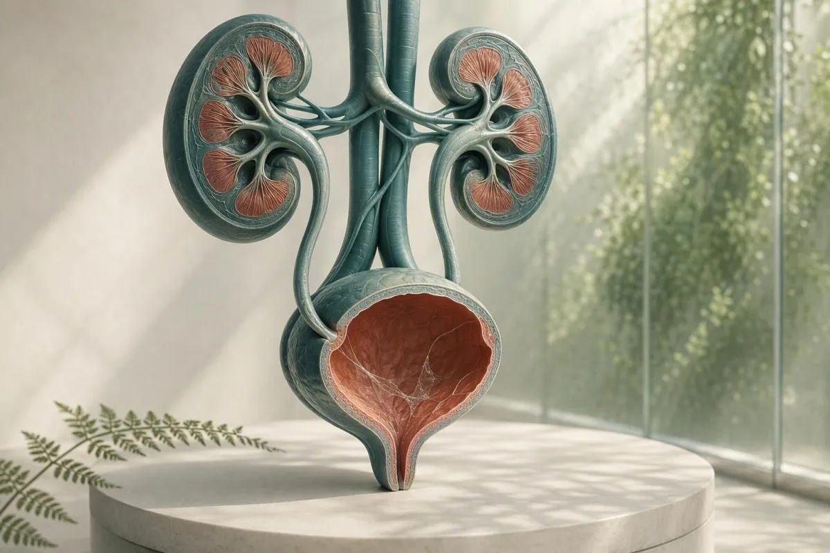
შარდის მიკროსკოპული ანალიზის ანგარიშში არსებული ლორწო, როგორც წესი, შეგროვების საკითხია და არა დიაგნოზი. შარდის ანალიზის შედეგები, სიმპტომები და სინჯის ხარისხი განსაზღვრავს, საჭიროა თუ არა მისი შემდგომი შეფასება.


- ლორწოს ძაფები თავისთავად ხშირად უვნებელია, განსაკუთრებით ერთჯერადი სუფთა სინჯისას, შარდგამომტანი სისტემის სიმპტომების გარეშე.
- პიურიის ზღვარი 5-ზე მეტი თეთრი უჯრედი მაღალი სიმძლავრის ველზე მიუთითებს შარდგამომტანი სისტემის ანთებაზე, მაგრამ არ ამტკიცებს ბაქტერიულ UTI-ს.
- კულტურის ტრიგერი არის ლორწო პლუს წვა, შარდვის იმპერატიული სურვილი, ცხელება, ტკივილი წელში, ნიტრატები ან მნიშვნელოვანი რაოდენობით თეთრი უჯრედები — არა მხოლოდ ლორწო.
- ბრტყელი ეპითელური უჯრედები დაახლოებით 10-ზე მეტი მაღალი სიმძლავრის ველზე ხშირად მიუთითებს სასქესო ორგანოების კანის დაბინძურებაზე და ამცირებს შარდის შედეგის სანდოობას.
- ხილული სისხლი კოლიკური გვერდითი ტკივილით საჭიროებს სასწრაფო შეფასებას ქვის არსებობისთვის, მაშინაც კი, თუ ლორწოც არის ანგარიშში.
- ორსულობა ცვლის თვალთვალის ზღურბლს, რადგან ასიმპტომური ბაქტერიურია ჩვეულებრივ იკვლევა შარდის კულტურით, არა მხოლოდ მიკროსკოპით.
- მოღრუბლული შარდი შეიძლება ასახავდეს კრისტალებს, კონცენტრირებულ შარდს, ვაგინალურ გამონადენს, სპერმას ან უჯრედებს; გარეგნობით ინფექციის დიაგნოზს ვერ სვამს.
- ნიმუშის გამეორება გამართლებულია, როდესაც ლორწო არის ერთადერთი უჩვეულო დასკვნა და შარდის შეგროვება არ იყო ფრთხილად გაკეთებული.
რას ნიშნავს ჩვეულებრივ შარდის ანალიზში ლორწოს ძაფები
შარდში ლორწოს ძაფები ჩვეულებრივ ნორმალური დამცავი სეკრეციის ან გენიტალური დაბინძურების ძაფებია და იზოლირებული შედეგი იშვიათად მიუთითებს თირკმელების დაავადებაზე. დასკვნა მნიშვნელოვანი ხდება, როდესაც ის ჩნდება შარდგამომყოფი სისტემის სიმპტომებთან, თეთრ უჯრედებთან, ბაქტერიებთან, სისხლთან ან ცილასთან ერთად.
ლაბორატორიები ლორწოს ამოიცნობენ, როგორც წვრილ, გამჭვირვალე ძაფებს, რომლებიც ჩანს მიკროსკოპის ქვეშ; ბევრი ანგარიში უბრალოდ აფასებს მას, როგორც იშვიათ, მცირე, ზომიერ ან ბევრს, ვიდრე რიცხვითი მითითების დიაპაზონის მინიჭებას. შარდი ბუნებრივად შეიცავს გლიკოპროტეინებით მდიდარ მასალას შარდგამომყოფი ტრაქტიდან, ხოლო ვაგინალური ან ურეთრალური სეკრეცია შეიძლება დაემატოს კოლექციის დროს.
ანგარიშის ფორმულირება ნაკლებად საგანგაშოა, ვიდრე ჟღერს. 2026 წლის 26 აგვისტოს მდგომარეობით, არ არსებობს დადასტურებული ლორწოს ძაფების რაოდენობა, რომელიც დიაგნოზს უსვამს შარდ-სასქესო ტრაქტის ინფექციას, ქვას, კიბოს ან თირკმლის უკმარისობას.; ეს არის კონტექსტუალური მიკროსკოპული დაკვირვება. ტესტ-ზოლისა და მიკროსკოპის ანგარიშის სხვა პუნქტების ფართო რუკისთვის იხილეთ ჩვენი შარდის ანალიზის სრული სახელმძღვანელო.
ჩემი კლინიკური გამოცდილებით, სიმპტომების გარეშე მყოფ ზრდასრულ ადამიანს, რომელსაც აღენიშნება “ზომიერი” ლორწო, 0-2 თეთრი უჯრედი მაღალი გადიდების ველზე, უარყოფითი ნიტრიტი და სისხლის არარსებობა, ჩვეულებრივ საუკეთესო საშუალებაა დამშვიდება ან ერთი სათანადოდ შეგროვებული განმეორებითი ნიმუში. დოქტორი თომას კლაინი ხშირად ხედავს ზედმეტ ანტიბიოტიკების შფოთვას, რომელიც გამოწვეულია ლაბორატორიული დროშის დაავადების ეტიკეტად შეცდომით.
Kantesti არის AI სისხლის ანალიზის განმარტება პლატფორმა, რომელსაც შეუძლია თირკმელებთან დაკავშირებული სისხლის მარკერები, როგორიცაა კრეატინინი და eGFR, მოათავსოს შარდის ანალიზის შედეგებთან ერთად - მაგრამ მას არ შეუძლია შარდ-სასქესო ტრაქტის ინფექციის დიაგნოზის დასმა მხოლოდ ლორწოს საფუძველზე. შარდის მიკროსკოპიის შედეგს სჭირდება კლინიკური ისტორია, რომელიც მას თან ახლდა.
რატომ აფიქსირებს ლაბორატორია მას
ლაბორატორიული საინფორმაციო სისტემები აფიქსირებენ ლორწოს, რადგან ის ვიზუალურად ჩანს და არა იმიტომ, რომ ლაბორატორიამ დაადგინა საშიში ზღვარი. ამრიგად, დროშა ნიშნავს “დაკვირვებულს” და არა “პათოლოგიურ დაავადების მარკერს”, ისევე როგორც იზოლირებულ კვალს, რომელიც არ საჭიროებს მკურნალობას.
საიდან მოდის ლორწო შარდის სინჯში
შარდში ლორწო შეიძლება წარმოიქმნას ურეთრიდან, ბუშტის ლორწოვანი გარსიდან, საშვილოსნოს ყელიდან ან საშოდან, სპერმიდან ან გარე სასქესო ორგანოებიდან კოლექციის დროს. მისი წყარო ხშირად იგულისხმება მიკროსკოპული ნიმუშის სხვა ნაწილებიდან, და არა თავად ძაფებიდან.
ბუშტსა და ურეთრას აქვს დამცავი ზედაპირი, რომელიც შეიცავს მუციინებს, მათ შორის უროპლაქინ-ასოცირებულ მასალას, რომელიც ამცირებს ხახუნს და მიკრობულ მიმაგრებას. მცირე რაოდენობით შეიძლება გამოიყოს შარდში დეჰიდრატაციის, ბოლო სექსუალური აქტივობის, მცირე გაღიზიანების შემდეგ, ან უბრალოდ იმიტომ, რომ დილის კონცენტრირებული ნიმუში აადვილებს ძაფების დანახვას.
მენსტრუაციული ან ვაგინალური გამონადენის მქონე ქალებში, ლეიკოციტები და ლორწო შეიძლება მოხვდეს კონტეინერში ბუშტიდან ამოსვლის გარეშე. 10-ზე მეტი ბრტყელი ეპითელური უჯრედი მაღალი გადიდების ველზე ზრდის დაბინძურების ალბათობას, თუმცა ლაბორატორიები იყენებენ განსხვავებულ მოხსენების ზღურბლებს. ჩვენი სახელმძღვანელო შარდის ეპითელური უჯრედების შესახებ ხსნის, თუ რატომ ცვლის ეს შედეგის სანდოობას.
სპერმა ასევე შეიძლება გამოიწვიოს ძაფისებრი მასალა ან მოღრუბლულობა ეაკულაციის შემდეგ რამდენიმე საათის განმავლობაში. ეს შარდს უსაფრთხოს არ ხდის, მაგრამ შეიძლება დაფაროს მიკროსკოპია; თუ კულტურა განიხილება, მე ზოგადად გირჩევთ ახალი ნიმუშის შეგროვებას სულ მცირე 24 საათის შემდეგ, თუ ეს შესაძლებელია.
რატომ ერყვნება სქესსა და ანატომიას ანგარიში
შარდის თასი არ იზოლირებს ბუშტს მიმდებარე ქსოვილებისგან. ამიტომ “ლორწოს არსებობა” უფრო ხშირად აღირიცხება გარეგანი სეკრეციის მქონე ნიმუშებში, ხოლო კათეტერიზებული ნიმუში ზოგჯერ გამოიყენება, როდესაც კლინიცისტებს სჭირდებათ უფრო სუფთა პასუხი.
როდის მიუთითებს ლორწოს ძაფები შარდგამომტანი სისტემის ინფექციაზე
ლორწოვანი ძაფები მხარს უჭერს შესაძლო UTI-ს მხოლოდ მაშინ, როდესაც თან ახლავს სიმპტომები და ობიექტური ანთებითი ნიშნები, განსაკუთრებით პიურია, ნიტრიტის დადებითი შედეგი ან დამაჯერებელი კულტურა. ლორწო თავისთავად არ არის UTI ტესტი.
სიმპტომური ქვედა სასქესო გზების UTI-ს ხშირად იწვევს წვა, გადაუდებლობა, სიხშირე, სუპრაპუბიკალური დისკომფორტი და ზოგჯერ შარდის ახალი სუნი. სათანადოდ შეგროვებულ ნიმუშში, 5-ზე მეტი თეთრი სისხლის უჯრედი მაღალი სიმძლავრის ველზე ხშირად მოიხსენიება როგორც პიურია; ის მხარს უჭერს შარდსასქესო ტრაქტის ანთებას, მაგრამ შეიძლება მოხდეს ქვების, სქესობრივი გზით გადამდები ინფექციების და დაბინძურების შემთხვევაშიც.
ნიტრიტი დადებითი შედეგისას ძალიან სპეციფიკურია, მაგრამ გამოტოვებს ორგანიზმებით გამოწვეულ ინფექციებს, რომლებიც არ ამცირებენ ნიტრატს, ხოლო ნეგატიური ნიტრიტის შედეგი არ გამორიცხავს UTI-ს. ლეიკოციტების ესთერაზა ავლენს თეთრი უჯრედების ფერმენტულ აქტივობას და შეიძლება იყოს ცრუ დადებითი, როდესაც ვაგინალური სეკრეცია აბინძურებს ნიმუშს; ჩვენი ახსნა ლეიკოციტური ესტერაზის შედეგებზე მოიცავს ამ ხაფანგებს.
IDSA-ს სახელმძღვანელო მითითებები ეწინააღმდეგება ასიმპტომური ბაქტერიურიის სკრინინგს ან მკურნალობას ჯანმრთელ, არაფეხმძიმეთა შორის, რადგან ანტიბიოტიკები ზიანს აყენებენ სარგებლის გარეშე (Nicolle et al., 2019). ეს პრინციპი განსაკუთრებით აქტუალურია ლორწოს მიმართ: წვის, ცხელების და დაგეგმილი უროლოგიური პროცედურის არარსებობა, როგორც წესი, არ ნიშნავს ანტიბიოტიკების დანიშვნას მხოლოდ იმიტომ, რომ მიკროსკოპის ველი არეულად გამოიყურებოდა.
როდესაც კულტურა უკეთესი ტესტია
შარდის კულტურა უფრო სასარგებლოა, ვიდრე განმეორებითი დიპსტიკი, როდესაც სიმპტომები გრძელდება, სიმპტომები მეორდება 4 კვირის განმავლობაში, არის ორსულობა, ეჭვმიტანილია პიელონეფრიტი, ან წინა ანტიბიოტიკებმა შესაძლოა შეცვალა შედეგი. შერეული ბაქტერიული ზრდა ხშირად მიუთითებს შეგროვების დაბინძურებაზე, ვიდრე ერთ შარდგამომყოფ ინფექციაზე; იხილეთ შარდის კულტურის ინტერპრეტაცია.
ლორწო, კრისტალები, ქვები და მექანიკური გაღიზიანება
ლორწოსთან ერთად ძლიერი ტალღისებური ტკივილი ან სისხლი შარდში შეიძლება მოხდეს საშარდე ქვებით, მაგრამ ლორწო არც ადასტურებს და არც გამორიცხავს ქვას. სისხლი, კრისტალები, ტკივილის ნიმუში, გამოსახულება და თირკმლის ფუნქცია უფრო მნიშვნელოვანია.
ქვა შეიძლება გაკაწროს ან დააკეტოს შარდსასქესო ლორწოვან გარსს, წარმოქმნის ერითროციტებს, ლეიკოციტებს და დამატებით ლორწოს. კლასიკური სიმპტომია მოულოდნელი კოლიკური ტკივილი, რომელიც ასხივებს საზარდულისკენ, ხშირად გულისრევით; თვალსაჩინო წითელი ან ჩაისფერი შარდი ზრდის გადაუდებლობას. დამატებითი ინფორმაცია შარდში სისხლის წითელი დროშები ვიდრე ვარაუდობთ, რომ ყოველი ვარდისფერი ნიმუში არის მარტივი ინფექცია.
კალციუმის ოქსალატის კრისტალები შეიძლება გამოჩნდეს ჯანმრთელ ადამიანებში, განსაკუთრებით კონცენტრირებულ მჟავე შარდში, ამიტომ ერთი ტიპის კრისტალი არ ამტკიცებს აქტიურ ქვას. ტკივილის, განმეორებითი ქვების ან ჰემატურიის მქონე პირებში, კლინიცისტებმა შეიძლება გამოიყენონ ულტრაბგერითი ან დაბალდოზით არა-კონტრასტული CT, ვიდრე დაეყრდნონ მხოლოდ ნალექს. ჩვენი რესურსი კალციუმის ოქსალატის კრისტალებზე დეტალურად აღწერს კრისტალის ანგარიშების შეზღუდვებს.
მნიშვნელოვანი გამონაკლისია ინფექციით გამოწვეული ობსტრუქცია: 38.0°C ან უფრო მაღალი ცხელება, ტკივილი ფლანგზე, ღებინება და შარდის გამოყოფის შეუძლებლობა საჭიროებს სასწრაფო შეფასებას. საზრუნავი არ არის ლორწო; ეს არის ინფიცირებული დაბლოკილი შარდსასქესო სისტემა, რომელიც შეიძლება სწრაფად გაუარესდეს.
გაღიზიანება კენჭის გარეშე
კათეტერის ბოლო გამოყენებამ, ბუშტის პროცედურებმა, ველოსიპედით აქტიურმა ტარებამ და მენჯის დასხივებამ შეიძლება გააღიზიანოს ქვედა ტრაქტი და გაზარდოს ლორწოს ან ლეიკოციტების რაოდენობა. კლინიცისტმა უნდა ინტერპრეტაცია გაუკეთოს ამ შედეგებს დროის მიხედვით, რადგან ინსტრუმენტაციიდან 48 საათის განმავლობაში აღებული ნიმუში არ არის ექვივალენტური რუტინულ სკრინინგის ნიმუშთან.
მოღრუბლული შარდის მნიშვნელობა: რისი თქმა შეუძლია გარეგნობას და რისი არა
მოღრუბლული შარდი შეიძლება გამოწვეული იყოს კონცენტრირებული მარილებით, კრისტალებით, ლორწოთი, უჯრედებით, გენიტალური გამონადენით ან ბაქტერიებით, ამიტომ მხოლოდ გარეგნობა ვერ დიაგნოსტიკოს ინფექციას. შარდი, რომელიც მოღრუბლულია, მაგრამ უმტკივნეულო და ხანმოკლეა, ხშირად არ არის გადაუდებელი.
ფოსფატის კრისტალებს შეუძლიათ ტუტე შარდი მოღრუბლული გახადონ გაცივების შემდეგ, ხოლო შარდმჟავას კრისტალებს შეუძლიათ მჟავე, კონცენტრირებული შარდი მოღრუბლული გახადონ. ოთახის ტემპერატურაზე 2 საათზე მეტხანს დატოვებულ ნიმუშს შეუძლია გახდეს უფრო მოღრუბლული უჯრედების დაშლის და ბაქტერიების გამრავლების გამო, ამიტომ სუფთა დამუშავება ან მაცივარში შენახვა მნიშვნელოვანია.
მოღრუბლულობა, დისურია, სასწრაფო შარდვა და პიურია საჭიროებს ტესტირებას; ფიზიკური დატვირთვის ან სითხის არასაკმარისი მიღების შემდეგ წარმოქმნილი მოღრუბლულობა ხშირად უმჯობესდება ჩვეულებრივი დატენიანებით. მიზნად დაისახეთ ღია ყვითელი შარდი, ვიდრე ზედმეტი წყლის მიღება – ძალიან გამჭვირვალე შარდი არ ნიშნავს, რომ თირკმელები ინფექციას “რეცხავენ”. ჩვენი დეტალური მიმოხილვა მოღრუბლული შარდის მიზეზების შესახებ განასხვავებს ვიზუალურ ნიშნებს სანდო ტესტებისგან.
კანტესტი არის AI სისხლის ტესტის ანალიზატორი განკუთვნილია სისხლის მარკერების კლინიკურ კონტექსტში ინტერპრეტაციისთვის; მაღალი კრეატინინი ან დაქვეითებული eGFR შარდის ანომალიებთან ერთად შეიძლება მოითხოვდეს სამედიცინო შეფასებას, მაშინ როდესაც მხოლოდ მოღრუბლული შარდი არ ადასტურებს თირკმელების ფილტრაციის დაქვეითებას. ძირითადი მეტაბოლური პანელი შეიძლება დაამატოს სასარგებლო კონტექსტი, როდესაც სიმპტომები მიუთითებს დეჰიდრატაციაზე ან ობსტრუქციაზე.
სუნი არ არის კულტურა
ძლიერი სუნის მქონე შარდი ხშირად ასახავს კონცენტრაციას, ასპარაგის მეტაბოლიტებს, B ვიტამინებს ან ძალიან დიდხანს დატოვებულ კონტეინერს. ახალი უსიამოვნო სუნი ცხელებით ან შარდვის სიმპტომებით არის მიზეზი შეფასების მისაღებად, მაგრამ სუნი არ არის მიკროსკოპისა და კულტურის შემცვლელი.
გენიტალური გამონადენი და სქესობრივი გზით გადამდები ინფექციები
ურეთრის ან ვაგინალური გამონადენი შეიძლება ჰგავდეს ლორწოს შარდში, ხოლო ახალი გამონადენი შარდის წვისას საჭიროებს სქესობრივი გზით გადამდები დაავადებების ტესტირებას, ასევე შარდის შეფასებას. შარდის ჩვეულებრივი კულტურა შეიძლება გამოტოვოს ქლამიდია და გონორეა.
ქლამიდიამ და გონორეამ შეიძლება გამოიწვიონ დისურია და სტერილური პიურია – თეთრი უჯრედები ბაქტერიების ჩვეულებრივი ზრდის გარეშე – განსაკუთრებით ახალი სექსუალური კონტაქტის შემდეგ. ნუკლეინის მჟავას ამპლიფიკაციის ტესტირება, ხშირად პირველი შარდის ნიმუშის გამოყენებით შუა ნაკადის ნიმუშის ნაცვლად, არის შესაბამისი ტესტი, რადგან სტანდარტული კულტურა მიზნად ისახავს სხვადასხვა მიკროორგანიზმებს.
ქალებში, ბაქტერიული ვაგინოზი, კანდიდოზი და საშვილოსნოს ყელის ანთება შეიძლება დაამატოთ გამონადენი შუა ნაკადის ჭიქაში; ვაგინალური ნაცხი ან გამოკვლევა შეიძლება უფრო ინფლაციური იყოს, ვიდრე შარდის მიკროსკოპიის გამეორება. მამაკაცებში, ხილული ურეთრის გამონადენი არ უნდა იყოს უგულებელყოფილი, როგორც “ლორწოს ძაფები”. პრაქტიკული განსხვავება არის გამონადენი, რომელიც აღინიშნება შარდვის გარეთ, ვიდრე ძაფები, რომლებსაც მხოლოდ ლაბორატორია იუწყება.
2021 წლის CDC სქესობრივი გზით გადამდები ინფექციების მკურნალობის სახელმძღვანელო რეკომენდაციას იძლევა NAAT-ზე დაფუძნებული ტესტირების შესაბამის ანატომიურ ადგილებში, ექსპოზიციის ისტორიის მიხედვით, არა მხოლოდ სიმპტომების (Workowski et al., 2021). თუ არსებობს მენჯის ტკივილი, სათესლე ჯირკვლის ტკივილი, ცხელება, ორსულობა ან შესაძლო თავდასხმა, მიმართეთ იმავე დღეს კლინიკურ რჩევას, ვიდრე თვითმკურნალობა დარჩენილი ანტიბიოტიკებით.
რატომ შეიძლება ანტიბიოტიკებმა დააბნიონ სურათი
კულტურამდე ანტიბიოტიკების 1-2 დოზის მიღებამაც კი შეიძლება ჩაახშოს ბაქტერიების ჩვეულებრივი ზრდა, მაშინ როდესაც შარდის სიმპტომები და თეთრი უჯრედები გრძელდება. უთხარით ექიმს ზუსტად რა პრეპარატი, დოზა და ბოლო დოზის დრო იქნა გამოყენებული; ეს დეტალი ცვლის ნეგატიური კულტურის ინტერპრეტაციას.
როგორ შევაგროვოთ შარდის სინჯი, რომელიც პასუხს გასცემს კითხვას
შუა ნაკადის სუფთა ნიმუში ამცირებს ლორწოს, ბრტყელ ეპითელურ უჯრედებს და შერეულ ბაქტერიულ ზრდას უკეთესად, ვიდრე ნაკადის პირველი ან ბოლო ნაწილის შეგროვება. ტექნიკა მარტივია, მაგრამ პირველი 2 წამი მნიშვნელოვან განსხვავებას ქმნის.
დაიბანეთ ხელები, გამოყავით სასქესო ორგანოების კანი ან მოაშორეთ ასო თუ კომფორტულად ხართ, გაწმინდეთ ნაკრების ინსტრუქციის მიხედვით, დაიწყეთ შარდვა ტუალეტში, შემდეგ შეაგროვეთ შუა ნაწილი, სანამ ჭიქა კანს შეეხება. 20–30 მლ ნიმუში ჩვეულებრივ საკმარისია; დიდი კონტეინერის პირამდე შევსება არ აუმჯობესებს ტესტს.
მიიტანეთ ჭიქა დაუყოვნებლივ, იდეალურად 1 საათის განმავლობაში; თუ დაგვიანება გარდაუვალია, შეინახეთ მაცივარში ლაბორატორიის ინსტრუქციის მიხედვით და დააბრუნეთ 24 საათის განმავლობაში. მაცივარში შენახვა ანელებს ბაქტერიების ზრდას, მაგრამ არ აღადგენს ნიმუშს, რომელიც უკვე დაბინძურდა. ეს განსაკუთრებით მნიშვნელოვანია, როდესაც მოთხოვნა მოიცავს კულტურას, დიპ-სტიკის ნაცვლად.
კანტესტი არის AI-ზე დაფუძნებული სისხლის ანალიზის (blood test analysis) ხელსაწყო გამოიყენება 127+ ქვეყნებში, და ჩვენი კლინიცისტის მიერ მიმოხილული სამუშაო პროცესები ნიმუშის ხარისხს ინტერპრეტაციის ნაწილად განიხილავს, არა როგორც მეორეხარისხოვან საკითხს. იგივე პრინციპი ვრცელდება ნებისმიერ ლაბორატორიულ მნიშვნელობაზე: ტექნიკურად ცუდი ნიმუში შეიძლება შეიცავდეს სამედიცინო თვალსაზრისით დამაჯერებელ, მაგრამ მცდარ შედეგს. იხილეთ ჩვენი ლაბორატორიული სიზუსტის საკონტროლო სია შეკითხვებისთვის, რომლებსაც უნდა დაუსვათ იზოლირებულ დროშაზე მოქმედებამდე.
არ შეაგროვოთ ამ სიტუაციებში, თუ შეგიძლიათ დაელოდოთ
მოერიდეთ ჩვეულებრივ ტესტირებას მძიმე მენსტრუაციის დროს, სქესობრივი აქტის შემდეგ დაუყოვნებლივ, ან ვაგინალური კრემების გამოყენების შემდეგ, თუ ექიმი სპეციალურად არ ითხოვს ამას. როდესაც ტესტირება არ შეიძლება დაელოდოს, აცნობეთ ლაბორატორიას ან ექიმს, რადგან ეს კონტექსტი შეიძლება აიხსნას ლორწოს, სისხლის წითელი უჯრედების ან გარე უჯრედების არსებობა.
როგორ კითხულობენ კლინიცისტები ლორწოს შარდის ანალიზის დანარჩენ შედეგებთან ერთად
შარდის ანალიზის ყველაზე სასარგებლო სურათი აერთიანებს სიმპტომებს თეთრ უჯრედებთან, სისხლის წითელ უჯრედებთან, ნიტრიტთან, ლეიკოციტ ესტერაზასთან, პროტეინთან, გლუკოზასთან, სპეციფიკურ სიმძიმესთან და ეპითელურ უჯრედებთან. ლორწო ამ ნიმუშში მცირე დამხმარე ნიშანია.
სუფთა ნიმუში ლორწოთი, 0–2 ლეიკოციტი მაღალი გადიდების ველზე, ნეგატიური ნიტრიტი და სისხლის არარსებობა, როგორც წესი, მკურნალობას არ საჭიროებს. ლორწო, რომელშიც 5-ზე მეტი ლეიკოციტია მაღალი გადიდების ველზე და ლეიკოციტების ესთერაზა დადებითია, ზრდის ანთების ალბათობას; ნიტრიტის ან ერთ-ორგანიზმიანი კულტურის დამატება ზრდის ნდობას, რომ ბაქტერიები იწვევენ ამას.
ცილას საკუთარი გზა სჭირდება. ცილის კვალი ცხელების, ვარჯიშის ან შარდის კონცენტრაციის შემდეგ შეიძლება დროებითი იყოს, მაგრამ მუდმივი ცილა უნდა გაიზომოს ალბუმინ-კრეატინინის თანაფარდობით და არა ლორწოს მიზეზით. ჩვენი სახელმძღვანელო ცილა შარდში ხსნის, რატომ არ არის დიპსტიკის ცილა და თირკმლის რისკი ურთიერთშემცვლელი.
სპეციფიკური სიმკვრივე 1.003-დან 1.030-მდე ხშირია მოზრდილებში, თუმცა საცნობი ინტერვალები ლაბორატორიის მიხედვით განსხვავდება. მაღალი სპეციფიკური სიმძიმე შეიძლება აკონცენტრირებდეს ლორწოს და ქმნიდეს უფრო დრამატულად გამომყურებელ ნალექს, ხოლო დაბალი სპეციფიკური სიმძიმე შეიძლება დაშალოს უჯრედები და გახადოს მიკროსკოპია მატყუარად ნათელი; ჩვენი სპეციფიკური სიმკვრივის სახელმძღვანელოში გვიჩვენებს, თუ როგორ მოქმედებს ჰიდრატაცია ინტერპრეტაციაზე.
უარყოფითი შედეგების მნიშვნელობა
უარყოფითი სისხლი, ცილა, ნიტრიტი და ლეიკოციტების ესთერაზა მნიშვნელოვნად ამცირებს შეშფოთებას ასიმპტომურ პაციენტში, მაშინაც კი, თუ ლორწო აღწერილია როგორც ზომიერი. არც ერთი ცალკეული უარყოფითი ტესტი არ არის სრულყოფილი, მაგრამ ეს ჯგუფი უფრო დამაიმედებელია, ვიდრე ლორწოს შეფასებაა შემაშფოთებელი.
როდის გავიმეოროთ ტესტი და როდის მოვითხოვოთ კულტურა
გაიმეორეთ შარდის ნიმუში, თუ ლორწო არის იზოლირებული, ეპითელური უჯრედები მიუთითებს დაბინძურებაზე ან ნიმუში ნაჩქარევად იყო აღებული; მოითხოვეთ კულტურა, თუ სიმპტომებია მუდმივი, განმეორებადი, მძიმე ან მაღალი რისკის. განმეორებითი ტესტი არ არის “არაფრის კეთება” - ეს ხშირად ყველაზე დიაგნოსტიკური შემდეგი ნაბიჯია.
არამსწრაფე ზრდასრულისთვის სიმპტომების გარეშე და მხოლოდ ლორწოს დროშით, განმეორებითი სუფთა შარდის ანალიზი 1-2 კვირის განმავლობაში გონივრულია, თუ საჭიროა დადასტურება; ბევრი ექიმი საერთოდ არ გაიმეორებს მას. არ მოექცეთ ლაბორატორიის დროშას ანტიბიოტიკებით ლოდინის დროს, თუ ექიმი არ განსაზღვრავს კლინიკურ ჩვენებას.
ანტიბიოტიკების მიღებამდე კულტურა განსაკუთრებით სასარგებლოა ცხელების, ტკივილის წელის არეში, ორსულობის, იმუნური დათრგუნვის, თირკმლის ტრანსპლანტაციის, შარდის კათეტერის გამოყენების, მამრობითი სქესის შარდ-სასქესო სისტემის სიმპტომების ან 4 კვირის განმავლობაში განმეორებითი სიმპტომების შემთხვევაში. Gupta et al. (2011) სახელმძღვანელო მხარს უჭერს კულტურას პიელონეფრიტის ეჭვის შემთხვევაში და იმ სიტუაციებში, როდესაც წინააღმდეგობა ან ალტერნატიული დიაგნოზი უფრო სავარაუდოა.
კულტურის რაოდენობა უნდა წაიკითხებოდეს შეგროვების ხარისხის გათვალისწინებით. ერთი ორგანიზმი 10^5 კოლონია-წარმომქმნელი ერთეული მილილიტრზე ტრადიციულად ბაქტერიურიის მხარდაჭერა ხდება სუფთა შუა შარდვის შარდში, მაგრამ სიმპტომატურ პაციენტებს შეიძლება ჰქონდეთ კლინიკურად მნიშვნელოვანი დაბალი რაოდენობა; “შერეული ფლორა” ჩვეულებრივ იწვევს ახალი ნიმუშის აღებას ფართო ანტიბიოტიკის ნაცვლად. შეადარეთ მიზნები შარდის ანალიზსა და კულტურაზე სანამ რომელიმე მათგანს მოითხოვთ.
ორსულობა მოითხოვს განსხვავებულ ზღვარს
ორსულობა გამონაკლისია, რადგან ასიმპტომურმა ბაქტერიურიამ შეიძლება გაზარდოს პიელონეფრიტის და ორსულობასთან დაკავშირებული არასასურველი შედეგების რისკი. ანტენატალური მოვლა ჩვეულებრივ იყენებს სკრინინგულ კულტურას ორსულობის ადრეულ ეტაპზე; მიკროსკოპით ნახული ლორწო ვერ შეცვლის ამ კულტურას.
როდის მატებს სისხლის ანალიზები თირკმელებისა და ინფექციის სასარგებლო კონტექსტს
სისხლის ანალიზები სასარგებლოა, როდესაც შარდში ლორწოს თან ახლავს ცხელება, ტკივილი წელის არეში, განმეორებითი ინფექციები, შეშუპება, შარდვის შემცირებული რაოდენობა ან მუდმივი ცილა ან სისხლი. კრეატინინი და eGFR აფასებენ ფილტრაციას, ხოლო CBC და C-რეაქტიული ცილა შეიძლება დაეხმაროს სისტემური დაავადების შეფასებას.
eGFR-ის მაჩვენებელი ქვემოთ 60 მლ/წთ/1.73 მ² მინიმუმ 3 თვის განმავლობაში აკმაყოფილებს ქრონიკული თირკმელების დაავადების ერთ კრიტერიუმს, მაგრამ ერთი დაბალი შედეგი დეჰიდრატაციის ან მწვავე დაავადების დროს არ არის საკმარისი ამ დიაგნოზის დასასმელად. კრეატინინი დამოკიდებულია კუნთების მასაზე, დიეტაზე და ზოგიერთ მედიკამენტზე, ამიტომ ტენდენციები და შარდის ალბუმინი ხშირად უფრო ინფორმატიულია, ვიდრე ერთი რიცხვი.
გაზრდილი თეთრი სისხლის უჯრედების რაოდენობა ან CRP შეიძლება მხარი დაუჭიროს ანთებით პროცესს, მაგრამ არ შეუძლია შარდსასქესო ტრაქტის წყაროდ იდენტიფიცირება. ცხელებით დაავადებულ ადამიანში ტკივილით წელის არეში, კლინიცისტებმა შეიძლება შეამოწმონ კრეატინინი გამოსახულების ან მედიკამენტების არჩევამდე, განსაკუთრებით თუ ღებინება ან ობსტრუქცია შეიძლება დააზიანოს თირკმელების ფუნქცია. მიმოხილვა ქრონიკული თირკმლის დაავადების სტადიებს eGFR და ალბუმინის კატეგორიებისთვის, რომლებსაც კლინიცისტები იყენებენ.
კანტესტი არის AI ლაბორატორიული ტესტის ინტერპრეტაციის სერვისზე რომელიც ასახავს კრეატინინის, eGFR, ელექტროლიტების, CBC და ანთებითი მარკერების ნიმუშებს, ვიდრე ერთი ლორწოს აღნიშვნის მკურნალობას, როგორც თირკმელების დიაგნოზს. ჩვენი მეთოდოლოგია ექვემდებარება კლინიკურ ვალიდაციის ზედამხედველობას, მაგრამ გადაუდებელი სიმპტომები მაინც საჭიროებს პირდაპირ სამედიცინო დახმარებას, არა აპლიკაციაზე დაფუძნებულ ინტერპრეტაციას.
პრაქტიკული დეჰიდრატაციის განსხვავება
დეჰიდრატაციამ შეიძლება გაზარდოს შარდის სპეციფიკური სიმძიმე და დროებით გაზარდოს კრეატინინი, განსაკუთრებით დიარეის, სიცხის ზემოქმედების ან ინტენსიური ვარჯიშის შემდეგ. შარდვის მუდმივად დაბალი რაოდენობა, თავბრუსხვევა ან eGFR-ის დაქვეითება რეჰიდრატაციის შემდეგ მოითხოვს ექიმის შეფასებას, განმეორებითი სახლის ტესტირების ნაცვლად.
სასწრაფო გაფრთხილების ნიშნები, რომლებსაც არ უნდა ველოდოთ განმეორებით ტესტს
მიმართეთ სასწრაფო შეფასებას შარდში ლორწოს არსებობისას 38.0°C ან უფრო მაღალი ცხელებით, ტკივილით წელის არეში, ღებინებით, შესამჩნევი სისხლით, შარდვის შეუძლებლობით, დაბნეულობით ან ორსულობასთან დაკავშირებული შარდვის სიმპტომებით. ეს კომბინაციები მნიშვნელოვანია, რადგან მათ შეუძლიათ მიუთითონ ზედა ტრაქტის ინფექცია, ობსტრუქცია ან სხვა მწვავე მდგომარეობა.
ცხელება ერთ-ერთ ზურგზე ან წელის ტკივილთან ერთად უფრო შემაშფოთებელია პიელონეფრიტისთვის, ვიდრე მარტივი ცისტიტისთვის, განსაკუთრებით შემცივნებით ან ღებინებით. დაგვიანებულმა მკურნალობამ შეიძლება გამოიწვიოს დეჰიდრატაცია, სეფსისი ან თირკმელების სტრესი; სახლში შარდის ნორმალურად მყოფი ნიმუში მას უსაფრთხოდ ვერ გამორიცხავს.
შესამჩნევი სისხლი უნდა შეფასდეს მაშინაც კი, როდესაც UTIs-ის ალბათობა ჩანს, განსაკუთრებით 35 წელზე უფროსი ასაკის, მწეველთა ან თუ სისხლდენა გრძელდება ინფექციის სიმპტომების ჩაცხრობის შემდეგ. AUA მიკროჰემატურიის სახელმძღვანელო რეკომენდაციას უწევს რისკზე დაფუძნებულ შეფასებას კეთილთვისებიანი ახსნის შემდეგ (Barocas et al., 2020); მუდმივი სისხლი არ აიხსნება ლორწოთი.
ბავშვები, 65 წელზე უფროსი ასაკის მოზრდილები, დიაბეტით დაავადებული ადამიანები, იმუნოსუპრესანტ მედიკამენტებზე მყოფი ადამიანები და მარტოხელა თირკმლის მქონე პირები იმსახურებენ შეფასების დაბალ ზღვარს. დოქტორ თომას კლაინის პრაქტიკული წესი მარტივია: თუ სიმპტომები მძიმდება 6-12 საათის განმავლობაში, ნუ დაელოდებით მეორე ჭიქას დამშვიდებისთვის.
გამოიძახეთ სასწრაფო დახმარება ახლა მძიმე ავადმყოფობისთვის
გამოიძახეთ სასწრაფო დახმარება ახალი დაბნეულობის, გულის წასვლის, ძლიერი სისუსტის, ტუჩების გალურჯების ან ნაცრისფერის, ძლიერი ქოშინის ან შარდვის სიმპტომებით სითხის შენარჩუნების შეუძლებლობის შემთხვევაში. ეს არის სისტემური საფრთხის ნიშნები, არა ჩვეულებრივი UTI სიმპტომები.
ლორწო შარდში ორსულობის, ბავშვობისა და შემდგომ პერიოდში
ორსულობა, ბავშვები და ხანდაზმულები საჭიროებენ უფრო ფრთხილად ინტერპრეტაციას, რადგან დაბინძურება ხშირია, მაგრამ ინფექციის გამოტოვების შედეგები შეიძლება უფრო დიდი იყოს. სიმპტომები და კულტურის ხარისხი რჩება უფრო ღირებულად, ვიდრე ლორწოს ხარისხი ყველა ასაკობრივ ჯგუფში.
ორსულობის დროს შარდვის ხშირი მოთხოვნილება შეიძლება იყოს ნორმალური, რაც სიმპტომებს ნაკლებად სპეციფიკურს ხდის; ცხელებამ, დიზურიამ ან ზურგის ტკივილმა უნდა გამოიწვიოს მეან-გინეკოლოგის ან კლინიცისტის შეფასება. სკრინინგ კულტურა, როგორც წესი, აღებულია ანტენატალური მეთვალყურეობის დასაწყისში, ხოლო განმეორებითი კულტურა შეიძლება გამოყენებულ იქნას მკურნალობის შემდეგ, კლინიცისტის გეგმის მიხედვით.
ბავშვებში, ტომრით აღებული შარდი მიდრეკილია დაბინძურებისკენ და, როგორც წესი, არ უნდა იქნას გამოყენებული უროინფექციის დიაგნოსტიკისთვის კულტურით. კათეტერიზებული ან ფრთხილად აღებული სუფთა შარდის ნიმუში შეიძლება იყოს საჭირო, როდესაც პატარა ბავშვს აქვს ცხელება აშკარა წყაროს გარეშე; ტომარაში არსებული ლორწო განსაკუთრებით არასპეციფიკურია.
ხანდაზმულ ასაკში ბაქტერიურია და პიურია უფრო ხშირია სიმპტომების გამოწვევის გარეშე. ახალმა დელირიუმმა თავისთავად უნდა გამოიწვიოს დეჰიდრატაციის, მედიკამენტების, ტკივილის, ყაბზობის და ინფექციის წყაროების ფართო სამედიცინო შეფასება, ვიდრე ავტომატური უროინფექციის მკურნალობა; იგივე თავშეკავება, როგორც რეკომენდებულია Nicolle et al. (2019) მიერ.
მენსტრუაცია და ჰორმონებთან დაკავშირებული ცვლილებები
მენსტრუალური სისხლი და საშვილოსნოს ყელის ლორწო შეიძლება რამდენიმე დღის განმავლობაში ამახინჯებდეს შარდის ანალიზს, ხოლო პოსტმენოპაუზის დროს ვაგინალური სიმშრალე შეიძლება გამოიწვიოს ადგილობრივი გაღიზიანება, რომელიც წააგავს შარდვისას წვის შეგრძნებას. თუ შედეგები და სიმპტომები არ ემთხვევა, კლინიცისტმა შეიძლება შეაფასოს გენიტალური მიზეზები განმეორებითი უროინფექციის მკურნალობის დანიშვნის ნაცვლად.
გავრცელებული შეცდომები ლაბორატორიული ანგარიშის ლორწოს ნახვის შემდეგ
ნუ დაიწყებთ დარჩენილი ანტიბიოტიკების გამოყენებას, შიდა წმენდას ან ლორწოს “გამოვლის” მცდელობას წყლის ექსტრემალური მიღებით, ერთჯერადი არასწორი შარდის ანალიზის შემდეგ. ამ ქმედებებმა შეიძლება გაართულოს კულტურა, დაარღვიოს ნორმალური ფლორა ან შეაფერხოს სწორი დიაგნოზი.
ანტიბიოტიკების მიღებამ კულტურის გარეშე შეიძლება მომდევნო ტესტი ცრუ-უარყოფითი გახადოს და გამოიწვიოს დიარეა, გამონაყარი, საფუარის სიმპტომები ან რეზისტენტობა. თუ სიმპტომები მსუბუქია, მაგრამ მუდმივი, როდესაც ეს პრაქტიკულია, ჯერ აიღეთ ნიმუში, შემდეგ კი მიჰყევით კლინიცისტის მკურნალობის გეგმას, რომელიც ეფუძნება სრულ სურათს.
მოცვის პროდუქტებმა შეიძლება ზომიერად შეამცირონ განმეორებითი არასპეციფიკური უროინფექციის რისკი ზოგიერთისთვის, მაგრამ ისინი არ მკურნალობენ ცხელებას, ფლანგის ტკივილს ან დადასტურებულ ზედა სასუნთქი გზების ინფექციას. მოერიდეთ მაღალი დოზით C ვიტამინს, როგორც თვითმკურნალობას: მას შეუძლია შეცვალოს ზოგიერთი ტესტ-ზოლის რეაქცია და შეიძლება გაზარდოს ოქსალატის დატვირთვა იმ პირებში, რომლებიც მიდრეკილნი არიან კალციუმ-ოქსალატის ქვების წარმოქმნისკენ.
არ შეამოწმოთ ტუალეტის თასი ძაფებისთვის. ტუალეტის ქაღალდის ბოჭკოები, საწმენდი ნარჩენები, გენიტალური სეკრეტები და წყლის დინება შეიძლება წააგავდეს ლორწოს; ლაბორატორიული ნიმუში, რომელიც აღებულია სტერილურ კონტეინერში, არის შესაფერისი ადგილი მის შესაფასებლად. დაკავშირებული ფერის ცვლილებებისთვის, ჩვენი შარდის ფერის სახელმძღვანელო გთავაზობთ უფრო სანდო ვიზუალურ კონტექსტს.
მედიკამენტების სია მნიშვნელოვანია
ფენაოპირიდინმა შეიძლება შარდი ნარინჯისფერი გახადოს და ხელი შეუშალოს ვიზუალურ ინტერპრეტაციას, ხოლო შარდმდენებმა შეიძლება შარდი კონცენტრირება ან განზავება დროის მიხედვით. აპარტამენტში მიიტანეთ რეცეპტით გამოწერილი მედიკამენტების, ურეცეპტოდ გაცემული პროდუქტების და დანამატების სია, დოზების ჩათვლით მგ-ში.
პრაქტიკული შემდგომი გეგმა შარდში ლორწოს ძაფებისთვის
უმეტეს ადამიანებს, რომლებსაც შარდში ლორწოს ძაფები აქვთ და სიმპტომები არ აქვთ, მკურნალობა არ სჭირდებათ; გონივრულია ფრთხილად განმეორებითი შარდის ანალიზი, თუ ნიმუში საეჭვო იყო. სიმპტომები ან თანმხლები ანომალიები განსაზღვრავს, არის თუ არა შესაბამისი კულტურა, სქესობრივი გზით გადამდები ინფექციების (STI) ტესტირება, ვიზუალიზაცია ან სისხლის ანალიზები.
ნაბიჯი 1: შეამოწმეთ წვა, შარდვის ხშირი მოთხოვნილება, ცხელება, ფლანგის ტკივილი, სისხლი შარდში, გამონადენი, ორსულობა, შარდსასქესო სისტემის ბოლო პროცედურები და სქესობრივი კონტაქტის ახალი შემთხვევა. ნაბიჯი 2: წაიკითხეთ ანგარიში თეთრი უჯრედების, ნიტრიტის, ლეიკოციტების ესთერაზას, სისხლის უჯრედების, ცილის, ბაქტერიების და ბრტყელი ეპითელიუმის უჯრედების შესახებ — არა მხოლოდ ლორწოს.
ნაბიჯი 3: თუ თავს კარგად გრძნობთ და ლორწო იზოლირებულია, აიღეთ ერთი შუა ნაკადის სუფთა შარდის ნიმუში 1-2 კვირის განმავლობაში მხოლოდ იმ შემთხვევაში, თუ თქვენი კლინიცისტი რეკომენდაციას მისცემს დადასტურებას. ნაბიჯი 4: თუ სიმპტომები არსებობს, იკითხეთ, უნდა აიღოთ თუ არა შარდის კულტურა მკურნალობამდე და არის თუ არა STI ტესტირება შესაბამისი; განმეორებითი ეპიზოდები უფრო საფუძვლიან განხილვას იმსახურებს, ვიდრე მესამე ემპირიული ანტიბიოტიკი.
Kantesti-ს შეუძლია მოაწყოს შესაბამისი სისხლის შედეგების ტენდენციები და კითხვები სამედიცინო შეხვედრისთვის, ხოლო ჩვენი სამედიცინო საკონსულტაციო საბჭო მხარს უჭერს კლინიცისტების მიერ მართვად უსაფრთხოების სტანდარტებს. სწორი საბოლოო მიზანი არ არის სრულყოფილად “სუფთა” მიკროსკოპული ანგარიში — ეს არის ახსნა, რომელიც შეესაბამება თქვენს სიმპტომებს, ნიმუშის ხარისხს და რისკ-ფაქტორებს.
კითხვები თქვენი შეხვედრისთვის
იკითხეთ, შეიცავდა თუ არა ნიმუში ბრტყელ ეპითელიუმის უჯრედებს, გაიზარდა თუ არა კულტურაში ერთი ორგანიზმი ან შერეული ფლორა, და დარჩა თუ არა სისხლი ან ცილა სიმპტომების ალაგების შემდეგ. ეს 3 კითხვა ხშირად იძლევა უფრო სასარგებლო პასუხებს, ვიდრე იმის კითხვა, თუ რამდენი ლორწო დაფიქსირდა.
კვლევის კონტექსტი და ლორწოს ანგარიშის ლიმიტები
ლაბორატორიების მიხედვით ლორწოს შეფასება სტანდარტიზებული არ არის, ამიტომ კლინიცისტებმა თავიდან უნდა აიცილონ “რამდენიმე”, “ზომიერი” ან “ბევრი” ტერმინების გამოყენება დაავადების სიმძიმის კატეგორიებად. მიკროსკოპის ტექნიკა, ნიმუშის ასაკი და ანგარიშგების პროგრამული უზრუნველყოფა შეიძლება შეიცვალოს ფორმულირება.
ზოგიერთი ლაბორატორია ნალექს ხელით ამოწმებს დაახლოებით 10–15 მლ შარდის ცენტრიფუგირების შემდეგ, ზოგი კი იყენებს ავტომატიზებულ ნაწილაკთა ანალიზს შერჩეული დროშების ხელით მიმოხილვით. ეს ვარიაცია ნიშნავს, რომ ერთი ლაბორატორიის “ზომიერი ლორწოს” შედეგი შეიძლება ზუსტად არ განმეორდეს სხვა ლაბორატორიაში, მაშინაც კი, როდესაც ადამიანის ჯანმრთელობა უცვლელია.
სასარგებლო ლაბორატორიული აზროვნება არის კითხვის დასმა, არის თუ არა შედეგი ანალიტიკურად რეალური, კლინიკურად მნიშვნელოვანი და განმეორებადი. ეს მიდგომა ნაცნობია სისხლის ანალიზისთვისაც: ჩვენი სისხლის ბიომარკერების სახელმძღვანელო ხსნის, რატომ განსაზღვრავს საცნობი ინტერვალები და პრე-ანალიტიკური პირობები, იმსახურებს თუ არა მოქმედებას მონიშნული მარკერი.
Kantesti AI იყენებს სტრუქტურირებულ ლაბორატორიულ კონტექსტს შედეგების ნიმუშების ახსნისთვის ძირითადი პანელების მიხედვით, მაგრამ შარდის მიკროსკოპია კვლავ მოითხოვს წყაროს სპეციფიკურ კლინიკურ განსჯას. მკითხველებისთვის, რომლებიც დაინტერესებულნი არიან, თუ როგორ ვაფასებთ ტექნიკურ მუშაობას და კლინიკურ საზღვრებს, ჩვენი AI ტექნოლოგიის გზამკვლევი აღწერს ჩვენი ინტერპრეტაციული მიდგომის მიღმა არსებულ უსაფრთხოების ზომებს.
როგორი ანგარიში უნდა იყოს მკაფიო
სასარგებლო ანგარიში მიუთითებს ნიმუშის ტიპს, შეგროვების თარიღს, მიკროსკოპულ ელემენტებს და ნებისმიერ კულტურულ ორგანიზმს და მგრძნობელობის შედეგებს, როდესაც კულტურა შესრულებულია. თუ ანგარიში მხოლოდ ამბობს “ლორწოს ძაფები არსებობს”, ის არასრულია დიაგნოზისთვის, მაგრამ ხშირად სრულიად ადეკვატურია დაბალი რისკის შემთხვევითი აღმოჩენისთვის.
ხშირად დასმული კითხვები
შარდში ლორწოს ძაფები ნორმალურია?
შარდში ლორწოს ძაფები ხშირად ნორმალურია ან გენიტალური სეკრეციის დაბინძურების გამო, განსაკუთრებით მაშინ, როდესაც ადამიანს არ აქვს შარდსასქესო სისტემის სიმპტომები. იზოლირებული მაჩვენებელი 0–2 თეთრი უჯრედით მაღალი გადიდების ველზე, უარყოფითი ნიტრიტით და სისხლის ან ცილის გარეშე, ჩვეულებრივ დაბალი რისკის შემცველია. ლაბორატორიები არ იყენებენ ვალიდირებულ ლორწოს ძაფების რაოდენობას საშარდე გზების ინფექციის (UTI) ან თირკმლის დაავადების დიაგნოსტიკისთვის. განმეორებითი შუა ნაკადის სუფთა ნიმუშის აღება გონივრულია, თუ თავდაპირველი შეგროვება ნაჩქარევი ან თვალსაჩინოდ დაბინძურებული იყო.
შარდში ლორწოს ძაფები ნიშნავს თუ არა, რომ მაქვს საშარდე გზების ინფექცია?
შარდში ლორწოს ძაფები თავისთავად არ ნიშნავს საშარ გზების ინფექციას (სგი). სგი-ის ალბათობა იზრდება, როდესაც წვის, შარდვის მწვავე მოთხოვნილების, ხშირი შარდვის, ცხელების ან პუბის ზემოთ ტკივილისას ვლინდება 5-ზე მეტი თეთრი უჯრედი მაღალი გადიდების ველზე, ლეიკოციტების ესთერაზას, ნიტრიტის დადებითი მაჩვენებელი ან დადებითი კულტურა. ნიტრიტის უარყოფითი მაჩვენებელი სრულად არ გამორიცხავს სგი-ს, რადგან ყველა ბაქტერია არ წარმოქმნის ნიტრიტს. კულტურა, როგორც წესი, უფრო ინფორმაციულია, ვიდრე ლორწოს შეფასება, როდესაც სიმპტომები გრძელდება ან მეორდება.
გაუწყლოებამ შეიძლება გამოიწვიოს ლორწო შარდში?
გაუწყლოებამ შეიძლება ლორწოს უფრო ადვილად ხილვადობა გამოიწვიოს, რადგან კონცენტრირებული შარდი შეიცავს ნაკლებ წყალს ნორმალურ სეკრეტებსა და ნალექთან მიმართებაში. მაღალი შარდის სპეციფიკური სიმძიმე, რომელიც ხშირად 1.025–1.030-ისკენ არის მიდრეკილი, შეიძლება თან ახლდეს ამ ეფექტს, თუმცა თავისთავად გაუწყლოებას არ ამტკიცებს. ნორმალური ჰიდრატაცია და სათანადოდ აღებული სინჯის გამეორება დააზუსტებს მონაცემებს. წყლის ჭარბი მიღება არ არის აუცილებელი და შეიძლება შექმნას ელექტროლიტური რისკები.
რას ნიშნავს ლორწო და თეთრი სისხლის უჯრედები შარდში?
შარდში ლორწოსა და თეთრი სისხლის უჯრედების არსებობა მიუთითებს შარდ- ან სასქესო გზების ანთებაზე, თუმცა ეს არ ადასტურებს ბაქტერიულ ინფექციას. 5-ზე მეტი თეთრი უჯრედი მაღალი გადიდების ველზე შეიძლება აღმოჩნდეს შარდ-სასქესო ტრაქტის ინფექციის, ქვების, სქესობრივი გზით გადამდები ინფექციების, ვაგინალური დაბინძურების ან შარდის ბუშტის ბოლოდროინდელი ინსტრუმენტაციის დროს. სიმპტომები, ნიტრიტი, ეპითელური უჯრედები და კულტურის შედეგები განსაზღვრავს შემდეგ ნაბიჯს. 38.0°C ან უფრო მაღალი სიცხე და გვერდის ტკივილი სასწრაფო შეფასებას მოითხოვს.
შეიძლება თუ არა ვაგინალური გამონადენი შარდის ანალიზში ლორწოს წარმოქმნას?
ვაგინალური გამონადენი შეიძლება მოხვდეს შარდის შუა ნაკადის სინჯში და ხშირად იწვევს ლორწოს, ბრტყელი ეპითელური უჯრედების და ზოგჯერ ლეიკოციტების გამოჩენას მიკროსკოპიის დროს. 10-ზე მეტი ბრტყელი უჯრედი მაღალი გადიდების ველზე ხშირად ზრდის დაბინძურების ალბათობას, თუმცა ლაბორატორიები განსხვავებულ ანგარიშებს აწარმოებენ. მენსტრუალური სისხლის დიდი ნაკადის გარეთ აღებული ფრთხილად აღებული სინჯი ამ პრობლემის შემცირებას უწყობს ხელს. თუ გამონადენი, ქავილი, სუნი ან მენჯის ტკივილი აღინიშნება, განმეორებითი შარდის კულტურები შეიძლება ნაკლებად სასარგებლო იყოს, ვიდრე ვაგინალური გამოკვლევა ან ნაცხი.
როდის უნდა ვიდარდო შარდში ლორწოს ძაფებისა და სისხლის გამო?
შარდში სისხლით შერეული ლორწოს ძაფები დაუყოვნებლივ უნდა შეფასდეს, განსაკუთრებით მაშინ, როდესაც სისხლდენა გრძელდება, აღინიშნება ტკივილი წელში, შედედება, ცხელება ან შარდვის გაძნელება. სისხლის მიზეზი შეიძლება იყოს კენჭი, საშარდე გზების ინფექცია, ტრავმა, თირკმელების დაავადება ან, ნაკლებად ხშირად, საშარდე გზების სიმსივნე; ლორწო არ მიუთითებს მიზეზზე. AUA რეკომენდაციას იძლევა მუდმივი მიკროსკოპული ჰემატურიის რისკზე დაფუძნებული შეფასების ჩატარების შესახებ დროებითი მიზეზების მოგვარების შემდეგ. გადაუდებელი შეფასება საჭიროა ცხელების, ღებინების, ძლიერი ტკივილის ან შარდის შეკავების შემთხვევაში.
მიიღეთ AI-ით გაძლიერებული სისხლის ანალიზის განმარტება დღეს
შეუერთდით 2 მილიონზე მეტ მომხმარებელს მთელ მსოფლიოში, რომლებიც ენდობიან Kantesti-ს ლაბორატორიული ანალიზების მყისიერ და ზუსტ განმარტებაში. ატვირთეთ თქვენი სისხლის ანალიზის შედეგები და მიიღეთ 15,000+ ბიომარკერების ყოვლისმომცველი ინტერპრეტაცია წამებში.
📚 მითითებული კვლევითი პუბლიკაციები
კლაინი, ტ., მიტჩელი, ს., და ვებერი, ჰ. (2026). aPTT-ის ნორმალური დიაპაზონი: D-დიმერი, ცილა C სისხლის შედედების სახელმძღვანელო. Kantesti AI სამედიცინო კვლევა.
კლაინი, ტ., მიტჩელი, ს., და ვებერი, ჰ. (2026). შრატის ცილების სახელმძღვანელო: გლობულინების, ალბუმინის და A/G თანაფარდობის სისხლის ანალიზი. Kantesti AI სამედიცინო კვლევა.
📖 გარე სამედიცინო წყაროები
⚕️ სამედიცინო პასუხისმგებლობის შეზღუდვა
ეს სტატია მხოლოდ საგანმანათლებლო მიზნებისთვისაა და არ წარმოადგენს სამედიცინო რჩევას. დიაგნოზისა და მკურნალობის გადაწყვეტილებებისთვის ყოველთვის მიმართეთ კვალიფიციურ ჯანდაცვის სპეციალისტს.
E-E-A-T სანდოობის სიგნალები
გამოცდილება
ექიმის ხელმძღვანელობით კლინიკური მიმოხილვა ლაბორატორიული ინტერპრეტაციის სამუშაო პროცესებზე.
ექსპერტიზა
ლაბორატორიული მედიცინის აქცენტი იმაზე, როგორ იქცევიან ბიომარკერები კლინიკურ კონტექსტში.
ავტორიტეტულობა
დაწერილია დოქტორ თომას კლაინის მიერ, მიმოხილვა: დოქტორ სარა მიტჩელისა და პროფესორ დოქტორ ჰანს ვებერის მიერ.
სანდოობა
მტკიცებულებებზე დაფუძნებული ინტერპრეტაცია, მკაფიო შემდგომი ნაბიჯებით, რათა შემცირდეს განგაშის დონე.