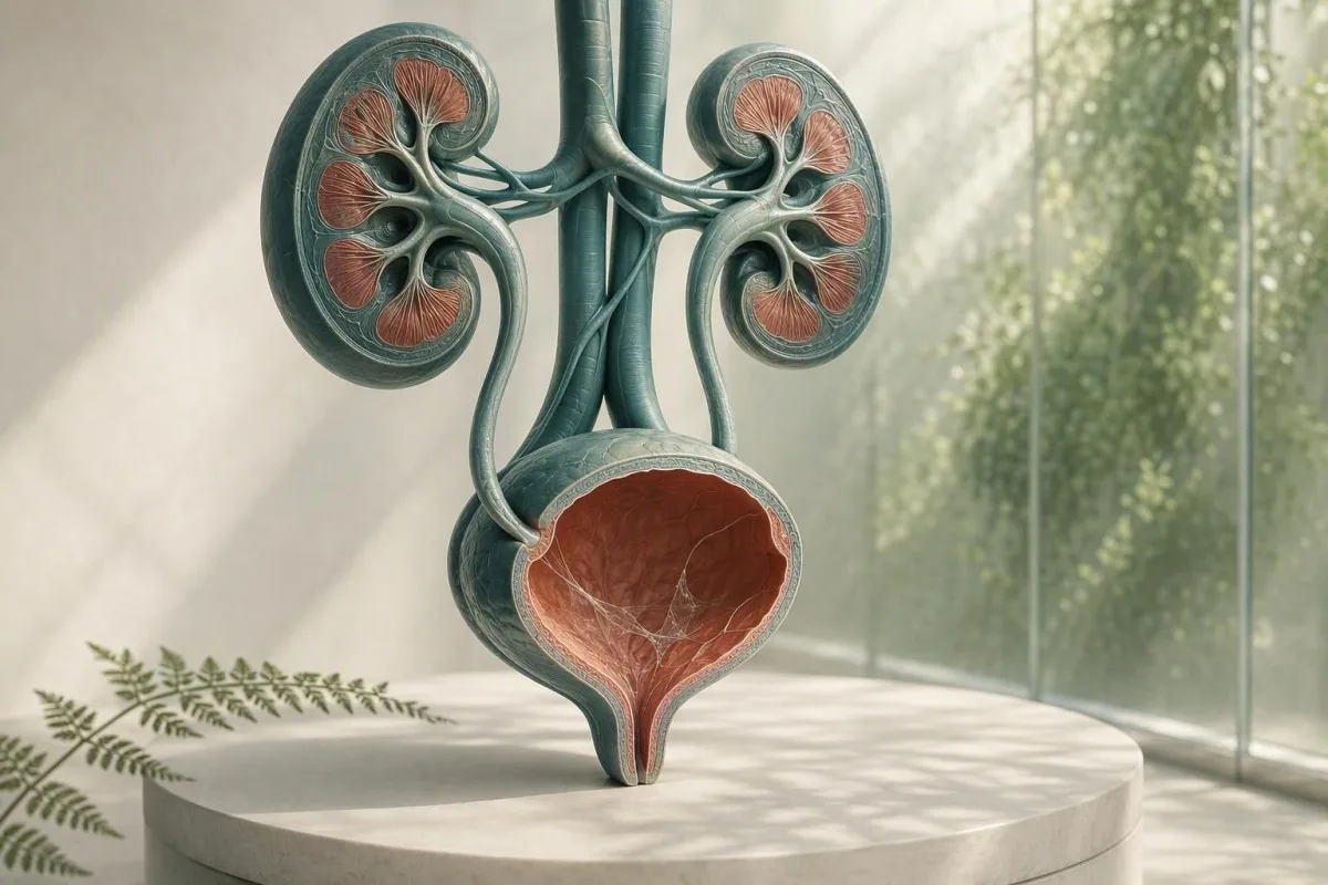
Ang mucus sa usa ka urine microscopy report kasagaran usa ka isyu sa pagkolekta, dili usa ka pagdayagnos. Ang naglibot nga mga nakaplagan sa urinalysis, mga sintomas, ug kalidad sa sample ang magdeterminar kung kini nagkinahanglan og dugang nga pagsusi.


- Mucus threads sa ilang kaugalingon kasagarang walay kadaot, labi na sa usa ka limpyo nga sample nga walay mga sintomas sa agianan sa ihi.
- Pyuria threshold nga labaw pa sa 5 ka puting selula matag high-power field nagsuporta sa panghubag sa agianan sa ihi apan wala magpamatuod sa usa ka bacterial UTI.
- Culture trigger mao ang mucus ug pagkasunog, pag-apura, hilanat, sakit sa kilid, nitrites, o dagkong puting selula—dili lang mucus.
- Squamous cells nga labaw sa gibana-bana nga 10 matag high-power field kasagaran nagpaila sa kontaminasyon sa kinatawo-panit ug naghimo sa resulta sa ihi nga dili kasaligan.
- Makita nga dugo nga adunay colicky side pain nagkinahanglan og dinalian nga pagtimbang-timbang alang sa usa ka bato, bisan kung ang mucus usab gitaho.
- Pagbuntis nagbag-o sa threshold alang sa follow-up tungod kay ang asymptomatic bacteriuria kasagaran gisusi gamit ang urine culture, dili lang sa microscopy.
- Maulap nga ihi makapakita sa mga kristal, konsentradong ihi, vaginal discharge, semilya, o mga selula; dili kini makadugtas sa impeksyon pinaagi sa panagway.
- Pag-usab sa sample makatarunganon kung ang uhog mao ra ang dili kasagaran nga nakit-an ug ang pagkolekta dili usa ka mabinantayon nga midstream clean-catch.
Unsa ang kasagarang gipasabut sa mucus threads sa urinalysis
Ang mga hilo sa uhog sa ihi kasagaran mga hibla sa normal nga proteksiyon nga sekresyon o kontaminasyon sa kinatawo, ug ang usa ka nahilit nga resulta panagsa ra nagpasinyas sa sakit sa amimika. Ang pagpangita nahimong makahuluganon kung kini makita nga adunay mga sintomas sa urinaryo, puti nga mga selula, bakterya, dugo, o protina.
Ang mga laboratoryo nag-ila sa uhog ingon nga malagkit, translucent nga mga hibla nga makita ubos sa microscopy; daghang mga report ang yano nga naggrado niini ingon nga talagsaon, pipila, kasarangan, o daghan kaysa paghatag usa ka numero nga reference range. Ang ihi natural nga adunay gamay nga kantidad sa materyal nga dato sa glycoprotein gikan sa urinary tract, ug ang vaginal o urethral secretions makadugang pa sa panahon sa pagkolekta.
Ang pulong sa report dili kaayo makahadlok kaysa sa paminaw niini. Sukad sa Agosto 26, 2026, walay balido nga ihap sa hilo sa uhog nga nag-diagnose sa UTI, bato, kanser, o pagkapakyas sa amimika; kini usa ka obserbasyon sa microscopic sa konteksto. Alang sa mas lapad nga mapa sa ubang mga butang sa usa ka dipstick ug microscope report, tan-awa ang among kompletong giya sa urinalysis.
Sa akong klinikal nga kasinatian, ang usa ka pasyente nga walay sintomas nga adunay uhog nga gimarkahan og “kasarangan,” 0-2 puti nga mga selula matag taas-nga-lig-on nga uma, negatibo nga nitrite, ug walay dugo kasagaran labing maayo nga gisilbi sa kasigurohan o usa ka husto nga nakolekta nga balik-balik nga sample. Si Dr. Thomas Klein kanunay nga makakita og walay kinahanglan nga kabalaka sa antibiotic nga motungha tungod kay ang usa ka bandila sa laboratoryo sayop nga giisip ingon nga usa ka label sa sakit.
Ang Kantesti usa ka AI platform sa pagsabot sa resulta sa blood test nga makapahimutang sa mga marker sa dugo nga may kalabotan sa amimika, sama sa creatinine ug eGFR, tupad sa usa ka pagpangita sa urinalysis—apan dili kini makadugtas sa UTI gikan lang sa uhog. Ang resulta sa urine microscopy nagkinahanglan sa klinikal nga istorya nga kauban niini.
Ngano nga gibandila kini sa usa ka lab
Ang mga sistema sa impormasyon sa laboratoryo nagbandila sa uhog tungod kay kini makita, dili tungod kay ang laboratoryo nagbutang ug usa ka peligrosong cutoff. Ang usa ka bandila busa nagpasabut nga “naobserbahan” imbes nga “dili normal nga marker sa sakit,” sama ra sa usa ka nahilit nga pagpangita sa pagsubay mahimong dili magkinahanglan og pagtambal.
Diin gikan ang mucus sa sample sa ihi
Ang uhog sa ihi mahimong gikan sa urethra, sapaw sa pantog, cervix o vagina, semilya, o ang gawas nga panit sa kinatawo sa panahon sa pagkolekta. Ang gigikanan niini kasagarang gibase sa nahibilin sa microscopic nga pattern kaysa sa mga hibla mismo.
Ang pantog ug urethra adunay proteksiyon nga sapaw sa ibabaw nga adunay mga mucin, lakip ang materyal nga nalambigit sa uroplakin, nga nagpamenos sa friction ug pagkadugtong sa microbial. Ang gagmay nga gidaghanon mahimong mahulog sa ihi pagkahuman sa dehydration, bag-o nga kalihokan sa sekso, gamay nga iritasyon, o yano tungod kay ang usa ka konsentradong specimen sa una sa buntag nagpadali sa pagtan-aw sa mga hibla.
Alang sa mga tawo nga nagregla o adunay vaginal discharge, ang leukocytes ug uhog mahimong makasulod sa tasa nga wala gikan sa pantog. Labaw pa sa 10 squamous epithelial cells matag high-power field naghimo sa kontaminasyon nga mas lagmit, bisan kung ang mga laboratoryo naggamit og lainlaing mga threshold sa pagreport. Ang among giya sa epithelial cells sa ihi nagpatin-aw ngano nga kini nagbag-o sa pagsalig sa usa ka resulta.
Ang semilya mahimo usab nga makahimo og stringy nga materyal o pagkamul-an sulod sa pipila ka oras human sa ejaculation. Dili kana makapahimo sa ihi nga dili luwas, apan kini makatago sa microscopy; kung gipamalandongan ang kultura, sa kasagaran akong tambagan ang pagkolekta og bag-ong sample labing menos 24 oras sa ulahi kung mahimo.
Nganong ang sekso ug anatomya makaimpluwensya sa report
Ang baso sa ihi dili makabulag sa pantog gikan sa kasikbit nga tisyu. Mao kini ang hinungdan nga ang “naay sipon” mas sagad ireport sa mga sample nga dunay gawas nga mga sekresyon, samtang ang usa ka catheterized specimen usahay gigamit kung ang mga clinician manginahanglan og mas limpyo nga tubag.
Kanus-a ang mucus threads nagpunting sa impeksyon sa agianan sa ihi
Ang mga hilo sa sipon mosuporta sa posibleng UTI kung kini kuyog sa mga sintomas ug objetibong mga pagpangita sa panghubag, labi na ang pyuria, nitrite positivity, o usa ka makapakombinsir nga kultura. Ang sipon sa iyang kaugalingon dili usa ka pagsulay sa UTI.
Ang usa ka symptomatic lower UTI sagad hinungdan sa pagsunog, pagkadinalian, kasubsob, suprapubic nga kasakit, ug usahay bag-ong baho sa ihi. Sa usa ka husto nga nakolekta nga sample, kapin sa 5 ka puti nga selula sa dugo kada high-power field kasagaran gitawag nga pyuria; kini mosuporta sa panghubag sa agianan sa ihi apan mahimo usab nga mahitabo sa mga bato, impeksyon nga nakuha pinaagi sa pakikipagtalik, ug kontaminasyon.
Ang nitrite taas kaayo ang pagka-espesipiko kung positibo apan makalimtan ang mga impeksyon nga gipahinabo sa mga organismo nga dili makapakunhod sa nitrate, ug ang negatibong resulta sa nitrite dili makapawala sa UTI. Ang leukocyte esterase nakamatikod sa kalihokan sa puting selula nga enzyme ug mahimong sayop nga positibo kung ang mga sekresyon sa kinatawo makontamina sa specimen; ang among pagpatin-aw sa mga resulta sa leukocyte esterase nagtabang sa pag-ila niadtong mga lit-ag.
Ang giya sa IDSA nagtambag batok sa pag-screen o pagtambal sa asymptomatic bacteriuria sa himsog nga mga hamtong nga dili mabdos tungod kay ang mga antibiotiko makadugang sa kadaot nga walay kaayohan (Nicolle et al., 2019). Kini nga prinsipyo ilabi na nga may kalabutan sa sipon: walay pagsunog, walay hilanat, ug walay giplano nga urolohiko nga pamaagi kasagaran nagpasabot ug walay antibiotiko tungod lang kay ang microscopy field morag tapulan.
Kanus-a ang kultura ang mas maayo nga pagsulay
Ang kultura sa ihi mas mapuslanon kaysa balik-balik nga dipstick kung ang mga sintomas magpadayon, ang mga sintomas mobalik sulod sa 4 ka semana, ang pagmabdos anaa, ang pyelonephritis gisuspetsahan, o ang kanhing mga antibiotiko mahimong nakapausab sa resulta. Ang sagol nga pagtubo sa bakterya sagad nagpaila sa kontaminasyon sa pagkolekta kaysa usa ka solong urinary pathogen; tan-awa ang pagpasabot sa urine culture.
Mucus, kristal, bato, ug mekanikal nga iritasyon
Ang sipon nga adunay grabeng balod nga sakit sa kilid o dugo sa ihi mahimong mahitabo sa urinary stone, apan ang sipon dili makakumpirma o makawagtang sa bato. Ang dugo, mga kristal, pattern sa kasakit, imaging, ug paggana sa kidney mas importante.
Ang usa ka bato mahimong makagisi o makababag sa linya sa agianan sa ihi, nga makahimo og mga pulang selula, mga puting selula, ug dugang nga sipon. Ang klasiko nga sintomas mao ang kalit nga sakit nga modagayday paingon sa singit, kanunay nga adunay kasukaon; ang makita nga pula o kolor tsa nga ihi nagpataas sa pagkadinalian. Basaha ang dugang pa mahitungod sa dugo sa ihi nga pula nga bandila kay sa pag-ingon nga ang matag pink nga sample kay usa ka simple nga impeksyon.
Ang mga kristal sa calcium oxalate mahimong makita sa mga himsog nga tawo, ilabi na sa konsentradong acidic nga ihi, busa ang usa ka tipo sa kristal dili makapamatuod sa usa ka aktibo nga bato. Sa usa ka tawo nga adunay kasakit, balik-balik nga mga bato, o nagpadayon nga haematuria, ang mga clinician mahimong mogamit og ultrasound o low-dose non-contrast CT kaysa mosalig lamang sa sediment. Ang among tinubdan sa mga kristal nga calcium oxalate nagdetalye sa mga limitasyon sa mga report sa kristal.
Usa ka importante nga eksepsyon mao ang pagbabag nga adunay impeksyon: hilanat nga 38.0°C o mas taas, sakit sa kilid, pagsuka, ug dili paglabay sa ihi naghatag ug emerhensya nga pagtimbang-timbang. Ang kabalaka dili ang sipon; kini usa ka nataptan nga nababagan nga sistema sa ihi, nga mahimong dali nga modaut.
Pagkalagot nga walay bato
Ang bag-o nga paggamit sa catheter, mga pamaagi sa pantog, kusog nga pagbisikleta, ug pelvic radiation mahimong makapalagot sa ubos nga agianan ug makadugang sa sipon o puti nga mga selula. Ang usa ka clinician kinahanglan maghubad niini nga mga resulta batok sa pag-oras, tungod kay ang usa ka sample nga nakolekta sulod sa 48 ka oras sa instrumentasyon dili katumbas sa usa ka regular nga screening specimen.
Kahulugan sa cloudy urine: unsay gisulti ug unsay dili nimo masulti
Ang madag-um nga ihi mahimong tungod sa konsentradong mga asin, mga kristal, sipon, mga selula, paggawas sa kinatawo, o bakterya, busa ang panagway lamang dili makadugno sa impeksyon. Ang ihi nga madag-um apan walay sakit ug mubo ra ang gidugayon kasagaran dili dinalian.
Ang mga kristal sa phosphate mahimong makapabaga sa alkalina nga ihi human kini mobugnaw, samtang ang mga kristal sa urate mahimong makapabaga sa acidic nga concentrated nga ihi. Ang usa ka sample nga ibilin sa temperatura sa kwarto sulod sa kapin sa 2 ka oras mahimong magkadaghan ang kabag-o samtang nagkadunot ang mga selula ug modaghan ang bakterya, mao nga hinungdanon ang pagproseso dayon o pagpabugnaw.
Ang kabag-o uban sa dysuria, urgency, ug pyuria angayan nga sulayan; ang kabag-o human sa ehersisyo o dili maayo nga pag-inom og pluwido kasagaran molambo sa ordinaryong hydration. Tumong sa ihi nga luspad nga dalag kaysa pagpilit sa sobra nga pag-inom og tubig—ang tin-aw kaayo nga ihi wala magpasabot nga ang mga amimoot “nagwawad-an” sa impeksyon. Ang among detalyado nga pagrepaso sa mga hinungdan sa madag-umon nga ihi nagbulag sa mga biswal nga timailhan gikan sa kasaligang mga pagsulay.
Si Kantesti usa ka AI blood test analyzer gidisenyo sa paghubad sa mga marka sa dugo sa clinical context; ang taas nga creatinine o ubos nga eGFR kauban sa mga abnormalidad sa ihi mahimong maghatag ug katarungan sa medikal nga pagrepaso, samtang ang baga nga ihi lamang wala magtukod ug nadaot nga pagsala sa amimoot. Ang usa ka batakang metabolic panel makadugang ug mapuslanong konteksto kung ang mga sintomas magpaila sa dehydration o obstruction.
Ang baho dili kultura
Ang kusog nga baho sa ihi kasagarang nagpabanaag sa konsentrasyon, mga metabolite sa asparagus, bitamina B, o usa ka sudlanan nga nagpabilin nga dugay kaayo. Ang bag-ong dili maayo nga baho nga adunay hilanat o mga sintomas sa urinaryo usa ka rason sa pagpangayo ug assessment, apan ang baho dili kapuli sa microscopy ug kultura.
Discharge sa kinatawo ug mga impeksyon nga makadala pinaagi sa pakighilawas
Ang urethral o vaginal discharge mahimong morag mucus sa ihi, ug ang bag-ong discharge nga adunay pagsunog sa urinaryo nagkinahanglan ug sexual-health testing ingon man usa ka urine assessment. Ang routine urine culture mahimong makalimtan ang chlamydia ug gonorrhoea.
Ang Chlamydia ug gonorrhoea mahimong hinungdan sa dysuria ug sterile pyuria—mga puti nga selula nga walay routine bacterial growth—labi na human sa bag-ong sexual exposure. Ang nucleic acid amplification testing, kasagarang naggamit ug first-catch urine kaysa midstream specimen, mao ang angay nga pagsulay tungod kay ang standard culture nagtarget sa ubang mga organismo.
Sa mga tawo nga adunay vagina, ang bacterial vaginosis, candidiasis, ug cervical inflammation mahimong makadugang og discharge sa midstream cup; ang vaginal swab o eksaminasyon mahimong mas impormatibo kaysa pag-usab sa urine microscopy. Sa mga tawo nga adunay penis, ang makita nga urethral discharge dili angay ipakaubos isip “mucus threads.” Ang praktikal nga kalainan mao ang discharge nga namatikdan sa gawas sa pag-ihi kumpara sa mga hilo nga gitaho lamang sa laboratoryo.
Ang 2021 CDC STI treatment guideline nagrekomenda sa NAAT-based testing sa mga may kalabotan nga anatomical sites sumala sa history sa exposure, dili lang mga sintomas (Workowski et al., 2021). Kung adunay pelvic pain, testicular pain, hilanat, pagmabdos, o posibleng pag-atake, mangayo ug samang adlaw nga clinical advice imbes nga mag-self-medicate sa nahabilin nga mga antibiotic.
Ngano nga ang mga antibiotic makalibog sa hulagway
Ang pag-inom bisan og 1 o 2 ka dosis sa antibiotic sa dili pa ang kultura mahimong makapugong sa ordinaryong bacterial growth samtang ang mga sintomas sa urinaryo ug mga puti nga selula magpadayon. Isulti sa clinician ang eksaktong tambal, dosis, ug oras sa katapusang dosis nga gigamit; kana nga detalye nagbag-o kung giunsa ang paghubad sa usa ka negatibo nga kultura.
Unsaon pagkolekta og sample sa ihi nga makatubag sa pangutana
Ang usa ka midstream clean-catch sample makapakunhod sa mucus, squamous cells, ug sagol nga bacterial growth kaysa sa pagkolekta sa una o katapusan nga bahin sa stream. Yano ang teknik, apan ang unang 2 segundos adunay dako nga kalainan.
Hugasi ang mga kamot, ibulag ang panit sa kinatawo o ibira ang foreskin kon komportable, limpyohan sumala sa mga instruksyon sa kit, sugdi ang pag-ihi sa kasilyas, dayon kolektaha ang tunga-tunga nga bahin nga walay paghikap sa sudlanan sa panit. Ang 20–30 mL nga sample kasagarang igo na; ang pagpuno sa usa ka dako nga sudlanan hangtod sa ngilit dili makapauswag sa pagsulay.
Ihatod ang sudlanan dayon, labing maayo sulod sa 1 ka oras; kung dili kalikayan ang paglangan, ibutang kini sa refrigerator sumala sa mga instruksyon sa laboratoryo ug ibalik kini sulod sa 24 oras. Ang pagpabugnaw makapahinay sa pagtubo sa bakterya apan dili makapahiuli sa usa ka sample nga nahugawan na. Kini ilabina importante kung ang hangyo naglakip sa kultura imbes nga dipstick lamang.
Si Kantesti usa ka AI-powered blood test analysis tool gigamit sa tibuok kalibutan, ug ang among mga clinician-reviewed workflow nagtratar sa kalidad sa specimen isip bahin sa interpretasyon imbes nga usa ka footnote. Ang parehas nga prinsipyo magamit sa bisan unsang bili sa laboratoryo: ang usa ka teknikal nga dili maayo nga sample mahimong makahimo usa ka medikal nga makapakombinsir apan makapahisalaag nga resulta. Tan-awa ang among checklist sa katukma sa lab alang sa mga pangutana nga ipangutana sa dili pa molihok sa usa ka isolated flag.
Ayaw pagkolekta sa kini nga mga sitwasyon kung mahimo ka maghulat
Likayi ang routine testing sa panahon sa bug-at nga menstrual flow, diha-diha dayon human sa pakighilawas, o human sa paggamit sa vaginal creams gawas kung ang clinician piho nga mohangyo niini. Kung dili mahimo ang pagsulay, isulti sa laboratoryo o clinician, tungod kay kana nga konteksto mahimong makapatin-aw sa mucus, pula nga mga selula, o external cells.
Unsaon pagbasa sa mga clinician ang mucus uban sa uban nga urinalysis
Ang labing mapuslanon nga urinalysis pattern nagkombinar sa mga sintomas sa mga puti nga selula, pula nga mga selula, nitrite, leukocyte esterase, protina, glucose, specific gravity, ug epithelial cells. Ang mucus usa ka gamay nga nagsuporta nga bahin sa kana nga sumbanan.
Ang usa ka limpyo nga sample nga adunay mucus, 0–2 puti nga selula matag high-power field, negatibo nga nitrite, ug walay dugo kasagaran dili kinahanglan og pagtambal. Ang mucus nga adunay labaw pa sa 5 puti nga selula matag high-power field ug positibo nga leukocyte esterase nagpataas sa posibilidad sa panghubag; ang pagdugang og nitrite o usa ka single-organism culture nagdugang sa pagsalig nga ang mga bakterya ang responsable.
Ang protina nagkinahanglan sa kaugalingon nga agianan. Ang trace protein pagkahuman sa hilanat, ehersisyo, o konsentrado nga ihi mahimong temporaryo, apan ang nagpadayon nga protina kinahanglan nga sukdon gamit ang albumin-to-creatinine ratio imbes nga ihalad sa mucus. Ang among giya sa protein sa ihi nagpatin-aw ngano nga ang dipstick protein ug ang peligro sa amimika dili mapulihan.
Ang specific gravity na 1.003 hangtod 1.030 kasagaran sa mga hamtong, bisan kung ang mga reperensya nagkalainlain sa matag laboratoryo. Ang taas nga espesipikong grabidad mahimong magkonsentrar sa mucus ug makahimo og mas dramatiko nga tan-awon nga sediment, samtang ang ubos nga espesipikong grabidad mahimong makadiskaril sa mga selula ug makapahimo sa microscopy nga malimbongon nga yano; ang among giya sa specific gravity nagpakita kung giunsa ang hydration makaapekto sa interpretasyon.
Ang bili sa negatibong mga resulta
Ang negatibong dugo, protina, nitrite, ug leukocyte esterase makahuluganon nga nagpaubos sa kabalaka sa usa ka tawo nga walay mga sintomas, bisan kung ang mucus gitaho nga kasarangan. Walay usa ka negatibong pagsulay ang perpekto, apan kini nga hugpong mas makapahupay kaysa sa usa ka grado sa mucus nga nakapaalarma.
Kanus-a balikon ang pagsulay ug kanus-a mag-order ug culture
Balika ang usa ka urine sample kung ang mucus mag-inusara, ang mga squamous cell nagpaila sa kontaminasyon, o ang pagkolekta gipadali; paghangyo og kultura kung ang mga sintomas magpadayon, balik-balik, grabe, o high-risk. Ang pag-usab sa pagsulay dili “pagbuhat og wala”—kini sagad ang labing diagnostic nga sunod nga lakang.
Alang sa usa ka dili mabdos nga hamtong nga walay mga sintomas ug mucus isip bugtong bandila, ang pag-usab sa clean-catch urinalysis sulod sa 1–2 ka semana makatarunganon kung gikinahanglan ang kasigurohan; daghang mga doktor dili na kini usbon. Ayaw pagtratar ang usa ka laboratory flag og mga antibiotic samtang naghulat gawas kung ang usa ka preskriber makaila sa usa ka klinikal nga indikasyon.
Ang kultura sa wala pa ang mga antibiotic labi na nga mapuslanon alang sa hilanat, kasakit sa kidney, pagmabdos, dili pag-ayo sa resistensya, pagbalhin sa amimika, paggamit sa catheter sa ihi, mga sintomas sa ihi sa lalaki, o mga sintomas nga mobalik sulod sa 4 ka semana. Ang giya ni Gupta et al. (2011) nagsuporta sa kultura sa gidudahang pyelonephritis ug mga sitwasyon diin ang resistensya o usa ka alternatibong diagnosis mas lagmit.
Ang mga ihap sa kultura kinahanglan basahon uban ang kalidad sa pagkolekta. Usa ka single organism sa 10^5 colony-forming units matag mL tradisyonal nga nagsusto sa bacteriuria sa limpyo nga tunga-tunga nga ihi, apan ang mga pasyente nga adunay sintomas mahimong adunay klinikal nga importante nga ubos nga ihap; ang “sagol nga flora” kasagaran nag-aghat ug bag-ong sample kaysa usa ka lapad nga antibiotic. Itandi ang mga katuyoan sa urinalysis ug culture sa wala pa mangayo bisan unsa.
Ang pagmabdos nanginahanglan og lahi nga threshold
Ang pagmabdos usa ka eksepsyon tungod kay ang asymptomatic bacteriuria mahimong makadugang sa risgo sa pyelonephritis ug dili maayo nga mga resulta sa pagmabdos. Ang antenatal care kasagarang naggamit og screening culture sayo sa pagmabdos; ang mucus sa microscopy dili makapuli niana nga culture.
Kanus-a ang mga blood test makadugang ug mapuslanon nga kahibalo sa kidney ug impeksyon
Ang mga pagsulay sa dugo mapuslanon kung ang mucus sa ihi adunay hilanat, kasakit sa kilid, balik-balik nga impeksyon, paghubag, pagkunhod sa output sa ihi, o padayon nga protina o dugo. Ang creatinine ug eGFR nagsusi sa pagsala, samtang ang CBC ug C-reactive protein makatabang sa pagbanabana sa systemic illness.
Ang eGFR nga ubos sa 60 mL/min/1.73 m² sulod sa labing menos 3 bulan nakab-ot ang usa ka criterion alang sa chronic kidney disease, apan ang usa ka ubos nga resulta sa panahon sa dehydration o acute illness dili igo aron himuon kana nga diagnosis. Ang creatinine naapektuhan sa muscle mass, pagkaon, ug pipila ka mga tambal, mao nga ang mga uso ug urine albumin kanunay mas mapuslanon kaysa usa ka numero.
Ang taas nga white blood cell count o CRP makasuporta sa usa ka inflammatory process apan dili makaila sa urinary tract isip tinubdan. Sa usa ka tawo nga adunay hilanat ug kasakit sa kilid, ang mga clinician mahimong mosusi sa creatinine sa wala pa mopili og imaging o mga tambal, labi na kung ang pagsuka o pagbara makapahinay sa function sa kidney. Review sa mga stage sa chronic kidney disease alang sa mga kategorya sa eGFR ug albumin nga gigamit sa mga clinician.
Si Kantesti usa ka AI lab test interpretation service nga nagpasiugda sa mga sumbanan sa creatinine, eGFR, electrolytes, CBC, ug inflammatory markers imbes nga pagtratar sa usa ka mucus notation isip usa ka diagnosis sa kidney. Ang among methodology gipailalom sa pagdumala sa balidasyon sa klinika, apan ang dinalian nga mga sintomas nagkinahanglan gihapon og direkta nga pag-atiman sa medisina, dili interpretasyon nga pinasikad sa app.
Usa ka praktikal nga kalainan sa dehydration
Ang dehydration mahimong makapataas sa urine specific gravity ug temporaryo nga makadugang sa creatinine, ilabi na human sa kalibanga, pagkaladlad sa kainit, o grabeng ehersisyo. Ang padayon nga ubos nga output sa ihi, pagkalipong, o pagkunhod sa eGFR human sa pag-rehydrate nagkinahanglan og pagrepaso sa clinician imbes nga balik-balik nga pagsulay sa balay.
Mga pasidaan sa dinalian nga mga senyales nga dili angay paabuton ang balik-balik nga pagsulay
Pangitaa ang pagtimbang-timbang dayon sa adlaw alang sa mucus sa ihi nga adunay hilanat nga 38.0°C o mas taas, kasakit sa kilid, pagsuka, makita nga dugo, kawalay katakus sa pag-ihi, kalibog, o mga sintomas sa urinaryo nga may kalabotan sa pagmabdos. Kini nga mga kombinasyon importante tungod kay mahimo kini nga magpaila sa impeksyon sa taas nga agianan, pagbara, o uban pang acute nga kondisyon.
Ang hilanat dugang ang usa ka kilid nga sakit sa likod o kilid mas gikabalak-an alang sa pyelonephritis kaysa simple nga cystitis, labi na sa pag-urog o pagsuka. Ang paglangan sa pagtambal mahimong mosangpot sa dehydration, sepsis, o stress sa kidney; ang usa ka normal nga tan-awon nga sample sa ihi sa balay dili luwas nga makawagtang niini.
Ang makita nga dugo kinahanglan nga susihon bisan kung ang UTI daw posible, labi na pagkahuman sa edad 35, sa mga nanigarilyo, o kung ang pagdugo nagpadayon kung ang mga sintomas sa impeksyon mohupas. Ang AUA microhaematuria guideline nagrekomenda sa pagsusi nga pinasikad sa risgo human masulbad ang mga benign nga pagpasabot (Barocas et al., 2020); ang padayon nga dugo dili mapasabot sa mucus.
Ang mga bata, mga hamtong nga kapin sa 65, mga tawo nga adunay diabetes, mga tawo nga nag-inom og immune-suppressing nga mga tambal, ug kadtong adunay usa ra ka kidney ang angay sa ubos nga threshold alang sa pagsusi. Ang lagda ni Dr. Thomas Klein sa praktis yano ra: kung ang mga sintomas nagkadako sulod sa 6-12 ka oras, ayaw paghulat og ikaduhang tasa aron maghatag og kasigurohan.
Tawag dayon sa emerhensya alang sa grabeng sakit
Tawag sa mga serbisyo sa emerhensya alang sa bag-ong kalibog, pagkalipong, grabeng kahuyang, asul o abo nga mga ngabil, grabeng kalisud sa pagginhawa, o kawalay katakus sa pagpugong sa mga likido nga adunay mga sintomas sa urinaryo. Kini mga systemic danger signs, dili ordinaryong mga sintomas sa UTI.
Mucus sa ihi atol sa pagmabdos, pagkabata, ug sa ulahi nga kinabuhi
Ang pagmabdos, mga bata, ug mga tigulang nanginahanglan og mas mabinantayon nga interpretasyon tungod kay kasagaran ang kontaminasyon apan ang mga sangputanan sa nawala nga impeksyon mahimong mas dako. Ang mga sintomas ug kalidad sa kultura nagpabilin nga mas bililhon kaysa sa grado sa mucus sa matag grupo sa edad.
Sa panahon sa pagmabdos, ang kasubsob sa pag-ihi mahimong normal, nga naghimo sa mga sintomas nga dili kaayo piho; hilanat, dysuria, o kasakit sa likod kinahanglan magpalihok sa pagtimbang-timbang sa obstetric o klinikal. Ang screening culture kasagarang kuhaon sayo sa prenatal care, ug ang balik-balik nga culture mahimong gamiton human sa pagtambal depende sa plano sa clinician.
Sa mga bata, ang ginadala sa bag nga ihi daling mahugawan ug dili angay gamiton nga nag-inusara aron madayagnos ang UTI gamit ang culture. Ang catheterized o maampingon nga nakuha nga limpyo-catch specimen mahimong gikinahanglan kung ang usa ka bata adunay hilanat nga walay klaro nga tinubdan; ang uhot sa bag nga specimen ilabi na dili piho.
Sa ulahi nga kinabuhi, ang bacteriuria ug pyuria mas komon nga dili hinungdan sa mga sintomas. Ang bag-ong delirium nga nag-inusara kinahanglan magpalihok sa usa ka halapad nga medikal nga pagtimbang-timbang alang sa dehydration, mga tambal, kasakit, constipation, ug mga tinubdan sa impeksyon kaysa awtomatikong pagtambal sa UTI; ang samang pagpugong nga girekomenda ni Nicolle et al. (2019) magamit.
Pagregla ug mga kausaban nga may kalabotan sa hormone
Ang dugo sa pagregla ug ang cervical mucus mahimong mag-usab sa urinalysis sulod sa pipila ka adlaw, ug ang postmenopausal vaginal dryness mahimong hinungdan sa lokal nga iritasyon nga morag pagsunog sa ihi. Kung ang mga resulta ug mga sintomas dili magkatugma, ang usa ka clinician mahimong motimbang-timbang sa mga hinungdan sa kinatawo kaysa magreseta og balik-balik nga pagtambal sa UTI.
Mga kasagarang sayop human makakita og mucus sa lab report
Ayaw pagsugod sa nahabilin nga mga antibiotic, paghinlo sa sulod, o pagsulay sa “pag-flush out” sa uhot nga adunay sobra nga pag-inom og tubig human sa usa ka abnormal nga tan-awon nga urinalysis. Kini nga mga aksyon mahimong makatabon sa usa ka culture, makadisturbo sa normal nga flora, o makadelay sa husto nga diagnosis.
Ang mga antibiotic nga gikuha nga walay culture mahimong maghimo sa sunod nga pagsulay nga bakak nga negatibo ug mahimong hinungdan sa kalibanga, pantal, mga sintomas sa yeast, o resistensya. Kung ang mga sintomas malumo apan nagpadayon, kuhaa una ang sample kung praktikal, dayon sundan ang plano sa pagtambal sa clinician base sa tibuok nga hulagway.
Ang mga produkto sa cranberry mahimong gamay nga makapakunhod sa peligro sa balik-balik nga uncomplicated UTI alang sa pipila ka mga tawo, apan dili kini makatambal sa hilanat, kasakit sa kilid, o usa ka napamatud-an nga impeksyon sa taas nga bahin sa agianan. Paglikay sa taas nga dosis sa bitamina C isip self-treatment: kini mahimong mag-usab sa pipila ka dipstick reactions ug mahimong makadugang sa oxalate burden sa mga tawo nga daling madala sa calcium oxalate stones.
Ayaw balik-balikon ang pag-inspeksyon sa kasilyas alang sa mga hilo. Ang mga lanot sa tissue sa kasilyas, mga residu sa paglimpyo, mga sekresyon sa kinatawo, ug ang pagkulo sa tubig mahimong morag uhot; ang usa ka laboratory sample nga gikuha sa usa ka sterile container mao ang angay nga lugar aron kini masusi. Alang sa may kalabutan nga mga kausaban sa kolor, among giya sa kolor sa ihi naghatag og mas kasaligang biswal nga konteksto.
Ang listahan sa tambal importante
Ang Phenazopyridine mahimong maghimo sa ihi nga orange ug makabalda sa biswal nga interpretasyon, samtang ang mga diuretic mahimong mag-concentrate o magdilute sa ihi depende sa oras. Pagdala ug lista sa mga reseta nga tambal, mga produkto nga over-the-counter, ug mga suplemento sa appointment, lakip ang mga dosis sa mg.
Usa ka praktikal nga follow-up plan alang sa mucus threads sa ihi
Kadaghanan sa mga tawo nga adunay mga hilo sa uhot sa ihi ug walay mga sintomas dili kinahanglan nga tambalan; usa ka maampingon nga balik-balik nga urinalysis makatarunganon kung ang sample kadudahan. Ang mga sintomas o mga kauban nga abnormalidad nagtino kung ang culture, STI testing, imaging, o mga pagsulay sa dugo ang angay.
Lakang 1: susiha ang pagsunog, pagkadinalian, hilanat, kasakit sa kilid, makit-an nga dugo, paggawas, pagmabdos, bag-o nga mga pamaagi sa urinary, ug bag-o nga sekswal nga pagkaladlad. Lakang 2: basaha ang report alang sa puting selula, nitrite, leukocyte esterase, pulang selula, protina, bakterya, ug squamous cells—dili lang uhot.
Lakang 3: kung maayo ang imong gibati ug ang uhot nag-inusara, pagkuha og usa ka midstream clean-catch sample sulod sa 1-2 ka semana kung ang imong clinician morekomendar og kumpirmasyon. Lakang 4: kung adunay mga sintomas, pangutana kung ang urine culture kinahanglan kuhaon sa wala pa ang pagtambal ug kung ang STI testing may kalabotan; ang balik-balik nga mga panghitabo angayan og dugang nga pagrepaso kaysa sa ikatulo nga empiric antibiotic.
Ang Kantesti makahan-ay sa may kalabotan nga mga trend sa resulta sa dugo ug mga pangutana alang sa usa ka medikal nga appointment, samtang ang among Lupon sa Medikal nga Magtatambag nagsuporta sa mga sumbanan sa kaluwasan nga gipangulohan sa clinician. Ang husto nga katapusan dili usa ka hingpit nga “limpyo” nga report sa microscopy—kini usa ka pagpatin-aw nga mohaum sa imong mga sintomas, kalidad sa specimen, ug mga risk factor.
Mga pangutana nga dad-on sa imong appointment
Pangutan-a kung ang specimen adunay squamous cells, kung ang culture nagpatubo og usa ka organismo o sagol nga flora, ug kung ang dugo o protina nagpadayon human nawala ang mga sintomas. Kini nga 3 ka pangutana kanunay makahatag og mas mapuslanon nga mga tubag kaysa pagpangutana kung unsa kadaghan ang uhot nga nakita.
Konteksto sa panukiduki ug ang mga limitasyon sa pagreport sa mucus
Ang paggrado sa uhot dili standardisado sa mga laboratoryo, mao nga ang mga clinician kinahanglan nga maglikay sa paggamit sa “gamay,” “moderate,” o “daghan” isip mga kategorya sa kagrabe sa sakit. Ang teknik sa mikroskopyo, edad sa espesimen, ug software sa pagreport mahimong mausab ang mga pulong.
Ang ubang mga laboratoryo nag-inspeksyon sa sediment human sa pag-centrifuge sa mga 10–15 mL nga ihi, samtang ang uban naggamit ug automated particle analysis uban ang manual review sa piniling mga flag. Kini nga kalainan nagpasabot nga ang usa ka “moderate mucus” nga resulta gikan sa usa ka laboratoryo mahimong dili motugma pag-ayo sa laing laboratoryo bisan kung ang panglawas sa tawo wala mausab.
Ang usa ka mapuslanon nga panghunahuna sa laboratoryo mao ang pagpangutana kung ang usa ka resulta ba ka analytical nga tinuod, ka clinical nga makahuluganon, ug ka reproducible. Kining paagiha pamilyar usab gikan sa pagsulay sa dugo: ang among giya sa blood biomarker nagpatin-aw nganong ang mga reference interval ug pre-analytic conditions nagtino kung ang usa ka gimarkahan nga marker angayan bang aksyunan.
Ang Kantesti AI naggamit ug structured laboratory context sa pagpatin-aw sa mga pattern sa resulta sa tibuok dagkong mga panel, apan ang urine microscopy nagkinahanglan gihapon ug kahanas sa paghukom base sa tinubdan. Alang sa mga magbabasa nga interesado kung unsaon nato pagtimbang-timbang ang teknikal nga pasundayag ug mga utlanan sa klinikal, ang among giya sa teknolohiya sa AI naghulagway sa mga panalipod luyo sa among pamaagi sa interpretasyon.
Unsa ang kinahanglan klarohon sa usa ka maayong report
Ang usa ka mapuslanon nga report nag-ila sa tipo sa espesimen, petsa sa pagkolekta, microscopic nga mga elemento, ug bisan unsang kultura nga organismo ug mga resulta sa pagkasensitibo kung himuon ang kultura. Kung ang report nag-ingon lamang nga “mucus threads present,” kini dili kompleto alang sa pagdayagnos apan kasagaran igo na alang sa usa ka low-risk incidental finding.
Kanunay nga Gipangutana nga mga Pangutana
Normal ra ba ang mga hilo sa mucus sa ihi?
Ang mga hilo sa mucus sa ihi sagad normal o tungod sa kontaminasyon gikan sa mga sekresyon sa kinatawo, labi na kung ang usa ka tawo walay mga sintomas sa ihi. Ang usa ka hilit nga nakaplagan nga adunay 0–2 ka puti nga selula matag high-power field, negatibo nga nitrite, ug walay dugo o protina kasagaran ubos ang risgo. Ang mga laboratoryo dili naggamit ug balido nga gidaghanon sa mucus-thread sa pagdayagnos sa UTI o sakit sa kidney. Ang pag-usab sa midstream clean-catch sample makatarunganon kung ang orihinal nga pagkolekta gipugos o nakit-an nga adunay kontaminasyon.
Ang mga hilo sa mucus sa ihi nagpasabot ba nga aduna koy UTI?
Ang mga hilo sa plema sa ihi dili sa ilang kaugalingon nagpasabot ug UTI. Ang UTI mahimong mas lagmit kung ang pagkasunog, pagdali, kasubsob, hilanat, o kasakit sa suprapubic mahitabo uban sa labaw sa 5 ka puti nga selula matag high-power field, positibo nga leukocyte esterase, nitrite, o positibo nga kultura. Ang negatibo nga nitrite dili hingpit nga makawagtang sa UTI tungod kay dili tanan nga bakterya naghimo og nitrite. Ang kultura kasagaran mas impormatibo kaysa sa paggradar sa plema kung ang mga sintomas magpadayon o mobalik.
Makapahinabo ba ang dehydration og mucus sa ihi?
Ang dehydration makapadali sa pagtan-aw sa mucus tungod kay ang concentrated nga ihi adunay gamay nga tubig sa palibot sa normal nga mga sekresyon ug sediment. Ang taas nga specific gravity sa ihi, kasagaran padulong sa 1.025–1.030, mahimong mouban niini nga epekto, bisan kung dili kini pamatuod sa dehydration sa iyang kaugalingon. Ang normal nga pag-rehydrate ug pag-usab sa usa ka husto nga nakolekta nga sample mahimong maklaro sa resulta. Ang grabeng pag-inom ug tubig dili kinahanglan ug mahimo’ng makamugna sa kaugalingon nga mga risgo sa electrolyte.
Unsay gipasabut sa mucus ug puti nga mga selula sa dugo sa ihi?
Ang mucus nga adunay puti nga mga selula sa dugo sa ihi nagpaila sa panghubag sa agianan sa ihi o kinatawo, apan dili kini makapamatuod sa impeksyon sa bakterya. Ang sobra sa 5 ka puti nga selula matag high-power field mahimong mahitabo sa UTI, bato, impeksyon nga nakuha pinaagi sa sekso, kontaminasyon sa kinatawo, o bag-o nga paggamit sa mga instrumento sa agianan sa ihi. Ang mga sintomas, nitrite, epithelial cells, ug mga resulta sa kultura ang magdeterminar sa sunod nga lakang. Ang hilanat nga 38.0°C o mas taas nga adunay kasakit sa kilid nanginahanglan og dinalian nga pagsusi.
Makakaapekto ba ang lagas sa puwerta sa mucus sa usa ka pagsulay sa ihi?
Ang vaginal discharge mahimo nga makasulod sa usa ka midstream nga sample sa ihi ug kasagarang hinungdan sa mucus, squamous epithelial cells, ug usahay white cells nga makita sa microscopy. Labaw sa 10 ka squamous cells matag high-power field kanunay nga naghimo sa kontaminasyon nga mas lagmit, bisan kung ang mga laboratoryo magkalainlain sa ilang pagreport. Ang usa ka maampingon nga clean-catch sample nga nakolekta sa gawas sa bug-at nga pagregla makapamenos niini nga isyu. Kung adunay discharge, pangangati, baho, o kasakit sa pelvis, ang usa ka vaginal assessment o swab mahimong mas mapuslanon kaysa sa balik-balik nga mga kultura sa ihi.
Kan ko mabalaka bahin sa mga tinuhod nga mucus ug dugo sa ihi?
Ang mga hilo sa mucus nga adunay makitang dugo sa ihi kinahanglan nga susihon dayon, labi na kung magpadayon ang dugo, adunay kasakit sa kilid, mga hugot, hilanat, o kalisud sa pag-ihi. Ang bato, UTI, samad, sakit sa kidney, o dili kasagaran nga pagtubo sa agianan sa ihi mahimong hinungdan sa dugo; ang mucus dili makaila kung unsa ang hinungdan. Girekomenda sa AUA ang pagsusi nga gibase sa risgo alang sa padayon nga microscopic haematuria pagkahuman matubag ang mga temporaryo nga hinungdan. Ang pagtimbang-timbang sa emerhensya angay alang sa hilanat, pagsuka, grabeng kasakit, o pagpugong sa ihi.
Karon na ang AI-Powered Blood Test Analysis
Apil sa kapin sa 2 milyon nga mga user sa tibuok kalibutan nga nagsalig sa Kantesti para sa dayon ug tukma nga pag-analisa sa lab test. I-upload ang imong resulta sa blood test ug makadawat og komprehensibong pagsabot sa 15,000+ nga mga biomarker sulod sa mga segundo.
📚 Mga Napangalan nga Research Publications
Klein, T., Mitchell, S., & Weber, H. (2026). Normal nga Sakop sa aPTT: D-Dimer, Giya sa Pag-ihap sa Dugo gamit ang Protina C. Kantesti AI Medical Research.
Klein, T., Mitchell, S., & Weber, H. (2026). Giya sa mga Protina sa Serum: Pagsulay sa Dugo sa mga Globulin, Albumin ug A/G Ratio. Kantesti AI Medical Research.
📖 Mga Panlabas nga Sanggunian sa Medisina
⚕️ Pagpasabot sa Medikal
Kini nga artikulo para sa katuyoan sa edukasyon ra ug dili kini mosangpot sa medikal nga tambag. Kanunay mokonsulta sa usa ka kwalipikado nga healthcare provider alang sa mga desisyon sa diagnosis ug pagtambal.
Mga E-E-A-T Trust Signals
Kasinatian
Pagsusi sa klinika nga gipangulohan sa doktor sa mga workflow sa interpretasyon sa lab.
Kahanas
Pokus sa medisina sa laboratoryo kung giunsa paglihok ang mga biomarker sa konteksto sa klinika.
Pagka-awtorisado
Gisulat ni Dr. Thomas Klein ug gisusi ni Dr. Sarah Mitchell ug Prof. Dr. Hans Weber.
Kasaligan
Interpretasyon nga base sa ebidensya, nga adunay klaro nga mga agianan sa sunod nga buhat aron makunhuran ang kabalaka.