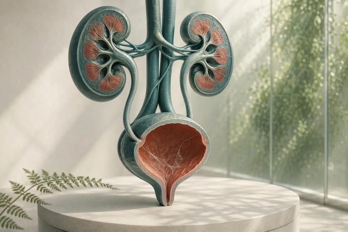
La muko sur urina mikroskopa raporto kutime estas kolekta problemo, ne diagnozo. La ĉirkaŭaj uranalizaj trovoj, simptomoj kaj kvalito de la specimeno determinas ĉu ĝi bezonas sekvadon.


- Mukaj fadenoj sole estas ofte sendanĝeraj, precipe en ununura pura kaptospecimeno sen urinanaj simptomoj.
- Püria sojlo de pli ol 5 blankaj ĉeloj po altpotenca kampo subtenas urinan vojan inflamon sed ne pruvas bakterian UTI.
- Kultura ellasilo estas muko plus brulado, urĝeco, febro, flankodoloro, nitritoj aŭ signifaj blankaj ĉeloj — ne muko sole.
- Skvamaj ĉeloj super ĉirkaŭ 10 po altpotenca kampo ofte indikas ĝenital-haŭtan kontaminadon kaj faras urinan rezulton malpli fidinda.
- Videbla sango kun kolikaflanka doloro bezonas rapidan taksadon por ŝtono, eĉ se muko ankaŭ estas raportita.
- Gravedeco ŝanĝas la sojlon por plua kontrolado ĉar senmalsana bakteriurio kutime estas ekzamenata per urinkulturo, ne nur per mikroskopio.
- Bulnura urino povas reflekti kristalojn, koncentritan urinon, vaginajn sekreciojn, semon aŭ ĉelojn; ĝi ne povas diagnozi infekton laŭ aspekto.
- Ripeti la specimenon estas racia kiam muko estas la nura nekutima trovo kaj kolekto ne estis zorgema mezrivera pura kaptado.
Kion mukaj fadenoj sur urianalizo kutime signifas
Mukaj fadenoj en urino estas kutime fadenoj de normala protekta sekrecio aŭ ĝenerala kontamino, kaj izola rezulto malofte signalas renan malsanon. La trovo fariĝas signifa kiam ĝi aperas kun urinsimptomoj, blankaj ĉeloj, bakterioj, sango aŭ proteino.
Laboratorioj identigas mukon kiel fadenecaj, diafanaj fadenoj vidataj sub mikroskopio; multaj raportoj simple klasifikas ĝin kiel malofta, malmulta, modera aŭ multaj prefere ol atribui nombran referencan gamon. Urino nature enhavas malgrandan kvanton da glikoproteino-riĉa materialo de la urina vojo, kaj vagaj aŭ uretralaj sekrecioj povas aldoni pli dum kolekto.
La raporta vortumo estas malpli alarmanta ol ĝi sonas. Ekde la 26-a de aŭgusto 2026, ne ekzistas validigita mukofadena kalkulo, kiu diagnozas UTI, ŝtonon, kanceron aŭ renan fiaskon; ĝi estas kunteksta mikroskopa observado. Por pli larĝa mapo de la aliaj eroj en dipstika kaj mikroskopa raporto, vidu nian kompleta gvidilo pri urinanalizo.
Laŭ mia klinika sperto, sensimptoma plenkreskulo kun muko markita “modera”, 0–2 blankaj ĉeloj po alt-povuma kampo, negativa nitrito, kaj sen sango kutime plej bone servas per konsolo aŭ unu ĝuste kolektita ripeto specimeno. D-ro Thomas Klein ofte vidas nenecesan antibiotikan timon aperi ĉar laboratorioflago estas erare konsiderata kiel malsanetikedo.
Kantesti estas AI-platformo por interpretado de sangotestoj, kiu povas meti renajn sangajn markilojn, kiel kreatinino kaj eGFR, apud urinan trovon—sed ĝi ne povas diagnozi UTI nur el muko. Urina mikroskopa rezulto bezonas la klinikan rakonton, kiu venis kun ĝi.
Kial laboratorio flagas ĝin
Laboratoriaj informaj sistemoj flagas mukon ĉar ĝi estas videble ĉeesta, ne ĉar la laboratorio establis danĝeran limon. Flago tial signifas “observita” prefere ol “nenormala malsanmarkilo”, simile al kiel izola spura trovo povas postuli neniun traktadon.
De kie devenas muko en urina specimeno
Muko en urino povas deveni de la uretro, la tegaĵo de la veziko, la kolo aŭ vagino, semo, aŭ la ekstera ĝenerala haŭto dum kolekto. Ties fonto ofte estas konkludita el la resto de la mikroskopa ŝablono prefere ol el la fadenoj mem.
La veziko kaj uretro havas protektan surfacan tavolon enhavantan mukinojn, inkluzive de uroplakin-asociita materialo, kiu reduktas froton kaj mikroban aliĝon. Malgrandaj kvantoj povas ŝuti en urinon post dehidratiĝo, lastatempa seksa agado, milda irito, aŭ simple ĉar koncentrita unua-matena specimeno faciligas vidi fadenojn.
Por personoj, kiuj menstruas aŭ havas vaginan sekrecion, leŭkocitoj kaj muko povas eniri la pokalon sen deveni de la veziko. Pli ol 10 skvamecaj epitelaj ĉeloj po alt-povuma kampo pli verŝajne signifas kontaminadon, kvankam laboratorioj uzas malsamajn raportajn sojlojn. Nia gvidilo pri epitelaj ĉeloj en urino klarigas kial tio ŝanĝas la konfidon en rezulto.
Spermo ankaŭ povas krei filiman materialon aŭ nebulecon dum pluraj horoj post ejakulado. Tio ne igas la urinon nesekura, sed ĝi povas obskurigi mikroskopion; se kulturo estas konsiderata, mi ĝenerale konsilas kolekti novan specimenon almenaŭ 24 horojn poste, kiam eble.
Kial sekso kaj anatomio influas la raporton
Urinpokalo ne izolas la vezikon de proksima histo. Tial “muko ĉeesta” estas raportita pli ofte en specimenoj kun eksteraj sekrecioj, dum kateterigita specimeno foje estas uzata kiam klinikistoj bezonas pli puran respondon.
Kiam mukaj fadenoj indikas urinan vojan infekton
Mukaj fadenoj subtenas eblan UTI nur kiam ili akompanas simptomojn kaj objektivajn inflamajn trovojn, precipe piurion, nitritan pozitivecon, aŭ konvinkan kulturon. Muko mem ne estas UTI-testo.
Simptoma malsupra UTI ofte kaŭzas bruladon, urĝecon, oftecon, subtakapan malkomforton, kaj foje novan urinsonoron. En ĝuste kolektita specimeno, pli ol 5 blankaj globuloj po alt-povuma kampo ofte nomiĝas piurio; ĝi subtenas inflamon de la urina vojo sed povas okazi ankaŭ kun ŝtonoj, sekse transdoneblaj infektoj kaj poluado.
Nitrito estas tre specifa kiam pozitiva sed maltrafas infektojn kaŭzitajn de organismoj, kiuj ne reduktas nitraton, kaj negativa nitrita rezulto ne ekskludas UTI. Leŭkocita esterazo detektas blanka-ĉelan enzim-agadon kaj povas esti false pozitiva kiam vaganaj sekrecioj poluas la specimenon; nia klarigo pri leŭkocita esterazo-rezultoj kovras tiujn kaptilojn.
La IDSA-gvidlinio konsilas kontraŭ ekzamenado aŭ traktado de sensimptoma bakteriurio ĉe sanaj ne-gravedaj plenkreskuloj, ĉar antibiotikoj aldonas damaĝon sen profito (Nicolle et al., 2019). Tiu principo estas precipe grava por muko: neniu brulado, neniu febro, kaj neniu planita urologia procedo kutime signifas neniun antibiotikon simple ĉar mikroskopa kampo aspektis malordigita.
Kiam kulturo estas la pli bona testo
Urinkulturo estas pli utila ol ripetita teststrieto kiam simptomoj daŭras, simptomoj rekuras ene de 4 semajnoj, gravedeco ĉeestas, pielonefrito estas suspektata, aŭ antaŭaj antibiotikoj eble ŝanĝis la rezulton. Miksita bakteria kresko ofte indikas kolekto-poluadon prefere ol ununura urina patogeno; vidu interpreto de urin-kulturo.
Muko, kristaloj, ŝtonoj kaj mekanika irito
Muko kun severa ondo-simila flankdoloro aŭ sango en urino povas okazi kun urina ŝtono, sed muko nek konfirmas nek ekskludas ŝtonon. Sango, kristaloj, dolormodelo, bildigo, kaj rena funkcio gravas pli.
Ŝtono povas skrapi aŭ bloki la urinan tegaĵon, produktante ruĝajn ĉelojn, blankajn ĉelojn, kaj kroman mukon. La klasika simptomo estas subita kolika doloro radianta al la ingveno, ofte kun naŭzo; videbla ruĝa aŭ te-kolora urino pliigas urĝecon. Legu pli pri sango en urino ruĝaj flagroj prefere ol supozi ke ĉiu rozkolora specimeno estas simpla infekto.
Kalciaj oksalataj kristaloj povas aperi ĉe sanaj homoj, precipe en koncentrita acida urino, do unu kristala tipo ne pruvas aktivan ŝtonon. Ĉe persono kun doloro, ripetitaj ŝtonoj, aŭ persista hematurio, klinikistoj povas uzi ultrasonon aŭ malalt-dozan ne-kontrastan CT prefere ol fidi nur je sedimento. Nia rimedo pri kristaloj de kalcia oksalato detaligas la limojn de kristalaj raportoj.
Gravaj esceptoj estas obstrukcio kun infekto: febro de 38.0°C aŭ pli alta, flankdoloro, vomado, kaj nekapablo pasi urinon postulas krizan taksadon. La zorgo ne estas la muko; ĝi estas infektita blokita urina sistemo, kiu povas rapide plimalboniĝi.
Irritacio sen ŝtono
Lastatempa kateteruzo, vezikaj proceduroj, vigla biciklado kaj pelva radiado povas iriti la malsupran vojon kaj pliigi mukon aŭ blankajn ĉelojn. Klinikisto devus interpreti tiujn rezultojn laŭ la tempo, ĉar specimeno kolektita ene de 48 horoj de instrumentado ne egalas al rutina ekzamena specimeno.
Nuba urina signifo: kion aspekto povas kaj ne povas diri al vi
Nebula urino povas esti kaŭzita de koncentritaj saloj, kristaloj, muko, ĉeloj, genitaj sekrecioj, aŭ bakterioj, do aspekto sola ne povas diagnozi infekton. Urino kiu estas nebula sed sendolora kaj mallongdaŭra estas kutime ne-urĝa.
Fosfatkristaloj povas igi alkalan urinon aspekti nubeca post kiam ĝi malvarmiĝas, dum uratkristaloj povas nebulegi acidan koncentritan urinon. Specimeno restanta ĉe ĉambra temperaturo dum pli ol 2 horoj povas fariĝi ĉiam pli turbida dum ĉeloj putriĝas kaj bakterioj multiĝas, tial freŝa prilaborado aŭ fridigo gravas.
Nubeco plus disuria, urĝeco, kaj piuria postulas testadon; nubeco post ekzercado aŭ malbona fluido-ingestaĵo ofte pliboniĝas kun ordinara hidratado. Celu urinon kiu estas pala flava prefere ol devigi troan akvo-ingestaĵon—tre klara urino ne signifas, ke la renoj “lavadas eksteren” infekton. Nia detala revizio de nubla urino kaŭzoj apartigas vidajn indikojn de fidindaj testoj.
Kantesti estas AI-sangotesta analizilo dizajnita por interpreti sangajn signojn en klinika kunteksto; alta kreatinino aŭ reduktita eGFR kune kun urinaj anomalioj povas pravigi medicinan revizion, dum nubeca urino sole ne establas difektitan renan filtradon. Baza metabola panelo povas aldoni utilan kuntekston kiam simptomoj sugestas dehidratiĝon aŭ obstrukcon.
Odoro ne estas kulturo
Forta-odoranta urino ofte reflektas koncentriĝon, metabolitojn de asparagoj, B-vitaminojn, aŭ ujon kiu staris tro longe. Nova malagrabla odoro kun febro aŭ urinaraj simptomoj estas kaŭzo serĉi taksadon, sed odoro ne estas anstataŭaĵo por mikroskopio kaj kulturo.
Ĝenitala eksceso kaj sekse transdoneblaj infektoj
Uretra aŭ vagina sekrecio povas simili mukon en urino, kaj nova sekrecio kun urina brulado bezonas sekso-sanititan testadon same kiel urinan taksadon. Rutina urinkulturo povas maltrafi Chlamydion kaj gonoreon.
Chlamidio kaj gonoreo povas kaŭzi disurion kaj sterilan piurion—blankaj ĉeloj sen rutina bacteria kresko—precipe post nova seksa eksponiĝo. Nukleacida amplifiko-testado, ofte uzante unuan kaptan urinon prefere ol mez-fluo specimeno, estas la taŭga testo ĉar norma kulturo celas malsamajn organismojn.
Ĉe homoj kun vagino, bakteria vaginosis, kandidiasis, kaj cervika inflamo povas aldoni sekrecion al mez-fluo pokalo; vagina swab aŭ ekzameno povas esti pli informaj ol ripeti urinan mikroskopion. Ĉe homoj kun peniso, videbla uretra sekrecio ne devus esti subtaksata kiel “mukaj fadenoj.” La praktika distingo estas sekrecio rimarkita ekster urinado kontraŭ fadenoj raportitaj nur de la laboratorio.
La 2021 CDC STI traktada gvidlinio rekomendas NAAT-bazitan testadon ĉe signifaj anatomiaj lokoj laŭ eksponiĝa historio, ne nur simptomoj (Workowski et al., 2021). Se pelva doloro, testika doloro, febro, gravedeco, aŭ ebla atako estas implikita, serĉu samtagan klinikan konsilon prefere ol mem-traktadon per restaj antibiotikoj.
Kial antibiotikoj povas konfuzi la bildon
Preni eĉ 1 aŭ 2 antibiotikajn dozojn antaŭ kulturo povas subpremi ordinaran bakterian kreskon dum urinaraj simptomoj kaj blankaj ĉeloj persistas. Diru al la kuracisto precize kiun drogon, dozon, kaj lastan doztagon oni uzis; tiu detalo ŝanĝas kiel negativa kulturo estas interpretita.
Kiel kolekti urinan specimenon, kiu respondas la demandon
Specimeno de mezfluo de pura kapto reduktas mukon, skvamajn ĉelojn, kaj miksitan bakterian kreskon pli bone ol kolekti la unuan aŭ finan parton de la fluo. La tekniko estas simpla, sed la unuaj 2 sekundoj faras neproporcian diferencon.
Lavu manojn, apartigu genitan haŭton aŭ retruu la antaŭhaŭton se konfortas, purigu laŭ la instrukcioj de la ilaro, komencu urini en la necesejon, poste kolektu la mezan parton sen ke la pokalo tuŝu haŭton. Specimeno de 20–30 mL estas kutime sufiĉa; plenigi grandan ujon ĝis la rando ne plibonigas la teston.
Liveru la pokalon senprokraste, ideale ene de 1 horo; se malfruo estas neevitebla, fridigu ĝin laŭ la instrukcioj de la laboratorio kaj redonu ĝin ene de 24 horoj. Fridigo malrapidigas bakterian kreskon sed ne restarigas specimenon kiu jam estis poluita. Tio estas precipe grava kiam la peto inkluzivas kulturon prefere ol nur dipstrion.
Kantesti estas AI-movita ilo por analizo de sangotestoj uzata tra 127+ landoj, kaj niaj klinikisto-reviziitaj laborfluoj traktas specimenan kvaliton kiel parton de interpretado prefere ol piednoto. La sama principo validas por ajna laboratorio-valoro: teknike malbona specimeno povas krei medicine konvinkan sed misgvidan rezulton. Vidu nian laborkontrolon pri precizeco por demandoj por demandi antaŭ ol agi pri izolita flago.
Ne kolektu dum ĉi tiuj situacioj se vi povas atendi
Evitu rutinajn testojn dum forta menstrua sangado, tuj post sekskuniĝo, aŭ post uzi vaginajn kremojn, krom se la kuracisto specife petas tion. Kiam testado ne povas atendi, diru al la laboratorio aŭ kuracisto, ĉar tiu kunteksto povas klarigi mukon, ruĝajn ĉelojn, aŭ eksterajn ĉelojn.
Kiel kuracistoj legas mukon kun la resto de urianalizo
La plej utila urinanala zoptipo kombinas simptomojn kun blankaj ĉeloj, ruĝaj ĉeloj, nitrito, lukoza esterazo, proteino, glukozo, specifa gravito, kaj epitelaj ĉeloj. Mukuso estas negrava apoga trajto en tiu zoptipo.
Pura specimeno kun mukuso, 0–2 blankaj ĉeloj per alt-potenca kampo, negativa nitrito, kaj sen sango kutime bezonas nenian traktadon. Mukuso kun pli ol 5 blankaj ĉeloj po alt-potenca kampo kaj pozitiva lukoza esterazo pliigas la probablecon de inflamo; aldoni nitriton aŭ ununuran organismokulturon pliigas la konfidon, ke bakterioj respondecas.
Proteino bezonas sian propran vojon. Spura proteino post febro, ekzercado, aŭ koncentrita urino povas esti provizora, sed persista proteino devus esti kvantigita per albumino-al-kreatinina proporcio prefere ol atribui al mukuso. Nia gvidilo al proteino en urino klarigas kial dipstika proteino kaj rena risko ne estas interŝanĝeblaj.
Specifa denseco de 1.003 ĝis 1.030 estas ofta ĉe plenkreskuloj, kvankam referencaj intervaloj varias laŭ laboratorio. Alta specifa gravito povas koncentri mukuson kaj krei pli dramecan sedimenton, dum malalta specifa gravito povas lizi ĉelojn kaj fari mikroskopion trompe pala; nia gvidilo pri specifa gravito montras kiel hidratiĝo influas interpreton.
La valoro de negativaj trovoj
Negativa sango, proteino, nitrito, kaj lukoza esterazo signife malaltigas zorgon ĉe persono sen simptomoj, eĉ se mukuso estas raportita kiel modera. Neniu ununura negativa testo estas perfekta, sed ĉi tiu grupo estas pli certiga ol mukusa grado estas zorgiga.
Kiam ripari la teston kaj kiam peti kulturon
Ripetu urinan specimenon kiam mukuso estas izolita, skvamaj ĉeloj sugestas kontaminaĵon, aŭ kolekto estis rapidita; petu kulturon kiam simptomoj estas persistaj, ripetiĝantaj, severaj, aŭ alt-riskaj. Ripeta testo ne estas “fari nenion”—ĝi estas ofte la plej diagnoza venonta paŝo.
Por ne graveda plenkreskulo sen simptomoj kaj mukuso kiel la sola signo, ripeta pura kapt-urinalizo ene de 1–2 semajnoj estas racia se certigo necesas; multaj klinikistoj ne ripetus ĝin entute. Ne traktu laboratorian signon per antibiotikoj dum atendado krom se preskribanto identigas klinikan indikon.
Kulturo antaŭ antibiotikoj estas precipe utila por febro, flankodoloro, gravedeco, imuna subpremado, rena transplantado, urina kateteruzo, masklaj urinsimptomoj, aŭ simptomoj kiuj revenas ene de 4 semajnoj. La gvidlinio de Gupta et al. (2011) subtenas kulturon en suspektata pielonefrito kaj situacioj kie rezisto aŭ alternativa diagnozo estas pli verŝajna.
Kulturoj devas esti legitaj kun kolektokvalito. Unuopa organisma je 10^5 kolonio-formaj unuoj por mL tradicie subtenis bakteriurion en pura mezflua urino, sed simptomaj pacientoj povas havi klinike signifajn pli malaltajn nombrojn; “miksa flaŭro” kutime instigas novan specimenon prefere ol larĝan antibiotikon. Komparu la celojn de urina analizo kaj kulturo antaŭ ol peti iun el la du.
Gravedeco postulas malsaman sojlon
Gravedeco estas escepto ĉar asimptoma bakteriurio povas pliigi la riskon de pielonefrito kaj malfavoraj gravedaj rezultoj. Antenata prizorgo ofte uzas ekranan kulturon frue en gravedeco; muko ĉe mikroskopio ne povas anstataŭigi tiun kulturon.
Kiam sangaj testoj aldonas utilan renan kaj infektan kuntekston
Sangaj testoj estas utilaj kiam muko en urino okazas kun febro, interna doloro, ripetitaj infektoj, ŝveliĝo, reduktita urina eligo, aŭ persista proteino aŭ sango. Kreatinino kaj eGFR taksas filtradon, dum CBC kaj C-reaktiva proteino povas helpi juĝi sisteman malsanon.
eGFR sub 60 mL/min/1.73 m² dum almenaŭ 3 monatoj plenumas unu kriterion por kronika rena malsano, sed ununura pli malalta rezulto dum dehidratiĝo aŭ akuta malsano ne sufiĉas por fari tiun diagnozon. Kreatinino estas influita de muskola maso, dieto, kaj kelkaj medikamentoj, do tendencoj kaj urina albumino ofte estas pli informaj ol unu nombro.
Leva blanka sangokula nombro aŭ CRP povas subteni inflaman procezon sed ne povas identigi la urinan vojon kiel la fonto. En febra persono kun interna doloro, klinikistoj povas kontroli kreatininon antaŭ ol elekti bildigon aŭ medikamentojn, precipe se vomado aŭ obstrukco povus difekti renan funkcion. Reviziu stadiojn de kronika rena malsano por la eGFR kaj albumino-kategorioj, kiujn klinikistoj uzas.
Kantesti estas AI-labora testa interpretservo kiu elstarigas ŝablonojn trans kreatinino, eGFR, elektrolitoj, CBC, kaj inflamo-markiloj prefere ol trakti ununuran mukan notacion kiel renan diagnozon. Nia metodaro estas submetita al klinika validiga kontrolado, sed urĝaj simptomoj tamen postulas rektan medicinan zorgon, ne apo-bazitan interpreton.
Praktika dehidratiĝa distingo
Dehidratiĝo povas levi urinan specifan graviton kaj provizore pliigi kreatininon, precipe post lakso, varmostreso, aŭ intensa ekzercado. Persista malalta urina eligo, kapturnoj, aŭ eGFR-malkresko post rehidratiĝo postulas klinikistan revizion prefere ol ripetitan hejman testadon.
Urĝaj avertsignoj, kiuj ne devas atendi ripetan teston
Serĉu samtempan urĝan taksadon por muko en urino kun febro de 38.0°C aŭ pli alta, interna doloro, vomado, videbla sango, malkapablo urini, konfuzeco, aŭ gravedec-rilataj urinsimptomoj. Tiuj kombinaĵoj gravas ĉar ili povas signali suprakambran infekton, obstrukcon, aŭ alian akutan kondiĉon.
Febro plus unuflanka dorsa aŭ interna doloro estas pli zorgiga por pielonefrito ol simpla cistito, precipe kun skuiĝantaj fridaj atakoj aŭ vomado. Malfrua traktado povas konduki al dehidratiĝo, sepso, aŭ rena streso; normal-aspekta urinspecimeno hejme ne sekure ekskludas ĝin.
Videbla sango devas esti taksata eĉ kiam UTI ŝajnas kredinda, precipe post 35 jaroj, ĉe fumantoj, aŭ se sangado daŭras post kiam infeksiaj simptomoj kvietiĝas. La AUA mikrohaematuria gvidlinio rekomendas risk-bazitan taksadon post kiam benignaj klarigoj estas traktitaj (Barocas et al., 2020); persista sango ne estas klarigita per muko.
Infanoj, plenkreskuloj super 65, homoj kun diabeto, homoj prenantaj imunosupresivajn medikamentojn, kaj tiuj kun sola reno meritas pli malaltan sojlon por taksado. La regulo de Dr. Thomas Klein en praktiko estas simpla: se simptomoj pliiĝas dum 6–12 horoj, ne atendu duan tason por doni trankvilon.
Voku krizajn servojn nun por severa malsano
Voku krizajn servojn por nova konfuzeco, sveno, severa malforteco, bluaj aŭ grizaj lipoj, severa manko de spiro, aŭ malkapablo reteni fluidojn kun urinsimptomoj. Tiuj estas sistemaj danĝosignoj, ne rutinaj UTI-simptomoj.
Muko en urino dum gravedeco, infanaĝo kaj pli posta vivo
Gravedeco, infanoj, kaj pliaĝuloj bezonas pli zorge interpreti, ĉar kontamino estas ofta, sed la konsekvencoj de maltrafita infekto povas esti pli grandaj. Simptomoj kaj kultura kvalito restas pli valoraj ol la muko-grado en ĉiu aĝokategorio.
Dum gravedeco, urinofrekvenco povas esti normala, kio faras simptomojn malpli specifaj; febro, disurio, aŭ dorsodoloro devus instigi tujan obstetran aŭ klinikan taksadon. Skriniga kulturo kutime estas akirita frue en antaŭnaska prizorgo, kaj ripetkulturo povas esti uzata post traktado depende de la plano de la klinikisto.
Ĉe infanoj, per saketo kolektita urino estas ema al kontamino kaj kutime ne devus esti uzata sole por diagnozi UTI per kulturo. Kateterigita aŭ zorge akirita pura kaptospecimeno povas esti necesa kiam juna infano havas febron sen klara fonto; muko en saketospecimeno estas precipe nespecifa.
En pli posta vivo, bakteriurio kaj piurio fariĝas pli oftaj sen kaŭzi simptomojn. Nova deliro sole devus ekigi larĝan medicinan taksadon por dehidratiĝo, medikamentoj, doloro, mallakso, kaj infektaj fontoj prefere ol aŭtomata UTI-traktado; la sama inhibicio rekomendita de Nicolle et al. (2019) validas.
Menstruado kaj hormonaj ŝanĝoj
Menstrua sango kaj cervika muko povas ŝanĝi urinan analizon dum pluraj tagoj, kaj postmenopaŭza vagina sekeco povas kaŭzi lokan iriton, kiu similas urinajn bruladon. Se rezultoj kaj simptomoj ne kongruas, klinikisto povas taksi genitajn kaŭzojn prefere ol preskribi ripetan UTI-traktadon.
Oftaj eraroj post vidado de muko sur laboratoria raporto
Ne komencu restaĵojn de antibiotikoj, purigu interne, aŭ provu “flulavi” mukon per ekstrema akvokonsumo post unu nekutime aspektanta urina analizo. Tiuj agoj povas obskuri kulturon, interrompi normalan flaŭron, aŭ prokrasti la ĝustan diagnozon.
Antibiotikoj prenitaj sen kulturo povas fari postan teston falsan negative kaj povas kaŭzi diareon, ekzemon, gistajn simptomojn, aŭ reziston. Se simptomoj estas mildaj sed persistentaj, akiru la specimenon unue kiam ajn praktike, tiam sekvu la traktan planon de klinikisto bazitan sur la tuta bildo.
Kranberaj produktoj povas modeste redukti la riskon de ripetiĝantaj nekomplikaj UTI por kelkaj homoj, sed ili ne traktas febron, flankodoloron, aŭ konfirmitan suprakorpan infekton. Evitu alt-dozan vitaminon C kiel mem-traktadon: ĝi povas ŝanĝi kelkajn dipstick-reagojn kaj povas pliigi oksalan ŝarĝon ĉe homoj inklinaj al kalcia oksalataj ŝtonetoj.
Ne ripetu inspekti la necesejan bovlon por fadenoj. Tolaĵpapero-fibroj, purigaj restaĵoj, genitaj sekrecioj, kaj akvoturbuleco povas imiti mukon; laboratorio-specimeno kolektita en sterila ujo estas la taŭga loko por taksi ĝin. Por rilataj kolorŝanĝoj, nia gvidilo pri urina koloro ofertas pli fidindan vidan kuntekston.
La medikamenta listo gravas
Fenazopiridino povas fari urinon oranĝa kaj malhelpi vidan interpreton, dum diuretikoj povas koncentri aŭ dilui urinon depende de la tempigo. Alportu liston de preskribitaj medikamentoj, senreceptaj produktoj, kaj suplementoj al la rendevuo, inkluzive de dozoj en mg.
Praktika sekva plano por mukaj fadenoj en urino
La plejmulto da homoj kun mukaj fadenoj en urino kaj sen simptomoj ne bezonas traktadon; zorgema ripeturina analizo estas racia se la specimeno estis pridubinda. Simptomoj aŭ kunaj nenormalaĵoj determinas ĉu kulturo, STI-testado, bildigo, aŭ sangaj testoj estas taŭgaj.
Paŝo 1: kontrolu bruladon, urĝecon, febron, flankodoloron, videblan sangon, elfluon, gravedecon, lastatempajn urinarajn procedojn, kaj lastatempan seksan eksponiĝon. Paŝo 2: legu la raporton por blankaj ĉeloj, nitrito, leŭkocita esterazo, ruĝaj ĉeloj, proteino, bakterioj, kaj skvamaj ĉeloj—ne nur muko.
Paŝo 3: se vi sentas vin bone kaj muko estas izolita, kolektu unu mez-fluajn purajn kaptospecimenon ene de 1–2 semajnoj nur se via klinikisto rekomendas konfirmon. Paŝo 4: se simptomoj ĉeestas, demandu ĉu urinkulturo devus esti kolektita antaŭ traktado kaj ĉu STI-testado estas grava; ripetiĝantaj epizodoj meritas pli deliberitan revizion ol tria empiria antibiotiko.
Kantesti povas organizi signifajn sangorezultajn tendencojn kaj demandojn por medicina rendevuo, dum nia Medicina Konsila Komisiono subtenas klinikist-gviditajn sekurecnormojn. La ĝusta fino ne estas perfekte “pura” mikroskopa raporto—ĝi estas klarigo, kiu konformas al viaj simptomoj, specimena kvalito, kaj riskfaktoroj.
Demandoj por alporti al via rendevuo
Demandu ĉu la specimeno enhavis skvamecajn ĉelojn, ĉu kulturo kreskigis unu organismon aŭ miksitan floron, kaj ĉu sango aŭ proteino persistis post kiam simptomoj malaperis. Tiuj 3 demandoj ofte produktas pli utilajn respondojn ol demandi kiom da mukozo estis vidita.
Esplora kunteksto kaj la limoj de mukra raportado
Mukoza gradigo ne estas normigita trans laboratorioj, tial kuracistoj devus eviti uzi “malmulte”, “modere”, aŭ “multe” kiel malsanaj severeckategorioj. Mikroskopa tekniko, specimena aĝo, kaj raporta programaro povas ĉiuj ŝanĝi la vortumon.
Iuj laboratorioj mane inspektas sedimenton post centrifugi ĉirkaŭ 10–15 mL da urino, dum aliaj uzas aŭtomatigitan partiklan analizon kun mana revizio de elektitaj flagoj. Tiu vario signifas, ke “modera mukozo” rezulto de unu laboratorio eble ne precize reproduktiĝas ĉe alia laboratorio eĉ kiam la sano de la persono estas senŝanĝa.
Utila laboratoria pensmaniero estas demandi, ĉu rezulto estas analize reala, klinike signifa, kaj reproduktebla. Tiu aliro estas konata el sangotestado ankaŭ: nia gvidilo pri sangaj biomarkiloj klarigas kial referencaj intervaloj kaj antaŭ-analitikaj kondiĉoj determinas ĉu flagita markilo meritas agon.
Kantesti AI uzas strukturitan laboratorian kuntekston por klarigi rezultajn padronojn trans ĉefaj paneloj, sed urina mikroskopio ankoraŭ postulas font-specifan klinikan juĝon. Por legantoj interesitaj pri kiel ni taksas teknikan rendimenton kaj klinikajn limojn, nia Gvidilo de AI-teknologio priskribas la sekurigilojn malantaŭ nia interpretada aliro.
Kion bona raporto devus klare montri
Utila raporto identigas la specimena tipo, kolekta dato, mikroskopiaj elementoj, kaj ajna kultura organismo kaj suskeptiveco-rezultoj kiam kulturo estas farita. Se la raporto nur diras “mukozaĵoj ĉeestaj”, ĝi estas nekompleta por diagnozo sed ofte tute adekvata por malalt-riska hazarda trovo.
Oftaj Demandoj
Ĉu mukaj fadenoj en urino estas normalaj?
Mukaj fadenoj en urino ofte estas normalaj aŭ pro poluado el genitalaj sekrecioj, precipe kiam homo ne havas urinduktajn simptomojn. Izolita trovaĵo kun 0–2 blankaj ĉeloj po alta-potenca kampo, negativa nitrito, kaj sen sango aŭ proteino kutime estas malalta risko. Laboratorioj ne uzas validigitan nombron de mukaj fadenoj por diagnozi UTI aŭ renan malsanon. Ripeta mezflua pura-kaptita specimeno estas racia, se la originala kolekto estis rapidema aŭ videble poluita.
Ĉu mukaj fadenoj en urino signifas, ke mi havas UTI?
Mukusaj fadenoj en urino ne mem signifas UTI. UTI pli probablas kiam brulado, urĝeco, ofteco, febro, aŭ suprapubia doloro okazas kun pli ol 5 blankaj ĉeloj per alt-potenca kampo, pozitiva leukocita esterazo, nitrito, aŭ pozitiva kulturo. Negativa nitrito ne plene ekskludas UTI ĉar ne ĉiuj bakterioj produktas nitriton. Kulturo estas kutime pli informacia ol mukusa gradado kiam simptomoj persistas aŭ rekuras.
Ĉu senhidratiĝo povas kaŭzi mukon en urino?
Senakvatio povas igi mukon pli facile videbla, ĉar koncentrita urino havas malpli da akvo ĉirkaŭ normalaj sekrecioj kaj sedimento. Alta urina specifa gravito, ofte ĉirkaŭ 1,025–1,030, povas akompani ĉi tiun efikon, kvankam ĝi ne pruvas senakvigon memstare. Normala rehidratiĝo kaj ripeto de taŭge kolektita specimeno povas klarigi la trovon. Ekstrema akvokonsumo estas nenecesa kaj povas krei proprajn riskajn elektrolitojn.
Kio signifas muko kaj blankaj globuloj en urino?
Mukuo plus blankaj globuloj en urino sugestas urinan aŭ genital-traktan inflamon, sed tio ne pruvas bakterian infekton. Pli ol 5 blankaj ĉeloj por alt-potenca kampo povas okazi kun UTI, ŝtonetoj, sekse transdonitaj infektoj, vagina kontamino, aŭ lastatempa urina instrumentado. Simptomoj, nitrito, epitelaj ĉeloj kaj kulturaj rezultoj determinas la sekvan paŝon. Febro de 38.0°C aŭ pli alta kun flank-doloro postulas urĝan taksadon.
Ĉu vagina sekreto povas kaŭzi mukon en urina testo?
Vaginala elaksaĵo povas eniri mezfluon urinspecimenon kaj ofte kaŭzas mukon, skvamajn epiteliajn ĉelojn, kaj foje blankajn ĉelojn aperi dum mikroskopio. Pli ol 10 skvamaj ĉeloj per alt-potenca kampo ofte pliigas la eblecon de poluado, kvankam laboratorioj malsamas pri siaj raportoj. Zorgema pura-kaptita specimeno kolektita ekstere de forta menstrua fluo povas redukti ĉi tiun problemon. Se elaksaĵo, jukado, odoro, aŭ pelva doloro ĉeestas, vagina taksado aŭ ŝvabo povas esti pli utila ol ripetaj urinokulturoj.
Kiam mi zorgu pri mukaj fadenoj kaj sango en urino?
Mukozaj fadenoj kun videbla sango en urino devas esti taksata senprokraste, precipe kiam la sango persistas, estas flankodoloro, sangokoagulaĵoj, febro, aŭ malfacileco urini. Ŝtono, URININFEKTO, vundo, rena malsano, aŭ malpli ofte kreskaĵo en la urina sistemo povas kaŭzi sangon; muko ne identigas kiun kaŭzon ĉeestas. La AUA rekomendas risk-bazitan taksadon por persista mikroskopa hematurio post kiam provizoraj kaŭzoj estas traktataj. Urĝa taksado estas konvena por febro, vomado, severa doloro, aŭ urinretentado.
Akiru hodiaŭ AI-movitan analizon de sangoanalizo
Aliĝu al pli ol 2 milionoj da uzantoj tutmonde, kiuj fidas je Kantesti por tuja, preciza analizo de laboratoriaj testoj. Alŝutu viajn rezultojn de sangoanalizo kaj ricevu ampleksan interpretadon de 15,000+-biomarkiloj en sekundoj.
📚 Referencitaj esplorpublikaĵoj
Klein, T., Mitchell, S., & Weber, H. (2026). Normala Gamo de aPTT: D-Dimero, Proteino C Gvidilo pri Sangokoaguliĝo. Kantesti AI Medicina Esploro.
Klein, T., Mitchell, S., & Weber, H. (2026). Gvidilo pri Serumaj Proteinoj: Globulinoj, Albumino kaj A/G-Proporcio Sangotesto. Kantesti AI Medicina Esploro.
📖 Eksteraj medicinaj referencoj
⚕️ Medicina Deklaro
Ĉi tiu artikolo estas nur por edukaj celoj kaj ne konsistigas medicinan konsilon. Ĉiam konsultu kvalifikitan sanprovizanton por diagnozaj kaj traktadaj decidoj.
Signaloj de fido E-E-A-T
Sperto
Kuracista gvidata klinika revizio de laboratoriaj interpretaj laborfluoj.
Kompetenteco
Laboratoria medicino fokusiĝas pri kiel biomarkiloj kondutas en klinika kunteksto.
Aŭtoritateco
Skribita de d-ro Thomas Klein kun revizio de d-ro Sarah Mitchell kaj prof. d-ro Hans Weber.
Fidindeco
Evident-bazita interpretado kun klaraj sekvaj vojoj por redukti alarmon.