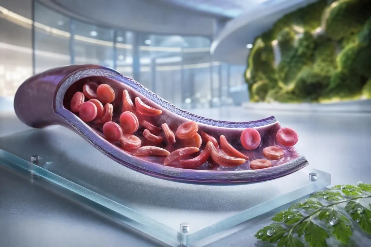
Sirpjveida šūnu anēmija nav viens tests, viena pazīme vai viens steidzamības līmenis. Šī pacientu orientētā rokasgrāmata atšķir nēsātāju statusu, apstiprinātu slimību, ikdienas uzraudzību un situācijas, kas prasa neatliekamo palīdzību.


- Sirpjveida šūnu pazīme (traits) nozīmē vienu HbS gēnu; tas parasti nerada hronisku anēmiju vai atkārtotas sāpju krīzes.
- Jaundzimušo skrīnings identificē lielāko daļu skarto zīdaiņu pirms simptomu parādīšanās, ļaujot agrīni uzsākt penicilīna un speciālista uzraudzību.
- Hemoglobīna elektroforēze atdala hemoglobīna veidus un palīdz apstiprināt HbSS, HbSC, HbS-beta talasēmiju vai nēsātāju statusu.
- Spēcīgas sāpes krūtīs, elpas trūkums, drudzis 38,5°C vai augstāks, apjukums vai jaunas vājuma pazīmes prasa steidzamu neatliekamās palīdzības novērtēšanu personai ar sirpjveida šūnu slimību.
- Akūts krūškurvja sindroms var sākties ar klepu, drudzi, sāpēm krūtīs vai zemu skābekļa līmeni un var pasliktināties vairākās stundās nekā dienās.
- Pēkšņs hemoglobīna samazinājums par 2 g/dL vai vairāk nocdot no dotās vērtības atšķirības no personas parastā līmeņa var signalizēt aplastisko krīzi, sekvestrāciju, asiņošanu vai paātrinātu hemolīzi.
- Retikulocītu skaits palīdz atšķirt aktīvu kaulu smadzeņu reakciju no aplastiskās krīzes; zems skaits anēmijas pasliktināšanās laikā ir brīdinājuma modelis.
- Hidroksilurīnviela ir slimību modificējošas zāles, nevis vienkārši pretsāpju zāles; uzraudzība parasti ietver pilnu asins analīzi ik pēc 4 nedēļām devas pielāgošanas laikā.
Ko nozīmē sirpjveida šūnu anēmija — un ko tā nenozīmē
Sirpjšūnu anēmija parasti attiecas uz HbSS slimību, kurā persona manto divus gēnus, kas ražo hemoglobīnu S; sarkanās šūnas var kļūt stīvas, priekšlaicīgi sadalīties un nosprostot mazos asinsvadus. Sirpjšūnu iezīme atšķiras: viens HbS gēns parasti ir pietiekams, lai to nodotu ģenētiski, bet nepietiekams, lai izraisītu hronisku anēmiju un atkārtotus vazo-okluzīvus epizodes, kas redzamas HbSS.
Sirpjšūnu slimība ir plašs termins, kas ietver HbSS, HbSC, HbS-beta-null talasēmiju un HbS-beta-plus talasēmiju. HbSS bieži izraisa visizteiktāko anēmiju, taču smagums ļoti atšķiras pat vienā ģimenē; hemoglobīna pamatlīmenis 7,5 g/dL var būt stabils vienam pieaugušajam un bīstams citam, ja tas ir strauji samazinājies.
Hemoglobīna S mutācija maina vienu aminoskābi beta-globulīnā: valīns aizstāj glutamīnskābi 6. pozīcijā. Zemā skābekļa līmenī, dehidratācijā, acidozē vai drudzī HbS molekulas var polimerizēties šūnas iekšienē. Šīs fiziskās izmaiņas izskaidro, kāpēc krīze var attīstīties pēc ilga lidojuma, kuņģa-zarnu trakta slimības vai nakts ar sliktu šķidruma uzņemšanu.
Kantesti AI ir AI asins analīžu analizators kas apvieno CBC, bilirubīnu, LDH, retikulocītu, kreatinīnu un iepriekšējos rezultātus vienā gareniskā skatā; tas nediagnosticē sirpjšūnu slimību tikai no CBC. Praktiskai atkārtotai hemoglobīna atskaišu apskatei skatiet ko HGB nozīmē.
Līdz 2026. gada 4. septembrim es joprojām redzu pacientus, kuriem nepareizi teikts, ka īpašība un slimība ir savstarpēji aizstājamas. Tās nav. Dr. Tomasa Kleina klīniskais noteikums ir vienkāršs: pieprasiet precīzu hemoglobīna genotipu, nevis neskaidru apgalvojumu, ka tests bija “pozitīvs”.”
Kāpēc genotips maina aprūpi
HbSC var būt augstāks hemoglobīna līmenis nekā HbSS, bet joprojām var izraisīt tīklenes komplikācijas, sāpes vai avaskulāru nekrozi. HbS-beta talasēmija var atgādināt īpašību vai HbSS atkarībā no tā, vai beta-globulīna ražošana ir daļēji klātesoša, tāpēc molekulārā testēšana dažreiz atrisina neskaidru elektroforēzes modeli.
Sirpjveida šūnu slimības simptomi pēc vecuma un pamata līmeņa
Sirpjšūnu slimības simptomi parasti ietver epizodiskas kaulu vai krūškurvja sāpes, nogurumu, dzelti, elpas trūkumu, aizkavētu augšanu un biežas infekcijas, bet ne divi cilvēki nepiedzīvo vienādu modeli. Simptomi parasti parādās pēc 4 līdz 6 dzīves mēnešiem, jo augļa hemoglobīns sākotnēji pasargā zīdaiņus no HbS polimerizācijas.
Daktilīts - roku vai pēdu pietūkums ar jutīgumu - var būt pirmais redzamais zīme zīdaiņiem. Skolas vecuma bērniem vēdera sāpes prasa īpašu uzmanību, jo aizcietējums, žultspūšļa slimība, liesas palielināšanās un vazo-oklūzija mājās var šķist pārsteidzoši līdzīgi.
Dzelte sirpjšūnu slimībā atspoguļo pastāvīgu sarkano asins šūnu sadalīšanos un tiešā bilirubīna paaugstināšanos; tas automātiski nenozīmē hepatītu. Tumšs urīns, bāla izkārnījumos, nieze, drudzis vai sāpes labajā vēdera augšējā kvadrantā maina šo novērtējumu un prasa aknu un žults cēloņu izvērtēšanu; mūsu ceļvedis uz tiešā bilirubīna modeļiem izskaidro atšķirību.
Pieaugušie var normalizēt sāpes, kas pakāpeniski kļuvušas biežākas. Manā pieredzē pāreja no divām pašregulētām epizodēm gadā uz divām mēneša epizodēm ir klīniski nozīmīga, pat ja neatliekamās palīdzības apmeklējumi nav palielinājušies. Jādokumentē miega traucējumi, darba kavēšana, erektīlas problēmas, slikts garastāvoklis un samazināta fiziskās slodzes tolerance, jo tie bieži vien ir priekšnoteikums oficiālai ārstēšanas maiņai.
Hemoglobīna koncentrācija no 6 līdz 9 g/dL var būt parasta stabilā stāvokļa diapazons HbSS gadījumā, savukārt vērtība zem 10 g/dL būtu negaidīta lielākajai daļai cilvēku bez slimības. Skaitlis ir svarīgs, bet izmaiņas no personīgās pamatvērtības ir svarīgākas.
Kam vajadzētu apsvērt sirpjveida šūnu nēsātāju skrīningu
Sirpjšūnu skrīnings ir pamatots pirms grūtniecības vai grūtniecības sākumā ikvienam, kura nesēja statuss nav zināms, neatkarīgi no izskata, uzvārda vai pieņemtas izcelsmes. Nesēja statuss ir iedzimts un bieži paliek nezināms līdz jaundzimušā skrīningam vai negaidīti anormālam hemoglobīna testam.
Ja abi bioloģiskie vecāki ir HbS vai beta-talasēmijas variantu nēsātāji, katrai grūtniecībai var būt 25% iespēja iedzimt klīniski nozīmīgu hemoglobīna stāvokli, atkarībā no attiecīgajiem variantiem. Nēsātājam var būt normāls enerģijas līmenis, normāli CBC rezultāti un nav sāpju epizodēs.
Pārbaudes ir īpaši noderīgas, ja viens partneris ir HbAS, HbC traits, beta-talasēmijas traits, neizskaidrojama mikrocitoze vai sirpjveida šūnu slimības ģimenes anamnēzē. Zems vidējais sarkano asinsķermenīšu tilpums nedrīkstētu tikt piedēvēts dzelzs deficītam, nepārbaudot feritīnu un neņemot vērā talasēmiju; salīdziniet MCV un MCH norādes.
Pasaules Veselības organizācijas 2022. gada globālais ziņojums lēš, ka hemoglobīna traucējumi joprojām ir nozīmīga iedzimtu slimību sloga sastāvdaļa, īpaši tur, kur jaundzimušo skrīninga pieejamība ir nevienmērīga. Viscienīgākā pieeja ir universāla pieeja informētai skrīningam, nevis etnisko piederību izmantošana kā vārtsargs.
Kantesti ir AI asins analīžu rezultātu platforma kas var izskaidrot ziņoto HbS rezultātu vienkāršā valodā, taču nēsātāja apstiprināšanai un reproduktīvai konsultēšanai ir nepieciešama akreditēta laboratorija un klīnicists. Ja pāris plāno grūtniecību, jautājiet, vai partneru testēšana un ģenētiskā konsultēšana var notikt pirms ieņemšanas, nevis pēc tam, kad rezultāts kļūst laika ziņā jutīgs.
Kā hemoglobīna elektroforēze apstiprina diagnozi
Hemoglobīna elektroforēze atdala hemoglobīna frakcijas un ir galvenais tests sirpjveida šūnu anēmijas vai traits apstiprināšanai, bieži kopā ar augstas veiktspējas šķidruma hromatogrāfiju un, ja nepieciešams, ģenētisko testēšanu. Rutīnas CBC nevar apstiprināt HbSS vai traits, jo dzelzs deficīts un talasēmija var radīt pārklājošus sarkano asinsķermenīšu indeksus.
Neārstētos HbSS gadījumā elektroforēze parasti parāda galvenokārt HbS bez HbA, lai gan HbF procentuālais daudzums atšķiras. HbAS parasti parāda gan HbA, gan HbS, bieži vien HbA ir augstāks nekā HbS; precīzi procenti var mainīties, ja ir līdzāspastopota alfa-talasēmija vai nesena transfūzija.
Nesena transfūzija var padarīt elektroforēzes rezultātu maldinošu aptuveni 3 mēnešus, jo donora HbA joprojām cirkulē. Tas ir arī iemesls, kāpēc HbA1c var nenovērtēt vidējo glikozes līmeni pēc transfūzijas vai augstas apgrozības hemolīzes gadījumā; skatiet kad HbA1c maldina.
Perifērajā smērē var būt mērķšūnas, polihromāzija un sirpjveida formas, taču mikroskopija atbalsta, nevis aizstāj frakciju testēšanu. Dr. Kleins ir redzējis, ka “normāli” neatliekamās palīdzības smērējumi kavē skaidrību, kad pacientam tika veikta transfūzija dažas nedēļas iepriekš – vienmēr informējiet laboratoriju un hematoloģijas komandu par transfūzijām.
Jaundzimušo skrīninga metodes identificē HbS pirms klīniskās slimības, taču rezultātam joprojām ir nepieciešama savlaicīga apstiprinoša testēšana. 2014. gada uz pierādījumiem balstītā vadlīnija, ko izstrādājusi Yawn et al., iesaka agrīni iesaistīt speciālistus, veicot profilaktiskus pasākumus pirms pirmās nopietnās komplikācijas (Yawn et al., 2014).
Asins rutīnas uzraudzība: ko klīnicisti faktiski izseko
Regulārā uzraudzība sirpjveida šūnu slimības gadījumā parasti ietver hemoglobīna, retikulocītu, balto asinsķermenīšu, trombocītu, bilirubīna, LDH, nieru funkciju un urīna proteīnu dinamiku — nevis vienu izolētu rezultātu. Drošākā salīdzināšana ir ar pacienta paša stabilu bāzes līniju, kas ideālā gadījumā ņemta, kad pacients ir vesels un nav nesen saņēmis transfūziju.
Hemolīze bieži izraisa paaugstinātu LDH, netiešo bilirubīnu un retikulocītus ar zemu haptoglobīna līmeni. Haptoglobīns var būt zems arī citu iemeslu dēļ, tostarp aknu slimības, tāpēc kombinētais modelis ir spēcīgāks nekā viens marķieris; mūsu haptoglobīna interpretācijas ceļvedi parāda, kāpēc.
Tikai retikulocītu procentuālā daļa var pārvērtēt kaulu smadzeņu reakciju nozīmīgas anēmijas gadījumā. Klīnicisti bieži aprēķina absolūto retikulocītu skaitu vai koriģēto retikulocītu reakciju; samazinošs absolūtais skaits pasliktinātas anēmijas laikā rada bažas par parvovīrusa B19 izraisītu aplastisko krīzi.
Kantesti ir ar AI balstīts asins analīzes rīks kas salīdzina atkārtotus hematoloģiskos rezultātus, nevis uzskata paaugstinātu LDH par atsevišķu diagnozi. Paraugs, kas caurulē ir iezīmēts kā hemolizēts, var falsificēt kālija un LDH līmeni, tāpēc pārsteidzošs rezultāts var prasīt atkārtotu analīzi; apskatiet atkārtotu testēšanu pēc hemolīzes.
Hidroksiurīnviela laika gaitā parasti palielina MCV un HbF, vienlaikus mēreni pazeminot neitrofilus efektīvā devā. Šīs izmaiņas var būt paredzamas, taču absolūtais neitrofilu skaits zem individuālās ārstēšanas sliekšņa vai strauji samazinošs trombocītu skaits prasa ārsta pārskatīšanu, nevis medikamentu pašregulāciju.
Kad sāpes ir vazo-okluzīva epizode — un kad tās nav
Vazo-okluzīvās sāpes bieži ir dziļas, stipras un lokalizētas mugurā, krūtīs, gurnos, ekstremitātēs vai vēderā, taču jaunas sāpes nedrīkst automātiski dēvēt par sirpjveida krīzi. Drudzis, fokāla pietūkums, ievainojums, vēdera saspringums vai vienpusēji neiroloģiski simptomi norāda uz problēmām, kas prasa citu izmeklēšanu.
Mājas sāpju plāns parasti ietver agrīnu šķidruma uzņemšanu, siltumu, izrakstīto pretsāpju līdzekļu lietošanu, atpūtu un personīgo eskalācijas slieksni, par kuru vienojušies ar sirpjveida komandu. Pārmērīgu šķidrumu dzeršana nav drošāka: cilvēki ar nieru darbības traucējumiem vai sirds slimībām var attīstīt šķidruma pārslodzi, īpaši akūtas slimības laikā.
Sāpes, kas paliek nekontrolējamas pēc parastā glābšanas plāna, neļauj organismam uzņemt šķidrumu vai zāles, vai ir atšķirīgas kvalitātes vai atrašanās vietas ziņā, jāmeklē neatliekama klīniskā palīdzība. Pietūcis, karsts locītava var liecināt par septisko artrītu vai kaulu infekciju, nevis nekomplicētu vazo-oklūziju; mūsu neizskaidrojamu sāpju testēšanas ceļvedis izklāsta noderīgas pirmās izmeklēšanas.
Neatliekamās palīdzības gadījumā savlaicīga pretsāpju līdzekļu lietošana un atkārtota novērtēšana ir svarīgāka nekā sāpju smaguma pierādīšana ar laboratorijas rādītāju. NICE vadlīnijas par akūtām sāpīgām sirpjveida epizodēm iesaka ātru pretsāpju līdzekļu novērtēšanu un biežu pārskatīšanu, jo nepietiekama ārstēšana pati par sevi veicina ilgstošu ciešanas un kavētu atveseļošanos (NICE, 2012).
Izvairieties no aukstām kompresēm tieši uz vazo-okluzīvo sāpju zonas, ja vien jūsu komanda tās īpaši neiesaka; aukstums var sašaurināt perifēros asinsvadus. Šī praktiskā detaļa šķiet nenozīmīga, taču daudzi pacienti man saka, ka neviens to nepaskaidroja līdz pat vairākām grūtām epizodēm.
Akūts krūškurvja sindroms: neatliekamā palīdzība, ko daudzi cilvēki palaidīs garām
Akūts krūškurvja sindroms ir jauns plaušu infiltrāts krūškurvja attēlveidošanā kopā ar elpošanas simptomiem, drudzi, krūškurvja sāpēm vai zemu skābekļa līmeni personai ar sirpjveida šūnu slimību, un tas ir neatliekama situācija. Tas var ātri pasliktināties, dažreiz pēc uzņemšanas sāpju epizodes dēļ, nevis brīdī, kad sākas krūškurvja simptomi.
Zvaniet neatliekamajai palīdzībai vai dodieties uz neatliekamās palīdzības nodaļu, ja rodas jauna elpas trūkums, krūškurvja sāpes, zilgani pelēkas lūpas, samaņas zudums, noturīgs klepus ar drudzi vai skābekļa piesātinājums zem personas noteiktā mērķa. Piesātinājums 92% var būt ievērojams kritums kādam, kam parasti ir 98%, savukārt dažiem pacientiem ir zemāka dokumentētā bāzes līnija; gan absolūtā vērtība, gan izmaiņas ir svarīgas.
Akūts krūškurvja sindroms var ietvert infekciju, tauku emboliju no kaulu smadzenēm, plaušu asinsvadu nosprostojumu, atelektāzi vai vairākus procesus vienlaicīgi. Klīnicisti parasti novērtē skābekļa līmeni, CBC, retikulocītus, kultūras drudža gadījumā, krūškurvja attēlveidošanu un dažreiz asinsgāzes; asins analīzēm elpas trūkuma gadījumā piešķir kontekstu šiem rezultātiem.
Pieaugušajiem opioīdu izraisīta pārmērīga sedācija, sekla elpošana sāpju dēļ un lieks intravenozais šķidrums var pasliktināt elpošanas stāvokli. Inhalācijas spirometrija hospitalizācijas laikā samazina plaušu komplikācijas izvēlētām sāpīgām epizodēm, lai gan tā neaizstāj neatliekamu novērtēšanu, kad simptomi jau ir sākušies.
Kato un kolēģu 2018. gada apskats apraksta akūtu krūškurvja sindromu kā galveno saslimstības un mirstības cēloni visā mūža garumā (Kato et al., 2018). Neautetiet paši, ja elpas trūkums, miegainība vai spiediens krūtīs ir ievērojams.
Drudzis, infekcija un liesas ārkārtas situācijas
Temperatūra 38,5°C (101,3°F) vai augstāka bērnam vai pieaugušajam ar sirpjveida šūnu anēmiju prasa steidzamu medicīnisku izmeklēšanu tajā pašā dienā, ja vien viņu speciālistu komanda nav noteikusi citu plānu. Bobu imūnās funkcijas funkcionāls zudums var izraisīt to, ka dažas bakteriālas infekcijas progresē daudz ātrāk nekā parasta vīrusu drudzis.
Drudzis, ko papildina letarģija, izsitumi, zems asinsspiediens, apjukums vai samazināta urīna izdalīšanās, prasa tūlītēju neatliekamo palīdzību. Normāls balto asinsķermenīšu skaits neuzticami neizslēdz nopietnu infekciju sirpjveida šūnu anēmijas gadījumā, īpaši slimības sākumā vai hidroksiurīnvielas terapijas laikā.
Bobu sekvestrācija visbiežāk notiek maziem bērniem un izraisa pēkšņus bobu palielināšanos, bālumu, vēdera pilnumu, ātru elpošanu un ātru hemoglobīna kritumu. Vecākiem dažreiz māca, kā sataustīt bērna bobus, taču jebkura jauna palielināšanās slimības laikā ir jāizmeklē steidzami, nevis jāveic atkārtotas mājas pārbaudes.
Hemoglobīna samazinājums par 2 g/dL vai vairāk zem parastā sākuma līmeņa ar palielinātiem bobiem rada bažas par sekvestrāciju. Turpretim smaga anēmija ar ļoti zemu retikulocītu skaitu var liecināt par aplastisko krīzi, kas bieži saistīta ar parvovīrusu B19; retikulocītu skaita interpretācija izskaidro kaulu smadzeņu signālu.
Sepse laboratorijas izmeklējumi var ietvert laktātu, kultūras, CBC, nieru testus un iekaisuma marķierus, taču lēmumi par ārstēšanu nekad nedrīkst kavēties, gaidot visu vērtību atgriešanos. Lai iegūtu kontekstu par steidzamiem modeļiem, lasiet sepse marķieru norādes.
Vakcīnas un profilaktiskie antibiotiku līdzekļi
Vakcīnas un bērnības penicilīna profilakse samazina risku, taču drudzi nepadara par zemu risku. Apstipriniet savu individuālo plānu ar hematologu vai pediatru komandu, īpaši pēc splenektomijas, ceļojuma, ja ģimenē ir jauns bērns vai mainās vietējais imunizācijas grafiks.
Insulta un neiroloģiskās brīdinājuma pazīmes prasa tūlītēju rīcību
Jebkura pēkšņa sejas noslīdēšana, rokas vai kājas vājums, runas grūtības, krampji, stipras neparastas galvassāpes, apjukums vai līdzsvara zudums sirpjveida šūnu anēmijas gadījumā ir jāuzskata par iespējamu insulta neatliekamo gadījumu. Nekavējoties zvaniet neatliekamās palīdzības dienestiem; negaidiet, lai redzētu, vai simptomi pāriet, vai vispirms dodiet pretsāpju līdzekli.
Bērniem ar HbSS vai HbS-beta-null talasēmiju no 2 līdz 16 gadu vecumam bieži piedāvādo pārmetrisko doplerogrāfijas ultraskaņas skrīningu. Nenormāls laika vidējais maksimālais ātrums 200 cm/sekundē vai augstāks identificē būtiski palielinātu insulta risku un prasa tūlītēju speciālista rīcību.
Hroniska transfūziju terapija var samazināt pirmā insulta risku bērniem ar nenormāliem doplerogrāfijas atklājumiem, taču tā rada papildu vajadzības: dzelzs pārslodzes uzraudzība, antivielu skrīnings un rūpīga donoru šūnu saskaņošana. Šī ir specializēta aprūpe, nevis rezultāts, ko pārvaldīt, izmantojot lietotni.
Pieaugušajiem var būt pārejoši neiroloģiski simptomi, kognitīvas izmaiņas vai kluss cerebrāls bojājums bez klasiska dramatiskā insulta. 10 minūšu vārdu atrašanas grūtību epizode joprojām prasa neatliekamo izmeklēšanu, jo pārejoši išēmiski simptomi var būt brīdinājums, nevis apliecinājums.
Dr. Thomas Klein iesaka ģimenēm saglabāt vietējo sirpjveida slimību neatliekamās palīdzības kontaktpersonu blakus parastajam sāpju plānam. Tas novērš vienu lēmumu dažu minūšu laikā, kad bērns vai pieaugušais var cīnīties, lai komunicētu.
Aknu, acu un plaušu aizsardzība starp krīzēm
Sirpjveida šūnu anēmija var bojāt nieres, tīkleni, plaušas un sirdi pat tad, ja sāpes ir reti, tāpēc regulāra orgānu uzraudzība ir ārstēšanas sastāvdaļa, nevis papildu iespēja. Urīna albumīna testēšana, asinsspiediena pārbaudes, acu izmeklējumi un simptomu vadīta plaušu izvērtēšana identificē komplikācijas pirms tās kļūst acīmredzamas.
Albuminūrija var būt agrīna sirpjveida nefropātijas pazīme. Urīna albumīna-kreatinīna attiecība 30 mg/g vai vairāk ir nenormāla lielākajai daļai pieaugušo un ir jāapstiprina ar atkārtotu testēšanu, jo drudzis, fiziskā slodze, menstruācijas un dehidratācija var to īslaicīgi paaugstināt.
Kreatinīns var izskatīties maldinoši normāls sirpjveida šūnu anēmijas gadījumā, jo palielināta tubulārā sekrēcija un zemāka muskuļu masa var maskēt samazinātu filtrāciju. Cistatīns C vai kombinētās formulas var sniegt noderīgu kontekstu, savukārt pastāvīgi putojošs urīns prasa izvērtēšanu; skatiet proteīna urīnā vadlīnijas.
Retinopātija ir īpaši svarīga HbSC slimības gadījumā, kas citādi var būt klīniski mierīgāka. Jauni peldoši punktiņi, zibšņi, aizkaram līdzīga redzes maiņa vai pēkšņs redzes miglošanās nepieciešama steidzama oftalmoloģiska izmeklēšana, nevis parasta vizīte pēc vairākām nedēļām.
Grūtniecība izraisa fizioloģisku hiperfiltrāciju un paaugstinātu mātes un augļa risku, tāpēc nieru un urīna sākotnējā novērtēšana jāveic agrīnā stadijā. Mūsu skaidrojums par grūtniecības GFR izmaiņām palīdz atšķirt paredzamās izmaiņas no satraucošas tendences.
Ārstēšanas izvēle, hidroksiurīnviela un transfūziju drošība
Hidroksiurīnviela daudziem cilvēkiem ar HbSS vai HbS-beta-null talasēmiju samazina sāpju epizodes un akūtu krūškurvja sindromu, palielinot augļa hemoglobīnu, taču tā prasa strukturētu uzraudzību. Transfūzijas var glābt dzīvību noteiktām komplikācijām, tomēr atkārtotas transfūzijas rada dzelzs uzkrāšanos un sarkano asinsķermenīšu antivielu riskus.
Hidroksiurīnvielu parasti titrē ar CBC un retikulocītu skaitu apmēram ik pēc 4 nedēļām, līdz tiek sasniegta stabila deva, pēc tam ilgākos intervālos, ko nosaka ārstējošā komanda. Augstāks MCV bieži ir gaidāms ārstēšanas efekts, savukārt smaga citopēnija prasa medikamentu pārskatīšanu un dažreiz pagaidu pārtraukšanu.
Vienkārša transfūzija palielina skābekli nesošo spēju, taču pārmērīga hemoglobīna koncentrācija var palielināt viskozitāti HbSS gadījumā. Daudzi akūtu protokolu gadījumā izvairās paaugstināt hemoglobīna līmeni pēc transfūzijas virs 10 g/dL, ja vien speciālists nav noteicis citu mērķi; apmaiņas transfūzija tiek izmantota noteiktām smagām komplikācijām.
Dzelzs pārslodze kļūst biežāka pēc atkārtotām transfūzijām, un to novērtē ar feritīna tendencēm, kā arī aknu vai sirds MRI, ja nepieciešams. Feritīns paaugstinās iekaisuma gadījumā, tāpēc viens augsts līmenis nav audu dzelzs slodzes pierādījums; mūsu dzelzs pētījumu ceļvedis izskaidro ierobežojumus.
Pierādījumi ir visspēcīgākie par hidroksiurīnvielu HbSS un HbS-beta-null slimības gadījumā; ieguvums HbSC joprojām ir joma ar mazāku noteiktību un vairāk individuālu lēmumu pieņemšanu. Medikamentu plānā jāiekļauj kontracepcijas un grūtniecības apspriešana, ja tas ir attiecināms, saistībā ar grūtībām ievērot režīmu un rakstisks plāns izlaistām devām.
Grūtniecība, auglība un ģimenes plānošana ar HbS
Grūtniecība ar sirpjšūnu slimību prasa agrīnu kopēju aprūpi no obstetrijas un hematoloģijas, jo paaugstināts sāpju epizodžu, anēmijas, trombozes, infekcijas, preeklampsijas, augļa augšanas aiztures un priekšlaicīgu dzemdību risks. Nēsātāju skrīnings ir svarīgs pirms grūtniecības, jo ģenētisks risks ir atkarīgs no abiem bioloģiskajiem vecākiem.
Vizītei pirms grūtniecības jāpārskata genotips, sākotnējais hemoglobīna līmenis, nieru darbība, urīna albumīns, asinsspiediens, transfūziju vēsture, sarkano asinsķermenīšu antivielas, vakcinācijas un medikamenti. Lēmumi par hidroksiurīnvielu grūtniecības laikā ir individuāli un jāpieņem kopā ar speciālistiem; nepārtrauciet vai nesāciet to atsākt tikai pēc interneta padoma.
Dzelzi nedrīkst automātiski izrakstīt zema hemoglobīna līmeņa gadījumā HbSS. Var būt arī dzelzs deficīts, īpaši grūtniecības vai smagas menstruālās asiņošanas gadījumā, taču feritīnam un transferīna piesātinājumam jāvadīs ārstēšanu, jo hronolīze pati par sevi nenozīmē dzelzs deficītu.
Ja abi vecāki ir atbilstošu hemoglobīna variantu nēsātāji, ģenētiskā konsultēšana var izskaidrot dabisko ieņemšanu, pirmsdzemdību diagnostikas iespējas, donoru izvēli un pirmsimplantācijas ģenētisko testēšanu, nevis virzot ģimeni uz vienu lēmumu. Mērķis ir informēta izvēle, nevis trauksme.
Citas praktiskas grūtniecības laboratorijas sarkanas brīdinājuma zīmes skatiet tā paša dienas grūtniecības laboratorijas bažas. Pacienta zināmais sākotnējais stāvoklis jāiekļauj katrā dzemdību nodaļas nodošanā; tas maina hemoglobīna rezultāta interpretāciju.
Laboratorijas rezultātu droša izmantošana starp vizītēm
Sirpjšūnu slimības laboratorijas rezultāts ir visnoderīgākais, ja tas tiek salīdzināts ar jūsu genotipu, stabilo sākotnējo stāvokli, simptomiem, ārstēšanu un nesenajām transfūzijām. Augsts bilirubīna līmenis vai zems hemoglobīns var būt paredzams vienam cilvēkam ar HbSS, taču steidzams tādam pašam cilvēkam, ja rezultāts strauji mainās.
Saglabājiet vienas lapas ierakstu par genotipu, parasto hemoglobīna līmeni, parasto skābekļa piesātinājumu, ja tas zināms, asins grupu un antivielām, pašreizējiem medikamentiem, transfūziju datumiem un neatliekamās palīdzības kontaktpersonām. Tas ietaupa laiku nepazīstamās neatliekamās palīdzības nodaļās un samazina iespēju, ka HbSS sākotnējais stāvoklis tiek kļūdaini uzskatīts par jaunatklātu anēmiju.
Kantesti ir AI laboratorijas testa interpretācijas pakalpojumā kas var sakārtot augšupielādētos laboratorijas pārskatus un atzīmēt tendences apspriešanai, tostarp hemolīzes marķierus, nieru tendences un medikamentu uzraudzību. Tas nevar redzēt jūsu krūškurvi, izmērīt skābekļa līmeni vai aizstāt neatliekamo aprūpi, kad simptomi norāda uz akūtu krūškurvja sindromu, sepsi vai insultu.
Rezultāts, kas ir ārpus laboratorijas atsauces intervāla, nav automātiski bīstams, un rezultāts, kas ir intervālā, joprojām var būt bīstams, ja tas ir krasi mainījies. Lai uzzinātu par ziņošanas karodziņiem un atsauces diapazoniem, lasiet rezultātu ārpus diapazona nozīme.
Kantesti klīniskā pieeja tiek pārskatīta, salīdzinot ar noteiktām metodēm un ierobežojumiem; lasītāji, kuri vēlas izprast mūsu drošības pasākumus, var iepazīties klīniskās validācijas standartiem. Nēsājiet oriģinālo PDF failu līdzi uz hematoloģijas vizīti — OCR ir noderīgs, taču avota ziņojums joprojām ir dokuments.
Skaidrs neatliekamās palīdzības rīcības plāns sirpjveida šūnu slimībai
Nekavējoties meklējiet neatliekamo palīdzību, ja rodas sāpes krūtīs vai elpas trūkums, drudzis 38,5°C vai augstāks, jauni neiroloģiski simptomi, samaņa zudums, stiprs bālums, nekontrolējamas sāpes, strauji palielināta vēdera apkārtmērs vai nespēja dzert un lietot medikamentus. Šīs brīdinājuma pazīmes var norādīt uz akūtu krūškurvja sindromu, sepsi, insultu, smagu anēmiju vai liesas sekvestrāciju.
Paziņojiet neatliekamās palīdzības personālam, ka Jums ir sirpjveida šūnu slimība, norādiet savu genotipu, ja tas ir zināms, un aprakstiet, kas atšķiras no Jūsu parastā epizodes. Miniet nesenu transfūziju, sākuma hemoglobīna līmeni, sāpju zāles, ko jau esat lietojis, norādot devas un laiku, alerģijas, grūtniecību un jebkādu iepriekšēju akūtu krūškurvja sindromu vai insultu.
Negaidiet, kamēr mājas pulsa oksimetrs kļūs nenormāls, ja krūškurvja simptomi pasliktinās. Patēriņa ierīces var rādīt neprecīzi, ja pirksti ir auksti, ja ir nagu lakas, kustības, slikta cirkulācija un daži ādas toņi; simptomi un klīniskā novērtēšana ir svarīgāki par vienu mierinošu displeju.
Ja rūpējaties par bērnu, lūdziet viņa komandu rakstisku informāciju par drudža slieksni, pretsāpju plānu un instrukcijām par liesas pārbaudi. Ja esat pieaugušais, glabājiet kopiju savā tālrunī un kopīgojiet to ar vienu uzticamu personu; ģimenes veselības uzskaites plānošana var to atvieglot.
Kantesti medicīnas komanda, ieskaitot mūsu Medicīnas konsultatīvā padome, atbalsta izglītību par laboratorijas interpretāciju, savukārt neatliekamās medicīniskās palīdzības lēmumi pieder klātienes klīniskajiem pakalpojumiem. Par mūsu organizāciju un klīnisko misiju skatiet mūsu komanda un pieeja.
Bieži uzdotie jautājumi
Kādas ir pirmās sirpjveida šūnu anēmijas pazīmes zīdaiņiem?
Pirmie sirpjveida šūnu anēmijas simptomi bieži parādās 4–6 mēnešu vecumā, kad dabiski samazinās augļa hemoglobīna līmenis. Raksturīgas agrīnas brīdinājuma pazīmes ir sāpīga plaukstu un pēdu pietūkums (dakrilīts), drudzis, neparasts nemiers, bālums, dzelte, slikta ēšana un vēdera pietūkums. Zīdainim ar sirpjveida šūnu slimību drudzis 38,5°C (101,3°F) vai augstāks prasa steidzamu medicīnisku izmeklēšanu tajā pašā dienā, jo nopietna infekcija var strauji progresēt. Jaundzimušo skrīnings identificē skartos zīdaiņus pirms simptomu parādīšanās, taču apstiprinoša testēšana un speciālista novērošana joprojām ir nepieciešama.
Vai cilvēkam ar sirds formas kroplības pazīmēm var būt simptomi?
Lielākajai daļai cilvēku ar sirpjveida šūnu pazīmi nav hroniskas anēmijas, rekurentu sāpju krīžu un normālas ikdienas fiziskās slodzes tolerances. Retas komplikācijas var rasties ārkārtējas dehidratācijas, spēcīgas karstuma iedarbības, lielā augstumā, ļoti intensīvas slodzes vai zema skābekļa vidē, un urīnā esošās asinis ir jāizvērtē, nevis jāuzskata par nekaitīgām. Pazīme nozīmē vienu HbS gēnu, savukārt sirpjveida šūnu anēmija parasti nozīmē HbSS slimību ar diviem bojātiem beta-globīna gēniem. Hemoglobīna elektroforēze vai līdzīgs frakcionēšanas tests var noskaidrot atšķirību.
Kāds tests apstiprina sirpjveida šūnu anēmiju?
Hemoglobīna elektroforēze, augstas veiktspējas šķidruma hromatogrāfija vai kapilārā elektroforēze apstiprina klātesošās hemoglobīna frakcijas un ir ļoti svarīga sirpjveida šūnu anēmijas diagnostikā. Neārstēta HbSS parasti uzrāda galvenokārt hemoglobīnu S bez hemoglobīna A, savukārt traucējumi parasti uzrāda gan hemoglobīnu A, gan hemoglobīnu S. CBC var noteikt anēmiju vai mazas sarkanās šūnas, bet nevar patstāvīgi diagnosticēt HbSS, HbSC vai traucējumus. Nesenā transfūzija var izkropļot frakciju rezultātus apmēram 3 mēnešus, tāpēc molekulārā testēšana var būt nepieciešama, ja modelis nav skaidrs.
Kad cilvēkam ar sirpjveida šūnu slimību vajadzētu doties uz neatliekamās palīdzības nodaļu?
Personai ar sirpjveida šūnu slimību jālūdz neatliekama medicīniskā palīdzība, ja temperatūra ir 38,5°C (101,3°F) vai augstāka, ja rodas sāpes krūtīs, elpas trūkums, zems skābekļa līmenis, pēkšņs vājums, runas grūtības, krampji, samaņas zudums, strauji progresējošs bālums, stipra vēdera uzpūšanšanās vai sāpes, kuras nevar kontrolēt ar esošo terapijas plānu. Šie simptomi var liecināt par akūtu krūškurvja sindromu, infekciju, insultu, liesas sekvestrāciju vai pēkšņu hemoglobīna samazināšanos. Nepieciešama arī jaunu vai atšķirīgu sāpju izvērtēšana, jo kaulu infekcija, žultspūšļa slimība, apendicīts un asins recekļi var imitēt vazo-okluzīvas sāpes. Neveiciet sev pašrocīgu braukšanu, ja neiroloģiski vai elpošanas simptomi ir izteikti.
Kāds hemoglobīna līmenis ir bīstams sirpjveida šūnu slimībai?
Nav viena konkrēta bīstama hemoglobīna skaitļa sirpjšūnu slimībai, jo daudziem HbSS pacientiem ir stabils pamata līmenis aptuveni 6–9 g/dL. Kritums par 2 g/dL vai vairāk salīdzinājumā ar individuālo parasto hemoglobīna līmeni bieži ir satraucošāks nekā absolūtā vērtība un var norādīt uz aplastisko krīzi, liesas sekvestrāciju, asiņošanu vai paātrinātu hemolīzi. Smagi simptomi, piemēram, elpas trūkums, samaņas zudums, sāpes krūtīs, ātra sirdsdarbība vai apjukums, prasa steidzamu izvērtēšanu jebkurā hemoglobīna līmenī. Transfūziju lēmumi ir atkarīgi no simptomiem, pamata līmeņa, anēmijas cēloņa, skābekļa līmeņa un hiperviskozitātes riska.
Vai hidroksilurīnviela izārstē sirpjveida šūnu anēmiju?
Hidroksiurīnviela neizārstē sirpjveida šūnu anēmiju, bet var samazināt sāpju epizodes, akūtu krūškurvja sindromu, transfūziju vajadzību un hospitalizācijas biežumu daudziem cilvēkiem ar HbSS vai HbS-beta-null talasēmiju. Tā darbojas daļēji, palielinot augļa hemoglobīnu, un parasti prasa ikmēneša (ik pēc 4 nedēļām) asins aina (CBC) un retikulocītu uzraudzību devas pielāgošanas laikā. Augošs MCV un HbF līmenis var norādīt uz bioloģisku efektu, lai gan neviens no šiem rezultātiem neliecina par pilnīgu zāļu lietošanas atbilstību. Ārstniecības metodes, piemēram, cilmes šūnu transplantācija un dažas gēnu terapijas, prasa ļoti specifisku atbilstības novērtēšanu un ilgtermiņa novērošanu.
Iegūstiet AI vadītu asins analīžu analīzi jau šodien
Pievienojieties vairāk nekā 2 miljoniem lietotāju visā pasaulē, kuri uzticas Kantesti tūlītējai, precīzai laboratorijas analīžu interpretācijai. Augšupielādējiet savas asins analīzes rezultātus un dažu sekunžu laikā saņemiet visaptverošu 15,000+ biomarķieru interpretāciju.
📚 Atsauces pētniecības publikācijas
Kleins, T., Mičels, S., un Vēbers, H. (2026). C3 C4 komplementa asins analīzes un ANA titra ceļvedis. Kantesti AI Medicīnas pētījumi.
Kleins, T., Mičels, S., un Vēbers, H. (2026). Nipah vīrusa asins analīze: agrīnas noteikšanas un diagnostikas ceļvedis 2026. gadam. Kantesti AI Medicīnas pētījumi.
📖 Ārējās medicīniskās atsauces
⚕️ Medicīniskā atruna
Šis raksts ir paredzēts tikai izglītojošiem nolūkiem un nav medicīnisks padoms. Vienmēr konsultējieties ar kvalificētu veselības aprūpes speciālistu, lai pieņemtu lēmumus par diagnostiku un ārstēšanu.
E-E-A-T uzticēšanās signāli
Pieredze
Ārstu vadīta klīniskā laboratorijas interpretācijas darbplūsmu pārskatīšana.
Ekspertīze
Laboratorijas medicīnas fokuss uz to, kā biomarķieri uzvedas klīniskā kontekstā.
Autoritāte
Sagatavojis Dr. Thomas Klein, pārskatījusi Dr. Sarah Mitchell un prof. Dr. Hans Weber.
Uzticamība
Uz pierādījumiem balstīta interpretācija ar skaidriem turpmākās rīcības ceļiem, lai mazinātu trauksmi.