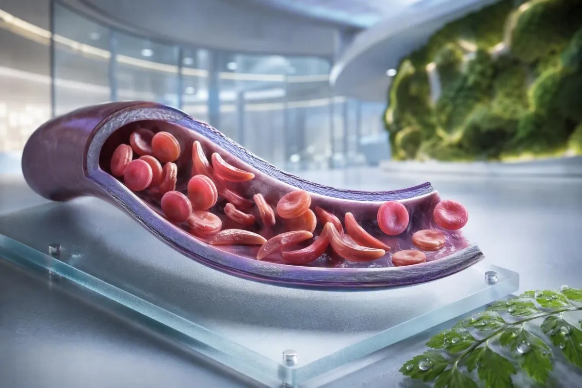
Серповидноклітинна анемія — це не один тест, один симптом чи один рівень терміновості. Цей посібник, орієнтований на пацієнта, розділяє носійство, підтверджений діагноз, рутинний моніторинг та ситуації, що потребують невідкладної допомоги.


- Серповидноклітинний слабкість означає один ген HbS; зазвичай він не викликає хронічної анемії або повторних больових кризів.
- Скринінг новонароджених виявляє більшість уражених немовлят до появи симптомів, дозволяючи рано розпочати прийом пеніциліну та подальше спостереження у спеціаліста.
- Електрофорез гемоглобіну розділяє типи гемоглобіну та допомагає підтвердити HbSS, HbSC, HbS-бета-таласемію або носійство.
- Сильний біль у грудях, задишка, температура 38,5°C або вище, сплутаність свідомості або нова слабкість потребують термінової невідкладної оцінки у особи з серповидноклітинною анемією.
- Гострий синдром грудної клітки може починатися з кашлю, гарячки, болю в грудях або низького рівня кисню і може погіршуватися протягом годин, а не днів.
- Раптове зниження гемоглобіну на 2 г/дл або більше від звичайного рівня людини може сигналізувати про апластичний криз, секвестрацію, кровотечу або прискорений гемоліз.
- Кількість ретикулоцитів допомагає відрізнити активну відповідь кісткового мозку від апластичного кризу; низький показник під час погіршення анемії є попереджувальним знаком.
- Гідроксисечовина є хворобомодифікуючим препаратом, а не просто знеболюючим засобом; моніторинг зазвичай включає повний аналіз крові кожні 4 тижні під час корекції дози.
Що означає серповидноклітинна анемія — і що не означає
Серпоподібноклітинна анемія зазвичай відноситься до захворювання HbSS, при якому людина успадковує два гени, що виробляють гемоглобін S; еритроцити можуть ставати жорсткими, передчасно руйнуватися та закупорювати дрібні судини. Серпоподібноклітинний траїт відрізняється: один ген HbS зазвичай достатній для передачі у спадок, але недостатній для спричинення хронічної анемії та рецидивуючих вазооклюзійних епізодів, що спостерігаються при HbSS.
Серпоподібноклітинна хвороба — це загальний термін, що включає HbSS, HbSC, HbS-бета-нульовий таласемію та HbS-бета-плюс таласемію. HbSS часто спричиняє найбільш виражену анемію, але тяжкість варіюється надзвичайно навіть у межах однієї родини; базовий рівень гемоглобіну 7,5 г/л може бути стабільним для одного дорослого і небезпечним для іншого, якщо він швидко знизився.
Мутація гемоглобіну S змінює одну амінокислоту в бета-глобіні: валін заміщує глутамінову кислоту в положенні 6. При низькому вмісті кисню, зневодненні, ацидозі або лихоманці молекули HbS можуть полімеризуватися всередині клітини. Ця фізична зміна пояснює, чому криз може розвинутися після тривалого перельоту, шлунково-кишкового захворювання або ночі з недостатнім споживанням рідини.
Kantesti AI — це Аналізатор крові зі штучним інтелектом що об'єднує CBC, білірубін, ЛДГ, ретикулоцити, креатинін та попередні результати в одному поздовжньому огляді; він не діагностує серпоподібноклітинну хворобу лише за CBC. Для практичного поглиблення знань про звітність щодо гемоглобіну див. що означає HGB.
Станом на 4 вересня 2026 року я все ще стикаюся з тим, що пацієнтам помилково кажуть, що траїт і хвороба взаємозамінні. Це не так. Клінічне правило доктора Томаса Кляйна просте: запитуйте точний генотип гемоглобіну, а не невизначену заяву про те, що тест був “позитивним”.”
Чому генотип змінює догляд
HbSC може мати вищий гемоглобін, ніж HbSS, але все ж спричиняти ретинальні ускладнення, біль або аваскулярний некроз. HbS-бета-таласемія може нагадувати траїт або HbSS залежно від того, чи присутнє часткове вироблення бета-глобіну, тому молекулярне тестування іноді вирішує неоднозначний електрофоретичний профіль.
Симптоми серповидноклітинної анемії за віком та базовими показниками
Симптоми серпоподібноклітинної хвороби зазвичай включають епізодичний біль у кістках або грудях, втому, жовтяницю, задишку, затримку росту та часті інфекції, але ніхто з двох людей не відчуває однакового патерну. Симптоми зазвичай з'являються після 4-6 місяців життя, оскільки фетальний гемоглобін спочатку захищає немовлят від полімеризації HbS.
Дактиліт — болюче набрякання рук або ніг — може бути першою помітною ознакою у немовлят. У дітей шкільного віку біль у животі потребує особливої уваги, оскільки запор, захворювання жовчного міхура, збільшення селезінки та вазооклюзія можуть відчуватися напрочуд схожими вдома.
Жовтяниця при серпоподібноклітинній хворобі відображає постійний розпад еритроцитів та підвищення непрямого білірубіну; це не обов'язково означає гепатит. Темна сеча, блідий кал, свербіж, лихоманка або біль у верхньому правому квадранті живота змінюють цю оцінку і вимагають оцінки печінкових та жовчних причин; наш посібник з патернів прямого білірубіну пояснює різницю.
Дорослі можуть нормалізувати біль, який поступово стає частішим. На мій досвід, перехід від двох епізодів на рік, керованих вдома, до двох епізодів на місяць є клінічно значущим, навіть якщо звернень до екстреної допомоги не збільшилося. Порушення сну, пропущена робота, еректильні симптоми, пригнічений настрій та зниження толерантності до фізичних навантажень слід документувати, оскільки вони часто передують офіційній зміні лікування.
Концентрація гемоглобіну від 6 до 9 г/л може бути звичайним стабільним діапазоном при HbSS, тоді як значення нижче 10 г/л буде неочікуваним у більшості людей без захворювання. Кількість має значення, але зміна від особистого базового рівня має більше значення.
Хто повинен розглянути скринінг носійства серповидноклітинної анемії
Скринінг на серпоподібноклітинну анемію є доцільним до вагітності або на ранніх термінах вагітності для будь-кого, чий носійський статус невідомий, незалежно від зовнішнього вигляду, прізвища чи припущеного походження. Носійський статус успадковується і часто залишається невідомим до скринінгу новонароджених або несподівано аномального тесту на гемоглобін.
Якщо обидва біологічні батьки є носіями варіанту, пов'язаного з HbS, або бета-таласемії, кожна вагітність може мати 25% шанс успадкування клінічно значущого гемоглобінопатії, залежно від залучених варіантів. Носій може мати нормальний рівень енергії, нормальні результати CBC та не мати анамнезу больових епізодів.
Скринінг особливо корисний, коли один з партнерів має HbAS, trait HbC, trait бета-таласемії, незрозумілу мікроцитоз або сімейний анамнез серповидноклітинної анемії. Низький середній об'єм еритроцитів не слід приписувати залізодефіциту без перевірки феритину та розгляду таласемії; порівняйте Підказки MCV та MCH.
Глобальний звіт Всесвітньої організації охорони здоров'я за 2022 рік оцінив, що гемоглобінопатії залишаються значним тягарем спадкових захворювань, особливо там, де доступ до скринінгу новонароджених є нерівномірним. Найбільш шанобливим підходом є універсальний доступ до інформованого скринінгу, а не використання етнічної приналежності як пропускного пункту.
Кантесті – це Платформа для розшифровки аналізу крові AI що може пояснити зазначений результат HbS простою мовою, але для підтвердження носійства та репродуктивного консультування потрібна акредитована лабораторія та клініцист. Якщо пара планує вагітність, запитайте, чи можна провести тестування партнера та генетичне консультування до зачаття, а не після того, як результат стане чутливим до часу.
Як електрофорез гемоглобіну підтверджує діагноз
Електрофорез гемоглобіну розділяє фракції гемоглобіну і є основним тестом для підтвердження серповидноклітинної анемії або trait, часто разом з високоефективною рідинною хроматографією та, за необхідності, генетичним тестуванням. Рутинний CBC не може підтвердити HbSS або trait, оскільки залізодефіцит та таласемія можуть призвести до перекриття показників еритроцитів.
При нелікованому HbSS електрофорез зазвичай показує переважно HbS без HbA, хоча відсоток HbF варіюється. HbAS зазвичай показує як HbA, так і HbS, часто з вищим відсотком HbA, ніж HbS; точні відсотки можуть змінюватися при співіснуючій альфа-таласемії або нещодавній трансфузії.
Нещодавня трансфузія може зробити результат електрофорезу оманливим приблизно протягом 3 місяців, оскільки донорський HbA все ще циркулює. Це також причина, чому HbA1c може занижувати середній рівень глюкози після трансфузії або при гемолізі з високим оборотом; див. коли HbA1c вводить в оману.
Периферійний мазок може показувати мішенеподібні еритроцити, поліхромазію та серпоподібні форми, але мікроскопія підтверджує, а не замінює фракційне тестування. Д-р Кляйн бачив “нормальні” екстрені мазки, що затримували з'ясування, коли пацієнту було проведено трансфузію за кілька тижнів до цього — завжди повідомляйте лабораторію та команду гематологів про трансфузії.
Методи скринінгу новонароджених ідентифікують HbS до клінічної хвороби, але результат все ще потребує своєчасного підтверджуючого тестування. Клінічне керівництво Yawn et al. за 2014 рік на основі доказів рекомендує залучення спеціалістів на ранніх стадіях життя, з профілактичними заходами, що починаються до першого серйозного ускладнення (Yawn et al., 2014).
Рутинний моніторинг крові: що насправді відстежують клініцисти
Звичайний моніторинг при серповидноклітинній анемії зазвичай відстежує рівень гемоглобіну, ретикулоцитів, лейкоцитів, тромбоцитів, білірубіну, ЛДГ, функцію нирок та білок у сечі — а не один ізольований результат. Найбезпечнішим порівнянням є зі стабільним базовим рівнем пацієнта, ідеально отриманим, коли він здоровий і нещодавно не отримував переливання.
Гемоліз часто викликає підвищення ЛДГ, непрямого білірубіну та ретикулоцитів з низьким рівнем гаптоглобіну. Гаптоглобін може бути низьким з інших причин, включаючи захворювання печінки, тому комбінований профіль є сильнішим за будь-який окремий маркер; наш гайд з інтерпретації haptoglobin показує чому.
Сам по собі відсоток ретикулоцитів може переоцінити відповідь кісткового мозку при значній анемії. Лікарі часто розраховують абсолютну кількість ретикулоцитів або скориговану відповідь ретикулоцитів; падіння абсолютної кількості при погіршенні анемії викликає занепокоєння щодо апластичного кризу, пов'язаного з парвовірусом B19.
Кантесті – це Інструмент для аналізу результатів аналізу крові на основі AI який порівнює повторні результати гематологічних досліджень, а не розглядає високу ЛДГ як самостійний діагноз. Зразок, позначений як гемолізований у пробірці, може хибно підвищити рівень калію та ЛДГ, тому дивовижний результат може вимагати повторного забору; перегляд повторне тестування після гемолізу.
Гідроксисечовина зазвичай з часом підвищує MCV та HbF, помірно знижуючи кількість нейтрофілів при ефективній дозі. Ці зміни можуть бути очікуваними, але абсолютна кількість нейтрофілів нижче індивідуального порогу лікування або швидко падаюча кількість тромбоцитів вимагає перегляду лікарем, а не самостійного коригування медикаментів.
Коли біль є вазооклюзивним кризом — а коли ні
Біль при вазооклюзії часто є глибоким, сильним і локалізується в спині, грудях, стегнах, кінцівках або животі, але новий біль не можна автоматично називати серповидноклітинним кризом. Гарячка, вогнищеве набрякання, травма, напруження м'язів живота або односторонні неврологічні симптоми вказують на проблеми, що потребують іншого обстеження.
Домашній план щодо знеболення зазвичай включає ранню гідратацію, тепло, призначену знеболювальну терапію, відпочинок та особистий поріг ескалації, узгоджений із серповидноклітинною командою. Вживання надмірної кількості рідини не є безпечнішим: люди з нирковою недостатністю або серцевими захворюваннями можуть розвинути перевантаження рідиною, особливо під час гострого захворювання.
Біль, який залишається неконтрольованим після звичайного плану невідкладної допомоги, заважає утримувати рідину або ліки, або відрізняється за якістю чи локалізацією, повинен спонукати до термінової медичної консультації. Набряклий, гарячий суглоб може свідчити про септичний артрит або інфекцію кісток, а не про неускладнену вазооклюзію; наші посібник з тестування незрозумілого болю окреслює корисні перші дослідження.
У невідкладній допомозі своєчасне знеболення та повторна оцінка мають більше значення, ніж доведення інтенсивності болю за допомогою лабораторного показника. Рекомендації NICE щодо гострих больових епізодів при серповидноклітинній анемії рекомендують швидку оцінку знеболення та часті огляди, оскільки недостатнє лікування саме по собі сприяє тривалому дистресу та затримці одужання (NICE, 2012).
Уникайте прикладання пакетів з льодом безпосередньо до ділянки вазооклюзійного болю, якщо ваша команда спеціально цього не рекомендує; холод може звужувати периферичні судини. Ця практична деталь здається незначною, але багато пацієнтів кажуть мені, що їм ніхто не пояснював це до кількох важких епізодів.
Гострий синдром грудної клітки: надзвичайна ситуація, яку багато хто пропускає
Гострий грудний синдром — це нова легенева інфільтрація на рентгенограмі грудної клітки плюс респіраторні симптоми, гарячка, біль у грудях або низький рівень кисню у людини із серповидноклітинною анемією, і це є надзвичайною ситуацією. Він може швидко погіршуватися, іноді після госпіталізації з приводу больового епізоду, а не в момент появи симптомів з боку грудної клітки.
Викликайте екстрену службу або зверніться до відділення невідкладної допомоги при новій задишці, болі в грудях, синьо-сірих губах, непритомності, тривалому кашлі з гарячкою або сатурації кисню нижче встановленої цільової норми. Сатурація 92% може бути значним падінням для когось, хто зазвичай має 98%, тоді як у деяких пацієнтів нижчий документально підтверджений базовий рівень; важливим є як абсолютне значення, так і зміна.
Гострий грудний синдром може включати інфекцію, жирову емболію з кісткового мозку, легеневу судинну обструкцію, ателектаз або кілька процесів одночасно. Лікарі зазвичай оцінюють оксигенацію, CBC, ретикулоцити, культури при гарячці, візуалізацію грудної клітки, а іноді й газовий склад крові; аналізів крові на задишку додає контекст до цих результатів.
У дорослих надмірна седація від опіоїдів, поверхневе дихання через біль та надлишок внутрішньовенної рідини можуть погіршити стан дихання. Стимулювальна спірометрія під час госпіталізації зменшує легеневі ускладнення при вибраних больових епізодах, хоча це не є заміною термінової оцінки, коли симптоми вже розпочалися.
Огляд Като та колег 2018 року описує гострий грудний синдром як основну причину захворюваності та смертності протягом усього життя (Kato et al., 2018). Не керуйте автомобілем самостійно, якщо задишка, сонливість або тиск у грудях значні.
Гарячка, інфекція та невідкладні стани селезінки
Температура 38,5°C (101,3°F) або вище у дитини чи дорослого із серпоподібноклітинною анемією потребує невідкладної медичної допомоги того ж дня, якщо їхня спеціалізована команда не надала іншого плану. Функціональна втрата імунної функції селезінки може призвести до того, що деякі бактеріальні інфекції прогресуватимуть набагато швидше, ніж звичайна вірусна лихоманка.
Лихоманка плюс млявість, висип, низький артеріальний тиск, сплутаність свідомості або зменшення виділення сечі потребують негайної невідкладної допомоги. Нормальний рівень лейкоцитів не виключає надійно серйозної інфекції при серпоподібноклітинній анемії, особливо на ранніх стадіях захворювання або під час лікування гідроксисечовиною.
Секвестрація селезінки найчастіше трапляється у маленьких дітей і викликає раптове збільшення селезінки, блідість, відчуття переповнення живота, прискорене дихання та швидке падіння рівня гемоглобіну. Батькам іноді показують, як пальпувати селезінку дитини, але будь-яке нове збільшення під час хвороби повинно призвести до термінової оцінки, а не до повторних домашніх перевірок.
Зниження рівня гемоглобіну на 2 г/дл або більше нижче звичайного базового рівня зі збільшеною селезінкою викликає занепокоєння щодо секвестрації. Натомість, важка анемія з дуже низьким рівнем ретикулоцитів може свідчити про апластичну кризу, часто пов'язану з парвовірусом B19; інтерпретація кількості ретикулоцитів пояснює сигнал кісткового мозку.
Лабораторні дослідження при сепсисі можуть включати лактат, посів, CBC, ниркові тести та запальні маркери, але рішення про лікування ніколи не повинні чекати, поки всі показники повернуться. Для контексту щодо термінових патернів, прочитайте підказки щодо маркерів сепсису.
Вакцини та профілактичні антибіотики
Вакцини та дитяча пеніцилінова профілактика знижують ризик, але не роблять лихоманку низькоризикованою. Підтвердьте свій індивідуальний план з гематологічною або педіатричною командою, особливо після спленектомії, подорожей, появи нової дитини або зміни місцевого календаря щеплень.
Інсульт та неврологічні попереджувальні ознаки потребують негайних дій
Будь-яке раптове опущення обличчя, слабкість руки чи ноги, труднощі з мовленням, судоми, сильний незвичайний головний біль, сплутаність свідомості або втрата рівноваги при серпоподібноклітинній анемії слід розглядати як можливу інсультну невідкладну допомогу. Негайно викличте службу екстреної допомоги; не чекайте, поки симптоми минуть, або не давайте знеболюючі ліки спочатку.
Дітям з HbSS або HbS-бета-нульовим таласемією зазвичай пропонують скринінг за допомогою ультразвуку транскраніального доплера у віці від 2 до 16 років. Аномальна середньочасова максимальна швидкість 200 см/сек або вище вказує на суттєво підвищений ризик інсульту та потребує негайних спеціалізованих дій.
Хронічна трансфузійна терапія може знизити ризик первинного інсульту у дітей з аномальними результатами доплерографії, але вона створює додаткові потреби: моніторинг перевантаження залізом, скринінг на антитіла та ретельний підбір донорських клітин. Це спеціалізована допомога, а не результат, яким можна керувати за допомогою програми.
Дорослі можуть мати транзиторні неврологічні симптоми, когнітивні зміни або безсимптомні церебральні ураження без класичного драматичного інсульту. 10-хвилинний епізод труднощів з пошуком слів все одно потребує невідкладної оцінки, оскільки транзиторні ішемічні симптоми можуть бути попередженням, а не заспокоєнням.
Доктор Томас Кляйн радить сім'ям зберегти місцевий номер екстреної допомоги при серпоподібноклітинній анемії поруч зі звичайним планом знеболення. Це усуває одне рішення протягом кількох хвилин, коли дитина або дорослий можуть мати труднощі з спілкуванням.
Захист нирок, очей та легень між кризами
Серпоподібноклітинна анемія може пошкоджувати нирки, сітківку, легені та серце навіть при рідкісному болю, тому регулярний нагляд за органами є частиною лікування, а не додатковим доповненням. Тестування сечі на альбумін, перевірка артеріального тиску, огляд очей та оцінка стану легень за симптомами виявляють ускладнення до того, як вони стануть очевидними.
Альбумінурія може бути ранньою ознакою серпоподібної нефропатії. Співвідношення альбуміну до креатиніну в сечі 30 мг/г або більше є аномальним у більшості дорослих і повинно бути підтверджено повторним тестуванням, оскільки лихоманка, фізичні навантаження, менструація та зневоднення можуть тимчасово його підвищити.
Креатинін може виглядати оманливо нормальним при серпоподібноклітинній анемії, оскільки підвищена канальцева секреція та менша м'язова маса можуть маскувати знижену фільтрацію. Цистатин С або комбіновані рівняння можуть додати корисний контекст, тоді як стійка піниста сеча заслуговує на оцінку; див. рекомендації щодо білка в сечі.
Ретинопатія є особливо важливою при хворобі HbSC, яка в іншому може бути клінічно менш вираженою. Нові "мушки", спалахи світла, поява завіси перед очима або раптове затуманення зору потребують термінового огляду офтальмологом, а не планового прийому через кілька тижнів.
Вагітність додає фізіологічну гіперфільтрацію та підвищує материнсько-фетальний ризик, тому початкова оцінка нирок та сечі повинна проводитися рано. Наше пояснення змін GFR при вагітності допомагає відрізнити очікувані зміни від тривожної тенденції.
Варіанти лікування, гідроксисечовина та безпека трансфузії
Гідроксисечовина зменшує кількість больових епізодів та гострого респіраторного дистрес-синдрому у багатьох людей з HbSS або бета-нульовим таласемією S, збільшуючи рівень фетального гемоглобіну, але потребує систематичного моніторингу. Трансфузія може врятувати життя при певних ускладненнях, але повторні трансфузії несуть ризик накопичення заліза та розвитку антитіл до еритроцитів.
Гідроксисечовину зазвичай титрують за допомогою CBC та підрахунку ретикулоцитів приблизно кожні 4 тижні до досягнення стабільної дози, а потім з довшими інтервалами, визначеними командою, що займається лікуванням. Підвищений MCV часто є очікуваним ефектом лікування, тоді як тяжка цитопенія потребує перегляду медикаментів і іноді тимчасової відміни.
Проста трансфузія підвищує киснево-транспортну здатність, але надмірна концентрація гемоглобіну може збільшити в'язкість при HbSS. Багато протоколів лікування гострих станів уникають підвищення посттрансфузійного гемоглобіну значно вище 10 г/дл, якщо тільки фахівець не встановить іншу мету; замісна трансфузія використовується для певних тяжких ускладнень.
Перевантаження залізом стає більш імовірним після повторних трансфузій і оцінюється за допомогою моніторингу рівня феритину, а також МРТ печінки або серця, коли це доречно. Феритин підвищується при запаленні, тому один високий показник не є доказом наявності заліза в тканинах; наші посібник з вивчення заліза пояснює межі.
Найбільш переконливі докази ефективності гідроксисечовини існують для HbSS та бета-нульової таласемії S; її користь при HbSC залишається областю з меншою визначеністю та більшим індивідуальним прийняттям рішень. План медикаментозного лікування повинен включати обговорення контрацепції та планування вагітності, де це доречно, бар'єри дотримання режиму та письмовий план дій у разі пропуску доз.
Вагітність, фертильність та планування сім’ї при HbS
Вагітність при серпоподібноклітинній анемії потребує раннього спільного спостереження акушера-гінеколога та гематолога, оскільки ризик больових нападів, анемії, тромбозу, інфекцій, прееклампсії, затримки росту плода та передчасних пологів є вищими. Скринінг носіїв є важливим до вагітності, оскільки генетичний ризик залежить від обох біологічних батьків.
Візит до вагітності повинен включати огляд генотипу, початкового рівня гемоглобіну, функції нирок, альбумінурії, артеріального тиску, історії трансфузій, антитіл до еритроцитів, вакцинацій та медикаментів. Рішення щодо гідроксисечовини під час вагітності приймаються індивідуально та повинні узгоджуватися з фахівцями; не припиняйте і не відновлюйте її, керуючись лише порадами з Інтернету.
Залізо не слід призначати автоматично при низькому рівні гемоглобіну при HbSS. Залізодефіцит може співіснувати, особливо під час вагітності або рясних менструальних кровотеч, але рівень феритину та насичення трансферину повинні керувати лікуванням, оскільки хромоліз сам по собі не дорівнює залізодефіциту.
Коли обидва батьки є носіями відповідних варіантів гемоглобіну, генетичне консультування може пояснити природне зачаття, варіанти пренатальної діагностики, донорські опції та передімплантаційну генетичну діагностику, не нав'язуючи родині жодного рішення. Мета – інформований вибір, а не тривога.
Щодо інших практичних лабораторних "червоних прапорців" під час вагітності див. термінові лабораторні показники вагітності. Відомий базовий стан пацієнта повинен бути включений до кожного звіту про вагітність; це змінює інтерпретацію результату гемоглобіну.
Безпечне використання лабораторних результатів між прийомами
Результат лабораторного дослідження при серпоподібноклітинній анемії найбільш корисний, коли він порівнюється з вашим генотипом, стабільним базовим показником, симптомами, лікуванням та нещодавніми трансфузіями. Високий рівень білірубіну або низький гемоглобін може бути очікуваним для однієї людини з HbSS, але терміновим для тієї ж людини, якщо результат раптово змінюється.
Зберігайте односторінковий запис про генотип, звичайний рівень гемоглобіну, звичайну насиченість киснем (якщо відомо), групу крові та антитіла, поточні ліки, дати трансфузій та контактну інформацію на випадок надзвичайної ситуації. Це економить час у незнайомих відділеннях невідкладної допомоги та зменшує ймовірність того, що базовий показник HbSS буде помилково прийнятий за нововиявлену анемію.
Кантесті – це сервісі інтерпретації тестів AI які можуть організовувати завантажені лабораторні звіти та виділяти патерни для обговорення, включаючи маркери гемолізу, тенденції показників функції нирок та моніторинг медикаментів. Вони не можуть побачити вашу грудну клітку, виміряти насиченість киснем або замінити невідкладну допомогу, коли симптоми вказують на гострий респіраторний дистрес-синдром, сепсис або інсульт.
Результат, що виходить за межі лабораторного референтного інтервалу, не є автоматично небезпечним, а результат у його межах все ще може бути небезпечним, якщо він різко змінився. Щоб дізнатися про прапорці звітності та референтні діапазони, прочитайте значення результату поза діапазоном.
клінічний підхід Kantesti перевіряється на відповідність визначеним методам та обмеженням; читачі, які хочуть зрозуміти наші запобіжні заходи, можуть переглянути стандартів клінічної валідації. Принесіть оригінальний PDF на прийом до гематолога — OCR корисний, але вихідний звіт залишається записом.
Чіткий план дій у надзвичайних ситуаціях при серповидноклітинній анемії
Негайно зверніться за екстреною допомогою у разі болю в грудях або задишки, температури 38,5°C або вище, нових неврологічних симптомів, непритомності, сильного блідості, неконтрольованого болю, швидкого збільшення живота або неможливості пити та приймати ліки. Ці тривожні ознаки можуть вказувати на гострий грудний синдром, сепсис, інсульт, тяжку анемію або секвестрацію селезінки.
Повідомте персонал екстреної допомоги, що у вас серпоподібноклітинна хвороба, вкажіть свій генотип, якщо він відомий, і опишіть, що відрізняється від вашого звичайного епізоду. Згадайте нещодавнє переливання крові, базовий рівень гемоглобіну, знеболюючі препарати, які вже приймалися, з дозами та часом, алергії, вагітність та будь-який попередній гострий грудний синдром або інсульт.
Не чекайте, поки домашній пульсоксиметр покаже відхилення, якщо симптоми з боку грудної клітки посилюються. Споживчі пристрої можуть неправильно зчитувати показники при холоді пальців, косметичних засобах для нігтів, рухах, поганому кровообігу та деяких тонах шкіри; симптоми та клінічна оцінка мають перевагу над одним заспокійливим дисплеєм.
Якщо ви доглядаєте за дитиною, попросіть її команду надати письмовий поріг температури, план знеболення та інструкції щодо перевірки селезінки. Якщо ви дорослий, збережіть копію на своєму телефоні та поділіться нею з однією довіреною особою; планування сімейного медичного запису може полегшити це.
Медична команда Kantesti, включаючи нашу Медична консультативна рада, підтримує освіту щодо інтерпретації лабораторних досліджень, тоді як рішення щодо екстрених станів належать до особистих клінічних послуг. Про нашу організацію та клінічну місію дивіться нашу команду та підхід.
Часті запитання
Які перші симптоми серповидноклітинної анемії у немовлят?
Перші симптоми серповидно-клітинної анемії часто з'являються у віці 4-6 місяців, коли природним чином знижується рівень фетального гемоглобіну. Болісні набряки рук і ніг, що називаються дактилітом, гарячка, незвичайна примхливість, блідість, жовтяниця, поганий апетит та здуття живота є поширеними ранніми попереджувальними ознаками. Гарячка 38,5°C (101,3°F) або вище у немовляти із серповидно-клітинною хворобою потребує термінової медичної оцінки того ж дня, оскільки серйозна інфекція може швидко прогресувати. Скринінг новонароджених виявляє уражених немовлят до появи симптомів, але підтверджувальне тестування та подальше спостереження фахівцем залишаються необхідними.
Чи може людина з серповидноклітинною анемією мати симптоми?
У більшості людей з серпоподібноклітинною ознакою немає хронічної анемії, рецидивуючих больових кризів та нормальної толерантності до повсякденних фізичних навантажень. Рідкісні ускладнення можуть виникати при екстремальному зневодненні, сильному перегріванні, на висоті, при дуже інтенсивних фізичних навантаженнях або в умовах низького вмісту кисню, і наявність крові в сечі слід оцінювати, а не вважати доброякісною. Ознака означає наявність одного гена HbS, тоді як серпоподібноклітинна анемія зазвичай означає захворювання HbSS з двома ураженими генами бета-глобіну. Електрофорез гемоглобіну або еквівалентний тест на фракціонування може уточнити відмінність.
Який тест підтверджує серповидноклітинну анемію?
Електрофорез гемоглобіну, високоефективна рідинна хроматографія або капілярний електрофорез підтверджують наявні фракції гемоглобіну і є ключовими для діагностики серповидноклітинної анемії. Нелікований HbSS зазвичай демонструє переважно гемоглобін S без гемоглобіну A, тоді як при носійстві зазвичай присутні як гемоглобін A, так і гемоглобін S. CBC може виявити анемію або дрібні еритроцити, але не може самостійно діагностувати HbSS, HbSC або носійство. Нещодавнє переливання крові може спотворити результати фракцій приблизно на 3 місяці, тому молекулярне тестування може бути необхідним, коли картина нечітка.
Коли людині із серпоподібноклітинною анемією слід звертатися до відділення невідкладної допомоги?
Особа із серпоподібноклітинною анемією повинна звернутися за невідкладною допомогою у разі лихоманки 38,5°C (101,3°F) або вище, болю в грудях, задишки, низького рівня кисню, раптової слабкості, труднощів з мовленням, судом, втрати свідомості, швидкого посилення блідості, сильного здуття живота або болю, який не контролюється встановленим планом лікування. Ці симптоми можуть вказувати на гострий грудний синдром, інфекцію, інсульт, спленомегалію, секвестрацію селезінки або раптове зниження рівня гемоглобіну. Новий або відмінний біль також потребує оцінки, оскільки кісткова інфекція, захворювання жовчного міхура, апендицит та тромби можуть імітувати вазооклюзійний біль. Не керуйте автомобілем самостійно, якщо неврологічні або дихальні симптоми є значними.
Який рівень гемоглобіну є небезпечним при серповидноклітинній анемії?
Немає єдиної небезпечної цифри гемоглобіну при серпоподібноклітинній анемії, оскільки багато людей з HbSS мають стабільний базовий рівень приблизно від 6 до 9 г/дл. Зниження на 2 г/дл або більше від звичайного гемоглобіну особи часто викликає більше занепокоєння, ніж абсолютне значення, і може вказувати на апластичний криз, секвестрацію селезінки, кровотечу або прискорений гемоліз. Важкі симптоми, такі як задишка, втрата свідомості, біль у грудях, прискорене серцебиття або сплутаність свідомості, потребують термінової оцінки при будь-якому рівні гемоглобіну. Рішення щодо переливання крові залежать від симптомів, базового рівня, причини анемії, оксигенації та ризику гіперв'язкості.
Чи лікує гідроксисечовина серпоподібноклітинну анемію?
Гідроксисечовина не виліковує серповидноклітинну анемію, але може зменшити епізоди болю, гострий грудний синдром, потребу в переливанні крові та використання стаціонару для багатьох людей з HbSS або HbS-бета-нульовим таласемією. Частково це відбувається шляхом збільшення рівня фетального гемоглобіну, і зазвичай потребує моніторингу **CBC** та ретикулоцитів приблизно кожні 4 тижні під час коригування дози. Зростання **MCV** та рівня HbF може вказувати на біологічну дію, хоча жоден з результатів не доводить повного дотримання режиму. Куративні підходи, такі як трансплантація стовбурових клітин та деякі генно-інженерні терапії, вимагають високоспеціалізованої оцінки відповідності критеріям та довгострокового спостереження.
Отримайте аналіз крові з підтримкою ШІ вже сьогодні
Приєднуйтесь до понад 2 мільйонів користувачів у всьому світі, які довіряють Kantesti для миттєвого та точного аналізу лабораторних тестів. Завантажте результати аналізу крові та отримайте комплексну інтерпретацію біомаркерів 15,000+ за секунди.
📚 Дослідження з посиланнями на публікації
Klein, T., Mitchell, S., & Weber, H. (2026). Керівництво до аналізу крові на комплемент C3 C4 та титр ANA. Медичні дослідження ШІ Kantesti.
Klein, T., Mitchell, S., & Weber, H. (2026). Аналіз крові на вірус Ніпах: посібник з раннього виявлення та діагностики 2026. Медичні дослідження ШІ Kantesti.
📖 Зовнішні медичні посилання
⚕️ Медична відмова від відповідальності
Ця стаття призначена лише для освітніх цілей і не становить медичної поради. Завжди консультуйтеся з кваліфікованим медичним працівником щодо рішень про діагностику та лікування.
Сигнали довіри E-E-A-T
Досвід
Лікарський клінічний огляд робочих процесів інтерпретації лабораторних показників.
Експертиза
Лабораторна медицина з акцентом на те, як біомаркери поводяться в клінічному контексті.
Авторитетність
Написано доктором Томасом Кляйном за редакційного перегляду докторки Сари Мітчелл і професора доктора Ганса Вебера.
Довірливість
Інтерпретація на основі доказів із чіткими шляхами подальших дій, щоб зменшити тривогу.