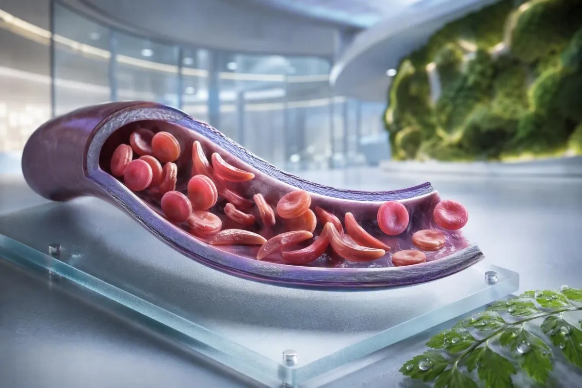
سِکل سیل انیمیا هڪ ٽيسٽ، هڪ علامت، يا ايمرجنسي جي هڪ سطح ناهي. هي مريض-پهريون گائيڊ ڪيريئر اسٽيٽس، تصديق ٿيل بيماري، معمول جي نگراني ۽ اهڙين حالتن کي الڳ ڪري ٿو جن کي ايمرجنسي ڪيئر جي ضرورت آهي.


- سِکل سیل ٽريٽ جو مطلب آهي هڪ HbS جين؛ اهو معمول طور تي دائمي انيميا يا بار بار ٿيندڙ درد جو سبب ناهي.
- نون ڄاول ٻارن جي اسڪريننگ گهڻو ڪري متاثر ٻارن کي علامتون شروع ٿيڻ کان اڳ سڃاڻي ٿو، جنهن سان پينسيلين ۽ اسپيشلسٽ فالو اپ جلد شروع ڪري سگهجي ٿو.
- Hemoglobin electrophoresis هيموگلوبن جا قسم الڳ ڪري ٿو ۽ HbSS، HbSC، HbS-beta thalassemia، يا ڪيريئر اسٽيٽس جي تصديق ڪرڻ ۾ مدد ڪري ٿو.
- سخت سينه جو درد، سانس جي تڪليف، 38.5°C يا ان کان وڌيڪ بخار، الجھن، يا نئين ڪمزوري سِکل سیل انیمیا واري شخص ۾ فوري ايمرجنسي تشخيص جي ضرورت آهي.
- اڪيوٽ چيسٽ سنڊروم ڪنهن به وقت شروع ٿي سگھي ٿو کنگهه، بخار، سينه جو درد، يا گهٽ آڪسيجن سان ۽ ڪلاڪن جي بدران ڏينهن ۾ وڌي سگهي ٿو.
- هيموگلوبن ۾ 2 g/dL يا ان کان وڌيڪ جي اوچتي گهٽتائي ڪنهن شخص جي عام سطح کان گهٽجڻ اَپلاسٽڪ بحران، سيڪوسٽريشن، رت وهڻ، يا تيز هيمولائسس جو اشارو ڏئي سگهي ٿو.
- ريٽيڪولوسائيٽ ڳڻپ فعال نڪشير جي ردعمل کي اَپلاسٽڪ بحران کان الڳ ڪرڻ ۾ مدد ڪري ٿو؛ وڌندڙ انيميا دوران گھٽ گڻت هڪ خبرداري نمونو آهي.
- هائيڊروڪسير يا هڪ بيماري تبديل ڪندڙ دوا آهي، نه رڳو درد جي دوا؛ خوراک جي ترتيب دوران هر 4 هفتي بعد مڪمل رت جي گڻت جي نگراني عام طور تي شامل آهي.
سِکل سیل انیمیا جو مطلب ڇا آهي - ۽ ڇا نه
سِڪل سيل انيميا عام طور تي HbSS بيماري ڏانهن اشارو ڪري ٿو, ، جتي هڪ شخص ٻه جين وراثت ۾ حاصل ڪري ٿو جيڪي هيموگلوبن S پيدا ڪن ٿا؛ ڳاڙهي رت جا خlii سخت ٿي سگهن ٿا، جلدي ڀڄي سگهن ٿا، ۽ ننڍڙن رت جي نالين کي رَڪاوٽ وجهي سگهن ٿا. سِڪل سيل ٽريٽ مختلف آهي، جتي هڪ HbS جين عام طور تي جينياتي طور تي منتقل ڪرڻ لاءِ ڪافي هوندو آهي پر HbSS ۾ ڏseenايل دائمي انيميا ۽ بار بار ٿيندڙ vasos-occlusive واقعن جو سبب بڻجڻ لاءِ ڪافي ناهي.
سِڪل سيل بيماري هڪ ڇتري جو اصطلاح آهي جنهن ۾ HbSS، HbSC، HbS-beta-zero thalassemia، ۽ HbS-beta-plus thalassemia شامل آهن. HbSS اڪثر ڪري سڀ کان وڌيڪ واضح انيميا جو سبب بڻجندي آهي، پر شدت هڪ ئي خاندان ۾ به تمام گهڻي مختلف هوندي آهي؛ 7.5 g/dL جو بنيادي هيموگلوبن هڪ بالغ لاءِ مستحڪم ۽ ٻئي لاءِ خطرناڪ ٿي سگهي ٿو جيڪڏهن اهو تيزيءَ سان گهٽجي ويو آهي.
هيموگلوبن S ميوٽيشن بيٽا-گلووبين ۾ هڪ امينو ايسڊ تبديل ڪري ٿو: ويلين پوزيشن 6 تي گلوٽامڪ ايسڊ جي جاءِ وٺي ٿو. گھٽ آڪسيجن، ڊيهائيڊريشن، اَسيڊوسس، يا بخار جي هيٺان، HbS ماليڪيول خlii جي اندر پوليمرائيز ٿي سگهن ٿا. اها جسماني تبديلي وضاحت ڪري ٿي ته ڊگهي فلائيٽ، گيسٽروئنٽيسٽائنل بيماري، يا خراب مائع جي مقدار جي رات کانپوءِ بحران ڪيئن پيدا ٿي سگهي ٿو.
Kantesti AI هڪ اي آءِ بلڊ ٽيسٽ اينالائيزر جيڪو CBC، بليروبين، LDH، رٽيڪولوسائٽ، ڪريٽينين، ۽ اڳئين نتيجن کي هڪ طولي واري ڏيک ۾ وجهي ٿو؛ اهو صرف CBC مان سِڪل سيل بيماري جو تشخيص نٿو ڪري. هيموگلوبن جي رپورٽنگ تي عملي ريفريشر لاءِ، ڏسو HGB جو مطلب ڇا آهي.
سيپٽمبر 4، 2026 تائين، مان اڃا تائين مريضن کي غلط طور تي ٻڌايو ويو ڏسان ٿو ته ٽريٽ ۽ بيماري هڪ ٻئي جي جاءِ تي استعمال ٿي سگهن ٿيون. اهي نه آهن. ڊاڪٽر ٿامس ڪلين جو ڪلينيڪل قاعدو سادو آهي: صحيح هيموگلوبن جينوٽائپ پڇو، نه هڪ مبهم بيان ته هڪ ٽيسٽ “مثبت” هئي.”
جينوٽائپ ڪيئن سنڀال تبديل ڪري ٿو
HbSC ۾ HbSS کان وڌيڪ هيموگلوبن ٿي سگهي ٿو پر پوءِ به ريٽينل پيچيدگيون، درد، يا ايوااسڪولر نڪروسس جو سبب بڻجي سگهي ٿو. HbS-beta-thalassemia ٽريٽ يا HbSS سان مشابهت رکي سگهي ٿي انحصار ڪندي ته بيٽا-گلووبين پيداوار جزوي طور تي موجود آهي يا نه، جنهن سبب مالڪيولر ٽيسٽنگ ڪڏهن ڪڏهن مبهم اليڪٽروفورسس پيٽرن کي حل ڪري ٿي.
سِکل سیل انیمیا جون علامتون عمر ۽ بيس لائن جي لحاظ کان
سِڪل سيل بيماري جي علامتن ۾ عام طور تي دوري واري هڏن يا سينه جو درد، ٿڪاوٽ، يرقان، ساهه کٽڻ، دير سان وڌڻ، ۽ بار بار انفيڪشن شامل آهن, ، پر ڪو به ٻه ماڻهو ساڳيو نمونو نه ٿا تجربو ڪن. علامتون عام طور تي 4 کان 6 مهينن جي ڄمار کانپوءِ ظاهر ٿين ٿيون ڇاڪاڻ ته جنين جو هيموگلوبن ابتدائي طور تي ٻارن کي HbS پوليمرائيزيشن کان بچائيندو آهي.
ڊڪٽائلائٽس - هٿن يا پيرن جي دردناڪ سوڄ - ننڍڙن ٻارن ۾ پهرين نظر ايندڙ نشاني ٿي سگهي ٿي. اسڪول جي عمر وارن ٻارن ۾، پيٽ جو درد خاص ڌيان جو مستحق آهي ڇاڪاڻ ته قبض، گال بلڊر جي بيماري، سِپلين جي وڌڻ، ۽ vasos-occlusion گهر ۾ حيرت انگيز طور تي ساڳيو محسوس ڪري سگهي ٿو.
سِڪل سيل بيماري ۾ يرقان ڳاڙهي رت جي مسلسل ڀڃڻ ۽ اڻ سڌي بليروبين جي وڌڻ کي ظاهر ڪري ٿو؛ اهو ضروري ناهي ته هيپاٽائٽس جو مطلب هجي. ڳاڙهي پيشاب، هلڪي ٻاجه، خارش، بخار، يا مٿئين ساڄي پيٽ جو درد هن تشخيص کي تبديل ڪري ٿو ۽ جگر ۽ بليري سببن جي جائزي جي لائق آهي؛ اسان جي گائيڊ ڏانهن سڌي بليروبين پيٽرن فرق واضح ڪري ٿو.
بالغن ۾ درد معمول تي اچي سگهي ٿو جيڪو تدريجي طور تي وڌيڪ بار بار ٿي ويو آهي. منهنجي تجربي ۾، سال ۾ ٻن گهر-انتظامي قسطن کان ٻن ماهوار قسطن تائين تبديلي ڪلينيڪل طور تي معنيٰ رکي ٿي جيتوڻيڪ ايمرجنسي دورا نه وڌيا هجن. ننڊ ۾ خلل، ڪم ڇڏڻ، عضوي جي علامتون، گهٽ مزاج، ۽ ورزش جي برداشت ۾ گهٽتائي دستاويز ڪئي وڃي ڇاڪاڻ ته اهي اڪثر علاج ۾ باضابطه تبديلي کان اڳتي وڌن ٿيون.
HbSS ۾ 6 کان 9 g/dL جو هيموگلوبن ڪنسنٽريشن هڪ عام مستحڪم حد ٿي سگهي ٿو، جڏهن ته 10 g/dL کان هيٺ هڪ قدر بيماري کان سواءِ گهڻن ماڻهن ۾ غير متوقع هوندو. انگ اکر اهم آهن، پر ذاتي بنيادي لڪير کان تبديلي وڌيڪ اهم آهي.
ڪنهن کي سِکل سیل ڪيريئر اسڪريننگ تي غور ڪرڻ گهرجي
سِڪل سيل اسڪريننگ حمل کان اڳ يا حمل جي شروعات ۾ ڪنهن به شخص لاءِ مناسب آهي جنهن جي ڪيريئر اسٽيٽس اڻڄاتل آهي, ، ظاهر، خانداني نالي، يا سمجهي ويندڙ نسليت کان سواءِ. ڪيريئر اسٽيٽس وراثت ۾ مليل آهي ۽ اڪثر ڪري نئون ڄاول اسڪرين يا غير متوقع طور تي غير معمولي هيموگلوبن ٽيسٽ تائين اڻڄاتل رهي ٿي.
جيڪڏهن ٻئي حياتياتي والدين HbS- سان لاڳاپيل يا بيٽا-ٿيلسيميا جي مختلف صورتون رکن ٿا، ته هر حمل ۾ ڪلينڪل طور تي اهم هيموگلوبين حالت وراثت ۾ ملڻ جو 25 سيڪڙو امڪان ٿي سگهي ٿو، جيڪي مختلف صورتون شامل آهن ان تي منحصر. هڪ ڪيريئر ۾ عام توانائي، عام CBC نتيجا، ۽ درد جي قسطن جي ڪا به تاريخ نٿي ٿي سگهي.
اسڪريننگ خاص طور تي مفيد آهي جڏهن هڪ ساٿي وٽ HbAS، HbC ٽريٽ، بيٽا-ٿيلسيميا ٽريٽ، ناقابل وضاحت مائڪروسائيٽوسس، يا سڪل سيل بيماري جي خانداني تاريخ هجي. هڪ گهٽ سراسري ڪورپاسڪولر واليم کي آئرن جي کوٽ جو الزام نه ڏيڻ گهرجي بغير فيريٽين چيڪ ڪرڻ ۽ ٿيلسيميا تي غور ڪرڻ جي؛ مقابلو ڪريو MCV ۽ MCH اشارا.
2022 جي ورلڊ هيلٿ آرگنائيزيشن جي عالمي رپورٽ جو اندازو آهي ته هيموگلوبين جي خرابين جو تعلق هڪ وڏي وراثت ۾ ملندڙ بيماري جو بار رهي ٿو، خاص طور تي جتي نون ڄاول ٻارن جي اسڪريننگ تائين رسائي غير مساوي آهي. سڀ کان وڌيڪ عزت وارو طريقو نسليت کي گيٽ ڪيپر طور استعمال ڪرڻ جي بدران باخبر اسڪريننگ تائين عالمگير رسائي آهي.
ڪينٽيسٽي هڪ آهي AI blood test interpretation پليٽ فارم جيڪو سادي زبان ۾ ٻڌايل HbS جي نتيجي جي وضاحت ڪري سگهي ٿو، پر ڪيريئر جي تصديق ۽ پيدائشي مشوري لاءِ تصديق ٿيل ليبارٽري ۽ ڊاڪٽر جي ضرورت آهي. جيڪڏهن ڪو جوڙو حمل جو منصوبو ٺاهي رهيو آهي، ته پڇو ته ڇا ساٿي جي ٽيسٽنگ ۽ جينياتي مشاورت نتيجي جي وقت جي حساس ٿيڻ کان پوءِ بجاءِ تصور کان اڳ ٿي سگهي ٿي.
هيموگلوبن اليڪٽروفورسس تشخيص جي تصديق ڪيئن ڪري ٿو
هيموگلوبين اليڪٽروفوريسس هيموگلوبين جي حصن کي الڳ ڪري ٿو ۽ سڪل سيل انيميا يا ٽريٽ جي تصديق ڪرڻ لاء هڪ بنيادي امتحان آهي, ، اڪثر ڪري اعليٰ ڪارڪردگي واري مائع ڪروماٽوگرافي ۽، جڏهن ضرورت هجي، جينياتي ٽيسٽنگ سان گڏ. هڪ معمولي CBC HbSS يا ٽريٽ جي تصديق نٿو ڪري سگهي ڇو ته آئرن جي کوٽ ۽ ٿيلسيميا ڳاڙهي رت جي سيلن جا اشارا پيدا ڪري سگهن ٿا.
اڻ علاج ٿيل HbSS ۾، اليڪٽروفوريسس عام طور تي HbS جي اڪثريت ڏيکاري ٿو بغير HbA جي، جيتوڻيڪ HbF جو سيڪڙو مختلف هوندو آهي. HbAS عام طور تي HbA ۽ HbS ٻنهي ڏيکاري ٿو، اڪثر ڪري HbA کان HbS وڌيڪ هوندو آهي؛ الفا-ٿيلسيميا يا تازي ٽرانسفيوژن جي گڏيل وراثت سان صحيح سيڪڙو تبديل ٿي سگهن ٿا.
تازي ٽرانسفيوژن تقريباً 3 مهينن تائين اليڪٽروفوريسس جي نتيجي کي گمراهه ڪندڙ بڻائي سگهي ٿي ڇو ته ڊونر HbA اڃا تائين گردش ۾ آهي. اهو ئي سبب آهي ته HbA1c ٽرانسفيوژن کان پوءِ يا اعليٰ-ٽرن اوور هيمولائسس ۾ سراسري گلوڪوز کي گهٽ ڪري سگهي ٿو؛ ڏسو جڏهن HbA1c گمراهه ڪري ٿو.
هڪ پردي جي ڊاکٽرن جي ڳوليءَ ۾ ٽارگيٽ سيلز، پوليوڪرومسيا، ۽ سڪل فارم ڏسي سگهجن ٿا، پر مائڪروسڪوپي حصي جي ٽيسٽنگ جي بدران مدد ڪري ٿي. ڊاڪٽر ڪلين ڏٺو آهي ته “عام” ايمرجنسي ڊاکٽرن جي ڳوليءَ جي تاخير ٿي وئي جڏهن ته مريض کي ڪجهه هفتا اڳ ٽرانسفيوژن ڪيو ويو هو - هميشه ليبارٽري ۽ هيماٽولوجي ٽيم کي ٽرانسفيوژن بابت ٻڌايو.
نون ڄاول ٻارن جي اسڪريننگ جا طريقا ڪلينڪل بيماري کان اڳ HbS جي سڃاڻپ ڪن ٿا، پر نتيجي کي اڃا تائين بروقت تصديقي ٽيسٽنگ جي ضرورت آهي. Yawn et al. پاران 2014 جي ثبوت-تي-بنياد تي هدايت زندگي جي شروعات ۾ ماهرن جي شموليت جي سفارش ڪري ٿي، پهرين وڏي پيچيدگي کان اڳ بچاءُ واري قدمن جي شروعات سان (Yawn et al., 2014).
معمول جي رت جي نگراني: ڊاڪٽر اصل ۾ ڇا ٽريڪ ڪن ٿا
سِڪل سيل جي بيماري ۾ معمول جي نگراني ۾ عام طور تي هيموگلوبن، ريٽيڪولوسائيٽس، سفيد خليه، پلٽليٽس، بليروبين، LDH، گردن جي ڪم، ۽ پيشاب ۾ پروٽين جو رجحان ڏسڻ ۾ اچي ٿو - نه ته ڪو هڪ الڳ نتيجو. سڀ کان محفوظ مقابلو مريض جي پنهنجي مستحڪم بنيادي حالت سان آهي، مثالي طور تي جڏهن هو صحتمند هجي ۽ تازو رت نه چڙهيو هجي.
هيموليسس اڪثر ڪري وڌايل LDH، غير سڌي بليروبين، ۽ ريٽيڪولوسائيٽس سان گهٽ هاپٽوگلوبن پيدا ڪري ٿو. هاپٽوگلوبن ٻين سببن جي ڪري گهٽجي سگهي ٿو، جنهن ۾ جگر جي بيماري شامل آهي، تنهن ڪري گڏيل نمونو ڪنهن به هڪ مارڪر کان وڌيڪ مضبوط آهي؛ اسان جو هپٽوگلوبن تفسير گائيڊ ڏيکاري ٿو ڇو.
ريٽيڪولوسائيٽس جو سيڪڙو اڪيلو اهم انميا ۾ نڪ جي جواب کي وڌيڪ بيان ڪري سگهي ٿو. ڊاڪٽر اڪثر ڪري مطلق ريٽيڪولوسائيٽس ڳڻپ يا درست ڪيل ريٽيڪولوسائيٽس جي جواب جو حساب ڪن ٿا؛ وڌندڙ انميا دوران گهٽجڻ وارو مطلق ڳڻپ پارو وائرس B19 سان لاڳاپيل aplastic بحران جو خدشو وڌائي ٿو.
ڪينٽيسٽي هڪ آهي AI-powered رت جي ٽيسٽ تجزيي جو اوزار جيڪو اعلي LDH کي الڳ تشخيص جي طور تي علاج ڪرڻ بجاءِ بار بار هيماتولوجي نتيجن جو مقابلو ڪري ٿو. ٽيوب ۾ هيموليزڊ جي طور تي نشان لڳل هڪ نمونو ڪوٽينين ۽ LDH کي ڪوڙي طور وڌائي سگهي ٿو، تنهن ڪري حيران ڪندڙ نتيجي کي ٻيهر ڊرا ڪرڻ جي ضرورت پيئي سگهي ٿي؛ جائزو هيموليسس کان پوءِ ٻيهر جانچ.
هائيڊروڪسريا عام طور تي وقت سان گڏ MCV ۽ HbF وڌائي ٿو، جڏهن ته اثرائتي دوز ۾ نيوٽروفيلس کي معمولي طور تي گهٽائي ٿو. اهي تبديليون متوقع ٿي سگهن ٿيون، پر انفرادي علاج جي حد کان هيٺ مطلق نيوٽروفيل ڳڻپ يا تيزيءَ سان گهٽجڻ وارو پلٽليٽ ڳڻپ ڊاڪٽر جي جائزي جي ضرورت آهي، نه ته دوائن کي پاڻ ۾ ترتيب ڏيڻ.
درد ڪڏهن واسو-اوڪليٽو ايپيسوڊ هوندو آهي - ۽ ڪڏهن نه
ويزو-اوڪلوسيو درد اڪثر ڪري گهرو، سخت، ۽ پٺي، سينه، هپ، عضون، يا پيٽ ۾ واقع هوندو آهي، پر نئين درد کي خودڪار طور تي سِڪل بحران جو ليبل نه لڳايو وڃي. بخار، مقامي سو، زخم، پيٽ جو ڍڪ، يا هڪ طرفي نيورولوجي علامات اهڙين مسئلن ڏانهن اشارو ڪن ٿا جن کي مختلف ورڪ اپ جي ضرورت آهي.
گهر جي درد جي منصوبي ۾ عام طور تي ابتدائي هائيڊريشن، گرمي، تجويز ڪيل دردناڪ، آرام، ۽ سِڪل ٽيم سان متفق ٿيل ذاتي واڌ جي حد شامل آهي. تمام گهڻو پاڻي پيئڻ محفوظ ناهي: گردن جي خرابي يا دل جي بيماري وارا ماڻهو مائع جي اوورلوڊ جو شڪار ٿي سگهن ٿا، خاص طور تي تيز بيماري دوران.
درد جيڪو معمول جي بچاءُ واري منصوبي کانپوءِ ڪنٽرول نه ٿئي، سيال يا دوائون رکڻ کان روڪي، يا معيار يا جڳهه ۾ مختلف هجي ته ان کي فوري طبي صلاح ڏيڻ گهرجي. سوجن وارو، گرم گڏيل سيپٽڪ گڏيل يا هڏن جي انفيڪشن جي نمائندگي ڪري سگهي ٿو نه ته غير پيچيده ويزو-اوڪلوزن؛ اسان جو اڻ ڄاتل درد جي جانچ جي رهنمائي مفيد پهرين تحقيقاتن جو خاڪو پيش ڪري ٿو.
ايمرجنسي ڪيئر ۾، وقت سر درد جي دوا ۽ ٻيهر تشخيص، ليڪن جي نمبر ذريعي درد جي شدت ثابت ڪرڻ کان وڌيڪ اهم آهن. اڪيوت درد واري سِڪل قسطن تي NICE جي رهنمائي تيز درد جي دوا جي تشخيص ۽ بار بار جائزو جي سفارش ڪري ٿي، ڇاڪاڻ ته گهٽ علاج خود ڊگهي تڪليف ۽ دير سان بحال ٿيڻ ۾ مدد ڪري ٿو (NICE، 2012).
ويزو-اوڪلوسيو درد جي علائقي تي سڌو سنئون ٿڌو پيڪن کان پاسو ڪريو جيستائين توهان جي ٽيم خاص طور تي انهن جي صلاح نه ڏئي؛ ٿڌ periferal رت جي نالن کي تنگ ڪري سگهي ٿو. اها عملي تفصيل ننڍڙي لڳي ٿي، پر ڪيترائي مريض مون کي ٻڌائين ٿا ته ڪنهن به ان کي ڪيترين ئي مشڪل قسطن کان پوءِ سمجهايو ئي ناهي.
اڪيوٽ چيسٽ سنڊروم: اها ايمرجنسي جنهن کي گهڻا ماڻهو وساري ڇڏيندا آهن
اڪيوت چيسٽ سنڊروم سِڪل سيل جي بيماري واري ماڻهوءَ ۾ چيسٽ تصوير تي نئون ڦڦڙن جو انفليٽريٽ، گڏوگڏ تنفس جي علامت، بخار، چيسٽ درد، يا گهٽ آڪسيجن، ۽ اها هڪ ايمرجنسي آهي. اها تيزيءَ سان خراب ٿي سگهي ٿي، ڪڏهن ڪڏهن درد جي قسط لاءِ داخل ٿيڻ کان پوءِ، نه ته جڏهن سينه جي علامت شروع ٿئي.
نئين سانس جي تڪليف، سينه جو درد، نيري-خاڪي لب، بي هوشي، بخار سان مسلسل کنگهه، يا ماڻهوءَ جي مقرر ڪيل حد کان هيٺ آڪسيجن جي ڇاپ لاء ايمرجنسي خدمتن کي سڏيو يا ايمرجنسي ڪيئر ڏانهن وڃو. 95% جي ڇاپ ڪنهن اهڙي ماڻهوءَ لاءِ وڏي گهٽتائي ٿي سگهي ٿي جيڪو عام طور تي 98% تي هجي، جڏهن ته ڪجهه مريضن جي هيٺيون دستاويزي بنيادي سطح هوندي آهي؛ مطلق قدر ۽ تبديلي ٻئي اهم آهن.
اڪيوت چيسٽ سنڊروم ۾ انفيڪشن، هڏن جي نڪ مان ٿلهي جي امبوليزيشن، پلمونري ويسڪولر رڪاوٽ، ايٽيليڪٽيسس، يا هڪ ئي وقت ڪيترائي عمل شامل ٿي سگهن ٿا. ڊاڪٽر عام طور تي آڪسيجنشن، CBC، ريٽيڪولوسائيٽس، جڏهن بخار هجي ته ڪلچر، چيسٽ تصوير، ۽ ڪڏهن ڪڏهن بلڊ گيس جو اندازو لڳائيندا آهن؛; ساهه کائڻ مشڪل لاء رت جا امتحان انهن نتيجن ۾ سياق و سباق شامل ڪري ٿو.
بالغن ۾، اوپيوڊس مان وڌيڪ آرام، درد مان هلڪي ساهه، ۽ وڌيڪ رگ ذريعي سيال تنفس جي حالت کي خراب ڪري سگهن ٿا. داخل ٿيڻ دوران انڪوٽ اسپيريٽري، چونڊيل درد واري قسطن ۾ پلمونري پيچيدگين کي گهٽائي ٿو، جيتوڻيڪ اها تيز تشخيص جو متبادل ناهي جڏهن علامتون اڳ ۾ ئي شروع ٿي چڪيون آهن.
ڪيٽو ۽ ساٿين پاران 2018 جي جائزي ۾ اڪيوت چيسٽ سنڊروم کي سڄي ڄم ۾ بيماري ۽ موت جو وڏو سبب قرار ڏنو ويو آهي (Kato et al.، 2018). جيڪڏهن سانس جي تڪليف، غنودگي، يا سينه جو دٻاءُ اهم هجي ته پاڻ ڊرائيو نه ڪريو.
بخار، انفيڪشن ۽ ڦڦڙن جون ايمرجنسيون
سکل سیل جي بيماري ۾ مبتلا ٻار يا بالغ ۾ 38.5°C (101.3°F) يا ان کان وڌيڪ گرمي پد جي صورت ۾، اها لازمي طور تي ساڳئي ڏينهن طبي جانچ جي ضرورت آهي، جيستائين انهن جي ماهر ٽيم طرفان ٻيو ڪو منصوبو نه ڏنو ويو هجي. طحال جي مدافعتي ڪم جي ڪارائتي نقصان ڪجهه بيڪٽيريل انفڪشن کي عام وائرل بخار جي ڀيٽ ۾ تمام تيزي سان وڌي سگهي ٿو.
بخار سان گڏ سستي، خارش، گهٽ بلڊ پريشر، الجھن، يا پيشاب جي گهٽتائي فوري طور تي ايمرجنسي ڪيئر جي ضرورت آهي. سفيد رت جي سيلن جي عام ڳڻپ، سکل سيل جي بيماري ۾ سنگين انفڪشن کي، خاص طور تي بيماري جي شروعات ۾ يا هائيڊروڪسيريو علاج دوران، قابل اعتماد طور تي رد نٿو ڪري.
طحال جي جڪڙجڻ (splenic sequestration) اڪثر ننڍن ٻارن ۾ ٿيندي آهي ۽ ان جي ڪري طحال ۾ اوچتو وڏو ٿيڻ، رنگت ڦڪي ٿيڻ، پيٽ ڀريل محسوس ٿيڻ، تيز سانس ۽ هيموگلوبن ۾ تيز گهٽتائي ٿئي ٿي. والدين کي ڪڏهن ڪڏهن پنهنجي ٻار جي طحال کي محسوس ڪرڻ سيکاريو ويندو آهي، پر بيماري سان گڏ ڪنهن به نئين وڏي ٿيڻ جي صورت ۾ گهر ۾ بار بار چڪاس ڪرڻ بدران تڪڙي جانچ ڪرڻ گهرجي.
طحال جي وڏي ٿيڻ سان گڏ معمول کان 2 g/dL يا ان کان وڌيڪ هيموگلوبن جي گهٽتائي جڪڙجڻ (sequestration) جي باري ۾ پريشان ڪندڙ آهي. ان جي برعڪس، تمام گهٽ ريٽيڪولوسائيٽ ڳڻپ سان شديد انيميا اپلاسٽڪ بحران (aplastic crisis) جو اشارو ڏئي سگهي ٿي، جيڪا اڪثر پاروائرس B19 سان لاڳاپيل هوندي آهي؛; ريٽيڪولوسائيٽ ڳڻپ جي تشريح بون ميرو جي سگنل جي وضاحت ڪري ٿو.
سيپسس ليبارٽريز ۾ ليڪٽيٽ، ڪلچرز، CBC، ڪڊني ٽيسٽ، ۽ سوزش جي نشانين شامل ٿي سگهن ٿا، پر علاج جا فيصلا ڪڏهن به هر قيمت جي واپس اچڻ جو انتظار نه ڪرڻ گهرجن. تڪڙي نمونن جي حوالي سان، پڙهو سيپسس مارڪر ڪلوز.
ويڪسين ۽ بچاءَ وارا اينٽي بايوٽيڪس
ويڪسين ۽ ٻارن جي پينسلين پروفائلڪسس خطري کي گهٽ ڪن ٿا پر بخار کي گهٽ خطري وارو نٿا بڻائين. پنهنجي انفرادي منصوبي کي هيماتولوجي يا پيڊياٽرڪ ٽيم سان تصديق ڪريو، خاص طور تي اسپلينڪٽومي (splenectomy)، سفر، نئين ٻار جي پيدائش، يا مقامي ٽيڪي جي شيڊول ۾ تبديلي کان پوءِ.
اسٽروڪ ۽ نيورولوجيڪل وارننگ سائن کي فوري عمل جي ضرورت آهي
سکل سيل جي بيماري ۾ منهن جو اوچتو ڦٽي وڃڻ، هٿ يا پيرن جي ڪمزوري، ڳالهائڻ ۾ مشڪل، دورو، شديد غير معمولي سر درد، الجھن، يا توازن وڃائڻ کي ممڪن اسٽروڪ ايمرجنسي طور علاج ڪيو وڃي. فوري طور تي ايمرجنسي خدمتن کي ڪال ڪريو؛ علامتون گذري وينديون يا درد جي دوا پهريان ڏيڻ جو انتظار نه ڪريو.
HbSS يا HbS-beta-zero ٿلاسيميا وارا ٻار عام طور تي 2 کان 16 سالن جي عمر تائين ٽرانسڪرينيل ڊاپلر الٽراسائونڊ اسڪريننگ لاءِ پيش ڪيا ويندا آهن. 200 سينٽي ميٽر في سيڪنڊ يا ان کان وڌيڪ جي غير معمولي وقت-اوسط وچولي وڌ ۾ وڌ رفتار اسٽروڪ جي خطري ۾ وڏي اضافي جي سڃاڻپ ڪري ٿي ۽ ان کي تڪڙي ماهر ڪارروائي جي ضرورت آهي.
مستقل ٽرانسفيوژن ٿراپي غير معمولي ڊاپلر نتيجن وارن ٻارن ۾ اسٽروڪ جي پهرين خطري کي گهٽائي سگهي ٿي، پر اهو اضافي ضرورتون پيدا ڪري ٿو: آئرن اوورلوڊ مانيٽرنگ، اينٽي باڊي اسڪريننگ، ۽ ڊونر سيلز جي احتياط سان ميلاپ. اها ماهرانه سنڀال آهي، ايپ ذريعي انتظام ڪرڻ جو نتيجو ناهي.
بالغن کي ڪلاسڪ ڊرامائي اسٽروڪ کان سواءِ عارضي نيورولوجيڪل علامتون، ادراڪي تبديليون، يا خاموش سريل نقصان ٿي سگهي ٿو. لفظ ڳولڻ ۾ مشڪل جو 10 منٽن جو دورو اڃا تائين ايمرجنسي جانچ جي ضرورت آهي ڇو ته عارضي اسڪيمڪ علامتون خبرداري ٿي سگهن ٿيون، اطمينان نه.
ڊاڪٽر ٿامس ڪلين خاندانن کي صلاح ڏئي ٿو ته هو معمول جي درد جي منصوبي جي برابر پنهنجي مقامي سکل ايمرجنسي رابطي جو نمبر محفوظ ڪن. اهو انهن ڪجهه منٽن دوران هڪ فيصلو ختم ڪري ٿو جڏهن ٻار يا بالغ ڳالهه ٻولهه ڪرڻ جي ڪوشش ڪري رهيا هجن.
بحرانن جي وچ ۾ ڦڦڙن، اکين ۽ ڦڦڙن جي حفاظت
سکل سيل جي بيماري جگر، ريٽنا، ڦڦڙن ۽ دل کي نقصان پهچائي سگهي ٿي جيتوڻيڪ درد گهٽ هجي، تنهنڪري معمول جي عضوي جي نگراني علاج جو حصو آهي نه ته هڪ اختياري اضافو. پيشاب جي البومين ٽيسٽنگ، بلڊ پريشر جي چڪاس، اکين جي معائنن، ۽ علامتن جي بنياد تي ڦڦڙن جي جانچ ان کان اڳ پيچيدگين جي سڃاڻپ ڪري ٿي جيئن اهي ظاهري ٿين.
البومينوريا (Albuminuria) سکل نيپروپيٿي (sickle nephropathy) جو ابتدائي نشانو ٿي سگهي ٿو. پيشاب جي البومين-ٽو-ڪريٽينين جو تناسب 30 mg/g يا ان کان وڌيڪ اڪثر بالغن ۾ غير معمولي هوندو آهي ۽ ان کي بار بار ٽيسٽ سان تصديق ڪرڻ گهرجي ڇاڪاڻ ته بخار، ورزش، حيض، ۽ dehydration ان کي عارضي طور تي وڌائي سگھن ٿا.
كريٽينين سکل سيل جي بيماري ۾ بظاهر عام نظر اچي سگهي ٿو ڇاڪاڻ ته وڌيل ٽيوبلر سڪريشن ۽ گهٽ عضلاتي ڪامي انهي جي گهٽتائي کي لڪائي سگهي ٿي. سسٽين سي (Cystatin C) يا گڏيل مساواتون ڪارائتو حوالي سان اضافو ڪري سگهن ٿيون، جڏهن ته مسلسل ڦوڳل پيشاب جانچ جي لائق آهي؛ ڏسو پيشاب ۾ پروٽين جي هدايت.
HbSC مرض ۾ ريٽينوپيٿي خاص طور تي اهم آهي، جيڪو ٻين لحاظن ۾ ڪلينڪل طور تي پرسکون ٿي سگهي ٿو. نوان فلوٽرز، چمڪ، پردي وانگر بصري تبديلي، يا اوچتو دھندلي نظر کي هفتن کان پوءِ معمول جي ملاقات جي بجاءِ فوري طور تي اکين جي ماهر کان چڪاس جي ضرورت آهي.
حمل ۾ فزيولوجيڪل هائيپرفلٽريشن ۽ ماءُ جي جنين جي وڌيڪ خطري شامل آهي، تنهنڪري گردن ۽ پيشاب جو بنيادي جائزو جلد ئي ٿيڻ گهرجي. اسان جي وضاحت حمل GFR تبديليون اميد ڪيل تبديلي کي پريشان ڪندڙ رجحان کان ڌار ڪرڻ ۾ مدد ڪري ٿي.
علاج جا اختيار، هائيڊروڪسيري ڊاڪٽر ۽ ٽرانسفيوژن سيفتي
هائيڊروڪسيريا ڪيترن ئي ماڻهن ۾ دردناڪ قسطن ۽ اڪيوت چيسٽ سنڊروم کي گهٽائي ٿو جن ۾ HbSS يا HbS-beta-zero تالاسيميا fetal هيموگلوبن وڌائڻ سان، پر ان کي منظم نگراني جي ضرورت آهي. ٽرانسفارميشن چونڊيل پيچيدگين لاءِ جان بچائڻ وارو ٿي سگهي ٿو، پر بار بار ٽرانسفارميشن لوھ جي لوڊنگ ۽ ريڊ-سيل اينٽي باڊي خطري کي آڻيندو آهي.
هائيڊروڪسيريا عام طور تي هر 4 هفتن ۾ CBC ۽ reticulocyte ڳڻپ سان گڏ مقرر ڪيو ويندو آهي جيستائين هڪ مستحڪم دوز تائين پهچي وڃي، پوءِ ڊگهي وقفي سان علاج ڪندڙ ٽيم طرفان طئي ڪيو ويندو آهي. هڪ اعلي MCV اڪثر ڪري هڪ اميد ڪيل علاج اثر آهي، جڏهن ته سخت سائيٽوپينيا کي دوا جي جائزي ۽ ڪڏهن ڪڏهن عارضي هولڊ جي ضرورت آهي.
سادي ٽرانسفارميشن آڪسيجن کڻڻ جي صلاحيت وڌائي ٿي، پر گهڻو هيموگلوبن ڪنسنٽريشن HbSS ۾ viscosity وڌائي سگهي ٿي. ڪيترائي اڪيوت پروٽوڪول 10 g/dL کان مٿي پوسٽ-ٽرانسفارميشن هيموگلوبن کي گهڻو وڌائڻ کان پاسو ڪندا آهن جيستائين ڪو ماهر مختلف مقصد مقرر نه ڪري؛ ايڪسچينج ٽرانسفارميشن چونڊيل سخت پيچيدگين لاءِ استعمال ڪيو ويندو آهي.
بار بار ٽرانسفارميشن کان پوءِ لوھ جي اوورلوڊ جو امڪان وڌي ٿو ۽ مناسب هجڻ تي فيرينٽين رجحانن سان گڏ جگر يا دل جي MRI سان اندازو لڳايو ويندو آهي. فيرينٽين سوزش سان وڌي ٿو، تنهنڪري هڪ واحد اعلي سطح ٽشو لوھ جي بوجھ جو ثبوت ناهي؛ اسان جي لوھ جي مطالعي جي ھدايت حدن (limits) کي بيان ڪري ٿي.
ثبوت HbSS ۽ HbS-beta-zero بيماري ۾ هائيڊروڪسيريا لاءِ مضبوط آهن؛ HbSC ۾ فائدو اڃا تائين گهٽ يقين ۽ وڌيڪ انفرادي فيصلا ڪرڻ جو علائقو آهي. هڪ دوا جي منصوبي ۾ حمل جي چرچائن جي صورت ۾، موافقت جي رڪاوٽن، ۽ غائب ڪيل دوز لاءِ لکيل منصوبي ۾ ڳالهه ٻولهه ۽ حمل جي ڳالهه ٻولهه شامل هجڻ گهرجي.
HbS سان حمل، زرخيزي ۽ خاندان جي منصوبابندي
سڪل سيل بيماري سان حمل ۾ اميدون، انيميا، ٿرومبوسس، انفيڪشن، پري-ايڪلمپسيا، جنين جي واڌ ويجهه جي ريسٽرڪشن ۽ وقت کان اڳ جنم جي خطري جي ڪري اميدون، انيميا، ٿرومبوسس، انفيڪشن، پري-ايڪلمپسيا، جنين جي واڌ ويجهه جي ريسٽرڪشن ۽ وقت کان اڳ جنم جي خطري جي ڪري اميدون، انيميا، ٿرومبوسس، انفيڪشن، پري-ايڪلمپسيا، جنين جي واڌ ويجهه جي ريسٽرڪشن ۽ وقت کان اڳ جنم جي خطري جي ڪري اميدون، انيميا، ٿرومبوسس، انفيڪشن، پري-ايڪلمپسيا، جنين جي واڌ ويجهه جي ريسٽرڪشن ۽ وقت کان اڳ جنم جي خطري جي ڪري اميدون، انيميا، ٿرومبوسس، انفيڪشن، پري-ايڪلمپسيا، جنين جي واڌ ويجهه جي ريسٽرڪشن ۽ وقت کان اڳ جنم جي خطري جي ڪري اميدون، انيميا، ٿرومبوسس، انفيڪشن، پري-ايڪلمپسيا، جنين جي واڌ ويجهه جي ريسٽرڪشن ۽ وقت کان اڳ جنم جي خطري جي ڪري اميدون، انيميا، ٿرومبوسس، انفيڪشن، پري-ايڪلمپسيا، جنين جي واڌ ويجهه جي ريسٽرڪشن ۽ وقت کان اڳ جنم جي خطري جي ڪري اميدون، انيميا، ٿرومبوسس، انفيڪشن، پري-ايڪلمپسيا، جنين جي واڌ ويجهه جي ريسٽرڪشن ۽ وقت کان اڳ جنم جي خطري جي ڪري اميدون، انيميا، ٿرومبوسس، انفيڪشن، پري-ايڪلمپسيا، جنين جي واڌ ويجهه جي ريسٽرڪشن ۽ وقت کان اڳ جنم جي خطري جي ڪري اميدون، انيميا، ٿرومبوسس، انفيڪشن، پري-ايڪلمپسيا، جنين جي واڌ ويجهه جي ريسٽرڪشن ۽ وقت کان اڳ جنم جي خطري جي ڪري اميدون، انيميا، ٿرومبوسس، انفيڪشن، پري-ايڪلمپسيا، جنين جي واڌ ويجهه جي ريسٽرڪشن ۽ وقت کان اڳ جنم جي خطري جي ڪري اميدون، انيميا، ٿرومبوسس، انفيڪشن، پري-ايڪلمپسيا، جنين جي واڌ ويجهه جي ريسٽرڪشن ۽ وقت کان اڳ جنم جي خطري جي ڪري اميدون، انيميا، ٿرومبوسس، انفيڪشن، پري-ايڪلمپسيا، جنين جي واڌ ويجهه جي ريسٽرڪشن ۽ وقت کان اڳ جنم جي خطري جي ڪري اميدون، انيميا، ٿرومبوسس، انفيڪشن، پري-ايڪلمپسيا، جنين جي واڌ ويجهه جي ريسٽرڪشن ۽ وقت کان اڳ جنم جي خطري جي ڪري اميدون، انيميا، ٿرومبوسس، انفيڪشن، پري-ايڪلمپسيا، جنين جي واڌ ويجهه جي ريسٽرڪشن ۽ وقت کان اڳ جنم جي خطري جي ڪري اميدون، انيميا، ٿرومبوسس، انفيڪشن، پري-ايڪلمپسيا، جنين جي واڌ ويجهه جي ريسٽرڪشن ۽ وقت کان اڳ جنم جي خطري جي ڪري اميدون، انيميا، ٿرومبوسس، انفيڪشن، پري-ايڪلمپسيا، جنين جي واڌ ويجهه جي ريسٽرڪشن ۽ وقت کان اڳ جنم جي خطري جي ڪري اميدون، انيميا، ٿرومبوسس، انفيڪشن، پري-ايڪلمپسيا، جنين جي واڌ ويجهه جي ريسٽرڪشن ۽ وقت کان اڳ جنم جي خطري جي ڪري اميدون، انيميا، ٿرومبوسس، انفيڪشن، پري-ايڪلمپسيا، جنين جي واڌ ويجهه جي ريسٽرڪشن ۽ وقت کان اڳ جنم جي خطري جي ڪري اميدون، انيميا، ٿرومبوسس، انفيڪشن، پري-ايڪلمپسيا، جنين جي واڌ ويجهه جي ريسٽرڪشن ۽ وقت کان اڳ جنم جي خطري جي ڪري اميدون، انيميا، ٿرومبوسس، انفيڪشن، پري-ايڪلمپسيا، جنين جي واڌ ويجهه جي ريسٽرڪشن ۽ وقت کان اڳ جنم جي خطري جي ڪري اميدون، انيميا، ٿرومبوسس، انفيڪشن، پري-ايڪلمپسيا، جنين جي واڌ ويجهه جي ريسٽرڪشن ۽ وقت کان اڳ جنم جي خطري جي ڪري اميدون، انيميا، ٿرومبوسس، انفيڪشن، پري-ايڪلمپسيا، جنين جي واڌ ويجهه جي ريسٽرڪشن ۽ وقت کان اڳ جنم جي خطري جي ڪري اميدون، انيميا، ٿرومبوسس، انفيڪشن، پري-ايڪلمپسيا، جنين جي واڌ ويجهه جي ريسٽرڪشن ۽ وقت کان اڳ جنم جي خطري جي ڪري اميدون، انيميا، ٿرومبوسس، انفيڪشن، پري-ايڪلمپسيا، جنين جي واڌ ويجهه جي ريسٽرڪشن ۽ وقت کان اڳ جنم جي خطري جي ڪري اميدون، انيميا، ٿرومبوسس، انفيڪشن، پري-ايڪلمپسيا، جنين جي واڌ ويجهه جي ريسٽرڪشن ۽ وقت کان اڳ جنم جي خطري جي ڪري اميدون، انيميا، ٿرومبوسس، انفيڪشن، پري-ايڪلمپسيا، جنين جي واڌ ويجهه جي ريسٽرڪشن ۽ وقت کان اڳ جنم جي خطري جي ڪري اميدون، انيميا، ٿرومبوسس، انفيڪشن، پري-ايڪلمپسيا، جنين جي واڌ ويجهه جي ريسٽرڪشن ۽ وقت کان اڳ جنم جي خطري جي ڪري اميدون، انيميا، ٿرومبوسس، انفيڪشن، پري-ايڪلمپسيا، جنين جي واڌ ويجهه جي ريسٽرڪشن ۽ وقت کان اڳ جنم جي خطري جي ڪري اميدون، انيميا، ٿرومبوسس، انفيڪشن، پري-ايڪلمپسيا، جنين جي واڌ ويجهه جي ريسٽرڪشن ۽ وقت کان اڳ جنم جي خطري جي ڪري اميدون، انيميا، ٿرومبوسس، انفيڪشن، پري-ايڪلمپسيا، جنين جي واڌ ويجهه جي ريسٽرڪشن ۽ وقت کان اڳ جنم جي خطري جي ڪري اميدون، انيميا، ٿرومبوسس، انفيڪشن، پري-ايڪلمپسيا، جنين جي واڌ ويجهه جي ريسٽرڪشن ۽ وقت کان اڳ جنم جي خطري جي ڪري اميدون، انيميا، ٿرومبوسس، انفيڪشن، پري-ايڪلمپسيا، جنين جي واڌ ويجهه جي ريسٽرڪشن ۽ وقت کان اڳ جنم جي خطري جي ڪري اميدون، انيميا، ٿرومبوسس، انفيڪشن، پري-ايڪلمپسيا، جنين جي واڌ ويجهه جي ريسٽرڪشن ۽ وقت کان اڳ جنم جي خطري جي ڪري اميدون، انيميا، ٿرومبوسس، انفيڪشن، پري-ايڪلمپسيا، جنين جي واڌ ويجهه جي ريسٽرڪشن ۽ وقت کان اڳ جنم جي خطري جي ڪري اميدون، انيميا، ٿرومبوسس، انفيڪشن، پري-ايڪلمپسيا، جنين جي واڌ ويجهه جي ريسٽرڪشن ۽ وقت کان اڳ جنم جي خطري جي ڪري اميدون، انيميا، ٿرومبوسس، انفيڪشن، پري-ايڪلمپسيا، جنين جي واڌ ويجهه جي ريسٽرڪشن ۽ وقت کان اڳ جنم جي خطري جي ڪري اميدون، انيميا، ٿرومبوسس، انفيڪشن، پري-ايڪلمپسيا، جنين جي واڌ ويجهه جي ريسٽرڪشن ۽ وقت کان اڳ جنم جي خطري جي ڪري اميدون، انيميا، ٿرومبوسس، انفيڪشن، پري-ايڪلمپسيا، جنين جي واڌ ويجهه جي ريسٽرڪشن ۽ وقت کان اڳ جنم جي خطري جي ڪري اميدون، انيميا، ٿرومبوسس، انفيڪشن، پري-ايڪلمپسيا، جنين جي واڌ ويجهه جي ريسٽرڪشن ۽ وقت کان اڳ جنم جي خطري جي ڪري اميدون، انيميا، ٿرومبوسس، انفيڪشن، پري-ايڪلمپسيا، جنين جي واڌ ويجهه جي ريسٽرڪشن ۽ وقت کان اڳ جنم جي خطري جي ڪري اميدون، انيميا، ٿرومبوسس، انفيڪشن، پري-ايڪلمپسيا، جنين جي واڌ ويجهه جي ريسٽرڪشن ۽ وقت کان اڳ جنم جي خطري جي ڪري اميدون، انيميا، ٿرومبوسس، انفيڪشن، پري-ايڪلمپسيا، جنين جي واڌ ويجهه جي ريسٽرڪشن ۽ وقت کان اڳ جنم جي خطري جي ڪري اميدون، انيميا، ٿرومبوسس، انفيڪشن، پري-ايڪلمپسيا، جنين جي واڌ ويجهه جي ريسٽرڪشن ۽ وقت کان اڳ جنم جي خطري جي ڪري اميدون، انيميا، ٿرومبوسس، انفيڪشن، پري-ايڪلمپسيا، جنين جي واڌ ويجهه جي ريسٽرڪشن ۽ وقت کان اڳ جنم جي خطري جي ڪري اميدون، انيميا، ٿرومبوسس، انفيڪشن، پري-ايڪلمپسيا، جنين جي واڌ ويجهه جي ريسٽرڪشن ۽ وقت کان اڳ جنم جي خطري جي ڪري اميدون، انيميا، ٿرومبوسس، انفيڪشن، پري-ايڪلمپسيا، جنين جي واڌ ويجهه جي ريسٽرڪشن ۽ وقت کان اڳ جنم جي خطري جي ڪري اميدون، انيميا، ٿرومبوسس، انفيڪشن، پري-ايڪلمپسيا، جنين جي واڌ ويجهه جي ريسٽرڪشن ۽ وقت کان اڳ جنم جي خطري جي ڪري اميدون، انيميا، ٿرومبوسس، انفيڪشن، پري-ايڪلمپسيا، جنين جي واڌ ويجهه جي ريسٽرڪشن ۽ وقت کان اڳ جنم جي خطري جي ڪري اميدون، انيميا، ٿرومبوسس، انفيڪشن، پري-ايڪلمپسيا، جنين جي واڌ ويجهه جي ريسٽرڪشن ۽ وقت کان اڳ جنم جي خطري جي ڪري اميدون، انيميا، ٿرومبوسس، انفيڪشن، پري-ايڪلمپسيا، جنين جي واڌ ويجهه جي ريسٽرڪشن ۽ وقت کان اڳ جنم جي خطري جي ڪري اميدون، انيميا، ٿرومبوسس، انفيڪشن، پري-ايڪلمپسيا، جنين جي واڌ ويجهه جي ريسٽرڪشن ۽ وقت کان اڳ جنم جي خطري جي ڪري اميدون، انيميا، ٿرومبوسس، انفيڪشن، پري-ايڪلمپسيا، جنين جي واڌ ويجهه جي ريسٽرڪشن ۽ وقت کان اڳ جنم جي خطري جي ڪري اميدون، انيميا، ٿرومبوسس، انفيڪشن، پري-ايڪلمپسيا، جنين جي واڌ ويجهه جي ريسٽرڪشن ۽ وقت کان اڳ جنم جي خطري جي ڪري اميدون، انيميا، ٿرومبوسس، انفيڪشن، پري-ايڪلمپسيا، جنين جي واڌ ويجهه جي ريسٽرڪشن ۽ وقت کان اڳ جنم جي خطري جي ڪري اميدون، انيميا، ٿرومبوسس، انفيڪشن، پري-ايڪلمپسيا، جنين جي واڌ ويجهه جي ريسٽرڪشن ۽ وقت کان اڳ جنم جي خطري جي ڪري اميدون، انيميا، ٿرومبوسس، انفيڪشن، پري-ايڪلمپسيا، جنين جي واڌ ويجهه جي ريسٽرڪشن ۽ وقت کان اڳ جنم جي خطري جي ڪري اميدون، انيميا، ٿرومبوسس، انفيڪشن، پري-ايڪلمپسيا، جنين جي واڌ ويجهه جي ريسٽرڪشن ۽ وقت کان اڳ جنم جي خطري جي ڪري اميدون، انيميا، ٿرومبوسس، انفيڪشن، پري-ايڪلمپسيا، جنين جي واڌ ويجهه جي ريسٽرڪشن ۽ وقت کان اڳ جنم جي خطري جي ڪري اميدون، انيميا، ٿرومبوسس، انفيڪشن، پري-ايڪلمپسيا، جنين جي واڌ ويجهه جي ريسٽرڪشن ۽ وقت کان اڳ جنم جي خطري جي ڪري اميدون، انيميا، ٿرومبوسس، انفيڪشن، پري-ايڪلمپسيا، جنين جي واڌ ويجهه جي ريسٽرڪشن ۽ وقت کان اڳ جنم جي خطري جي ڪري اميدون، انيميا، ٿرومبوسس، انفيڪشن، پري-ايڪلمپسيا، جنين جي واڌ ويجهه جي ريسٽرڪشن ۽ وقت کان اڳ جنم جي خطري جي ڪري اميدون، انيميا، ٿرومبوسس، انفيڪشن، پري-ايڪلمپسيا، جنين جي واڌ ويجهه جي ريسٽرڪشن ۽ وقت کان اڳ جنم جي خطري جي ڪري اميدون، انيميا، ٿرومبوسس، انفيڪشن، پري-ايڪلمپسيا، جنين جي واڌ ويجهه جي ريسٽرڪشن ۽ وقت کان اڳ جنم جي خطري جي ڪري اميدون، انيميا، ٿرومبوسس، انفيڪشن، پري-ايڪلمپسيا، جنين جي واڌ ويجهه جي ريسٽرڪشن ۽ وقت کان اڳ جنم جي خطري جي ڪري اميدون، انيميا، ٿرومبوسس، انفيڪشن، پري-ايڪلمپسيا، جنين جي واڌ ويجهه جي ريسٽرڪشن ۽ وقت کان اڳ جنم جي خطري جي ڪري اميدون، انيميا، ٿرومبوسس، انفيڪشن، پري-ايڪلمپسيا، جنين جي واڌ ويجهه جي ريسٽرڪشن ۽ وقت کان اڳ جنم جي خطري جي ڪري اميدون، انيميا، ٿرومبوسس، انفيڪشن، پري-ايڪلمپسيا، جنين جي واڌ ويجهه جي ريسٽرڪشن ۽ وقت کان اڳ جنم جي خطري جي ڪري اميدون، انيميا، ٿرومبوسس، انفيڪشن، پري-ايڪلمپسيا، جنين جي واڌ ويجهه جي ريسٽرڪشن ۽ وقت کان اڳ جنم جي خطري جي ڪري اميدون، انيميا، ٿرومبوسس، انفيڪشن، پري-ايڪلمپسيا، جنين جي واڌ ويجهه جي ريسٽرڪشن ۽ وقت کان اڳ جنم جي خطري جي ڪري اميدون، انيميا، ٿرومبوسس، انفيڪشن، پري-ايڪلمپسيا، جنين جي واڌ ويجهه جي ريسٽرڪشن ۽ وقت کان اڳ جنم جي خطري جي ڪري اميدون، انيميا، ٿرومبوسس، انفيڪشن، پري-ايڪلمپسيا، جنين جي واڌ ويجهه جي ريسٽرڪشن ۽ وقت کان اڳ جنم جي خطري جي ڪري اميدون، انيميا، ٿرومبوسس، انفيڪشن، پري-ايڪلمپسيا، جنين جي واڌ ويجهه جي ريسٽرڪشن ۽ وقت کان اڳ جنم جي خطري جي ڪري اميدون، انيميا، ٿرومبوسس، انفيڪشن، پري-ايڪلمپسيا، جنين جي واڌ ويجهه جي ريسٽرڪشن ۽ وقت کان اڳ جنم جي خطري جي ڪري اميدون، انيميا، ٿرومبوسس، انفيڪشن، پري-ايڪلمپسيا، جنين جي واڌ ويجهه جي ريسٽرڪشن ۽ وقت کان اڳ جنم جي خطري جي ڪري اميدون، انيميا، ٿرومبوسس، انفيڪشن، پري-ايڪلمپسيا، جنين جي واڌ ويجهه جي ريسٽرڪشن ۽ وقت کان اڳ جنم جي خطري جي ڪري اميدون، انيميا، ٿرومبوسس، انفيڪشن، پري-ايڪلمپسيا، جنين جي واڌ ويجهه جي ريسٽرڪشن ۽ وقت کان اڳ جنم جي خطري جي ڪري اميدون، انيميا، ٿرومبوسس، انفيڪشن، پري-ايڪلمپسيا، جنين جي واڌ ويجهه جي ريسٽرڪشن ۽ وقت کان اڳ جنم جي خطري جي ڪري اميدون، انيميا، ٿرومبوسس، انفيڪشن، پري-ايڪلمپسيا، جنين جي واڌ ويجهه جي ريسٽرڪشن ۽ وقت کان اڳ جنم جي خطري جي ڪري اميدون، انيميا، ٿرومبوسس، انفيڪشن، پري-ايڪلمپسيا، جنين جي واڌ ويجهه جي ريسٽرڪشن ۽ وقت کان اڳ جنم جي خطري جي ڪري اميدون، انيميا، ٿرومبوسس، انفيڪشن، پري-ايڪلمپسيا، جنين جي واڌ ويجهه جي ريسٽرڪشن ۽ وقت کان اڳ جنم جي خطري جي ڪري اميدون، انيميا، ٿرومبوسس، انفيڪشن، پري-ايڪلمپسيا، جنين جي واڌ ويجهه جي ريسٽرڪشن ۽ وقت کان اڳ جنم جي خطري جي ڪري اميدون، انيميا، ٿرومبوسس، انفيڪشن، پري-ايڪلمپسيا، جنين جي واڌ ويجهه جي ريسٽرڪشن ۽ وقت کان اڳ جنم جي خطري جي ڪري اميدون، انيميا، ٿرومبوسس، انفيڪشن، پري-ايڪلمپسيا، جنين جي واڌ ويجهه جي ريسٽرڪشن ۽ وقت کان اڳ جنم جي خطري جي ڪري اميدون، انيميا، ٿرومبوسس، انفيڪشن، پري-ايڪلمپسيا، جنين جي واڌ ويجهه جي ريسٽرڪشن ۽ وقت کان اڳ جنم جي خطري جي ڪري اميدون، انيميا، ٿرومبوسس، انفيڪشن، پري-ايڪلمپسيا، جنين جي واڌ ويجهه جي ريسٽرڪشن ۽ وقت کان اڳ جنم جي خطري جي ڪري اميدون، انيميا، ٿرومبوسس، انفيڪشن، پري-ايڪلمپسيا، جنين جي واڌ ويجهه جي ريسٽرڪشن ۽ وقت کان اڳ جنم جي خطري جي ڪري اميدون، انيميا، ٿرومبوسس، انفيڪشن، پري-ايڪلمپسيا، جنين جي واڌ ويجهه جي ريسٽرڪشن ۽ وقت کان اڳ جنم جي خطري جي ڪري اميدون، انيميا، ٿرومبوسس، انفيڪشن، پري-ايڪلمپسيا، جنين جي واڌ ويجهه جي ريسٽرڪشن ۽ وقت کان اڳ جنم جي خطري جي ڪري اميدون، انيميا، ٿرومبوسس، انفيڪشن، پري-ايڪلمپسيا، جنين جي واڌ ويجهه جي ريسٽرڪشن ۽ وقت کان اڳ جنم جي خطري جي ڪري اميدون، انيميا، ٿرومبوسس، انفيڪشن، پري-ايڪلمپسيا، جنين جي واڌ ويجهه جي ريسٽرڪشن ۽ وقت کان اڳ جنم جي خطري جي ڪري اميدون، انيميا، ٿرومبوسس، انفيڪشن، پري-ايڪلمپسيا، جنين جي واڌ ويجهه جي ريسٽرڪشن ۽ وقت کان اڳ جنم جي خطري جي ڪري اميدون، انيميا، ٿرومبوسس، انفيڪشن، پري-ايڪلمپسيا، جنين جي واڌ ويجهه جي ريسٽرڪشن ۽ وقت کان اڳ جنم جي خطري جي ڪري اميدون، انيميا، ٿرومبوسس، انفيڪشن، پري-ايڪلمپسيا، جنين جي واڌ ويجهه جي ريسٽرڪشن ۽ وقت کان اڳ جنم جي خطري جي ڪري اميدون، انيميا، ٿرومبوسس، انفيڪشن، پري-ايڪلمپسيا، جنين جي واڌ ويجهه جي ريسٽرڪشن ۽ وقت کان اڳ جنم جي خطري جي ڪري اميدون، انيميا، ٿرومبوسس، انفيڪشن، پري-ايڪلمپسيا، جنين جي واڌ ويجهه جي ريسٽرڪشن ۽ وقت کان اڳ جنم جي خطري جي ڪري اميدون، انيميا، ٿرومبوسس، انفيڪشن، پري-ايڪلمپسيا، جنين جي واڌ ويجهه جي ريسٽرڪشن ۽ وقت کان اڳ جنم جي خطري جي ڪري اميدون، انيميا، ٿرومبوسس، انفيڪشن، پري-ايڪلمپسيا، جنين جي واڌ ويجهه جي ريسٽرڪشن ۽ وقت کان اڳ جنم جي خطري جي ڪري اميدون، انيميا، ٿرومبوسس، انفيڪشن، پري-ايڪلمپسيا، جنين جي واڌ ويجهه جي ريسٽرڪشن ۽ وقت کان اڳ جنم جي خطري جي ڪري اميدون، انيميا، ٿرومبوسس، انفيڪشن، پري-ايڪلمپسيا، جنين جي واڌ ويجهه جي ريسٽرڪشن ۽ وقت کان اڳ جنم جي خطري جي ڪري اميدون، انيميا، ٿرومبوسس، انفيڪشن، پري-ايڪلمپسيا، جنين جي واڌ ويجهه جي ريسٽرڪشن ۽ وقت کان اڳ جنم جي خطري جي ڪري اميدون، انيميا، ٿرومبوسس، انفيڪشن، پري-ايڪلمپسيا، جنين جي واڌ ويجهه جي ريسٽرڪشن ۽ وقت کان اڳ جنم جي خطري جي ڪري اميدون، انيميا، ٿرومبوسس، انفيڪشن، پري-ايڪلمپسيا، جنين جي واڌ ويجهه جي ريسٽرڪشن ۽ وقت کان اڳ جنم جي خطري جي ڪري اميدون، انيميا، ٿرومبوسس، انفيڪشن، پري-ايڪلمپسيا، جنين جي واڌ ويجهه جي ريسٽرڪشن ۽ وقت کان اڳ جنم جي خطري جي ڪري اميدون، انيميا، ٿرومبوسس، انفيڪشن، پري-ايڪلمپسيا، جنين جي واڌ ويجهه جي ريسٽرڪشن ۽ وقت کان اڳ جنم جي خطري جي ڪري اميدون، انيميا، ٿرومبوسس، انفيڪشن، پري-ايڪلمپسيا، جنين جي واڌ ويجهه جي ريسٽرڪشن ۽ وقت کان اڳ جنم جي خطري جي ڪري اميدون، انيميا، ٿرومبوسس، انفيڪشن، پري-ايڪلمپسيا، جنين جي واڌ ويجهه جي ريسٽرڪشن ۽ وقت کان اڳ جنم جي خطري جي ڪري اميدون، انيميا، ٿرومبوسس، انفيڪشن، پري-ايڪلمپسيا، جنين جي واڌ ويجهه جي ريسٽرڪشن ۽ وقت کان اڳ جنم جي خطري جي ڪري اميدون، انيميا، ٿرومبوسس، انفيڪشن، پري-ايڪلمپسيا، جنين جي واڌ ويجهه جي ريسٽرڪشن ۽ وقت کان اڳ جنم جي خطري جي ڪري اميدون، انيميا، ٿرومبوسس، انفيڪشن، پري-ايڪلمپسيا، جنين جي واڌ ويجهه جي ريسٽرڪشن ۽ وقت کان اڳ جنم جي خطري جي ڪري اميدون، انيميا، ٿرومبوسس، انفيڪشن، پري-ايڪلمپسيا، جنين جي واڌ ويجهه جي ريسٽرڪشن ۽ وقت کان اڳ جنم جي خطري جي ڪري اميدون، انيميا، ٿرومبوسس، انفيڪشن، پري-ايڪلمپسيا، جنين جي واڌ ويجهه جي ريسٽرڪشن ۽ وقت کان اڳ جنم جي خطري جي ڪري اميدون، انيميا، ٿرومبوسس، انفيڪشن، پري-ايڪلمپسيا، جنين جي واڌ ويجهه جي ريسٽرڪشن ۽ وقت کان اڳ جنم جي خطري جي ڪري اميدون، انيميا، ٿرومبوسس، انفيڪشن، پري-ايڪلمپسيا، جنين جي واڌ ويجهه جي ريسٽرڪشن ۽ وقت کان اڳ جنم جي خطري جي ڪري اميدون، انيميا، ٿرومبوسس، انفيڪشن، پري-ايڪلمپسيا، جنين جي واڌ ويجهه جي ريسٽرڪشن ۽ وقت کان اڳ جنم جي خطري جي ڪري اميدون، انيميا، ٿرومبوسس، انفيڪشن، پري-ايڪلمپسيا، جنين جي واڌ ويجهه جي ريسٽرڪشن ۽ وقت کان اڳ جنم جي خطري جي ڪري اميدون، انيميا، ٿرومبوسس، انفيڪشن، پري-ايڪلمپسيا، جنين جي واڌ ويجهه جي ريسٽرڪشن ۽ وقت کان اڳ جنم جي خطري جي ڪري اميدون، انيميا، ٿرومبوسس، انفيڪشن، پري-ايڪلمپسيا، جنين جي واڌ ويجهه جي ريسٽرڪشن ۽ وقت کان اڳ جنم جي خطري جي ڪري اميدون، انيميا، ٿرومبوسس، انفيڪشن، پري-ايڪلمپسيا، جنين جي واڌ ويجهه جي ريسٽرڪشن ۽ وقت کان اڳ جنم جي خطري جي ڪري اميدون، انيميا، ٿرومبوسس، انفيڪشن، پري-ايڪلمپسيا، جنين جي واڌ ويجهه جي ريسٽرڪشن ۽ وقت کان اڳ جنم جي خطري جي ڪري اميدون، انيميا، ٿرومبوسس، انفيڪشن، پري-ايڪلمپسيا، جنين جي واڌ ويجهه جي ريسٽرڪشن ۽ وقت کان اڳ جنم جي خطري جي ڪري اميدون، انيميا، ٿرومبوسس، انفيڪشن، پري-ايڪلمپسيا، جنين جي واڌ ويجهه جي ريسٽرڪشن ۽ وقت کان اڳ جنم جي خطري جي ڪري اميدون، انيميا، ٿرومبوسس، انفيڪشن، پري-ايڪلمپسيا، جنين جي واڌ ويجهه جي ريسٽرڪشن ۽ وقت کان اڳ جنم جي خطري جي ڪري اميدون، انيميا، ٿرومبوسس، انفيڪشن، پري-ايڪلمپسيا، جنين جي واڌ ويجهه جي ريسٽرڪشن ۽ وقت کان اڳ جنم جي خطري جي ڪري اميدون، انيميا، ٿرومبوسس، انفيڪشن، پري-ايڪلمپسيا، جنين جي واڌ ويجهه جي ريسٽرڪشن ۽ وقت کان اڳ جنم جي خطري جي ڪري اميدون، انيميا، ٿرومبوسس، انفيڪشن، پري-ايڪلمپسيا، جنين جي واڌ ويجهه جي ريسٽرڪشن ۽ وقت کان اڳ جنم جي خطري جي ڪري اميدون، انيميا، ٿرومبوسس، انفيڪشن، پري-ايڪلمپسيا، جنين جي واڌ ويجهه جي ريسٽرڪشن ۽ وقت کان اڳ جنم جي خطري جي ڪري اميدون، انيميا، ٿرومبوسس، انفيڪشن، پري-ايڪلمپسيا، جنين جي واڌ ويجهه جي ريسٽرڪشن ۽ وقت کان اڳ جنم جي خطري جي ڪري اميدون، انيميا، ٿرومبوسس، انفيڪشن، پري-ايڪلمپسيا، جنين جي واڌ ويجهه جي ريسٽرڪشن ۽ وقت کان اڳ جنم جي خطري جي ڪري اميدون، انيميا، ٿرومبوسس، انفيڪشن، پري-ايڪلمپسيا، جنين جي واڌ ويجهه جي ريسٽرڪشن ۽ وقت کان اڳ جنم جي خطري جي ڪري اميدون، انيميا، ٿرومبوسس، انفيڪشن، پري-ايڪلمپسيا، جنين جي واڌ ويجهه جي ريسٽرڪشن ۽ وقت کان اڳ جنم جي خطري جي ڪري اميدون، انيميا، ٿرومبوسس، انفيڪشن، پري-ايڪلمپسيا، جنين جي واڌ ويجهه جي ريسٽرڪشن ۽ وقت کان اڳ جنم جي خطري جي ڪري اميدون، انيميا، ٿرومبوسس، انفيڪشن، پري-ايڪلمپسيا، جنين جي واڌ ويجهه جي ريسٽرڪشن ۽ وقت کان اڳ جنم جي خطري جي ڪري اميدون، انيميا، ٿرومبوسس، انفيڪشن، پري-ايڪلمپسيا، جنين جي واڌ ويجهه جي ريسٽرڪشن ۽ وقت کان اڳ جنم جي خطري جي ڪري اميدون، انيميا، ٿرومبوسس، انفيڪشن، پري-ايڪلمپسيا، جنين جي واڌ ويجهه جي ريسٽرڪشن ۽ وقت کان اڳ جنم جي خطري جي ڪري اميدون، انيميا، ٿرومبوسس، انفيڪشن، پري-ايڪلمپسيا، جنين جي واڌ ويجهه جي ريسٽرڪشن ۽ وقت کان اڳ جنم جي خطري جي ڪري اميدون، انيميا، ٿرومبوسس، انفيڪشن، پري-ايڪلمپسيا، جنين جي واڌ ويجهه جي ريسٽرڪشن ۽ وقت کان اڳ جنم جي خطري جي ڪري اميدون، انيميا، ٿرومبوسس، انفيڪشن، پري-ايڪلمپسيا، جنين جي واڌ ويجهه جي ريسٽرڪشن ۽ وقت کان اڳ جنم جي خطري جي ڪري اميدون، انيميا، ٿرومبوسس، انفيڪشن، پري-ايڪلمپسيا، جنين جي واڌ ويجهه جي ريسٽرڪشن ۽ وقت کان اڳ جنم جي خطري جي ڪري اميدون، انيميا، ٿرومبوسس، انفيڪشن، پري-ايڪلمپسيا، جنين جي واڌ ويجهه جي ريسٽرڪشن ۽ وقت کان اڳ جنم جي خطري جي ڪري اميدون، انيميا، ٿرومبوسس، انفيڪشن، پري-ايڪلمپسيا، جنين جي واڌ ويجهه جي ريسٽرڪشن ۽ وقت کان اڳ جنم جي خطري جي ڪري اميدون، انيميا، ٿرومبوسس، انفيڪشن، پري-ايڪلمپسيا، جنين جي واڌ ويجهه جي ريسٽرڪشن ۽ وقت کان اڳ جنم جي خطري جي ڪري اميدون، انيميا، ٿرومبوسس، انفيڪشن، پري-ايڪلمپسيا، جنين جي واڌ ويجهه جي ريسٽرڪشن ۽ وقت کان اڳ جنم جي خطري جي ڪري اميدون، انيميا، ٿرومبوسس، انفيڪشن، پري-ايڪلمپسيا، جنين جي واڌ ويجهه جي ريسٽرڪشن ۽ وقت کان اڳ جنم جي خطري جي ڪري اميدون، انيميا، ٿرومبوسس، انفيڪشن، پري-ايڪلمپسيا، جنين جي واڌ ويجهه جي ريسٽرڪشن ۽ وقت کان اڳ جنم جي خطري جي ڪري اميدون، انيميا، ٿرومبوسس، انفيڪشن، پري-ايڪلمپسيا، جنين جي واڌ ويجهه جي ريسٽرڪشن ۽ وقت کان اڳ جنم جي خطري جي ڪري اميدون، انيميا، ٿرومبوسس، انفيڪشن، پري-ايڪلمپسيا، جنين جي واڌ ويجهه جي ريسٽرڪشن ۽ وقت کان اڳ جنم جي خطري جي ڪري اميدون، انيميا، ٿرومبوسس، انفيڪشن، پري-ايڪلمپسيا، جنين جي واڌ ويجهه جي ريسٽرڪشن ۽ وقت کان اڳ جنم جي خطري جي ڪري اميدون، انيميا، ٿرومبوسس، انفيڪشن، پري-ايڪلمپسيا، جنين جي واڌ ويجهه جي ريسٽرڪشن ۽ وقت کان اڳ جنم جي خطري جي ڪري اميدون، انيميا، ٿرومبوسس، انفيڪشن، پري-ايڪلمپسيا، جنين جي واڌ ويجهه جي ريسٽرڪشن ۽ وقت کان اڳ جنم جي خطري جي ڪري اميدون، انيميا، ٿرومبوسس، انفيڪشن، پري-ايڪلمپسيا، جنين جي واڌ ويجهه جي ريسٽرڪشن ۽ وقت کان اڳ جنم جي خطري جي ڪري اميدون، انيميا، ٿرومبوسس، انفيڪشن، پري-ايڪلمپسيا، جنين جي واڌ ويجهه جي ريسٽرڪشن ۽ وقت کان اڳ جنم جي خطري جي ڪري اميدون، انيميا، ٿرومبوسس، انفيڪشن، پري-ايڪلمپسيا، جنين جي واڌ ويجهه جي ريسٽرڪشن ۽ وقت کان اڳ جنم جي خطري جي ڪري اميدون، انيميا، ٿرومبوسس، انفيڪشن، پري-ايڪلمپسيا، جنين جي واڌ ويجهه جي ريسٽرڪشن ۽ وقت کان اڳ جنم جي خطري جي ڪري اميدون، انيميا، ٿرومبوسس، انفيڪشن، پري-ايڪلمپسيا، جنين جي واڌ ويجهه جي ريسٽرڪشن ۽ وقت کان اڳ جنم جي خطري جي ڪري اميدون، انيميا، ٿرومبوسس، انفيڪشن، پري-ايڪلمپسيا، جنين جي واڌ ويجهه جي ريسٽرڪشن ۽ وقت کان اڳ جنم جي خطري جي ڪري اميدون، انيميا، ٿرومبوسس، انفيڪشن، پري-ايڪلمپسيا، جنين جي واڌ ويجهه جي ريسٽرڪشن ۽ وقت کان اڳ جنم جي خطري جي ڪري اميدون، انيميا، ٿرومبوسس، انفيڪشن، پري-ايڪلمپسيا، جنين جي واڌ ويجهه جي ريسٽرڪشن ۽ وقت کان اڳ جنم جي خطري جي ڪري اميدون، انيميا، ٿرومبوسس، انفيڪشن، پري-ايڪلمپسيا، جنين جي واڌ ويجهه جي ريسٽرڪشن ۽ وقت کان اڳ جنم جي خطري جي ڪري اميدون، انيميا، ٿرومبوسس، انفيڪشن، پري-ايڪلمپسيا، جنين جي واڌ ويجهه جي ريسٽرڪشن ۽ وقت کان اڳ جنم جي خطري جي ڪري اميدون، انيميا، ٿرومبوسس، انفيڪشن، پري-ايڪلمپسيا، جنين جي واڌ ويجهه جي ريسٽرڪشن ۽ وقت کان اڳ جنم جي خطري جي ڪري اميدون، انيميا، ٿرومبوسس، انفيڪشن، پري-ايڪلمپسيا، جنين جي واڌ ويجهه جي ريسٽرڪشن ۽ وقت کان اڳ جنم جي خطري جي ڪري اميدون، انيميا، ٿرومبوسس، انفيڪشن، پري-ايڪلمپسيا، جنين جي واڌ ويجهه جي ريسٽرڪشن ۽ وقت کان اڳ جنم جي خطري جي ڪري اميدون، انيميا، ٿرومبوسس، انفيڪشن، پري-ايڪلمپسيا، جنين جي واڌ ويجهه جي ريسٽرڪشن ۽ وقت کان اڳ جنم جي خطري جي ڪري اميدون، انيميا، ٿرومبوسس، انفيڪشن، پري-ايڪلمپسيا، جنين جي واڌ ويجهه جي ريسٽرڪشن ۽ وقت کان اڳ جنم جي خطري جي ڪري اميدون، انيميا، ٿرومبوسس، انفيڪشن، پري-ايڪلمپسيا، جنين جي واڌ ويجهه جي ريسٽرڪشن ۽ وقت کان اڳ جنم جي خطري جي ڪري اميدون، انيميا، ٿرومبوسس، انفيڪشن، پري-ايڪلمپسيا، جنين جي واڌ ويجهه جي ريسٽرڪشن ۽ وقت کان اڳ جنم جي خطري جي ڪري اميدون، انيميا، ٿرومبوسس، انفيڪشن، پري-ايڪلمپسيا، جنين جي واڌ ويجهه جي ريسٽرڪشن ۽ وقت کان اڳ جنم جي خطري جي ڪري اميدون، انيميا، ٿرومبوسس، انفيڪشن، پري-ايڪلمپسيا، جنين جي واڌ ويجهه جي ريسٽرڪشن ۽ وقت کان اڳ جنم جي خطري جي ڪري اميدون، انيميا، ٿرومبوسس، انفيڪشن، پري-ايڪلمپسيا، جنين جي واڌ ويجهه جي ريسٽرڪشن ۽ وقت کان اڳ جنم جي خطري جي ڪري اميدون، انيميا، ٿرومبوسس، انفيڪشن، پري-ايڪلمپسيا، جنين جي واڌ ويجهه جي ريسٽرڪشن ۽ وقت کان اڳ جنم جي خطري جي ڪري اميدون، انيميا، ٿرومبوسس، انفيڪشن، پري-ايڪلمپسيا، جنين جي واڌ ويجهه جي ريسٽرڪشن ۽ وقت کان اڳ جنم جي خطري جي ڪري اميدون، انيميا، ٿرومبوسس، انفيڪشن، پري-ايڪلمپسيا، جنين جي واڌ ويجهه جي ريسٽرڪشن ۽ وقت کان اڳ جنم جي خطري جي ڪري اميدون، انيميا، ٿرومبوسس، انفيڪشن، پري-ايڪلمپسيا، جنين جي واڌ ويجهه جي ريسٽرڪشن ۽ وقت کان اڳ جنم جي خطري جي ڪري اميدون، انيميا، ٿرومبوسس، انفيڪشن، پري-ايڪلمپسيا، جنين جي واڌ ويجهه جي ريسٽرڪشن ۽ وقت کان اڳ جنم جي خطري جي ڪري اميدون، انيميا، ٿرومبوسس، انفيڪشن، پري-ايڪلمپسيا، جنين جي واڌ ويجهه جي ريسٽرڪشن ۽ وقت کان اڳ جنم جي خطري جي ڪري اميدون، انيميا، ٿرومبوسس، انفيڪشن، پري-ايڪلمپسيا، جنين جي واڌ ويجهه جي ريسٽرڪشن ۽ وقت کان اڳ جنم جي خطري جي ڪري اميدون، انيميا، ٿرومبوسس، انفيڪشن، پري-ايڪلمپسيا، جنين جي واڌ ويجهه جي ريسٽرڪشن ۽ وقت کان اڳ جنم جي خطري جي ڪري اميدون، انيميا، ٿرومبوسس، انفيڪشن، پري-ايڪلمپسيا، جنين جي واڌ ويجهه جي ريسٽرڪشن ۽ وقت کان اڳ جنم جي خطري جي ڪري اميدون، انيميا، ٿرومبوسس، انفيڪشن، پري-ايڪلمپسيا، جنين جي واڌ ويجهه جي ريسٽرڪشن ۽ وقت کان اڳ جنم جي خطري جي ڪري اميدون، انيميا، ٿرومبوسس، انفيڪشن، پري-ايڪلمپسيا، جنين جي واڌ ويجهه جي ريسٽرڪشن ۽ وقت کان اڳ جنم جي خطري جي ڪري اميدون، انيميا، ٿرومبوسس، انفيڪشن، پري-ايڪلمپسيا، جنين جي واڌ ويجهه جي ريسٽرڪشن ۽ وقت کان اڳ جنم جي خطري جي ڪري اميدون، انيميا، ٿرومبوسس، انفيڪشن، پري-ايڪلمپسيا، جنين جي واڌ ويجهه جي ريسٽرڪشن ۽ وقت کان اڳ جنم جي خطري جي ڪري اميدون، انيميا، ٿرومبوسس، انفيڪشن، پري-ايڪلمپسيا، جنين جي واڌ ويجهه جي ريسٽرڪشن ۽ وقت کان اڳ جنم جي خطري جي ڪري اميدون، انيميا، ٿرومبوسس، انفيڪشن، پري-ايڪلمپسيا، جنين جي واڌ ويجهه جي ريسٽرڪشن ۽ وقت کان اڳ جنم جي خطري جي ڪري اميدون، انيميا، ٿرومبوسس، انفيڪشن، پري-ايڪلمپسيا، جنين جي واڌ ويجهه جي ريسٽرڪشن ۽ وقت کان اڳ جنم جي خطري جي ڪري اميدون، انيميا، ٿرومبوسس، انفيڪشن، پري-ايڪلمپسيا، جنين جي واڌ ويجهه جي ريسٽرڪشن ۽ وقت کان اڳ جنم جي خطري جي ڪري اميدون، انيميا، ٿرومبوسس، انفيڪشن، پري-ايڪلمپسيا، جنين جي واڌ ويجهه جي ريسٽرڪشن ۽ وقت کان اڳ جنم جي خطري جي ڪري اميدون، انيميا، ٿرومبوسس، انفيڪشن، پري-ايڪلمپسيا، جنين جي واڌ ويجهه جي ريسٽرڪشن ۽ وقت کان اڳ جنم جي خطري جي ڪري اميدون، انيميا، ٿرومبوسس، انفيڪشن، پري-ايڪلمپسيا، جنين جي واڌ ويجهه جي ريسٽرڪشن ۽ وقت کان اڳ جنم جي خطري جي ڪري اميدون، انيميا، ٿرومبوسس، انفيڪشن، پري-ايڪلمپسيا، جنين جي واڌ ويجهه جي ريسٽرڪشن ۽ وقت کان اڳ جنم جي خطري جي ڪري اميدون، انيميا، ٿرومبوسس، انفيڪشن، پري-ايڪلمپسيا، جنين جي واڌ ويجهه جي ريسٽرڪشن ۽ وقت کان اڳ جنم جي خطري جي ڪري اميدون، انيميا، ٿرومبوسس، انفيڪشن، پري-ايڪلمپسيا، جنين جي واڌ ويجهه جي ريسٽرڪشن ۽ وقت کان اڳ جنم جي خطري جي ڪري اميدون، انيميا، ٿرومبوسس، انفيڪشن، پري-ايڪلمپسيا، جنين جي واڌ ويجهه جي ريسٽرڪشن ۽ وقت کان اڳ جنم جي خطري جي ڪري اميدون، انيميا، ٿرومبوسس، انفيڪشن، پري-ايڪلمپسيا، جنين جي واڌ ويجهه جي ريسٽرڪشن ۽ وقت کان اڳ جنم جي خطري جي ڪري اميدون، انيميا، ٿرومبوسس، انفيڪشن، پري-ايڪلمپسيا، جنين جي واڌ ويجهه جي ريسٽرڪشن ۽ وقت کان اڳ جنم جي خطري جي ڪري اميدون، انيميا، ٿرومبوسس، انفيڪشن، پري-ايڪلمپسيا، جنين جي واڌ ويجهه جي ريسٽرڪشن ۽ وقت کان اڳ جنم جي خطري جي ڪري اميدون، انيميا، ٿرومبوسس، انفيڪشن، پري-ايڪلمپسيا، جنين جي واڌ ويجهه جي ريسٽرڪشن ۽ وقت کان اڳ جنم جي خطري جي ڪري اميدون، انيميا، ٿرومبوسس، انفيڪشن، پري-ايڪلمپسيا، جنين جي واڌ ويجهه جي ريسٽرڪشن ۽ وقت کان اڳ جنم جي خطري جي ڪري اميدون، انيميا، ٿرومبوسس، انفيڪشن، پري-ايڪلمپسيا، جنين جي واڌ ويجهه جي ريسٽرڪشن ۽ وقت کان اڳ جنم جي خطري جي ڪري اميدون، انيميا، ٿرومبوسس، انفيڪشن، پري-ايڪلمپسيا، جنين جي واڌ ويجهه جي ريسٽرڪشن ۽ وقت کان اڳ جنم جي خطري جي ڪري اميدون، انيميا، ٿرومبوسس، انفيڪشن، پري-ايڪلمپسيا، جنين جي واڌ ويجهه جي ريسٽرڪشن ۽ وقت کان اڳ جنم جي خطري جي ڪري اميدون، انيميا، ٿرومبوسس، انفيڪشن، پري-ايڪلمپسيا، جنين جي واڌ ويجهه جي ريسٽرڪشن ۽ وقت کان اڳ جنم جي خطري جي ڪري اميدون، انيميا، ٿرومبوسس، انفيڪشن، پري-ايڪلمپسيا، جنين جي واڌ ويجهه جي ريسٽرڪشن ۽ وقت کان اڳ جنم جي خطري جي ڪري اميدون، انيميا، ٿرومبوسس، انفيڪشن، پري-ايڪلمپسيا، جنين جي واڌ ويجهه جي ريسٽرڪشن ۽ وقت کان اڳ جنم جي خطري جي ڪري اميدون، انيميا، ٿرومبوسس، انفيڪشن، پري-ايڪلمپسيا، جنين جي واڌ ويجهه جي ريسٽرڪشن ۽ وقت کان اڳ جنم جي خطري جي ڪري اميدون، انيميا، ٿرومبوسس، انفيڪشن، پري-ايڪلمپسيا، جنين جي واڌ ويجهه جي ريسٽرڪشن ۽ وقت کان اڳ جنم جي خطري جي ڪري اميدون، انيميا، ٿرومبوسس، انفيڪشن، پري-ايڪلمپسيا، جنين جي واڌ ويجهه جي ريسٽرڪشن ۽ وقت کان اڳ جنم جي خطري جي ڪري اميدون، انيميا، ٿرومبوسس، انفيڪشن، پري-ايڪلمپسيا، جنين جي واڌ ويجهه جي ريسٽرڪشن ۽ وقت کان اڳ جنم جي خطري جي ڪري اميدون، انيميا، ٿرومبوسس، انفيڪشن، پري-ايڪلمپسيا، جنين جي واڌ ويجهه جي ريسٽرڪشن ۽ وقت کان اڳ جنم جي خطري جي ڪري اميدون، انيميا، ٿرومبوسس، انفيڪشن، پري-ايڪلمپسيا، جنين جي واڌ ويجهه جي ريسٽرڪشن ۽ وقت کان اڳ جنم جي خطري جي ڪري اميدون، انيميا، ٿرومبوسس، انفيڪشن، پري-ايڪلمپسيا، جنين جي واڌ ويجهه جي ريسٽرڪشن ۽ وقت کان اڳ جنم جي خطري جي ڪري اميدون، انيميا، ٿرومبوسس، انفيڪشن، پري-ايڪلمپسيا، جنين جي واڌ ويجهه جي ريسٽرڪشن ۽ وقت کان اڳ جنم جي خطري جي ڪري اميدون، انيميا، ٿرومبوسس، انفيڪشن، پري-ايڪلمپسيا، جنين جي واڌ ويجهه جي ريسٽرڪشن ۽ وقت کان اڳ جنم جي خطري جي ڪري اميدون، انيميا، ٿرومبوسس، انفيڪشن، پري-ايڪلمپسيا، جنين جي واڌ ويجهه جي ريسٽرڪشن ۽ وقت کان اڳ جنم جي خطري جي ڪري اميدون، انيميا، ٿرومبوسس، انفيڪشن، پري-ايڪلمپسيا، جنين جي واڌ ويجهه جي ريسٽرڪشن ۽ وقت کان اڳ جنم جي خطري جي ڪري اميدون، انيميا، ٿرومبوسس، انفيڪشن، پري-ايڪلمپسيا، جنين جي واڌ ويجهه جي ريسٽرڪشن ۽ وقت کان اڳ جنم جي خطري جي ڪري اميدون، انيميا، ٿرومبوسس، انفيڪشن، پري-ايڪلمپسيا، جنين جي واڌ ويجهه جي ريسٽرڪشن ۽ وقت کان اڳ جنم جي خطري جي ڪري اميدون، انيميا، ٿرومبوسس، انفيڪشن، پري-ايڪلمپسيا، جنين جي واڌ ويجهه جي ريسٽرڪشن ۽ وقت کان اڳ جنم جي خطري جي ڪري اميدون، انيميا، ٿرومبوسس، انفيڪشن، پري-ايڪلمپسيا، جنين جي واڌ ويجهه جي ريسٽرڪشن ۽ وقت کان اڳ جنم جي خطري جي ڪري اميدون، انيميا، ٿرومبوسس، انفيڪشن، پري-ايڪلمپسيا، جنين جي واڌ ويجهه جي ريسٽرڪشن ۽ وقت کان اڳ جنم جي خطري جي ڪري اميدون، انيميا، ٿرومبوسس، انفيڪشن، پري-ايڪلمپسيا، جنين جي واڌ ويجهه جي ريسٽرڪشن ۽ وقت کان اڳ جنم جي خطري جي ڪري اميدون، انيميا، ٿرومبوسس، انفيڪشن، پري-ايڪلمپسيا، جنين جي واڌ ويجهه جي ريسٽرڪشن ۽ وقت کان اڳ جنم جي خطري جي ڪري اميدون، انيميا، ٿرومبوسس، انفيڪشن، پري-ايڪلمپسيا، جنين جي واڌ ويجهه جي ريسٽرڪشن ۽ وقت کان اڳ جنم جي خطري جي ڪري اميدون، انيميا، ٿرومبوسس، انفيڪشن، پري-ايڪلمپسيا، جنين جي واڌ ويجهه جي ريسٽرڪشن ۽ وقت کان اڳ جنم جي خطري جي ڪري اميدون، انيميا، ٿرومبوسس، انفيڪشن، پري-ايڪلمپسيا، جنين جي واڌ ويجهه جي ريسٽرڪشن ۽ وقت کان اڳ جنم جي خطري جي ڪري اميدون، انيميا، ٿرومبوسس، انفيڪشن، پري-ايڪلمپسيا، جنين جي واڌ ويجهه جي ريسٽرڪشن ۽ وقت کان اڳ جنم جي خطري جي ڪري اميدون، انيميا، ٿرومبوسس، انفيڪشن، پري-ايڪلمپسيا، جنين جي واڌ ويجهه جي ريسٽرڪشن ۽ وقت کان اڳ جنم جي خطري جي ڪري اميدون، انيميا، ٿرومبوسس، انفيڪشن، پري-ايڪلمپسيا، جنين جي واڌ ويجهه جي ريسٽرڪشن ۽ وقت کان اڳ جنم جي خطري جي ڪري اميدون، انيميا، ٿرومبوسس، انفيڪشن، پري-ايڪلمپسيا، جنين جي واڌ ويجهه جي ريسٽرڪشن ۽ وقت کان اڳ جنم جي خطري جي ڪري اميدون، انيميا، ٿرومبوسس، انفيڪشن، پري-ايڪلمپسيا، جنين جي واڌ ويجهه جي ريسٽرڪشن ۽ وقت کان اڳ جنم جي خطري جي ڪري اميدون، انيميا، ٿرومبوسس، انفيڪشن، پري-ايڪلمپسيا، جنين جي واڌ ويجهه جي ريسٽرڪشن ۽ وقت کان اڳ جنم جي خطري جي ڪري اميدون، انيميا، ٿرومبوسس، انفيڪشن، پري-ايڪلمپسيا، جنين جي واڌ ويجهه جي ريسٽرڪشن ۽ وقت کان اڳ جنم جي خطري جي ڪري اميدون، انيميا، ٿرومبوسس، انفيڪشن، پري-ايڪلمپسيا، جنين جي واڌ ويجهه جي ريسٽرڪشن ۽ وقت کان اڳ جنم جي خطري جي ڪري اميدون، انيميا، ٿرومبوسس، انفيڪشن، پري-ايڪلمپسيا، جنين جي واڌ ويجهه جي ريسٽرڪشن ۽ وقت کان اڳ جنم جي خطري جي ڪري اميدون، انيميا، ٿرومبوسس، انفيڪشن، پري-ايڪلمپسيا، جنين جي واڌ ويجهه جي ريسٽرڪشن ۽ وقت کان اڳ جنم جي خطري جي ڪري اميدون، انيميا، ٿرومبوسس، انفيڪشن، پري-ايڪلمپسيا، جنين جي واڌ ويجهه جي ريسٽرڪشن ۽ وقت کان اڳ جنم جي خطري جي ڪري اميدون، انيميا، ٿرومبوسس، انفيڪشن، پري-ايڪلمپسيا، جنين جي واڌ ويجهه جي ريسٽرڪشن ۽ وقت کان اڳ جنم جي خطري جي ڪري اميدون، انيميا، ٿرومبوسس، انفيڪشن، پري-ايڪلمپسيا، جنين جي واڌ ويجهه جي ريسٽرڪشن ۽ وقت کان اڳ جنم جي خطري جي ڪري اميدون، انيميا، ٿرومبوسس، انفيڪشن، پري-ايڪلمپسيا، جنين جي واڌ ويجهه جي ريسٽرڪشن ۽ وقت کان اڳ جنم جي خطري جي ڪري اميدون، انيميا، ٿرومبوسس، انفيڪشن، پري-ايڪلمپسيا، جنين جي واڌ ويجهه جي ريسٽرڪشن ۽ وقت کان اڳ جنم جي خطري جي ڪري اميدون، انيميا، ٿرومبوسس، انفيڪشن، پري-ايڪلمپسيا، جنين جي واڌ ويجهه جي ريسٽرڪشن ۽ وقت کان اڳ جنم جي خطري جي ڪري اميدون، انيميا، ٿرومبوسس، انفيڪشن، پري-ايڪلمپسيا، جنين جي واڌ ويجهه جي ريسٽرڪشن ۽ وقت کان اڳ جنم جي خطري جي ڪري اميدون، انيميا، ٿرومبوسس، انفيڪشن، پري-ايڪلمپسيا، جنين جي واڌ ويجهه جي ريسٽرڪشن ۽ وقت کان اڳ جنم جي خطري جي ڪري اميدون، انيميا، ٿرومبوسس، انفيڪشن، پري-ايڪلمپسيا، جنين جي واڌ ويجهه جي ريسٽرڪشن ۽ وقت کان اڳ جنم جي خطري جي ڪري اميدون، انيميا، ٿرومبوسس، انفيڪشن، پري-ايڪلمپسيا، جنين جي واڌ ويجهه جي ريسٽرڪشن ۽ وقت کان اڳ جنم جي خطري جي ڪري اميدون، انيميا، ٿرومبوسس، انفيڪشن، پري-ايڪلمپسيا، جنين جي واڌ ويجهه جي ريسٽرڪشن ۽ وقت کان اڳ جنم جي خطري جي ڪري اميدون، انيميا، ٿرومبوسس، انفيڪشن، پري-ايڪلمپسيا، جنين جي واڌ ويجهه جي ريسٽرڪشن ۽ وقت کان اڳ جنم جي خطري جي ڪري اميدون، انيميا، ٿرومبوسس، انفيڪشن، پري-ايڪلمپسيا، جنين جي واڌ ويجهه جي ريسٽرڪشن ۽ وقت کان اڳ جنم جي خطري جي ڪري اميدون، انيميا، ٿرومبوسس، انفيڪشن، پري-ايڪلمپسيا، جنين جي واڌ ويجهه جي ريسٽرڪشن ۽ وقت کان اڳ جنم جي خطري جي ڪري اميدون، انيميا، ٿرومبوسس، انفيڪشن، پري-ايڪلمپسيا، جنين جي واڌ ويجهه جي ريسٽرڪشن ۽ وقت کان اڳ جنم جي خطري جي ڪري اميدون، انيميا، ٿرومبوسس، انفيڪشن، پري-ايڪلمپسيا، جنين جي واڌ ويجهه جي ريسٽرڪشن ۽ وقت کان اڳ جنم جي خطري جي ڪري اميدون، انيميا، ٿرومبوسس، انفيڪشن، پري-ايڪلمپسيا، جنين جي واڌ ويجهه جي ريسٽرڪشن ۽ وقت کان اڳ جنم جي خطري جي ڪري اميدون، انيميا، ٿرومبوسس، انفيڪشن، پري-ايڪلمپسيا، جنين جي واڌ ويجهه جي ريسٽرڪشن ۽ وقت کان اڳ جنم جي خطري جي ڪري اميدون، انيميا، ٿرومبوسس، انفيڪشن، پري-ايڪلمپسيا، جنين جي واڌ ويجهه جي ريسٽرڪشن ۽ وقت کان اڳ جنم جي خطري جي ڪري اميدون، انيميا، ٿرومبوسس، انفيڪشن، پري-ايڪلمپسيا، جنين جي واڌ ويجهه جي ريسٽرڪشن ۽ وقت کان اڳ جنم جي خطري جي ڪري اميدون، انيميا، ٿرومبوسس، انفيڪشن، پري-ايڪلمپسيا، جنين جي واڌ ويجهه جي ريسٽرڪشن ۽ وقت کان اڳ جنم جي خطري جي ڪري اميدون، انيميا، ٿرومبوسس، انفيڪشن، پري-ايڪلمپسيا، جنين جي واڌ ويجهه جي ريسٽرڪشن ۽ وقت کان اڳ جنم جي خطري جي ڪري اميدون، انيميا، ٿرومبوسس، انفيڪشن، پري-ايڪلمپسيا، جنين جي واڌ ويجهه جي ريسٽرڪشن ۽ وقت کان اڳ جنم جي خطري جي ڪري اميدون، انيميا، ٿرومبوسس، انفيڪشن، پري-ايڪلمپسيا، جنين جي واڌ ويجهه جي ريسٽرڪشن ۽ وقت کان اڳ جنم جي خطري جي ڪري اميدون، انيميا، ٿرومبوسس، انفيڪشن، پري-ايڪلمپسيا، جنين جي واڌ ويجهه جي ريسٽرڪشن ۽ وقت کان اڳ جنم جي خطري جي ڪري اميدون، انيميا، ٿرومبوسس، انفيڪشن، پري-ايڪلمپسيا، جنين جي واڌ ويجهه جي ريسٽرڪشن ۽ وقت کان اڳ جنم جي خطري جي ڪري اميدون، انيميا، ٿرومبوسس، انفيڪشن، پري-ايڪلمپسيا، جنين جي واڌ ويجهه جي ريسٽرڪشن ۽ وقت کان اڳ جنم جي خطري جي ڪري اميدون، انيميا، ٿرومبوسس، انفيڪشن، پري-ايڪلمپسيا، جنين جي واڌ ويجهه جي ريسٽرڪشن ۽ وقت کان اڳ جنم جي خطري جي ڪري اميدون، انيميا، ٿرومبوسس، انفيڪشن، پري-ايڪلمپسيا، جنين جي واڌ ويجهه جي ريسٽرڪشن ۽ وقت کان اڳ جنم جي خطري جي ڪري اميدون، انيميا، ٿرومبوسس، انفيڪشن، پري-ايڪلمپسيا، جنين جي واڌ ويجهه جي ريسٽرڪشن ۽ وقت کان اڳ جنم جي خطري جي ڪري اميدون، انيميا، ٿرومبوسس، انفيڪشن، پري-ايڪلمپسيا، جنين جي واڌ ويجهه جي ريسٽرڪشن ۽ وقت کان اڳ جنم جي خطري جي ڪري اميدون، انيميا، ٿرومبوسس، انفيڪشن، پري-ايڪلمپسيا، جنين جي واڌ ويجهه جي ريسٽرڪشن ۽ وقت کان اڳ جنم جي خطري جي ڪري اميدون، انيميا، ٿرومبوسس، انفيڪشن، پري-ايڪلمپسيا، جنين جي واڌ ويجهه جي ريسٽرڪشن ۽ وقت کان اڳ جنم جي خطري جي ڪري اميدون، انيميا، ٿرومبوسس، انفيڪشن، پري-ايڪلمپسيا، جنين جي واڌ ويجهه جي ريسٽرڪشن ۽ وقت کان اڳ جنم جي خطري جي ڪري اميدون، انيميا، ٿرومبوسس، انفيڪشن، پري-ايڪلمپسيا، جنين جي واڌ ويجهه جي ريسٽرڪشن ۽ وقت کان اڳ جنم جي خطري جي ڪري اميدون، انيميا، ٿرومبوسس، انفيڪشن، پري-ايڪلمپسيا، جنين جي واڌ ويجهه جي ريسٽرڪشن ۽ وقت کان اڳ جنم جي خطري جي ڪري اميدون، انيميا، ٿرومبوسس، انفيڪشن، پري-ايڪلمپسيا، جنين جي واڌ ويجهه جي ريسٽرڪشن ۽ وقت کان اڳ جنم جي خطري جي ڪري اميدون، انيميا، ٿرومبوسس، انفيڪشن، پري-ايڪلمپسيا، جنين جي واڌ ويجهه جي ريسٽرڪشن ۽ وقت کان اڳ جنم جي خطري جي ڪري اميدون، انيميا، ٿرومبوسس، انفيڪشن، پري-ايڪلمپسيا، جنين جي واڌ ويجهه جي ريسٽرڪشن ۽ وقت کان اڳ جنم جي خطري جي ڪري اميدون، انيميا، ٿرومبوسس، انفيڪشن، پري-ايڪلمپسيا، جنين جي واڌ ويجهه جي ريسٽرڪشن ۽ وقت کان اڳ جنم جي خطري جي ڪري اميدون، انيميا، ٿرومبوسس، انفيڪشن، پري-ايڪلمپسيا، جنين جي واڌ ويجهه جي ريسٽرڪشن ۽ وقت کان اڳ جنم جي خطري جي ڪري اميدون، انيميا، ٿرومبوسس، انفيڪشن، پري-ايڪلمپسيا، جنين جي واڌ ويجهه جي ريسٽرڪشن ۽ وقت کان اڳ جنم جي خطري جي ڪري اميدون، انيميا، ٿرومبوسس، انفيڪشن، پري-ايڪلمپسيا، جنين جي واڌ ويجهه جي ريسٽرڪشن ۽ وقت کان اڳ جنم جي خطري جي ڪري اميدون، انيميا، ٿرومبوسس، انفيڪشن، پري-ايڪلمپسيا، جنين جي واڌ ويجهه جي ريسٽرڪشن ۽ وقت کان اڳ جنم جي خطري جي ڪري اميدون، انيميا، ٿرومبوسس، انفيڪشن، پري-ايڪلم. ڄاولد جي ڄمڻ کان اڳ جينٽڪ اسڪريننگ اهم آھي ڇاڪاڻ ته جينياتي خطرو ٻنهي ماءُ پيءَ تي دارومدار رکي ٿو.
حمل کان اڳ جي دوري ۾ جينوٽائپ، بيسلائن هيموگلوبن، گردن جي ڪم، پيشاب ۾ البومين، بلڊ پريشر، ٽرانسفيوژن جي تاريخ، رت جي ڳاڙهن سيلن جي اينٽي باڊيز، وينٽيليشن، ۽ دوائن جو جائزو وٺڻ گهرجي. حمل ۾ هائيڊروڪسائيوريا جا فيصلا انفرادي طور تي ڪيا ويندا آهن ۽ ماهرن سان گڏ ڪيا ويندا آهن؛ صرف انٽرنيٽ جي صلاح جي بنياد تي ان کي بند يا ٻيهر شروع نه ڪريو.
HbSS ۾ گهٽ هيموگلوبن لاءِ لوهه پاڻمرادو تجويز نه ڪيو وڃي. آئرن جي کوٽ موجود ٿي سگهي ٿي، خاص طور تي حمل يا ڳري حيض جي رت وهڻ سان، پر فيراٽين ۽ ٽرانسفرين سيچوريشن کي علاج جي رهنمائي ڪرڻ گهرجي ڇاڪاڻ ته دائمي هيمولائيسز اڪيلو آئرن جي کوٽ جي برابر ناهي.
جڏهن ٻئي والدين لاڳاپيل هيموگلوبن جا مختلف قسم کڻندا آهن، جينياتي مشوري قدرتي تصور، حمل کان اڳ جي تشخيصي پسندن، ڊونر جي اختيارن، ۽ امپلانٽيشن کان اڳ جينياتي ٽيسٽنگ جي وضاحت ڪري سگهي ٿي بغير ڪنهن خاندان کي هڪ فيصلي طرف ڌڪڻ جي. مقصد ڄاڻ بابت چونڊڻ آهي، نه پريشان ڪرڻ.
ٻين عملي حملن جي ليبارٽري جي ڳاڙهن ڌاڳن لاءِ، ڏسو ساڳئي ڏينهن حمل جي ليب جي خدشات. مريض جي ڄاتل بيسلائن کي هر ماءُ جي هٿيار ۾ شامل ڪيو وڃي؛ اهو هيموگلوبن جي نتيجي جي تشريح کي تبديل ڪري ٿو.
اپائنٽمنٽس جي وچ ۾ ليبارٽري نتيجن کي محفوظ طور تي استعمال ڪرڻ
سکل سيل ليبارٽري جو نتيجو توهان جي جينوٽائپ، مستحڪم بيسلائن، علامتن، علاج، ۽ تازي ٽرانسفيوژن سان مقابلي ۾ سڀ کان وڌيڪ ڪارائتو آهي. HbSS واري ماڻهوءَ لاءِ اعليٰ بليروبين يا گهٽ هيموگلوبن متوقع ٿي سگهي ٿو پر ساڳئي ماڻهوءَ لاءِ ايمرجنسي آھي جيڪڏهن نتيجو اوچتو تبديل ٿئي.
جينوٽائپ، عام هيموگلوبن، عام آڪسيجن سيچوريشن جيڪڏهن ڄاتل هجي، بلڊ گروپ ۽ اينٽي باڊيز، موجوده دوائون، ٽرانسفيوژن جون تاريخون، ۽ ايمرجنسي رابطن جو هڪ صفحي جو رڪارڊ رکو. اهو اڻڄاتل ايمرجنسي ڊپارٽمنٽن ۾ وقت بچائي ٿو ۽ ان موقعي کي گھٽائي ٿو ته HbSS بيسلائن کي نئين دريافت ٿيل انيميا سمجهيو وڃي.
ڪينٽيسٽي هڪ آهي AI ليب ٽيسٽ جي تشريح واري سروس جيڪا اپلوڊ ڪيل ليبارٽري رپورٽس کي منظم ڪري سگهي ٿي ۽ بحث لاءِ نمونن کي ڌاڳو ڪري سگهي ٿي، جنهن ۾ هيمولائيسز مارڪرز، گردن جي رجحانات، ۽ دوا جي نگراني شامل آھي. اھو توھان جي سينه کي نٿو ڏسي سگھي، آڪسيجنشن کي ماپي نٿو سگھي، يا ايمرجنسي ڪيئر کي تبديل نٿو ڪري سگھي جڏھن علامتون ايڪيوٽ چيسٽ سنڊروم، سيپسس، يا اسٽروڪ ڏانهن اشارو ڪن.
ليبارٽري جي حوالي واري حد کان ٻاهر نتيجو خود بخود خطرناڪ ناهي، ۽ ان جي اندر نتيجو اڃا تائين خطرناڪ ٿي سگهي ٿو جيڪڏهن ان ۾ تيز تبديلي آئي هجي. رپورٽنگ پرچم ۽ حوالي جي حدن جي وضاحت لاءِ، پڙهو حد کان ٻاهر نتيجو جو مطلب.
Kantesti جو ڪلينڪل طريقو مقرر ڪيل طريقن ۽ حدن جي خلاف جائزو ورتو وڃي ٿو؛ پڙهندڙ جيڪي اسان جي حفاظت کي سمجهڻ چاهيندا آهن، جائزو وٺي سگهن ٿا ڪلينڪل ويليڊيشن معيارن. پنهنجي هيماتولوجي اپائنٽمينٽ ۾ اصل PDF آڻيو—OCR مفيد آهي، پر ذريعو رپورٽ رڪارڊ رهي ٿو.
سِکل سیل انیمیا لاءِ هڪ واضح ايمرجنسي عمل جو منصوبو
سينه جي درد يا ساهه کڻڻ ۾ مشڪلات، 38.5°C يا ان کان وڌيڪ بخار، نئين نيورولوجيڪل علامتون، بي هوشي، سخت خوني رنگت، بي قابو درد، تيزيءَ سان وڌندڙ پيٽ، يا پيئڻ ۽ دوا وٺڻ ۾ ناڪاميءَ لاءِ هاڻي فوري مدد وٺو. اهي خبرداري جون نشانيون ايڪيوٽ چيسٽ سنڊروم، سيپسس، اسٽروڪ، سخت انيميا، يا اسپلينڪ سيڪويسٽريشن جي نشاندهي ڪري سگهن ٿيون.
ايمرجنسي اسٽاف کي ٻڌايو ته توهان کي سڪل سيل بيماري آهي، پنهنجو جينوٽائپ بيان ڪريو جيڪڏهن معلوم هجي، ۽ بيان ڪريو ته توهان جي عام قسط کان ڇا مختلف آهي. تازي ٽرانسفيوژن، بيسلين هيموگلوبن، درد جي دوائن جو ذڪر ڪريو جيڪي اڳ ۾ ئي دوز ۽ وقت سان ورتيون ويون آهن، الرجي، حمل، ۽ ڪا به اڳوڻي ايڪيوٽ چيسٽ سنڊروم يا اسٽروڪ.
گهر جي پلس آڪسي ميٽر جي غير معمولي ٿيڻ جو انتظار نه ڪريو جيڪڏهن سينه جي علامتون وڌي رهيون آهن. صارف ڊوائيس ٿڌن آڱرين، ناخن جي شين، حرڪت، خراب گردش، ۽ ڪجهه چمڙي جي رنگن سان غلط پڙهي سگهن ٿا؛ علامتون ۽ ڪلينڪل تشخيص هڪ تسلي بخش ڊسپلي کان وڌيڪ اهم آهن.
جيڪڏهن توهان ٻار جي سنڀال ڪريو ٿا، انهن جي ٽيم کان لکيل بخار جي حد، درد جي دوا جي منصوبي، ۽ اسپلين-چيڪ جي هدايتن لاءِ پڇو. جيڪڏهن توهان بالغ آهيو، پنهنجي فون ۾ هڪ ڪاپي رکو ۽ هڪ ڀروسي واري شخص سان شيئر ڪريو؛; خاندان جي صحت رڪارڊ جي منصوبه بندي ان کي آسان بڻائي سگهي ٿو.
Kantesti جو طبي عملو، جنهن ۾ اسان جو طبي صلاحڪار بورڊ, شامل آهي، ليبارٽري جي تشريح جي ዙrd گرد تعليم جي حمايت ڪري ٿو، جڏهن ته ايمرجنسي فيصلا منهن-۾-منهن ڪلينڪل خدمتن سان تعلق رکن ٿا. اسان جي تنظيم ۽ ڪلينڪل مشن بابت، ڏسو اسان جي ٽيم ۽ طريقي.
وچان وچان سوال ڪرڻ
ٻارن ۾ سڪل سيل انيميا جا پهرين علامتون ڇا آهن؟
سِکل سیل انیمیا جا پھریا علامات اڪثر 4 کان 6 مهينن جي عمر کان پوءِ ظاهر ٿين ٿا، جڏهن فيٽل هيموگلوبن قدرتي طور گهٽجي ويندو آهي. هٿن ۽ پيرن ۾ دردناڪ سوڄ، جنهن کي ڊڪٽيليٽس چئبو آهي، تپ، غير معمولي بيچيني، پيلي رنگت، سَٻَک، خراب کاڌو کائڻ، ۽ ڦاٽل پيٽ انيڪ جي شروعاتي ڄاڻ ڏيندڙ عام نشانيون آهن. سِکل سیل بيماريءَ ۾ مبتلا ٻارن ۾ 38.5°C (101.3°F) يا ان کان وڌيڪ تپ لڳڻ سبب سنگين انفيڪشن جلدي وڌي سگهي ٿو، ان ڪري فوري طور ساڳئي ڏينهن طبي چڪاس جي ضرورت آهي. نون ڄاول ٻارن جي اسڪريننگ جي ذريعي علامات شروع ٿيڻ کان اڳ ئي متاثر ٻارن جي سڃاڻپ ٿي ويندي آهي، پر تصديقي ٽيسٽنگ ۽ ماهر جي نگراني ضروري رهي ٿي.
ڇا سِڪل سيل ٽريٽ هجڻ سان به علامتون ظاهر ٿي سگهن ٿيون؟
sickle cell trait وارن گهڻا ماڻهو ڪنهن دائمي انيميا، درد جي مڪرري حملن، ۽ روزاني جي عام ورزش جي برداشت کان خالي هوندا آهن. غير معمولي پيچيدگيون انتهائي خشڪي، شديد گرمي، اونچائي، تمام سخت ڪوشش، يا آڪسيجن جي گهٽتائي واري ماحول ۾ ٿي سگهن ٿيون، ۽ پيشاب ۾ رت جو جائزو وٺڻ گهرجي بجاءِ ان کي معمولي سمجهيو وڃي. Trait جو مطلب آهي هڪ HbS جين، جڏهن ته sickle cell anemia جو مطلب عام طور تي HbSS بيماري آهي جنهن ۾ ٻه متاثر ٿيل بيٽا-گلوبين جين هوندا آهن. Hemoglobin electrophoresis يا هڪ برابر fractionation ٽيسٽ فرق کي واضح ڪري سگهي ٿي.
سِکل سیل انیمیا جي تصديق ڪهڙي ٽيسٽ ڪندي آهي؟
هيموگلوبين اليڪٽروفورسس، هاءِ پرفارمنس مائع ڪروماتوگرافي، يا ڪيپلري اليڪٽروفورسس موجود هيموگلوبين فرڪشنز جي تصديق ڪن ٿا ۽ سڪل سيل انيميا جي تشخيص ۾ اهم آهن. اڻ علاجيل HbSS عام طور تي هيموگلوبين S سان گڏ هيموگلوبين A کان سواءِ ڏيکاري ٿو، جڏهن ته ٽريٽ عام طور تي هيموگلوبين A ۽ هيموگلوبين S ٻنهي سان ڏيکاري ٿو. CBC انيميا يا ننڍڙا ڳاڙها بلڊ سيلز سڃاڻي سگهي ٿو پر HbSS، HbSC، يا ٽريٽ جي آزادانه تشخيص نٿو ڪري سگهي. تازي رت جي منتقلي اٽڪل 3 مهينن تائين فرڪشن جي نتيجن کي خراب ڪري سگهي ٿي، تنهنڪري جڏهن نمونو غير واضح هجي ته ماليڪيولر ٽيسٽنگ جي ضرورت ٿي سگهي ٿي.
sickle cell disease ۾ مبتلا ماڻهوءَ کي ڪڏهن ايمرجنسي ڊپارٽمينٽ وڃڻ گهرجي؟
سِڪل سيل بيماريءَ واري ماڻهوءَ کي 38.5°C (101.3°F) يا ان کان وڌيڪ بُخار، سينه جو درد، ساه کائڻ ۾ تڪليف، آڪسيجن جي گھٽتائي، اوچتي ڪمزوري، ڳالهائڻ ۾ تڪليف، مَراض، بيهوشي، جلد بي رنگ ٿيڻ، پيٽ جو سخت ڦوٽو، يا اڳتي وڌيل علاج سان به نه وڃڻ واري درد جي صورت ۾ ايمرجنسي طور طبيب کان چڪاس ڪرائڻ گهرجي. اهي علامتون اڪيوٽ چيسٽ سنڊروم، انفيڪشن، اسٽروڪ، اسپلينڪ سڪويسٽريشن، يا هيموگلوبن ۾ اوچتي گهٽتائي جو اشارو ڏئي سگهن ٿيون. نئون يا مختلف درد پڻ چڪاس جي ضرورت آهي ڇو ته هڏن جو انفيڪشن، گال بلېڊر جي بيماري، اپنڊيسايٽس، ۽ رت جا ڄمڻ واسي ويزو-اوڪلو سيو درد جيان ٿي سگهن ٿا. جيڪڏهن دماغي يا تنفسي علامتون اهم آهن ته پاڻ ڊرائيونگ نه ڪريو.
sickle cell disease ۾ خطرناڪ هيموگلوبن جي سطح ڇا آهي؟
سکل سیل بيماري ۾ ھيموگلوبن جي ڪا به ھڪڙي خطرناڪ ماپ ناھي ڇو ته HbSS واري گھڻن ماڻھن ۾ اٽڪل 6 کان 9 g/dL جي وچ ۾ ھڪڙي مستحڪم بنيادي ماپ ھوندي آھي. ڪنھن فرد جي معمول جي ھيموگلوبن مان 2 g/dL يا ان کان گھٽ جي گهٽتائي اڪثر ڪري مطلق قدر کان وڌيڪ پريشان ڪندڙ ھوندي آھي ۽ اها aplastic crisis، splenic sequestration، رت وهڻ، يا تيز hemolysis جي نشاندهي ڪري سگهي ٿي. سانس جي تڪليف، بيھوشي، سيني ۾ درد، تيز دل جي ڌڙڪن، يا مونجهاري جھڙيون سخت علامتون ڪنھن به ھيموگلوبن جي سطح تي فوري طور تي جانچڻ جي ضرورت آھي. Transfusion جا فيصلا علامتن، بنيادي ماپ، انيميا جي سبب، آڪسيجن جي سطح، ۽ hyperviscosity جي خطري تي منحصر آھن.
ڇا هائيڊروڪسيري sickle cell anemia جو علاج ڪري ٿو؟
هائيڊروڪسيوريا سڪل سيل انيميا جو علاج نٿو ڪري، پر اهو HbSS يا HbS-beta-zero ٿلاسيميا وارن ڪيترين ئي ماڻهن لاءِ درد جي قسطن، اڪيوٽ چيسٽ سنڊروم، رت جي منتقلي جي ضرورتن ۽ اسپتالن ۾ داخل ٿيڻ گهٽائي سگھي ٿو. اهو جزوي طور تي ڳپل هيموگلوبن وڌائڻ سان ڪم ڪري ٿو ۽ عام طور تي خوراک کي ترتيب ڏيڻ دوران تقريباً هر 4 هفتن ۾ CBC ۽ ريٽيڪولوسائيٽ نگراني جي ضرورت هوندي آهي. وڌندڙ MCV ۽ HbF سطح جيواتي اثر جو اشارو ڏئي سگھي ٿو، جيتوڻيڪ ڪو به نتيجو مڪمل پابندي ثابت نٿو ڪري. علاج وارا طريقا جهڙوڪر اسٽيم-سيل ٽرانسپلانٽيشن ۽ ڪجهه جين-تي-بنياد تي علاج تمام خاص قابليت جي تشخيص ۽ ڊگهي مدي تائين نگراني جي ضرورت رکن ٿا.
اڄ ئي AI-طاقتور خون جي جاچ جو تجزيو حاصل ڪريو
دنيا ڀر ۾ 2 ملين کان وڌيڪ استعمال ڪندڙن ۾ شامل ٿيو جيڪي فوري ۽ درست ليب ٽيسٽ تجزيو لاءِ Kantesti تي ڀروسو ڪن ٿا. پنهنجا خون جي جاچ جا نتيجا اپلوڊ ڪريو ۽ سيڪنڊن ۾ 15,000+ بائيو مارڪرز جي جامع تشريح حاصل ڪريو.
📚 حوالا ڏنل تحقيقي اشاعتون
ڪلين، ٽي.، مچل، ايس.، ۽ ويبر، ايڇ. (2026). C3 C4 Complement Blood Test ۽ ANA Titer گائيڊ. Kantesti AI Medical Research.
ڪلين، ٽي.، مچل، ايس.، ۽ ويبر، ايڇ. (2026). نيپا وائرس بلڊ ٽيسٽ: ابتدائي سڃاڻپ ۽ تشخيص گائيڊ 2026. Kantesti AI Medical Research.
📖 ٻاهرين طبي حوالا
⚕️ طبي دستبرداري
هي آرٽيڪل صرف تعليمي مقصدن لاءِ آهي ۽ طبي مشورو نٿو بڻجي. تشخيص ۽ علاج جي فيصلن لاءِ هميشه ڪنهن قابل صحت فراهم ڪندڙ سان صلاح ڪريو.
E-E-A-T اعتماد جا سگنل
تجربو
ڊاڪٽر جي نگرانيءَ هيٺ ليبارٽري نتيجن جي تشريح واري عمل جو جائزو.
ماهر
ليبارٽري دوائن جو ڌيان ان ڳالهه تي ته بايو مارڪرز ڪلينڪل حوالي سان ڪيئن رويو ڏيکارين ٿا.
اختيار
ڊاڪٽر ٿامس ڪلين لکيو، ۽ ڊاڪٽر ساره مچل ۽ پروف. ڊاڪٽر هانس ويبر طرفان جائزو ورتل.
اعتبار
ثبوتن تي ٻڌل تشريح، جنهن سان خبرداري گهٽائڻ لاءِ واضح پيرويءَ جا رستا موجود هجن.